of October 6, 2013.
This information is current as
Lymphocyte-Endothelial Cell Cross-Talk
and Angiogenesis Depends on
Bussolino
Mitola, Mario P. Colombo, Guido Forni and Federico
Marina Strasly, Federica Cavallo, Massimo Geuna, Stefania
http://www.jimmunol.org/content/166/6/3890
2001; 166:3890-3899; ;
J Immunol
References
http://www.jimmunol.org/content/166/6/3890.full#ref-list-1
, 44 of which you can access for free at:
cites 71 articles
This article
Subscriptions
http://jimmunol.org/subscriptions
is online at:
The Journal of Immunology
Information about subscribing to
Permissions
http://www.aai.org/ji/copyright.html
Submit copyright permission requests at:
Email Alerts
http://jimmunol.org/cgi/alerts/etoc
Receive free email-alerts when new articles cite this article. Sign up at:
Print ISSN: 0022-1767 Online ISSN: 1550-6606.
Immunologists All rights reserved.
Copyright © 2001 by The American Association of
9650 Rockville Pike, Bethesda, MD 20814-3994.
The American Association of Immunologists, Inc.,
is published twice each month by
The Journal of Immunology
by guest on October 6, 2013 http://www.jimmunol.org/ Downloaded from by guest on October 6, 2013 http://www.jimmunol.org/ Downloaded from by guest on October 6, 2013 http://www.jimmunol.org/ Downloaded from by guest on October 6, 2013 http://www.jimmunol.org/ Downloaded from by guest on October 6, 2013 http://www.jimmunol.org/ Downloaded from by guest on October 6, 2013 http://www.jimmunol.org/ Downloaded from by guest on October 6, 2013 http://www.jimmunol.org/ Downloaded from by guest on October 6, 2013 http://www.jimmunol.org/ Downloaded from by guest on October 6, 2013 http://www.jimmunol.org/ Downloaded from by guest on October 6, 2013 http://www.jimmunol.org/ Downloaded from by guest on October 6, 2013 http://www.jimmunol.org/ Downloaded from
IL-12 Inhibition of Endothelial Cell Functions and
Angiogenesis Depends on Lymphocyte-Endothelial Cell
Cross-Talk
1
Marina Strasly,* Federica Cavallo,
†Massimo Geuna,
‡Stefania Mitola,* Mario P. Colombo,
§Guido Forni,
†and Federico Bussolino
2*
In vivo IL-12-dependent tumor inhibition rests on the ability of IL-12 to activate a CD8-mediated cytotoxicity, inhibit angiogenesis, and cause vascular injury. Although in vivo studies have shown that such inhibition stems from complex interactions of immune cells and the production of IFN-␥ and other downstream angiostatic chemokines, the mechanisms involved are still poorly defined. Here we show that IL-12 activates an anti-angiogenic program in Con A-activated mouse spleen cells (activated spc) or human PBMC (activated PBMC). The soluble factors they release in its presence arrest the cycle of endothelial cells (EC), inhibit in vitro angiogenesis, negatively modulate the production of matrix metalloproteinase-9, and the ability of EC to adhere to vitronectin and up-regulate ICAM-1 and VCAM-1 expression. These effects do not require direct cell-cell contact, yet result from continuous interaction between activated lymphoid cells and EC. We used neutralizing Abs to show that the IFN-inducible protein-10 and monokine-induced by IFN-␥ chemokines are pivotal in inducing these effects. Experiments with nu/nu mice, nonobese diabetic-SCID mice, or activated spc enriched in specific cell subpopulations demonstrated that CD4ⴙ, CD8ⴙ, and NK cells are all needed to mediate the full anti-angiogenetic effect of IL-12. The Journal of Immunology, 2001, 166: 3890 –3899.
T
he progression of tumors is largely dependent on their vascularization (1, 2). The term angiogenic switch de-scribes the expression of specific genes that alter the bal-ance between pro- and anti-angiogenic molecules produced by tu-mor cells themselves and by cells of host microenvironment (3–5). This event is critical, as it results in vascularization of a primary tumor and its growth and metastatic spreading.Besides stromal cells (5), lymphocytes and monocytes/macro-phages infiltrating tumor play a major role in regulating angiogen-esis. T cells may favor angiogenesis by producing endothelial growth factors (6, 7) and by releasing metalloproteinases (MMPs)3
that participate in capillary formation (8). Monocytes/macrophages produce direct and indirect inducers of angiogenesis, including TNF-␣, NO, IL-8, platelet-activating factor, vascular endothelial growth factors (VEGFs), and hepatocyte growth factor (9, 10), as
well as angiogenic inhibitors, such as angiostatin, inhibitory che-mokines, and thrombospondin (9 –12).
IL-12 has been recently demonstrated to be a component of the complex signal network between lymphoid cells and neoplastic cells. Its systemic or local administration in tumor-bearing mice results in up-regulation of VCAM-1 on the endothelial cell (EC) surface, recruits leukocytes to the tumor site, alters tumor capil-laries activated by polymorphonuclear cells, and leads to ischemic-hemorrhagic necrosis of the tumor. (13–21). Furthermore, an early effect of IL-12 on tumor behavior is inhibition of tumor angiogen-esis resulting in ischemic necrosis (13, 17, 18, 22–25).
As EC do not respond directly to IL-12 (25), its anti-angiogenic effect appears to depend on a series of downstream mediators. In vivo experiments suggest that IL-12 promotes Th1 responses re-sulting in IFN-␥ production. This, in turn, orchestrates the produc-tion of secondary chemokines. IFN-inducible protein-10 (IP-10) and monokine induced by IFN-␥ (MIG), in fact, display anti-angiogenic properties (22, 25–27) and down-modulate the activity of angiogenic inducers (25).
Our data show that the presence of IL-12 during the activation of mouse spleen cells (activated spc) or human PBMC (activated PBMC) is of critical importance for their ability to modulate en-dothelium functions. A clearer picture is also offered of a few of the features of IL-12-lymphoid cell-EC ping-pong-like interactions that result in inhibition of neoangiogenesis and tumor hemorrhag-ic-ischemic necrosis.
Materials and Methods
Endothelial cells
The murine heart microvascular EC (H.end) cell line (28) was immortal-ized by mT Ag of polyomavirus and maintains the in vitro features of EC, including normal cell growth rate; cobblestone-like morphology; expres-sion of vascular endothelial-cadherin, CD-31, and von Willebrand factor-related Ag; and the ability to respond to inflammatory cytokines IL-1 and TNF-␣ and produce chemokines and platelet-activating factor (28–30). These cells do not produce IFN-␥ even after coculture with activated lym-phocytes, as assessed by 30 cycles of RT-PCR (data not shown). H.end
*Institute for Cancer Research and Treatment and Department of Genetics, Biology, and Biochemistry, University of Torino, Candiolo, Italy;†Department of Clinical and
Biological Sciences, University of Torino, Orbassano, Italy;‡Institute for Cancer
Research and Treatment and Ordine del Mauriziano, Laboratory of Tumor Immunol-ogy, Candiolo, Italy; and§Immunotherapy and Gene Therapy Unit, Istituto
Nazion-aletumori, Milan, Italy
Received for publication August 28, 2000. Accepted for publication January 10, 2001. The costs of publication of this article were defrayed in part by the payment of page charges. This article must therefore be hereby marked advertisement in accordance with 18 U.S.C. Section 1734 solely to indicate this fact.
1This work was supported by the Italian Association for Cancer Research, Istituto
Superiore di Sanita` (Programma Nazionale di Ricerca sull’AIDS: Patogenesi, Immu-nita` e Vaccino per l’AIDS (40B.19) e Patologia, Clinica e Terapia dell’AIDS (30B.9), Ministero dell’ Universita` e della Ricerca Scientifica e Tecnologica (60% and Pro-grammi di Ricerca di Rilevante Interesse Nazionale–1998, 1999, and 2000), Regione Piemonte, and Centro Nazionale delle Ricerche (P.F. Biotecnologie). M.S. is sup-ported by a fellowship from the Italian Association for Cancer Research.
2Address correspondence and reprint requests to Dr. Federico Bussolino, Instituto
Ricerca e Cura del Cancro, s.p. 142, Km. 3,95, 10060 Candiolo (To), Italy. E-mail address: fbussolino@ ircc.unito.it
3Abbreviations used in this paper: MMP, metalloproteinase; EC, endothelial cells;
TSA, transplantable mammary adenocarcinoma; BrdU, 5-bromodeoxyuridine; IP-10, IFN-inducible protein-10; MIG, monokine induced by IFN-␥; spc, mouse spleen cell; VEGF, vascular endothelial growth factor; NOD, nonobese diabetic.
cells were cultured in DMEM (Sigma, St. Louis, MO) supplemented with 2 mM glutamine (Sigma), 10% FBS (Life Technologies, Paisley, U.K.), 100 U/ml of penicillin, and 100g/ml of streptomycin (Sigma).
Myco-plasma contamination was periodically checked with a PCR kit
(Strat-agene, La Jolla, CA) with negative results. Human EC were isolated from umbilical cord veins, characterized, and grown in medium 199 (Sigma) containing 20% FBS as previously described (31). They were used at early passages (I-IV).
Activation of spc
Spc were obtained from normal and IFN-␥⫺/⫺BALB/c, C57BL/6, CD1
nu/nu mice (Charles River, Calco, Italy), and nonobese diabetic (NOD)/
Ltsz scid/scid mice (The Jackson Laboratory, Bar Harbor, ME). In selected experiments spc were obtained from mice bearing an s.c. transplantable mammary adenocarcinoma (TSA) (17) that were treated, or not treated, with 0.1g of recombinant murine IL-12 (kindly provided by Michael Brunda, Hoffmann-La Roche, Nutley, NJ) i.p. daily for 5 or 10 days. Spc (2⫻ 106/ml) were activated by overnight culture in RPMI 1640
supple-mented with 10% FBS, 100 U/ml of penicillin, 10g/ml of streptomycin, and 2.5⫻ 10⫺5M 2-ME (Sigma; RPMI 1640 complete medium) contain-ing 2g/ml Con A (Sigma) in the absence (activated spc) or the presence (activated spc-IL-12) of 10 ng/ml murine IL-12 (kindly provided by Mi-chael Brunda, Hoffmann-La Roche, Nutley, NJ). In a few experiments spc were enriched in T and NK cells through passage on a nylon wool column as previously described in detail (32). Column-emerging cells were acti-vated with Con A with or without IL-12 (10 ng/ml) in the presence or the absence of adherent BALB/c peritoneal macrophages obtained by perito-neal washes of BALB/c mice as previously described (33). In other exper-iments depletion of CD4⫹, CD8⫹, and NK cells was performed using the MiniMACS magnetic separation system (Miltenyi Biotec, Bergsch Glad-bach, Germany). BALB/c activated spc were first labeled with anti-CD4 (L3T4 mAb) or anti-CD8 (Lyt-2 mAb) and then with indirect goat anti-rat IgG microbeads (Miltenyi Biotec). C57BL/6 activated spc were first la-beled with anti-NK (PK 136; PharMingen, San Diego, CA) and then with these microbeads (Miltenyi Biotec). Activated spc and activated spc-IL-12 were washed and resuspended at 2⫻ 106/ml of RPMI 1640 complete
medium or RPMI 1640 complete medium supplemented with 10 ng/ml IL-12, respectively.
Activation of human PBMC
Human PBMC were from buffy coats of healthy blood donors obtained through the courtesy of Centro Trasfusionale AVIS (Torino, Italy). Blood was washed once with cold PBS at 400⫻ g to remove plasma and platelets and then was centrifuged on Histopaque 1077 (Sigma) at 600⫻ g for 30 min at room temperature. Cells were collected at the interface, washed twice with PBS, and resuspended in RPMI 1640 complete medium. Cells (1.5⫻ 106/ml) were activated for 24 h with Con A (1.5g/ml) in the
absence (activated PBMC) or the presence (activated PBMC-IL-12) of 10 ng/ml human IL-12 (R&D Systems, Wiesbaden-Nordenstadt, Germany). Activated PBMC and activated PBMC-IL-12 were then, respectively, re-suspended in RPMI 1640 complete medium alone or supplemented with 10 ng/ml IL-12.
Coculture of EC and spc or human PBMC
Coculture experiments were performed in six-well plate Transwell systems (0.4m; Falcon, Plymouth, U.K.) with mouse H.end cells (3 ⫻ 104) or EC
(6⫻ 104) plated at the bottom of the wells. Murine spc (2⫻ 106/ml) and
human PBMC (1.5–3⫻ 106/ml) were seeded onto the inserts. Activated
spc and human PBMC were cultured in RPMI 1640 complete medium. Activated spc-IL-12 and activated PBMC-IL-12 were cultured in complete medium supplemented with 10 ng/ml IL-12. In a few cases cocultures were performed in the presence of the rabbit serum anti-MIG (1/100 final dilu-tion) and anti-IP-10 (1/200 final diludilu-tion), mAb anti-murine TNF-␣ (cloneV1q; 10g/ml final dilution; American Type Cell Culture Collec-tion, Manassas, VA), anti-murine GM-CSF (clones H10650 and HB10651; 10g/ml final dilution; American Type Cell Culture Collection, used with permission from J. A. Abrams, DNAX, Palo Alto, CA), rat mAb (IgG1) anti-murine IFN-␥ (clone XMG 1.2; final dilution 10 g/ml; PharMingen), or rabbit serum (1/50 final dilution; Sigma), and rat IgG1 and IgG2a (1/50 final dilution; PharMingen). At the end of incubation, EC were detached in PBS containing 2 mM EGTA (Sigma) for adhesion assay or with trypsin (Life Technologies) for in vitro angiogenesis assay and FACS analysis.
Proliferation assay
Proliferation of H.end and human EC in the cocultured system was mea-sured by evaluating cell number by crystal violet staining (31). Briefly cells were washed twice with PBS, fixed with 2.5% glutaraldehyde for 20 min at room temperature, and stained with 0.5 ml crystal violet (0.1% in 20% methyl alcohol solution). After washes, color was developed in 10% acetic acid and read at 540 nm on a microplate reader (model HTS7000; Perkin-Elmer, Boston, MA). A calibration curve was set up with known numbers of cells, and a linear correlation between absorbance and cell counts was established up to 1⫻ 105cells. To assay mitogenic activity of IL-12, H.end
cells and human EC were seeded in 48-well plates (1⫻ 104H.end and
2.5⫻ 104human EC/well) and allowed to attach for 24 h. The cells were
then starved in medium 199 containing 1% FBS for 24 h, and IL-12 or VEGF-A165(R&D Systems) were added to the wells in medium containing
2.5% FBS every 2 days. Cells were counted on day 5 as previously detailed.
Adhesion assay
EC adhesion was assayed as previously described (34). Suspended cells in medium 199 containing 1% FBS were plated (3⫻ 104) in 96-well plates
coated with vitronectin (Sigma; overnight at 4°C, 25g/ml), washed, and then incubated at room temperature with 1% human serum albumin (Sig-ma) for 2 h. After 1-h incubation at 37°C cells were washed, fixed, stained with crystal violet, and processed as above.
Chemotaxis assay
Cell migration assay was performed using a 48-well microchemotaxis chamber (Neuroprobe, Pleasanton, CA). Polyvinylpyrrolidone-free poly-carbonate filters (Nucleopore, Corning Costar, Cambridge, MA) with a pore size of 5m were coated with 1% gelatin for 10 min at room tem-perature and equilibrated in medium 199 supplemented with 1% FBS (31). Indicated concentrations of IL-12 or VEGF-A165were placed in the lower
compartment of a Boyden chamber, and 1.25⫻ 105H.end or human EC
were seeded in the upper compartment. Cells were allowed to migrate for 7 h at 37°C in a humidified atmosphere with 5% CO2. The filter was then
removed, and cells on the upper side were scraped off with a rubber po-liceman. Migrated cells were fixed in methanol, stained with Giemsa so-lution (Diff-Quick; Baxter Diagnostics, Rome, Italy), and counted from five random high power fields (magnification,⫻100) in each well.
Matrigel morphogenetic assay
Matrigel (Collaborative Biomedical Products, Becton Dickinson, Milan, Italy; not purified from contaminant growth factors; 0.2 ml) was added to
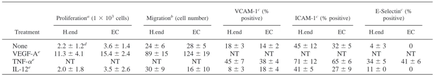
Table I. Effect of IL-12 on EC functions
Treatment
Proliferationa(1⫻ 103cells) Migrationb(cell number)
VCAM-1c(%
positive) ICAM-1c(% positive)
E-Selectinc(% positive)
H.end EC H.end EC H.end EC H.end EC H.end EC
None 2.2⫾ 1.2d 3.6⫾ 1.4 24⫾ 6 28⫾ 5 18⫾ 3 14⫾ 2 45⫾ 12 32⫾ 5 4⫾ 3 0
VEGF-Ae 11.3⫾ 4.1 15.4⫾ 2.4 89⫾ 15 124⫾ 19 NT NT NT NT NT NT
TNF-␣e NT NT NT NT 45⫾ 7 38⫾ 4 71⫾ 12 65⫾ 6 34⫾ 5 41⫾ 6
IL-12e 2.0⫾ 1.8 3.5⫾ 2.6 30⫾ 9 16⫾ 10 8⫾ 3 18⫾ 4 41⫾ 5 27⫾ 9 11⫾ 0 0
aQuiescent cells were stimulated every 2 days with VEGF-A
165or with murine or human IL-12, and cells were counted at day 5.
bCell migration was evaluated after 7-h stimulation by Boyden’s chamber technique.
cAdhesion molecule expression was evaluated after 24-h (VCAM-1 and ICAM-1) or 6-h cell stimulation. dMean⫾ SD of three experiments done in triplicate.
eVEGF-A
each well of a 48-well plate and incubated at 37°C for 30 min to allow gel formation. Human EC (2⫻ 104/well) were plated onto Matrigel and
cocul-tured with human PBMC. After 48-h incubation in 5% CO2humidified
atmosphere at 37°C, the cell three-dimensional organization was examined under an inverted phase contrast photomicroscope (DM-IBM model; Leica Microsystems, Wetzlar, Germany) and then photographed. This Matrigel allows spontaneous in vitro angiogenesis (35).
Gel zymography
MMPs activity was assayed by zymography of EC proteins (36). EC were lysed in 50 mM Tris, 300 mM NaCl, and 1% Triton X-100, pH 7.5, and 10
g of proteins were separated by 10% SDS-PAGE impregnated with
gel-atin (1 mg/ml; Sigma) in nonreducing conditions. Gels were washed twice for 20 min each time with 50 mM Tris, and 2.5% Triton X-100, pH 7.5, and incubated overnight at 37°C in 40 mM Tris, 200 mM NaCl, 10 mM CaCl2,
and 0.02% NaN3, pH 7.5, with or without 5 mM EDTA. Clear bands,
identifying the positions of 72- and 92-kDa gelatinases, were visualized on the blue background after staining with 0.25% Coomassie blue R250 and destaining with 50% methanol and 10% acetic acid.
Expression of adhesion molecules
Single-cell suspensions of H.end or human EC (1⫻ 105/ml) recovered
from cocultures or adherent cell cultures stimulated with IL-12 or human or murine TNF-␣ (both from R&D Systems; 10 ng/ml) for appropriate times were respectively incubated in PBS containing 1% FBS (30 min in ice) with rat anti-mouse mAb against VCAM-1 (Clone M/K-2; 0.1g/ sample), ICAM-1 (Clone KAT-1; 0.1g/sample; R&D Systems), E-selec-tin (Clone 10E9; 0.5 g/sample; provided by E. Dejana, Istituto Mario Negri, Milan, Italy), or mouse anti-human mAbs against VCAM-1 (Clone 51-10C9; 0.1g/sample), ICAM-1 (Clone HA58; 0.1 g/sample; PharM-ingen), and E-selectin (Clone 10C10; 0.2g/sample; R&D Systems). Cells were then washed twice with ice-cold PBS and labeled with appropriate FITC-conjugated secondary Abs (R&D Systems). The secondary Ab alone was used as a control. FACS analysis was performed with a FACScan flow cytometer (FACSCalibur, Becton Dickinson).
Apoptosis assays
H.end and human EC apoptosis was studied by exposure in the outer mem-brane of phosphatidylserine assayed using an annexin V-FITC kit (Bender Medsystem, Wien, Austria) and propidium iodide staining by FACS anal-ysis. The two annexin V-positive quadrants of the analysis were taken as the apoptotic fraction. Apoptosis was also studied by detection of cytosolic histone-associated DNA fragments with a Cell Death Detection ELISA kit (Roche, Mannheim, Germany). The absorbance was measured at 405 nm with a microplate reader (Perkin-Elmer). DNA laddering was studied on 1⫻ 106cells lysed in 20 mM Tris-HCl, pH 7.4, containing 5 mM EDTA
and 0.4% Triton X-100 (37). Cytosolic DNA was extracted with phenol/ chloroform/isoamylic alcohol (25/24/1) and was precipitated with
ammo-nium acetate (7.5 M; pH 7.5). Ethanol-precipitated DNA pellets were treated with pancreatic RNase (200 ng for 15 min at 37°C) in Tris-borate EDTA buffer. Samples were electrophoresed on an agarose gel (Bio-Rad, Hercules, CA) containing ethidium bromide (1.8%) to visualize DNA fragmentation.
Cell cycle analysis
Murine H.end and human EC were suspended in 50l of PBS, fixed in 1 ml of 70% ice-cold ethanol for 30 min on ice, stained with 0.5 ml of propidium iodide (100 g/ml; Sigma) containing 5 g/ml pancreatic RNase (Stratagene) for 20 min at 37°C. After gating out cellular aggregates and debris, propidium iodide fluorescence was measured using a FACScan flow cytometer, and cell cycle analysis was performed with the Mod Sit LT program (Verity Software House, Topsham, ME).
DNA synthesis was measured by 5-bromodeoxyuridine (BrdU) uptake and compared with DNA content as determined by propidium iodide up-take (38). Briefly, adherent cells were incubated with 30M BrdU (Sigma; 1 h at 37°C). Detached cells were fixed as described above, washed, and suspended in PBS. Cells were denatured in 1 ml of 2 N HCl for 20 min at room temperature and then neutralized with 1 ml of 0.1 M Na2B4O7for 5
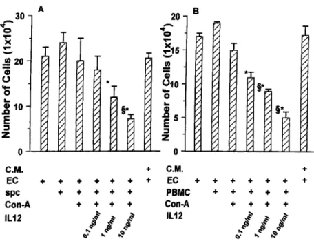
min at room temperature. The cells were centrifuged and incubated (10 min at 45°C) in 2 ml of PBS containing 5% Tween 20 and 0.1% BSA (Sigma), then centrifuged and incubated with 10g of fluoresceinated mAb anti-FIGURE 1. Cocultures between EC and spc and
human PBMC. The growth of H.end cells (A) and of human EC (B) was evaluated after 72 h of cocultures with differently treated spc or PBMC or with con-ditioned medium (C.M.) from cultures of activated spc-IL-12 and activated PBMC-IL-12. The mean⫾ SD of five experiments performed in triplicate are shown.ⴱ and §, p ⬍ 0.05 vs cocultures of EC with nonactivated spc and PBMC and with activated spc and activated PBMC, respectively, evaluated by Stu-dent-Neuman-Keuls test performed after ANOVA (A, F⫽ 10.75; B, F ⫽ 17.08).
FIGURE 2. Growth of murine H.end cells cocultured with spc from TSA mammary adenocarcinoma-bearing BALB/C mice treated with sys-temic IL-12. Mice bearing TSA tumor were treated with vehicle or with 0.1
g of murine IL-12 i.p. daily for 5 or 10 days. The results from 72-h
cultures are expressed as the mean⫾ SD of three animals. Student’s t test was used for evaluation of statistical significance.
BrdU (PharMingen) in 0.2 ml of PBS for 30 min at room temperature in the dark. After washes the cells were kept overnight at 4°C in the dark to favor partial DNA renaturation. Staining with propidium iodide and FACS anal-ysis were performed as described above. At least 10,000 events were analyzed.
Results
Effect of IL-12 on EC
In the first series of experiments we observed that the presence of 0.1–100 ng/ml of IL-12 in the culture medium did not alter the expression of adhesion molecules or the proliferative and migra-tory ability of both murine H.end and human EC. By contrast, the addition of VEGF-A165 induced proliferation and migration of
both cells, while the addition of TNF-␣ up-regulated their mem-brane expression of ICAM-1, VCAM-1, and E-selectin (Table I). As these results apparently challenge many data on the effects of IL-12 on vascular system (13, 17, 18, 22–25), we next investigated in a coculture system whether the effects of IL-12 were, in fact, mediated by lymphoid cells.
Activated spc-IL-12 inhibit EC proliferation
First, the proliferation of H.end cells cocultured with spc, activated spc, and activated spc-IL-12 was evaluated. No inhibition of H.end proliferation was observed except when they were cultured with activated spc-IL-12. The decrease in H.end cell proliferation was directly proportional to the amount of IL-12. The stronger inhibi-tion was found in the presence of 10 ng/ml IL-12. In this case, inhibition was already evident after 24 h of coculture (data not shown) and peaked at 72 h (Fig. 1A). Similar results were obtained by coculturing human PBMC with human EC (Fig. 1B). Human EC were even more sensitive than mouse H.end cells to the inhi-bition by activated PBMC-IL-12.
Although conditioned medium from activated spc-IL-12 was not sufficient to mimic the inhibition observed in the cocultures (Fig. 1), arrest of either mouse H.end or human EC proliferation did not require direct cell contacts because it occurred when the two cell types were separated by a semipermeable membrane.
IL-12 impairs the in vivo growth of several murine tumors as well as that of TSA adenocarcinoma by inhibiting angiogenesis and causing vascular necrosis (16, 17, 39). To evaluate whether this in vivo situation is mirrored by our culture system, the ability of spc from mice bearing TSA tumor and treated systemically with IL-12 to reduce H.end proliferation was tested. A significant im-pairment of proliferation was found in cocultures with spc from mice bearing TSA adenocarcinoma that received 10 daily injec-tions of 100 pg of IL-12 (Fig. 2).
Activated spc-IL-12 and activated PBMC-IL-12 arrest the cell cycle of EC
The observed inhibition of EC by mouse activated spc-IL-12 and human activated PBMC-IL-12 could depend either on cell death or cell cycle arrest. As no toxic effect in the cocultures (i.e., lactate dehydrogenase release and trypan blue exclusion) were observed, it was first evaluated whether the reduced number of EC was due to an increase in their apoptosis. In contrast with etoposide (40), which markedly increased human EC apoptosis (positive control), no enhanced EC apoptosis was found after 72-h culture with hu-man activated PBMC-IL-12 (Table II). This result was assessed with four independent methods, namely by evaluating the binding of fluoresceinated annexin V to phosphatidylserine expressed in the outer membrane, the presence of nucleosome by measuring histone-associated DNA fragments, the presence of a hypodiploid
Table II. Effect of human PBMC cells stimulated by IL-12 on EC apoptosis
EC Cocultured witha Annexin V staining
(% positive cells)
Nucleosome ELISA (OD at 405 nm)
Propidium iodide staining (% hypodiploid cells) None 0.61⫾ 0.20b 0.401⫾ 0.04 3.19⫾ 1.20 PBMC 0.96⫾ 0.50 0.352⫾ 0.05 5.70⫾ 1.89 Activated PBMC 1.23⫾ 0.54 0.36⫾ 0.02 7.81⫾ 2.54 Activated PBMC IL-12c 0.74⫾ 0.77 0.34⫾ 0.07 8.06⫾ 1.77 Etoposide 5M 17.4⫾ 1.56 2.68⫾ 0.12 53.12⫾ 9.50
aHuman EC were cocultured with 2⫻ 106human PBMCs for 72 h in transwells.
bMean⫾ SD of three measures in one experiment representative of three independent experiments. cIL-12 was used at 10 ng/ml.
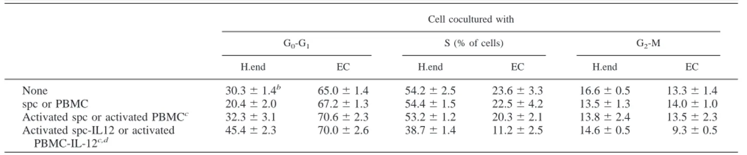
Table III. Analysis of cell cycle in murine microvascular H.end cells and human ECa
Cell cocultured with
G0-G1 S (% of cells) G2-M
H.end EC H.end EC H.end EC
None 30.3⫾ 1.4b 65.0⫾ 1.4 54.2⫾ 2.5 23.6⫾ 3.3 16.6⫾ 0.5 13.3⫾ 1.4
spc or PBMC 20.4⫾ 2.0 67.2⫾ 1.3 54.4⫾ 1.5 22.5⫾ 4.2 13.5⫾ 1.3 14.0⫾ 1.0 Activated spc or activated PBMCc 32.3⫾ 3.1 70.6⫾ 2.3 53.2⫾ 1.2 20.3⫾ 2.1 13.8⫾ 2.4 13.5⫾ 2.3
Activated spc-IL12 or activated PBMC-IL-12c,d
45.4⫾ 2.3 70.0⫾ 2.6 38.7⫾ 1.4 11.2⫾ 2.5 14.6⫾ 0.5 9.3⫾ 0.5
aCell cycle analysis was performed on H.end cells or human EC cells respectively cocultured with murine or human lymphomononuclear cells for 72 h. At the end of cocultures cells were labeled with BrdU, stained with fluorosceinated Ab anti-BrdU and propidium iodide, and then analyzed by FACS.
bMean⫾ SD of three samples from one typical experiments of three. cspc and PBMC were respectively used with H.end and human EC. dHuman or murine IL-12 were used at 10 ng/ml.
cell population by propidium iodide staining (Table II), and cyto-solic DNA laddering (data not shown). No apoptosis increase was also observed after 24- and 48-h cocultures of activated spc-IL-12 and H.end cells (data not shown). DNA synthesis in human EC and H.end cells was measured by evaluating BrdU uptake during S phase compared with DNA content by FACS analysis. Table III shows that a reduced number of human EC entered S phase after coculture for 72 h with human activated PBMC-IL-12. Similar
results were obtained with H.end cells cocultured with activated spc-IL-12 (Table III).
Activated spc-IL-12 and activated human PBMC-IL-12 inhibit EC adhesion
As adhesion to extracellular matrix is crucial for survival of EC and nascent vessels (41), and impairment of integrin-mediated en-dothelial adhesion may even result in tumor regression (42, 43), the effect of activated spc-IL-12 and PBMC-IL-12 on adhesive properties of EC was next evaluated. A dramatically reduced ca-pacity of murine and human EC to adhere to vitronectin, the ligand of␣v3integrin (44) was found; by contrast, no modulation of EC
adhesion ability was found following their culture with condi-tioned medium from activated spc-IL-12 or PBMC-IL-12 cultured alone (Fig. 3).
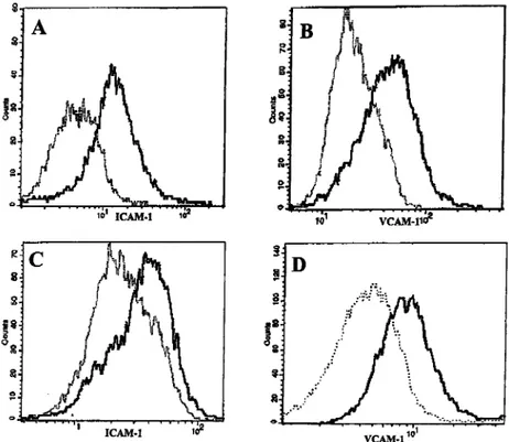
Activated spc-IL-12 and activated human PBMC-IL-12 regulate adhesion molecule expression
In mice treated with systemic IL-12, the expression of VCAM-1 and, to a lesser extent, of ICAM-1 is consistently increased on EC lining tumor capillaries (13, 17). Our cocultures confirm these re-sults. Factors released by both human and murine activated spc-IL-12 and activated PBMC-spc-IL-12 increase the expression of VCAM-1 and ICAM-1 (Fig. 4), but not that of E-selectin (data not shown) on human EC and mouse H.end cells, respectively.
Activated PBMC-IL-12 impair in vitro angiogenesis of human EC
To gain an insight into the effect of IL-12 on angiogenesis, human EC were cultured on a three-dimensional matrix protein gel (Ma-trigel) (45– 47), where they gave rise spontaneously to an orga-nized meshwork of anastomosing cord-like structures (Fig. 5A). Although coculture with PBMC or activated PBMC did not mod-ify the EC angiogenetic ability (Fig. 5, B and C), their coculture with activated PBMC-IL-12 dramatically inhibited it. By failing to establish contacts to neighboring cells, EC were unable to form continuous tubes (Fig. 5D).
FIGURE 3. Adhesion to vitronectin of murine H.end and human EC cocultured with spc and PBMC. Adhesion of H.end cells (A) and human EC (B) was evaluated after 72 h of cocultures with differently treated spc and PBMC or with conditioned medium (C.M.) from cultures of activated spc-IL-12 and activated PBMC-IL-12. At the end of cocultures, EC were detached, and 3⫻ 104cells were seeded on 96-well plates coated with
vitronectin. After 1-h incubation at 37°C, cells were washed, fixed, and stained by crystal violet, and the absorbance was read at 540 nm. The adhesion of EC to BSA, used as a negative control, was 0.05⫾ 0.003 (mean⫾ SD of four experiments performed in triplicate). ⴱ, p ⬍ 0.05 vs cocultures of EC with activated spc and activated PBMC, evaluated by Student-Neuman-Keuls test performed after ANOVA (A, F⫽ 295.11; B,
F⫽ 171.98).
FIGURE 4. Expression of adhesion molecules on human end murine EC cocultured with spc and PBMC. FACS analysis of ICAM-1 (A and C) and VCAM-1 (B and D) in H.end (A and B) and human EC (C and D) cocultured for 72 h with activated spc-IL-12 (dotted lines) or with activated spc-IL-12 and activated PBMC-IL-12 (continuous lines). The fluorescence intensity of both molecules analyzed in unstimulated H.end cells or human EC or cocultured with spc and PBMC alone was similar to that ob-tained after coculture with activated spc and PBMC. Results are representative of four independent experiments.
Production of MMPs by EC is a crucial event during the pro-gression phase of angiogenesis. It is characterized by cell sprouting and matrix remodeling and regulates in vitro angiogenesis (10, 48). To assess the production of MMPs by cocultured human EC, the presence of the bioactive proteases was assessed by zymography in EC lysate. We found that EC did not show constitutive production of MMP-9 (97 kDa). The gelatinolytic activity appeared after co-culture with PBMC, but was not modified by activated PBMC. In contrast, the proteolytic activity was consistently reduced in co-culture with activated PBMC-IL-12. MMP-2 (72 kDa) was present in untreated EC, but its production was not affected by the cocul-tures (Fig. 6).
Cell populations responsible for the inhibition of EC proliferation
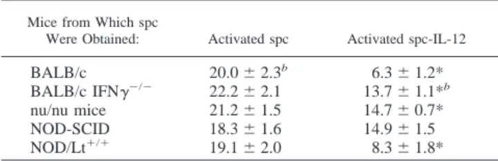
To define the cell population mostly responsible for the inhibition of EC proliferation, activated spc-IL-12 from variously immuno-deficient mice were first tested. Activated spc-IL-12 from nu/nu mice lacking T cell functions and displaying enhanced NK activity (49) displayed a reduced ability to inhibit mouse H.end growth (36% inhibition) compared with normal mice (70% inhibition). Activated spc-IL-12 from NOD-SCID mice exhibiting low NK
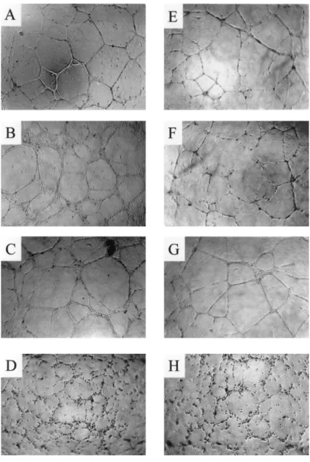
FIGURE 5. Formation of tube-like structures by hu-man EC cocultured with PBMC. Huhu-man EC (2⫻ 104)
were plated onto Matrigel and cocultured with human PBMC. After 48-h incubation the cell three-dimensional organization was examined under an inverted phase contrast photomicroscope. A, EC alone; B, EC cocul-tured with PBMC; C, EC coculcocul-tured with activated PBMC; D, EC cocultured with activated PBMC-IL-12;
E, EC cocultured with activated PBMC-IL-12 in
pres-ence of anti-MIG Ab; F, EC cocultured with activated PBMC-IL-12 in the presence of anti-IP-10 Ab; G, EC cocultured with activated PBMC-IL-12 in the presence of anti-MIG and anti-IP-10 Abs; H, EC cocultured with activated PBMC-IL-12 in the presence of irrelevant Ig. Magnification,⫻10. Results are representative of three independent experiments.
FIGURE 6. MMP-2 and MMP-9 production by human EC cocultured with human PBMC. Cell lysates (30g) from EC alone or cocultured for 72 h with activated PBMC or activated PBMC-IL-12 were analyzed by gelatin zymography as detailed in Materials and Methods. The MMP-9 (upper band) and MMP-2 (lower band) were located at⬃97 and 72 kDa, respectively. Results are representative of two independent experiments.
activity and defective for macrophage function (50) did not impair the H.end proliferation rate, while activated spc-IL-12 from NOD/Lt⫹/⫹mice, displaying normal immune functions, inhibited H.end proliferation as did those from normal mice (Table IV).
In other experiments, activated spc-IL-12 were selectively de-pleted of CD4⫹, CD8⫹, or NK cells using specific mAbs and the MiniMACS magnetic separation system (Miltenyi Biotec; Table V). The absence of CD4⫹, CD8⫹, NK cells, and macrophages during cocultures partially decreased the inhibition of H.end pro-liferation. In contrast, spc passed through a nylon wool column had a reduced inhibitory effect on H.end proliferation, which was re-versed by addition of peritoneal macrophages (Table V). Together these results strongly suggest that distinct activated spc-IL-12 sub-populations play a role in the inhibition of EC proliferation.
Factors leading to the inhibition of EC proliferation
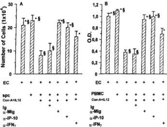
In many experimental systems in vivo the anti-tumor activity of IL-12 appears to rest on its ability to induce downstream produc-tion of IP-10 and MIG as a consequence of its inducproduc-tion of IFN-␥ production (13, 17, 21, 23, 51). To evaluate the downstream me-diators involved in the inhibition of EC in the cocultures with activated spc-IL-12, different cytokine-neutralizing mAbs were used. Although the presence of nonspecific rabbit IgG (Fig. 7), anti-TNF (or anti-GM-CSF mAb), either alone or in combination, did not affect the inhibitory activity of murine activated spc-IL-12 on H.end proliferation (data not shown), it was abolished when MIG and-IP-10 were neutralized by specific Abs (Fig. 7). Simi-larly, anti-MIG and anti-IP-10 Abs reduced the inhibitory effect of activated PBMC-IL-12 on human EC adhesion (Fig. 7) and in vitro angiogenesis (Fig. 5, E–G). Surprisingly, a neutralizing anti-IFN-␥
mAb added to the cocultures alone (Fig. 7) or in combination with anti-TNF-␣ or anti-GM-CSF mAbs (data not shown) at the begin-ning or every 12 h only partially abolished the inhibition. More-over, a partial inhibition of H.end proliferative ability was still evident in the presence of activated spc-IL-12 from IFN-␥⫺/⫺ mice (Table IV).
Discussion
The experiments presented here show that both spc and human PBMC activated by Con A in the presence of IL-12 markedly inhibit the proliferation, matrix adhesion, ability to differentiate into tube-like structure, and expression of VCAM-1 and ICAM-1 of human and murine EC. The strength of this inhibition makes it the in vitro equivalent of the marked anti-angiogenic and angio-static activity of IL-12 observed in vivo (13, 17, 18, 22–25). Use of a Transwell to impede physical contact between lymphoid cells and EC allowed investigation of the inhibiting factors released in the medium. IL-12 alone had no effect on EC functions, whose inhibition, in effect, stems from factors released by activated spc-IL-12 and activated PBMC-spc-IL-12. Normal, nonactivated spc or PBMC are devoid of this inhibitory activity or even enhance EC proliferation. In a few cases a minor inhibition was observed in the presence of activated spc and activated PBMC. In all cases this inhibition was markedly and significantly amplified by the pres-ence of IL-12, suggesting that this cytokine specifically induces an anti-angiogenic program in human and murine lymphoid cells. Murine H.end cells are a suitable model for investigation of the activities of microvascular EC (28 –30, 52). Their growth inhibi-tion as well as that of human EC in the presence of factors released by activated spc-IL-12 and activated PBMC-IL-12 does not cor-relate with a major increase in their apoptosis, but with their de-layed entry into S phase. This finding fits in well with recent data showing that tumor vessels have a diminished proliferation index after treatment with IL-12 (25). Reduced proliferation of EC was associated with a decrease in their ability to adhere to vitronectin, the ligand of␣v3integrin (44). In vivo adhesion of EC to this
substrate provides anti-apoptotic signals to nascent vessels (42, 53) through suppression of p53 and p53-inducible cell cycle inhibitor p21WAF1/CIP1 and elevation of the Bcl-2:Bax ratio (54). Fur-thermore integrins containing␣vsubunit mediate the progression
through G1 phase by protracted activation of Jun NH2-terminal
kinase (55). Coculture of EC with activated spc-IL-12 and acti-vated PBMC-IL-12 may first result in changes in␣v3affinity for Table IV. Effect of spc from immunodeficient mice on H.end growtha
Mice from Which spc
Were Obtained: Activated spc Activated spc-IL-12
BALB/c 20.0⫾ 2.3b 6.3⫾ 1.2*
BALB/c IFN␥⫺/⫺ 22.2⫾ 2.1 13.7⫾ 1.1*b
nu/nu mice 21.2⫾ 1.5 14.7⫾ 0.7* NOD-SCID 18.3⫾ 1.6 14.9⫾ 1.5 NOD/Lt⫹/⫹ 19.1⫾ 2.0 8.3⫾ 1.8*
aActivated spc or activated spc-IL-12 (2⫻ 106/ml) were cocultured for 72 h with
3⫻ 104H.end cells.
bEC number (⫻ 104) expressed as the mean⫾ SD of three independent
exper-iments. ANOVA, F⫽ 32.16. *, p ⬍ 0.05 vs activated spc (Student-Neuman-Keuls test).
Table V. Effect of spc sub-population on murine EC growth in coculturea
Cell Type
Cell Treatment
None Con A⫹ IL-12 EC number (1⫻ 104)
spc 18.0⫾ 2.3b 5.3⫾ 1.2*
Nylon wool spcc 17.0⫾ 1.2 12.4⫾ 0.3*§
Nylon wool spc supplemented with macrophagesd 20.4⫾ 0.2 7.2⫾ 3.0*
spc depleted of CD4⫹cells 18.3⫾ 1.8 16.4⫾ 1.8§ spc depleted of CD8⫹cells 17.2⫾ 0.8 11.4⫾ 2.1*§ spc depleted of NK cells 21.1⫾ 1.1 13.0⫾ 0.8*§
aspc (2⫻ 106/ml) were activated as detailed in Materials and Methods and cocultured for 72 h with H.end cells (3⫻ 104).
bMean⫾ SD of three experiments. ANOVA, F ⫽ 67.51. * and §, respectively, indicate p ⬍ 0.05 vs unstimulated cells and vs activated Ly-IL-12 (Student-Neuman-Keuls test).
cspc (2⫻ 106) were layered on mouse peritoneal macrophages (2⫻ 104/cm2) and incubated overnight in RPMI 1640
complete medium containing 2g/ml Con A in the absence or in the presence of IL-12 as detailed in Materials and Methods. dMacrophages depleted spc by passage on nylon wool column (2⫻ 106) were layered on mouse peritoneal macrophages
(2⫻ 104/cm2) and incubated overnight in RPMI 1640 complete medium containing 2g/ml Con A in the absence or in the
presence of IL-12 as detailed in Materials and Methods.
the matrix ligand, followed by impairment of EC adhesion ma-chinery and cell cycle arrest. Of interest, tumor-associated blood vessels of colon carcinoma cells transduced with IL-12 have re-duced expression of␣v3integrin even when injected in IFN-␥⫺/⫺
mice (15). The inhibitory effect of activated PBMC-IL-12 on EC adhesion may explain their inability to differentiate into tube-like structures when grown on a three-dimensional gel of extracellular matrix. The failure of EC to engage extracellular matrix proteins through␣v3integrin inhibits in vitro angiogenesis (56, 57).
Besides these effects on EC proliferation and matrix adhesion, activated spc-IL-12 and activated PBMC-IL-12 up-regulated the expression of VCAM-1 and ICAM-1 on EC surface. These adhe-sion molecules are involved in lymphocyte recruitment (58) and are also up-regulated in tumor vessels of mice receiving systemic IL-12 (13, 17). Additional impairment of tumor vasculature may occur when lymphoid cells are allowed to touch endothelium, as occurs in vivo (59).
Investigation of the nature of the inhibitory factors responsible for these effects exerted by activated spc-IL-12 and activated PBMC-IL-12 showed that anti-IP-10 and anti-MIG Abs greatly reduced the inhibition of EC proliferation, adhesion, and in vitro angiogenesis. IP-10 and MIG are IFN-␥-induced chemokines that play a crucial role in the anti-tumor effect of IL-12. Their expres-sion is up-regulated in tumors growing in IL-12-treated mice (13, 17, 23, 25), while their neutralization by specific Abs prevents the curative effect of IL-12 (21). They also inhibit the chemotaxis of EC and the angiogenesis induced by fibroblast growth factor 2 or by IL-8 in vivo (26). These effects appear to be mediated by their interaction with heparan sulfates (60) rather than by their ability to interact with CXC chemokine receptor 3. Several reports have demonstrated that this receptor is absent or very poorly expressed on human EC (61– 64), while one study found evidence of its expression in murine EC (65). Human microvascular EC from der-mis also express CXC chemokine receptor 3. However, the active concentration of IP-10 (5⫻ 10⫺4M) is too high to be reached in vivo (64). Impairment of EC functions requires a continuous
in-teraction between activated spc-IL-12 and activated PBMC-IL-12 and EC, as shown by the lack of inhibitory activity of conditioned medium from both activated spc-IL-12 and activated PBMC-IL-12 (Figs. 1 and 3) and of recombinant MIG and IP-10 in the absence of activated lymphoid cells (M. Strasly, unpublished observations). In our murine cocultures established in the presence of IL-12 both lymphoid cells and EC expressed MIG and IP-10 (F. Cavallo, unpublished observations). This expression is mostly due to the high titers of IFN-␥ elicited by IL-12 on lymphoid cells (66). The influence of lymphoid cells on EC functions thus rests on a ping-pong of soluble signals of lymphoid and endothelial origin. A pos-sible scenario emerging from our data is that MIG and IP-10 do not directly cause the inhibition of EC growth. Instead, MIG and IP-10 of both endothelial and lymphoid origins appear to act primarily on T and NK cells. Following this additional signal, lymphoid cells activate a sustained release of factors that cause EC growth arrest. Depletion of T and NK cells abolishes the inhibitory activity of activated spc-IL-12. This scenario is also supported by in vivo data from Tosato’s group underlying the importance of NK and T cells in the anti-angiogenic loop triggered by IL-12 (63, 66). Removal of the inhibitory activity of activated spc-IL-12 by either anti-MIG or anti-IP-10 mAb suggests that these chemokines also have an independent effect, as reported in other systems (67). Besides their overlapping redundant functions, MIG and IP-10 may trigger unique intracellular signals that differentiate their activity. In vivo, the different patterns of MIG and IP-10 expression suggest that they may trigger nonredundant functions (67). Another possibility connected to the redundancy of the chemokine/cytokine system is that other molecules produced in our coculture system might in-teract with CXC receptor 3 and modify its sensitivity to MIG or IP-10.
Moroever, MIG and IP-10 do not seem to be the sole molecules that play a role in inducing the inhibition of EC functions in our cocultures. Actually, recombinant MIG and IP-10 are necessary, but not sufficient, for the elicitation of the anti-angiogenic activity, and other molecules appear to be needed.
The antitumor activity of IL-12, including inhibition of angio-genesis, is largely dependent on its ability to induce IFN-␥ released by NK cells (22, 23, 25, 27) and to activate a perforin-mediated cytolytic pathway (68, 69). Surprisingly, our data obtained with neutralizing anti-IFN-␥ mAb and with spc from IFN-␥⫺/⫺ mice showed that in the absence of IFN-␥ the inhibition of EC prolif-eration is not completely abrogated. This suggests that other mol-ecules may replace IFN-␥ in our system. Germane to our obser-vation is the finding that in IFN-␥⫺/⫺mice, IL-12 is still able to induce an effective rejection of tumor cells engineered to release IL-12 (15). The presence of IL-12 during the activation of spc and PBMC is critical for the induction of their anti-angiogenic pro-gram. The way in which activated spc-IL-12 and activated PBMC-IL-12 inhibit EC is distinct from that of endotoxin-primed PBMC. Here, EC undergo cycle arrest and apoptosis through a pathway that requires PBMC-EC contact, expression of TNF-␣ on the lym-phoid cell surface and TGF-␣ secretion (70). Similarly to endotoxin-primed PBMC, PBMC activated by Con A adhere to EC and induce their detachment from the matrix, but not their apoptosis (71).
The fact that IL-12 reduces angiogenesis in SCID mice, NK cell-deficient beige mice, and nu/nu mice (22) indicates that its inhibition of neoangiogenesis is not necessarily mediated by a sin-gle immune cell population, although NK recruitment and cyto-toxicity are needed (63). In our cocultures, total inhibition of EC growth required cooperation between two cell populations, each necessary but not in itself sufficient. Because both spc and human PBMC require prior activation by Con A before IL-12 priming, it would seem that the anti-angiogenic effect of IL-12 is not directly
FIGURE 7. Effects of neutralizing Abs anti-MIG, IP-10, and IFN-␥ on H.end proliferation (A) and human EC adhesion to vitronectin (B). Cells were cocultured for 72 h with spc and PBMC or activated spc-IL-12 and activated PBMC-IL-12 in the presence of specific Abs or irrelevant Ig and processed for the cell count and the adhesion assays as detailed in Figs. 1 and 4. Shown are the mean⫾ SD of four experiments performed in trip-licate.ⴱ and §, p ⬍ 0.05 vs cocultures of EC with activated spc-IL-12 and activated PBMC-IL-12 in the presence of nonimmune Ig or Ab anti-IFN-␥, respectively, evaluated by Student-Neuman-Keuls test performed after ANOVA (A, F⫽ 15.58; B, F ⫽ 182.79).
activated by IL-12 on the cells that constitutively express the IL-12 receptor. Con A activation presumably up-regulates expression of the inducible IL-12 B2 receptor on T cells. Indeed, the T helper function is required, as spc from nu/nu mice or spc depleted of CD4⫹cells were less able to inhibit EC growth. Similar findings were observed on depletion of CD8⫹or NK cells. Moreover, ac-tivated spc-IL-12 from NOD-SCID mice, which are deficient in T cell, B cell, and macrophage function and display low NK activity (50), were virtually unable to inhibit EC growth. Furthermore, ny-lon wool column-emerging BALB/c cells were less active than total spc. Their activation in the presence of peritoneal macro-phages fully restores their inhibitory potential on EC. By costimu-lating T cell activation, macrophages induce IL-12 receptor ex-pression (72).
These findings suggest that an early circuit triggered by IL-12 requires a ping-pong of soluble signals (such as MIG and IP-10) between spc and EC, whose outcome is functional impairment of EC activities. EC can no longer sustain neoangiogenesis and as-sume a phenotype more prone to tissue invasion by leukocytes that amplify vascular damage in vivo (73). These alterations of EC functions appear to form the basis of IL-12-mediated ischemic-hemorrhagic rejection of tumors in vivo.
Acknowledgments
We thank Drs. J. Farber (National Institutes of Health, Bethesda, MD), T. A. Hamilton (Cleveland Clinic Foundation, Cleveland, OH), and E. Dejana (Istituto Mario Negri, Milan, Italy) for providing fundamental reagents.
References
1. Folkman, J. 1995. Angiogenesis in cancer, vascular, rheumatoid and other dis-ease. Nat. Med. 1:27.
2. Zetter, B. R. 1998. Angiogenesis and tumor metastasis. Annu. Rev. Med. 49:407. 3. Hanahan, D., and J. Folkman. 1996. Patterns and emerging mechanisms of the
angiogenic switch during tumorigenesis. Cell 86:353.
4. Mantovani, A., B. Bottazzi, F. Colotta, S. Sozzani, and L. Ruco. 1992. The origin and function of tumor-associated macrophages. Immunol. Today 13:265. 5. Fukumura, D., R. Xavier, T. Sugiura, Y. Chen, E. Park, N. Lu, M. Selig,
G. Nielsen, T. Taksir, R. K. Jain, et al. 1998. Tumor induction of VEGF promoter activity in stromal cells. Cell 94:715.
6. Freeman, M. R., F. X. Schneck, M. L. Gagnon, C. Corless, S. Soker, K. Niknejad, G. E. Peoples, and M. Klagsbrun. 1995. Peripheral blood T lymphocytes and lymphocytes infiltrating human cancers express vascular endothelial growth fac-tor: a potential role for T cells in angiogenesis. Cancer Res. 55:4140. 7. Peoples, G. E., S. Blotnick, K. Takahashi, M. R. Freeman, M. Klagsbrun, and
T. J. Eberlein. 1995. T lymphocytes that infiltrate tumors and atherosclerotic plaques produce heparin-binding epidermal growth factor-like growth factor and basic fibroblast growth factor: a potential pathologic role. Proc. Natl. Acad. Sci. USA 92:6547.
8. Mach, F., U. Schonbeck, R. P. Fabunmi, C. Murphy, E. Atkinson, J. Y. Bonnefoy, P. Graber, and P. Libby. 1999. T lymphocytes induce endothelial cell matrix metalloproteinase expression by a CD40L-dependent mechanism: im-plications for tubule formation. Am. J. Pathol. 154:229.
9. Mantovani, A., F. Bussolino, and M. Introna. 1997. Cytokine regulation of en-dothelial cell function: from molecular level to the bedside. Immunol. Today 18:231.
10. Bussolino, F., A. Mantovani, and G. Persico. 1997. Molecular mechanisms of blood vessel formation. Trends Biochem. Sci. 22:251.
11. Rollins, B. J. 1997. Chemokines. Blood 90:909.
12. Falcone, D. J., K. M. Khan, T. Layne, and L. Fernandes. 1998. Macrophage formation of angiostatin during inflammation: a byproduct of the activation of plasminogen. J. Biol. Chem. 273:31480.
13. Boggio, K., G. Nicoletti, E. Di Carlo, F. Cavallo, L. Landuzzi, C. Melani, M. Giovarelli, I. Rossi, P. Nanni, C. De Giovanni, et al. 1998. Interleukin 12-mediated prevention of spontaneous mammary adenocarcinomas in two lines of Her-2/neu transgenic mice. J. Exp. Med. 188:589.
14. Boggio, K., E. Di Carlo, S. Rovero, F. Cavallo, E. Quaglino, P. L. Lollini, P. Nanni, G. Nicoletti, S. Wolf, P. Musiani, et al. 2000. Ability of systemic interleukin-12 to hamper progressive stages of mammary carcinogenesis in HER2/neu transgenic mice. Cancer Res. 60:359.
15. Zilocchi, C., A. Stopacciaro, C. Chiodoni, M. Parenza, N. Terrazzini, and M. P. Colombo. 1998. Interferon␥-independent rejection of interleukin 12-trans-duced carcinoma cells requires CD⫹T cells and granulocyte/macrophage colony-stimulating factor. J. Exp. Med. 188:133.
16. Cavallo, F., P. Signorelli, M. Giovarelli, P. Musiani, A. Modesti, M. J. Brunda, M. P. Colombo, and G. Forni. 1997. Antitumor efficacy of adenocarcinoma cells
engineered to produce IL-12 or other cytokines compared with exogenous IL-12. J. Natl. Cancer Inst. 89:1049.
17. Cavallo, F., E. Di Carlo, M. Butera, R. Verrua, M. P. Colombo, P. Musiani, and G. Forni. 1999. Immune events associated with the cure of established tumors and spontaneous metastases by local and systemic interleukin 12. Cancer Res. 59: 414.
18. Coughlin, C. M., K. E. Salhany, M. Wysocka, E. Aruga, H. Kurzawa, A. E. Chang, C. A. Hunter, J. C. Fox, G. Trinchieri, and W. M. F. Lee. 1998. Interleukin-12 and interleukin-18 synergistically induce murine tumor regression which involves inhibition of angiogenesis. J. Clin. Invest. 101:1441. 19. Tasaki, K., Y. Yoshida, T. Maeda, M. Miyauchi, K. Kawamura, K. Takenaga,
H. Yamamoto, T. Kouzu, T. Asano, T. Ochiai, et al. 2000. Protective immunity is induced in murine colon carcinoma cells by the expression of interleukin-12 or interleukin-18, which activate type 1 helper T cells. Cancer Gene Ther. 7:247. 20. Sunamura, M., L. Sun, L. Lozonschi, D. G. Duda, T. Kodama, G. Matsumoto, H. Shimamura, K. Takeda, M. Kobari, H. Hamada, et al. 2000. The antiangio-genesis effect of interleukin 12 during early growth of human pancreatic cancer in SCID mice. Pancreas 20:227.
21. Tannenbaum, C. S., R. Tubbs, D. Armstrong, J. H. Finke, R. M. Bukowski, and T. A. Hamilton. 1998. The CXC chemokines IP-10 and Mig are necessary for IL-12-mediated regression of the mouse RENCA tumor. J. Immunol. 161:927. 22. Voest, E. E., B. M. Kenyon, M. S. O’Reilly, G. Truitt, R. J. D’Amato, and
J. Folkman. 1995. Inhibition of angiogenesis in vivo by interleukin 12. J. Natl. Cancer Inst. 87:581.
23. Coughlin, C. M., K. E. Salhany, M. S. Gee, D. C. LaTemple, S. Kotenko, X. Ma, G. Gri, M. Wysocka, J. E. Kim, L. Liu, et al. 1998. Tumor cell responses to IFN␥ affect tumorigenicity and response to IL-12 therapy and antiangiogenesis. Immu-nity 9:25.
24. Gee, M. S., C. J. Koch, S. M. Evans, W. T. Jenkins, C. H. Pletcher, Jr., J. S. Moore, H. K. Koblish, J. Lee, E. M. Lord, G. Trinchieri, et al. 1999. Hy-poxia-mediated apoptosis from angiogenesis inhibition underlies tumor control by recombinant interleukin 12. Cancer Res. 59:4882.
25. Duda, D. G., M. Sunamura, L. Lozonschi, T. Kodama, S. Egawa, G. Matsumoto, H. Shimamura, K. Shibuya, K. Takeda, and S. Matsuno. 2000. Direct in vitro evidence and in vivo analysis of the antiangiogenesis effects of interleukin 12. Cancer Res. 60:1111.
26. Strieter, R. M., P. J. Polverini, S. L. Kunkel, D. A. Arenberg, M. D. Burdick, J. Kasper, J. Dzuiba, J. Van Damme, A. Walz, D. Marriott, et al. 1995. The functional role of the ELR motif in CXC chemokine-mediated angiogenesis. J. Biol. Chem. 270:27348.
27. Sgadari, C., A. L. Angiolillo, and G. Tosato. 1996. Inhibition of angiogenesis by interleukin-12 is mediated by the interferon-inducible protein 10. Blood 87:3877. 28. Giraudo, E., M. Arese, C. Toniatti, M. Strasly, L. Primo, A. Mantovani, G. Ciliberto, and F. Bussolino. 1996. IL-6 is an in vitro and in vivo autocrine growth factor for middle T antigen-transformed endothelial cells. J. Immunol. 157:2618.
29. Bussolino, F., M. De Rossi, A. Sica, F. Colotta, J. M. Wang, E. Bocchietto, I. M. Padura, A. Bosia, E. Dejana, and A. Mantovani. 1991. Murine endothelioma cell lines transformed by polyoma middle T oncogene as target for and producers of cytokines. J. Immunol. 147:2122.
30. Dong, Q. G., A. Graziani, C. Garlanda, R. W. De Calmanovici, M. Arese, R. Soldi, A. Vecchi, A. Mantovani, and F. Bussolino. 1996. Anti-tumor activity of cytokines against opportunistic vascular tumors in mice. Int. J. Cancer 65:700. 31. Bussolino, F., M. F. Di Renzo, M. Ziche, E. Bocchietto, M. Olivero, L. Naldini, G. Gaudino, L. Tamagnone, A. Coffer, and P. M. Comoglio. 1992. Hepatocyte growth factor is a potent angiogenic factor which stimulates endothelial cell motility and growth. J. Cell Biol. 119:629.
32. Forni, G., and M. Giocarelli. 1984. In vitro reeducated T helper cells from sar-coma-bearing mice inhibit sarcoma growth in vivo. J. Immunol. 132:527. 33. Giovarelli, M., S. Landolofo, A. C. Whitmore, and G. Forni. 1982. Rous sarcoma
virus induced tumors in mice: macrophage mediated natural cytotoxicity. Eur. J. Cancer Clin. Oncol. 18:307.
34. Defilippi, P., G. Truffa, G. Stefanutto, F. Altruda, L. Silengo, and G. Tarone. 1991. Tumor necrosis factor␣ and interferon ␥ modulates the expression of the vitronectin receptor (integrin3) in human endothelial cells. J. Biol. Chem. 266:
7638.
35. Marconcini, L., S. Marchio`, L. Morbidelli, E. Cartocci, A. Albini, M. Ziche, F. Bussolino, and S. Oliviero. 1999. c-fos-induced growth factor/vascular endo-thelial growth factor-D induces angiogenesis in vivo and in vitro. Proc. Natl. Acad. Sci. USA 96:9671.
36. Fisher, S. J., and Z. Werb. 1995. The catabolism of extracellular matrix compo-nents. In Extracellular Matrix Proteins: A Practical Approach. M. A. Haralson and J. R. Hassel, eds. IRL Press at Oxford University Press, Oxford, p. 261.
37. Mandriota, S. J., and M. S. Pepper. 1997. Vascular endothelial growth factor-induced in vitro angiogenesis and plasminogen activator expression are depen-dent on endogenous basic fibroblast growth factor. J. Cell. Sci. 110:2293. 38. Mazzini, G., M. Danova, C. Ferrari, M. Giordano, P. Dionigi, and A. Riccardi.
1996. Cell proliferation and ploidy of human solid tumours: methodological ex-perience with in vivo bromodeoxyuridine and DNA flow cytometry. Anal. Cell. Pathol. 10:101.
39. Brunda, M. J., L. Luistro, R. R. Warrier, R. B. Wright, B. R. Hubbard, M. Murphy, S. F. Wolf, and M. K. Gately. 1993. Antitumor and antimetastatic activity of interleukin 12 against murine tumors. J. Exp. Med. 178:1223. 40. Sun, X. M., P. Carthew, D. Dinsdale, G. M. Snowden, and G. M. Cohen. 1994.
The involvement of apoptosis in etoposide-induced thymic atrophy. Toxicol. Appl. Pharmacol. 128:78.
41. Brooks, P. C. 1996. Role of integrins in angiogenesis. Eur. J. Cancer 32A:2423. 42. Brooks, P. C., A. M. P. Montgomery, M. Rosenfeld, R. A. Reisfeld, T. Hu, G. Klier, and D. A. Cheresh. 1994. Integrin␣v3antagonists promote tumor
regression by inducing apoptosis of angiogenic blood vessels. Cell 79:1157. 43. Brooks, P. C., S. Stromblad, R. Klemke, D. Visscher, F. H. Sarkar, and
D. A. Cheresh. 1995. Antiintegrin␣v3blocks human breast cancer growth and
angiogenesis in human skin. J. Clin. Invest. 96:1815.
44. Kuhn, K., and J. Eble. 1994. The structural bases of integrin-ligand interactions. Trends Cell Biol. 4:256.
45. Montesano, R., L. Orci, and P. Vassalli. 1983. In vitro rapid organization of endothelial cells into capillary-like networks is promoted by collagen matrices. J. Cell Biol. 97:1648.
46. Grant, D. S., P. I. Lelkes, K. Fukuda, and H. K. Kleinman. 1991. Intracellular mechanisms involved in basement membrane induced blood vessels differentia-tion in vitro. In Vitro Cell Dev. Biol. 27A:327.
47. Ilan, N., S. Mahooti, and J. A. Madri. 1998. Distinct signal transduction pathways are utilized during the tube formation and survival phases of in vitro angiogen-esis. J. Cell Sci. 111:3621.
48. Hiroaka, N., E. Allen, I. J. Apel, M. R. Gyeto, and S. J. Weiss. 1998. Matrix metalloproteinases regulates neovascularization by acting as pericellular fibrino-lysis. Cell 95:365.
49. Kindred, B. 1979. Nude mice in immunology. Prog. Allergy 26:137. 50. Shultz, L. D., P. A. Schweitzer, S. W. Christianson, B. Gott, I. B. Schweitzer,
B. Tennent, S. McKenna, L. Mobraaten, T. V. Rajan, D. L. Greiner, et al. 1995. Multiple defects in innate and adaptive immunologic function in NOD/LtSz-scid mice. J. Immunol. 154:180.
51. Bukowski, R. M., P. Rayman, L. Molto, C. S. Tannenbaum, T. Olencki, D. Peereboom, R. Tubbs, D. McLain, G. T. Budd, T. Griffin, et al. 1999. Inter-feron-␥ and CXC chemokine induction by interleukin 12 in renal cell carcinoma. Clin. Cancer Res. 5:2780.
52. Ghigo, D., M. Arese, R. Todde, A. Vecchi, F. Silvagno, C. Costamagna, Q. D. Dong, M. Alessio, R. Heller, R. Soldi, et al. 1995. Middle T antigen-transformed endothelial cells exhibit an increased activity of nitric oxide syn-thase. J. Exp. Med. 181:9.
53. Brooks, P. C., R. A. F. Clark, and D. A. Cheresh. 1994. Requirement of vascular integrin␣v3for angiogenesis. Science 264:569.
54. Stromblad, S., J. C. Becker, M. Yebra, P. C. Brooks, and D. A. Cheresh. 1996. Suppression of p53 activity and p21WAF/CIP1 expression by vascular cell in-tegrin avb3 during angiogenesis. J. Clin. Invest. 98:426.
55. Oktay, M., K. K. Wary, M. Dans, R. B. Birge, and F. G. Giancotti. 1999. Integrin-mediated activation of focal adhesion kinase is required for signaling to Jun NH2-terminal kinase and progression through the G1phase of the cell cycle.
J. Cell Biol. 145:1461.
56. Yeh, C. H., H. C. Peng, and T. F. Huang. 1998. Accutin, a new disintegrin, inhibits angiogenesis in vitro and in vivo by acting as integrin␣v3antagonist
and inducing apoptosis. Blood 92:3268.
57. Sheu, J. R., M. H. Yen, Y. C. Kan, W. C. Hung, P. T. Chang, and H. N. Luk. 1997. Inhibition of angiogenesis in vitro and in vivo: comparison of the relative activities of triflavin, an Arg-Gly-Asp-containing peptide and anti-␣v3integrin
monoclonal antibody. Biochim. Biophys. Acta 1336:445.
58. Springer, T. A. 1994. Traffic signals for lymphocyte recirculation and leukocyte emigration: the multistep paradigm. Cell 76:301.
59. Colombo, M. P., L. Lombardi, C. Melani, M. Parenza, C. Baroni, L. Ruco, and A. Stopacciaro. 1996. Hypoxic tumor cell death and modulation of endothelial adhesion molecules in the regression of G-CSF transduced tumors. Am. J. Pathol. 148:473.
60. Luster, A. D., L. D. Grrenberg, and P. Leder. 1995. The IP-10 chemokine binds to a specific cell surface heparan sulfate site shared with platelet factor 4and inhibits endothelial cell proliferation. J. Exp. Med. 182:219.
61. Gupta, S. K., P. G. Lysko, K. Pillarisetti, E. Ohlstein, and J. M. Stadel. 1998. Chemokine receptors in human endothelial cells: functional expression of CXCR4 and its transcriptional regulation by inflammatory cytokines. J. Biol. Chem. 273:4282.
62. Murdoch, C., P. N. Monk, and A. Finn. 1999. CXC chemokine receptor expres-sion on human endothelial cells. Cytokine 11:704.
63. Yao, L., C. Sgadari, K. Furuke, E. T. Bloom, J. Teruya-Feldstein, and G. Tosato. 1999. Contribution of natural killer cells to inhibition of angiogenesis by inter-leukin-12. Blood 93:1612.
64. Salcedo, R., J. H. Resau, D. Halverson, E. A. Hudson, M. Dambach, D. Powell, K. Wasserman, and J. J. Oppenheim. 2000. Differential expression and respon-siveness of chemokine receptors (CXCR1–3) by human microvascular endothe-lial cells and umbilical vein endotheendothe-lial cells. FASEB J. 14:2055.
65. Soto, H., W. Wang, R. M. Strieter, N. G. Copeland, D. J. Gilbert, N. A. Jenkins, J. Hedrick, and A. Zlotnik. 1998. The CC chemokines 6Ckine binds the CXC chemokine receptor CXCR3. Proc. Natl. Acad. Sci. USA 95:8205.
66. Kanegane, C., C. Sgadari, H. Kanegane, J. Teruya-Feldstein, L. Yao, G. Gupta, J. M. Farber, F. Liao, L. Liu, and G. Tosato. 1998. Contribution of the CXC chemokines IP-10 and Mig to the antitumor effects of IL-12. J. Leukocyte Biol. 64:384.
67. Farber, J. M. 1997. Mig and IP-10: CXC chemokines that target lymphocytes. J. Leukocyte Biol. 61:246.
68. Tannenbaum, C. S., N. Wicker, D. Armstrong, R. Tubbs, J. Finke, R. M. Bukowski, and T. A. Hamilton. 1996. Cytokine and chemokine expression in tumors of mice receiving systemic therapy with IL-12. J. Immunol. 156:693. 69. Hashimoto, W., T. Osaki, H. Okamura, P. D. Robbins, M. Kurimoto, S. Nagata, M. T. Lotze, and H. Tahara. 1999. Differential antitumor effects of administration of recombinant IL-18 or recombinant IL-12 are mediated primarily by Fas-Fas ligand- and perforin-induced tumor apoptosis, respectively. J. Immunol. 163:583. 70. Lindner, H., E. Holler, B. Ertl, M. G., M. Schregimann, I. Klanke, S. Schultz-Hector, and G. Eisner. 1997. Peripheral blood mononuclear cells in-duced programmed cell death in human endothelial cells and may prevent repair: role of cytokines. Blood 89:1931.
71. Phan, C., A. W. McMahon, R. C. Nelson, J. F. Elliott, and A. G. Murray. 1999. Activated lymphocytes promote endothelial cell detachment from matrix: a role for modulation of endothelial cell beta 1 integrin affinity. J. Immunol. 163:4557. 72. Igarashi, O., S. Imajoh-Ohmi, H. Nariuchi, and H. Yamana. 1998. IL-12 receptor (IL-12R) expression mRNA in CD4⫹T cells and accumulation of IL-12R1 and IL-12R2 by costimulation with B7-2 molecules. J. Immunol. 160:1638. 73. Stopacciaro, A., C. Melani, M. Parenza, A. Mastracchio, C. Bassi, C. Baroni,
G. Parmiani, and M. P. Colombo. 1993. Regression of an established tumor genetically modified to release granulocyte colony-stimulating factor requires granulocyte-T cell cooperation and T cell-produced interferon␥. J. Exp. Med. 178:151.