FACOLTÀ DI SCIENZE MATEMATICHE FISICHE E NATURALI Scuola Dottorale in Biologia
Sezione Biologia Applicata alla Salute dell’Uomo XXV ciclo
(A.A. 2011/2012)
Response to DNA damage in terminally differentiated muscle cells Risposta al danno al DNA in cellule muscolari terminalmente differenziate
Dottoranda: Chiara Ferretti
Tutor: Docente guida: Prof. Antonio Antoccia Dott.ssa Paola Fortini
INDEX
RIASSUNTO 5
SUMMARY 9
I. INTRODUCTION 13
1. DNA damage 13
2. DNA repair capacity during cell differentiation 13 2.1 Homologous recombination and non-homologous end
joining 13
2.2 Nucleotide excision repair 17
2.3 Base excision repair 20
2.4 Mismatch repair 22
3. DNA damage signalling 24
3.1 DNA damage response in post-mitotic cells 26
4. DNA damage and cell death 27
4.1 Apoptosis 27 4.2. Autophagy 29 II. RESULTS 33 III. DISCUSSION 53 IV. CONCLUSIONS 59 V. REFERENCES 61 ACKNOWLEDGEMENTS 77
RIASSUNTO
Nel muscolo scheletrico, il mantenimento e l’integrità del tessuto sono garantite da cellule staminali adulte, note come cellule satelliti, localizzate sotto la lamina basale (Shi X. et al., 2006). In questo studio cellule satelliti (mioblasti) di topo sono state indotte a differenziare in vitro in miociti mononucleati che raggiungono il differenziamento terminale fondendo in miotubi multinucleati. Questo modello è stato utilizzato per analizzare la capacità di riparazione del DNA e la DNA Damage Response (DDR) dopo trattamento con agenti genotossici in cellule muscolari post-mitotiche. Il sistema di differenziamento in
vitro ben ricapitola il processo in vivo come dimostrato dall’uscita irreversibile
dal ciclo cellulare, dalla repressione di geni associati alla proliferazione cellulare e dall'espressione di geni specifici del tessuto muscolare.
Studi precedenti condotti nel nostro laboratorio hanno dimostrato una forte riduzione nell’efficienza di riparazione del danno al DNA tramite il sistema di riparazione per escissione di basi (BER) in cellule muscolari post-mitotiche rispetto alla loro controparte proliferante (Narciso L. et al., 2007). A livello molecolare questa de-regolazione è dovuta alla down-regolazione di XRCC1 e alla mancanza della DNA ligasi I, componenti chiave dello short- e del long-patch BER, i due sub-pathways di tale via di riparazione. La ridotta capacità riparativa si traduce nell’accumulo di lesioni al DNA dopo esposizione a perossido di idrogeno, rilevate tramite il test della cometa e corrispondenti a una più lenta defosforilazione dell’istone 2AX.
Questo studio inizia con un’ulteriore caratterizzazione del BER tramite l’analisi dell’espressione genica di altri enzimi chiave quali le DNA glicosilasi, responsabili del riconoscimento e della rimozione di basi specifiche danneggiate od erroneamente incorporate nel DNA. I risultati mostrano una forte down-regolazione, nelle cellule muscolari terminalmente differenziate, di Ung2 e Myh che codificano rispettivamente per l’uracile DNA glicosilasi 2 (UNG2) e l’adenina DNA glicosilasi (MYH). La de-regolazione di UNG2 e MYH è in linea con il ben noto coinvolgimento di questi due enzimi nella rimozione di lesioni che si formano durante la replicazione del DNA. La down-regolazione di UNG2 è stata confermata a livello funzionale tramite saggi di attività.
In linea con questi risultati ed estendendo il nostro studio ad altri tipi di danno, abbiamo osservato che in seguito ad esposizione a metil-metan-sulfonato (MMS), un agente alchilante le cui lesioni sono substrato per il BER, i miotubi accumulano nel DNA rotture a singolo filamento (SSB) e mostrano l’attivazione della DDR tramite fosforilazione dell’istone 2AX, rafforzando quanto osservato dopo danno ossidativo. Sorprendentemente, nonostante il danno accumulato a carico del DNA, le cellule muscolari post-mitotiche sono estremamente resistenti alla citotossicità indotta da una varietà di agenti che producono SSB. Abbiamo inoltre osservato che la fosforilazione dell’istone 2AX, successiva all’induzione di SSB, nei miotubi è ampiamente ATM-dipendente. Tuttavia, la cascata di segnalazione a valle di ATM è difettiva, come indicato dalla mancata
stabilizzazione e fosforilazione di p53, fenomeni che, come atteso, si osservano dopo trattamento dei mioblasti proliferanti. Pur stabilizzando p53 con la nutlina non si osserva morte cellulare nei miotubi dopo esposizione ad agenti genotossici che inducono SSB, indicando che tale resistenza sia dovuta ad un difetto di segnalazione a valle di p53. Al contrario, quando i miotubi vengono trattati con la doxorubicina (un inibitore della topoisomerasi2) e con il menadione (un inibitore della DNA polimerasi mitocondriale) la risposta citotossica è paragonabile a quanto osservato nei mioblasti ed è accompagnata dalla stabilizzazione e attivazione di p53. Nei miotubi l’ulteriore stabilizzazione di p53 con la nutlina determina, come atteso, un aumento dell’effetto citotossico indotto dalla doxorubicina, mentre l’uso dell’inibitore del proteasoma, MG132, attenua la letalità da essa indotta. Questi dati sono in linea con il meccanismo citotossico proposto per questo inibitore della topoisomerasi 2 che prevede l’esposizione delle rotture a doppio filamento solamente dopo rimozione degli “addotti enzima-DNA” tramite degradazione proteasomale. In netto contrasto, l’uso di MG132 incrementa la morte cellulare dopo trattamento con menadione, impedendo probabilmente la degradazione di p53. Esperimenti analoghi sono stati condotti in cellule muscolari derivate da topi privi di p53. I risultati hanno mostrato che la via di morte cellulare indotta dalla doxorubicina e dal menadione è strettamente p53-dipendente. Inaspettatamente, nessuna induzione di Bax e p21, geni normalmente attivati da p53, è stata osservata nelle cellule post-mitotiche dopo esposizione a questi agenti, lasciando aperta la domanda su quale fosse il meccanismo di morte cellulare attivato dai miotubi.
La nostra attenzione si è focalizzata sull’autofagia, un processo fisiologico deputato alla degradazione di proteine a emivita breve e di organelli e componenti cellulari danneggiati. Studi recenti indicano che l’autofagia è importante per il differenziamento cellulare, ma il suo ruolo in risposta al danno al DNA è meno chiaro. Per studiare il contributo dell'autofagia nella morte/sopravvivenza dei miotubi dopo stress genotossico, abbiamo prima di tutto analizzato il livello basale di autofagia durante il processo di differenziamento muscolare. Dal momento che l'autofagia è un processo dinamico, abbiamo utilizzato inibitori della funzionalità lisosomale che impediscono il completamento del processo autofagico portando all’accumulo del marcatore di membrana degli autofagosomi/autolisosomi, noto come LC3 II. Tale approccio sperimentale ha dimostrato chiaramente l’induzione dell’autofagia durante il differenziamento muscolare, sia come accumulo di LC3 II che come degradazione di p62, un altro marcatore di autofagia la cui digestione è testimonianza di un regolare flusso autofagico. Sorprendentemente abbiamo trovato che mTOR, il regolatore negativo dell’autofagia, è mantenuto attivo durante il differenziamento, suggerendo che il processo autofagico che accompagna il differenziamento muscolare sia indipendente dall’inattivazione di mTOR, analogamente a quanto osservato in cellule muscolari immortalizzate C2C12 (Tanida I. et al., 2006). Per verificare le eventuali conseguenze sul differenziamento muscolare dell’attivazione dell’autofagia dipendente
dall’inattivazione di mTOR, abbiamo indotto il differenziamento muscolare in presenza di rapamicina, un noto inibitore di mTOR. Tale trattamento non comporta alcun cambiamento nei livelli proteici di marcatori del differenziamento quali miosina (MHC) e desmina. Si osserva invece, come atteso, un aumento nell’induzione dell'autofagia, dimostrato dall'incremento dei livelli LC3 II e dalla diminuzione di p62. Nell’insieme questi dati suggeriscono che, sebbene il processo autofagico dipendente dall’inattivazione di mTOR sia funzionale nelle cellule muscolari, non è questa la via responsabile dell’autofagia basale che affianca il processo di differenziamento.
Nel corso di questo studio abbiamo inoltre osservato che durante il differenziamento muscolare si assiste a un riarrangiamento dell’espressione genica che determina la down-regolazione di p53 e di altri geni coinvolti nella DDR. Dal momento che la degradazione di p53 è stata descritta come necessaria per l’induzione di autofagia (Tasdemir E. et al., 2008), abbiamo testato l'effetto della stabilizzazione di p53 sia sul differenziamento muscolare che sul processo autofagico. La stabilizzazione di p53 con la nutlina non influenza il processo di differenziamento e non interferisce con l'induzione di autofagia. Si può anche escludere un ritardo nel flusso autofagico, in quanto la degradazione di p62 si verifica sia in presenza che in assenza di nutlina. Inoltre, l’autofagia si induce anche durante il differenziamento di cellule p53-/- escludendo un contributo di p53 nella regolazione dell’autofagia basale di cellule muscolari.
Infine, per indagare se l'autofagia sia necessaria per il completamento del programma miogenico, abbiamo down-regolato Beclin 1, uno dei geni chiave dell’autofagia, tramite small interfering RNA (siRNA). I nostri risultati indicano che il silenziamento di Beclin 1 durante il differenziamento muscolare induce cambiamenti sia a livello del differenziamento che della risposta autofagica. In miotubi down-regolati per Beclin1 si osserva infatti sia una ridotta espressione di MHC che un ridotto accumulo di LC3 II. Inoltre, la down-regolazione di
Beclin 1 causa una stabilizzazione di p62 indicando un blocco nel flusso
autofagico. È interessante notare che l’assenza della proteina Beclin 1 sembra interferire non con l’efficienza di differenziamento (nuclei appartenenti a cellule MHC positive/nuclei totali), quanto piuttosto con la capacità dei miociti di fondersi in miotubi (nuclei all'interno dei miotubi/numero dei miotubi totali) suggerendo che l’autofagia possa essere coinvolta in questa fase del processo di differenziamento muscolare.
Rimane ancora da chiarire se l'autofagia sia implicata come meccanismo di sopravvivenza che contribuisce alla resistenza delle cellule post-mitotiche in seguito a stress genotossico. In questo senso il processo autofagico potrebbe rappresentare una strategia volte al mantenimento dell’omeostasi e dell’integrità del tessuto muscolare. La caratterizzazione dei processi di morte cellulare dopo induzione di danno e dell’autofagia basale nelle cellule muscolari terminalmente differenziate potrebbe contribuire a fare luce sui meccanismi coinvolti nell’insorgenza di diverse miopatie, come la Distrofia Muscolare di Duchenne (DMD), che presentano accumulo di organelli danneggiati, tra cui anche
mitocondri. Molto recentemente l’autofagia è stata proposta come target terapeutico per la DMD (Pauly M. et al., 2012; De Palma C. et al., 2012) che potrebbe limitare il danno alle fibre muscolari promuovendo lo smaltimento degli organelli difettivi e riducendo i processi infiammatori.
SUMMARY
In skeletal muscle, tissue maintenance and repair are ensure by adult stem cells (satellite cells) that reside under the basal lamina (Shi X. et al., 2006). In this study mouse satellite cells (myoblasts) were induced to differentiate in vitro into mononucleated myocytes which fuse into multinucleated myotubes. This model was used to analyse DNA repair capacity and DDR after genotoxic insults in post-mitotic muscle cells. By genome-wide analysis it has been confirmed that the in vitro differentiation system completely recapitulates the in vivo process being characterised by irreversible cell cycle withdrawal, repression of cell proliferation-associated genes and expression of muscle-specific genes.
It has been described that Base Excision Repair (BER) efficiency is reduced in post-mitotic muscle cells when compared to their proliferating counterpart (Narciso L. et al., 2007). This impairment can be ascribed to the lack of DNA ligase I and the downregulation of XRCC1. Consistently, myotubes accumulate DNA lesions, detected by comet assay and H2AX phosphorylation, upon hydrogen peroxide exposure.
We carried out a further characterization of BER, analysing the gene expression of several DNA glycosylases, the enzymes involved in the first step of BER. Interestingly, we found that Ung2 (uracil-DNA glycosylase2) and Myh (human mutY homolog) were clearly downregulated in post-mitotic muscle cells in agreement with the involvement of these two enzymes in the removal of lesions formed during S-phase. The activity of UNG2 was also impaired in myotubes as demonstrated by a functional assay with a specific substrate, further confirming a clear downregulation of this cell cycle-associated DNA glycosylase in terminally differentiated cells.
Extending our analysis to different DNA damaging agents, we observed that the exposure of myotubes to methyl methanesulfonate (MMS), a single strand break (SSB)-inducer, led to DNA damage accumulation which was mirrored by phosphorylation of H2AX. Surprisingly, we found that post-mitotic muscle cells were extraordinarily resistant to the killing effects of a variety of SSB-inducers despite the accumulation of DNA damage. We demonstrate that, upon SSB induction, H2AX phosphorylation occurs in myotubes and is largely ATM-dependent. However, DNA damage signalling cascade downstream of ATM is defective as shown by lack of p53 increase and phosphorylation. The stabilization of p53 by nutlin-3 was ineffective in activating the cell death pathway indicating that the resistance to SSB inducers is due to defective p53 downstream signalling. Conversely, doxorubicin (a Topoisomerase2 inhibitor) and menadione (an inhibitor of the mitochondrial DNA polymerase) were able to activate p53 and to kill myotubes suggesting that specific type of damage are required to induce cell death. We show that cell killing is p53-dependent although a restriction of p53 activated genes is observed.
In order to gain insights into the cell death pathways active in post-mitotic muscle cells, experiments were carried out to characterise autophagy as a
cellular stress response. Recent evidence indicate that autophagy is important for cell differentiation but its role in response to DNA damage is less clear. To investigate the contribution of autophagy in cell death or survival of myotubes upon genotoxic stress, we first analysed the basal level of autophagy during muscle differentiation. Since autophagy is a dynamic process, we exploited the use of lysosomal inibitors which prevent degradation of the autophagic marker LC3 II. We observed accumulation of LC3 II and degradation of p62, a specific substrate for autophagy, as myoblasts differentiated into myotubes. As expected p62 degradation was affected by lysosomal inibitors. Surprisingly, we found that mTOR, the negative regulator of autophagy, was not inactivated suggesting that during in vitro muscle differentiation autophagy is induced and is independent of mTOR inactivation accordingly with findings in C2C12 cells (Tanida I. et al., 2006).
To further characterize the autophagic pathway, rapamicin, a well known mTOR inhibitor, was added during myoblasts differentiation. No effect was observed on the expression of myosin heavy chain (MHC) and desmin, markers of muscle differentiation. Rapamycin increased autophagy induction as shown by the increase in LC3 II levels and by the decrease of p62.
Thus, we found that the mTOR inactivation-dependent autophagic pathway is functional in muscle cells although is not the pathway underlying the basal autophagy and is not involved in differentiation.
We demonstrated that during differentiation a significant gene reprogramming occurs and involves p53 downregulation in myotubes compared to myoblasts. Since it was described that p53 degradation is required for autophagy induction (Tasdemir E. et al., 2008), we tested the effect of p53 stabilization by nutlin-3 on both muscle differentiation and autophagy. We observed that p53 stabilization did not affect the differentiation process and did not interfere with the induction of autophagy. We can also exclude a delay in the autophagic flux since degradation of p62 occurred both in the presence and in the absence of nutlin-3. Moreover, we found that autophagy was also triggered during differentiation in a p53 null background indicating that p53 had no role in autophagy induction in murine muscle cells.
Finally, to investigate if autophagy was required to myogenic program, we downregulated Beclin 1, one of the key autophagic genes, by small interfering RNA (siRNA). Interestingly, the silencing of Beclin 1 reduced both the protein expression of MHC and the accumulation of LC3 II compared to non-silenced myotubes. Moreover, Beclin 1 siRNA induced a lack of p62 degradation indicating a block in the autophagic flux. Although the autophagy impairment observed, the phosphorylation of the mTOR target S6 was not affected indicating that mTOR was still active. Interestingly, the efficiency of differentiation, (MHC positive nuclei/total nuclei) was not influenced by Beclin
1 downregulation whereas the fusion index (nuclei inside myotubes/number of
myotubes) was reduced suggesting that autophagy could be involved in in this step during muscle differentiation process.
Whether autophagy is implicated as a pro-survival mechanism in the resistance of post-mitotic cells to different genotoxic insults is currently under investigation. It is possible that the autophagic process could acts as a strategy to maintain tissue homeostasis and integrity.
The comprehension of the mechanisms underlying the cell death pathway and the characterization of basal autophagy in muscle cells may contribute to shed light on the process involved in several myopaties, such as Duchenne Muscular Dystrophy (DMD). Indeed, very recent studies identify autophagy as a potential therapeutic target for ameliorating DMD (Pauly M. et al., 2012; De Palma C. et al., 2012) by removal of defective mitochondria and inhibition of muscle damage and inflammation.
I. INTRODUCTION
1. DNA DAMAGE
Maintenance of genetic stability is crucial to ensure tissues preservation and homeostasis during the lifespan. Alteration of DNA sequence can lead to expression of non functional protein limiting cellular performance or can cause the activation of oncogenes and/or the inactivation of oncosuppressor genes inducing uncontrolled cell proliferation. The biological effects of the persistence of DNA damage depend on the occurrence of the lesion in somatic or germinal cell lineages and are represented by tumors, premature aging and genetic diseases.
DNA is constantly subjected to genotoxic stress due to the exposure to environmental agents, such as UV light, ionizing radiation (IR) and chemical pollutants, or to endogenous compounds generated as by-products of oxidative metabolism. Depending on the time maintenance and function of a specific cell type the risk of accumulating DNA damage may vary. For instance damage to proliferating/stem cells if not repaired can lead to mutation amplification or propagation through the processes of self-renewal and differentiation, respectively whereas damage to post-mitotic cells can affect mostly tissue homeostasis. To counteract these effects cells are provided of a variety of DNA repair mechanisms and signalling pathways that ensure that DNA damage is repaired. Emerging evidence suggests that proliferating cells address DNA damage differently from their differentiated counterpart and this issue will be the subject of the next sections.
2. DNA REPAIR CAPACITY DURING CELL DIFFERENTIATION
Cells have evolved several mechanisms of DNA repair to ensure genome integrity: homologous recombination (HR), non-homologous end joining (NHEJ), nucleotide excision repair (NER), base excision repair (BER) and mismatch repair (MMR).
Depending on cell-type, differentiation stage and age, cells may have different need to withstand genotoxic stress and therefore might have different priorities in the use of the various DNA repair pathways.
2.1 Homologous recombination and non-homologous end joining
Double strand breaks (DSB) represent the most lethal form of DNA lesion, so an accurate mechanism to repair DSB is required to preserve genome stability. Mammalian cells present two major pathways for repair of DNA DSB:
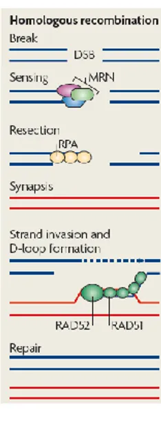
homologous recombination (HR) and non-homologous end joining (NHEJ) (reviewed by Bernstein K.A. and Rothstein R., 2009). In HR, an undamaged DNA strand containing hundreds of base pairs of sequence homology is used as a template to replace an adjacent damaged one. For this reason HR is a high-fidelity mechanism. Because sister chromatids are available as templates upon DNA replication, HR is active mainly in S/G2 phases of the cell cycle and therefore it is precluded in non-cycling cells. In mammals the MRN complex (that consists of Mre11-Rad50-Nbs1) together with the DNA endonuclease CtIP does the initial processing of the DNA ends. Following resection, the exposed ssDNA is coated by replication protein A (RPA), which recruits the Rad52 epistasis group of proteins to enable Rad51 filament formation (Fig. 1). Then, Rad51 facilitates the formation of a physical connection between the invading DNA substrate and homologous duplex DNA template, leading to the generation of heteroduplex DNA (D-loop). Here, Rad51–dsDNA filaments are formed by accommodating both the invading and donor ssDNA strands within the filament. Finally, Rad51 dissociates from dsDNA to expose the 3’-OH required for DNA synthesis using the invading 3’-end as a primer.
The breast cancer susceptibility genes BRCA1 and BRCA2 are also involved in facilitating orderly HR.
Figure 1. Homologous recombination repair. After the recognition of the DNA lesion, the MRN complex is recruited to DSB and generates single-stranded DNA (ssDNA) by resection. ssDNA is coated by RPA and bound by Rad51 and Rad52 leading to homologous template invasion and D-loop formation (modified from Misteli T. et al., 2009).
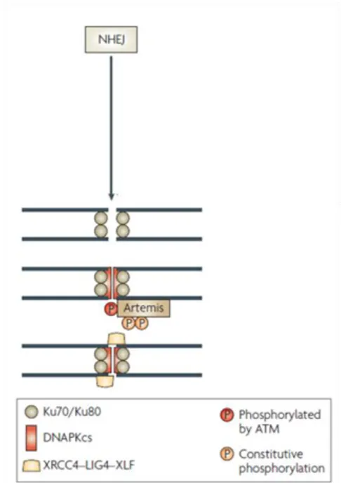
In contrast to HR, NHEJ requires little or no sequence homology for efficient repair and can be error-prone. This mechanism is active during all cell cycle phases and is the predominant pathway for DSB repair in mammalian cells. NHEJ results in minimal DNA end processing and requires at least seven proteins: Ku70, Ku80, the DNA-dependent protein kinase catalytic subunit (DNA-PKcs), Artemis, X-ray cross complementing 4 (XRCC4), XRCC4- like factor (XLF) and DNA ligase IV (LIGIV) (reviewed by Lieber M.R. et al., 2008). The basic mechanism of NHEJ has been worked out in great detail (Fig. 2).
Figure 2. Players of non-homologous end joining. Ku70 and Ku80 form a heterodimer (Ku) which binds to and encircles broken DNA ends. Ku, besides protecting DNA ends from exonucleolytic attack, also recruits DNA-PKcs, a phosphoinositol-3-like family serine/threonine protein kinase. Together, Ku70, Ku80 and DNA-PKcs form the DNA-dependent protein kinase complex (DNA-PK) and the assembly of this trimeric complex on the ends of double-stranded DNA activates the kinase activity of DNA-PKcs. DNA-PKcs, in turn, phosphorylates and activates the nuclease Artemis, which facilitates ‘‘cleaning up’’ of the ends. As a final step, ligation of the broken ends is catalyzed by the trimeric LIG IV complex, which consists of the catalytic core, DNA ligase IV, and its two accessory factors, XLF and XRCC4 (modified from Löbrich M. et al., 2007).
Beside this canonical NHEJ, another mechanism named backup NHEJ (B-NHEJ) (Wang H. et al., 2003; Nussenzweig A. et al., 2007) or microhomology-mediated end joining (MMEJ) (McVey M. et al., 2008) has been described but the mechanism, the regulation and the factors involved in it remain elusive.
Defects in DSB repair lead to premature aging, neurodegeneration and increased cancer susceptibility. Mutations in the HR proteins BRCA1/2 that cause increased breast and ovarian cancers in women have been linked to the accumulation of genetically unstable mammary stem cells (SC) (Liu P. et al., 2008) and mutations in model mice defective in NHEJ components have been shown to display hematopoietic SC (HSC) self-renewal defects (Kenyon J. et al., 2007; Rossi D. J. et al., 2007).
DSB can be lethal to a cell and proliferating/stem cells present an efficient response to this type of damage. The basal level of γ-H2AX foci (a DSB marker) and the expression of several DSB repair genes, as BRCA1, XRCC5 and Rad51, are higher in embryonic stem cells (ESC) when compared to fibroblasts (Tichy E. D. et al., 2008; Maynard S. et al., 2008). Furthermore, ESC are suggested to repair DSB more quickly than mouse 3T3 cells after ionizing radiation exposure (Tichy E. D. et al., 2008).
Proliferating/stem cells mainly rely on HR rather than NHEJ for high-fidelity DSB repair. A comparative analysis of the DSB repair capacity of cycling and non-dividing cells in mouse shows that HR is the major mechanism to repair DSB in ESC with a minimal role played by NHEJ. In contrast, when ESC are induced to differentiate, NHEJ becomes the predominant pathway and HR is reduced. The different use of NHEJ seems to be due to the change of the protein level of DNA ligase IV during the differentiation process. Indeed, the abundance of ligase IV is low in ESC and increased in mouse embryonic fibroblasts (MEF) concomitantly with NHEJ activity (Tichy E. D. et al., 2010). Also in human cells HR is the election pathway to repair DSB and its efficiency decreases when human embryonic stem cells (hESC) are induced to differentiate in neural progenitors and in astrocytes (Adams B.R. et al., 2010). Differently from mouse, hESC also utilize NHEJ, despite the preferential use of HR. The selective requirement for HR or NHEJ during nervous system development has been analysed by an elegant study (Orii K. E. et al., 2006). By using mice carrying a germ line disruption of XRCC2 (HR defective) and DNA ligase IV (NHEJ defective) the two pathways for DSB repair are found to be spatio-temporally distinct: HR inactivation leads to abundant apoptosis in proliferating neural precursor cells and resultant lethality around embryonic days 9-10 (E9-E10), whereas the disruption of NHEJ has deleterious consequences only at later developmental stages (not before E12). Since the late stages of the embryogenesis are characterized by massive differentiation, these results suggest that the HR pathway has an essential protective role against the lethality of DSB in proliferating cells becoming dispensable in post-mitotic cells where NHEJ is the repair pathway of election. To further testify the key role of the NHEJ in differentiated long-lived cells a faster DSB repair kinetics in adipocytes compared to their proliferating precursors is found after exposure to a radiomimetic chemical or ionizing radiation (Meulle A. et al., 2008). In this case, the increased ability of adipocytes to repair DSB is mainly ascribed to the upregulation of DNA-PK expression and activity.
Interestingly, a recent study shows that the induction of NHEJ after H2O2 treatment (working concentration close to concentration in normal brain) in postmitotic neurons requires G0 exit mediated by pRb phosphorylation directed by cyclin C. Abrogation of G0-G1 transition by silencing of cyclin C compromises NHEJ activation while forcing G1 entry determines NHEJ induction even in absence of DNA damage (Tomashevski A. et al., 2010). So, the re-entering into the cell cycle to activate NHEJ can explain the expression of cell cycle markers observed in damaged neurons of normal brain (Schmetsdorf S. et al., 2009).
In the hematopoietic system, the induction of NHEJ in stem cells can be detrimental for genome stability. Hematopoietic development starts from largely quiescent, slowly cycling HSC that, in response to environmental stimuli, are able to give rise to a series of proliferating committed progenitors and mature cells. In contrast to human HSC (hHSC) that undergo apoptosis upon IR exposure, murine HSC (mHSC) survive and repair DNA damage by error-prone NHEJ that can lead to accumulation of gene aberrations. Differently, myeloid progenitors from mouse mainly die after IR treatment and surviving cells use high-fidelity HR (Naka K. et al., 2011; Mohrin M. et al., 2010).
2.2 Nucleotide excision repair
The removal of UV light-induced photoproducts, bulky chemical adducts and intra-strand DNA cross-links depends on nucleotide excision repair (NER). This mechanism consists of three sub-pathways, namely global genome repair (GGR), transcription-coupled repair (TCR) and domain-associated repair (DAR). While GGR operates on the entire genome, TCR and DAR participate in the repair of active genes. In particular, TCR provides efficient repair of the transcribed strand only and DAR repairs both strands of active genes and is detectable only when GGR is attenuated (Nouspikel T., 2009a).
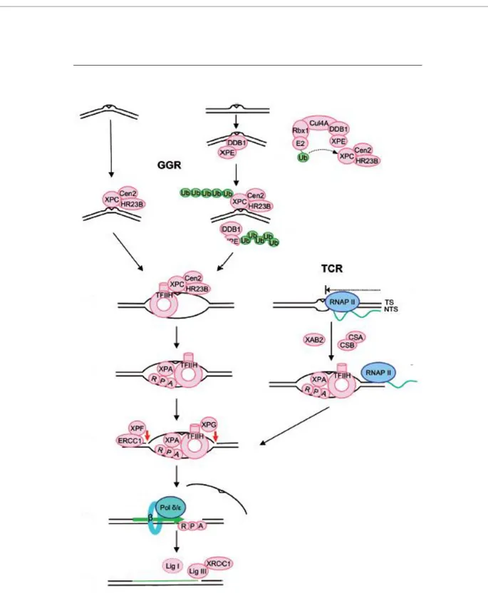
NER is well characterised at mechanistic level and it is known that during GGR the double helix distortion caused by DNA damage is sensed by the heterotrimer XPC/HR23B/Centrin2, with the contribution of the DDB heterodimer for some lesions. In the next step a denaturation bubble is opened around the lesion by the transcription factor TFIIH and later the damaged strand is nicked by XPG on the 3’ side of the lesion and by the heterodimer ERCC1/XPF on the 5’ side. Finally, after the displacement of an oligonucleotide encompassing the lesion, the undamaged strand is used as a template to fill the gap. The TCR mechanism is very similar, differing only in the recognition step that involves RNA polymerase II (RNAP II) rather than XPC and DDB complexes (Fig. 3).
Figure 3. Sub-pathways of NER. In GGR the lesion sensing involves XPC/HR23B/Cen2 whereas in TCR DNA damage is recognized by RNAP II. Subsequently TFIIH opens a denaturation bubble around the lesion, XPG nicks at the 3’ and ERCC1/XPF at the 5’. The resulting gap is filled by DNA polymerase δ or ε. The nick is sealed by ligase III with a minor contribution of ligase I in replicating cells (modified from Nouspikel T., 2009b).
CS proteins associate with UV-stalled RNAPII and are required for efficient TC-NER by recruiting NER factors and chromatin remodelers (Fousteri M. et al., 2008). The importance of functional NER is illustrated by genetic disorders like Cockayne syndrome (CS) and trichothiodistrophy (TTD) defective in TC-NER, and Xeroderma pigmentosum (XP) characterized by GGR defects or total NER deficiencies.
Pioneering studies carried out by Hanawalt and co-workers in several human terminally differentiated tissues, i.e. striated muscle, macrophages and neurons, show that NER is generally attenuated during cell differentiation. More precisely, in terminally differentiated cells GGR is lower than the undifferentiated counterpart whereas, within active genes, not only the transcribed, but also the non-transcribed strand are efficiently repaired (Nouspikel T., 2007; Nouspikel T., 2009a).At molecular level the attenuation of GGR could be ascribed to the incomplete phosphorylation of ubiquitin-activating E1 enzyme Ube1 upon differentiation that could lead to a reduction in the ubiquitination of TFIIH decreasing its activity in GGR (Nouspikel T. et al., 2006). This phenomenon also occurs in human B lymphocytes in which NER at global level is downregulated (Hyka-Nouspikel N. et al., 2011).B lymphocytes are G0-arrested cells that retain the ability to re-enter the cell cycle upon a proper stimulus. Like terminally differentiated cells, in these quiescent B cells a reduction in phosphorylation of Ube1 and GGR impairment is observed, while transcribed genes are efficiently repaired by TCR and DAR. Interestingly, upon proliferation, an increase in phosphorylation of Ube1 and NER recovery is detectable. It is important to note that NER impairment during quiescence could lead to DNA damage accumulation at global genome level that would increase the likelihood of mutation fixation upon proliferative stimuli promoting B cell malignancies or abnormal immune functions (Hyka-Nouspikel N. et al., 2011). The different contribution of NER sub-pathways in protection during differentiation is also detected analysing UV sensitivity of stem cells and differentiated cells (de Waard H. et al., 2008). UV sensitivity of MEF depends on the capacity of perform TC-NER, as shown by marked UV-sensitivity of
Xpa-/- and Csb-/- MEF compared to GGR defective MEF. Conversely, GGR is the main determinant of UV sensitivity in ESC as shown by severe UV sensitivity of Xpa-/- and Xpc-/- ESC compared to Csb-/- TC-NER defective ESC. Altogether these studies suggest that NER downregulation is a common feature in G0-arrested cells, either quiescent or terminally differentiated. It is reasonable that non-dividing cells, which only have to maintain genomic integrity of transcribed genes and preserve tissue specificity, do not need a severe surveillance of the entire genome but an efficient repair of active genes. In contrast, ESC cannot rely on only TCR or DAR because the accumulation of damage and mutations in non-transcribed genes would be detrimental for a proper development and lead to genetic instability.
2.3 Base excision repair
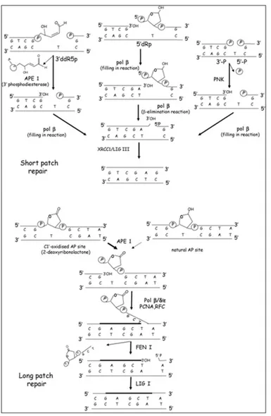
Structurally non-distorting lesions in DNA, such as alkylated, oxidised bases and abasic sites are mainly processed by BER. The first step of BER implies the removal of the damaged base by specific DNA glycosylases which give rise to abasic sites rapidly converted into SSB by an AP endonuclease. After that, the polymerization step involves either the short patch BER (SP-BER) or the long patch BER (LP-BER) pathway that differ for the repair-patch length and the specific players involved (Fig. 4). DNA polymerase β and DNA ligase III are responsible for the filling-in and the sealing step of the SP-BER, respectively, whereas, in LP-BER, these reactions are catalysed by the PCNA-dependent DNA polymerases δ/ε and/or DNA polymerase β and DNA ligase I (Fortini P. et al., 2007).
BER is the repair mechanism of election for oxidative DNA damage that is also produced spontaneously as a consequence of aerobic metabolism. Post-mitotic tissues such as muscle and brain present high metabolism and therefore are particularly subjected to ROS-induced damage. In skeletal muscle the BER efficiency has been addressed by Narciso and coworkers using an in vitro murine system which recapitulates the in vivo differentiation process (Narciso L. et al., 2007). Muscle satellite cells (myoblasts) isolated from mouse thigh are adult stem cells that are able to proliferate and differentiate, in appropriate culture medium, in multinucleated myotubes. A comparative analysis of BER capacity in myoblasts versus myotubes shows a clear impairment of BER in post-mitotic muscle cells. Both the SP-BER and LP-BER sub-pathways are compromised although the LP-BER, which shares several partners with DNA replication, is more severely affected. At molecular level the BER impairment can be ascribed to the nearly complete lack of DNA ligase I and to the strong down-regulation of XRCC1, a scaffold protein known to be essential for DNA ligase III stabilization. Consistently, XRCC1 is a transcriptional target for FoxM1 and E2F1 which activate several cell cycle genes and are both downregulated upon cell cycle exit in myotubes (Fortini P. et al., 2010). In contrast, downregulation of XRCC1 is not observed in post-mitotic neural cells where this enzyme has a protective role against the cytotoxicity induced by oxidative DNA damage (Kulkarni A. et al., 2008). Moreover, XRCC1 seems to be important during neurogenesis since its inactivation leads to loss of cerebellar interneurons and abnormal hippocampal functions (Lee Y. et al., 2009).
Since the mitochondrial genome is a susceptible target for oxidative damage its repair should play an important role particularly in post-mitotic tissues with high metabolism. It has been recently discovered that repair in mitochondria occurs not only by SP-BER but also by LP-BER. The LP-BER is involved in the processing of oxidative lesions (Sung J. S. et al., 2005) and requires the nuclease activity of FEN1 to repair damage both in nuclear and mitochondrial genome (Liu P. et al., 2008; Szczesny B. et al., 2008). FEN1 is not only involved in LP-BER, but it is also essential for DNA replication.
Figure 4. Schematic representation of short patch and long patch BER repair (Fortini P. et al., 2007).
FEN1 is strongly downregulated in terminally differentiated cells. How this might impact on the efficiency of mitochondrial BER in differentiated tissues under normal and pathological conditions is currently under investigation. A comparison of BER efficiency between stem and differentiated cells shows that the former have a higher BER activity and a faster repair after oxidative
stress compared to the latter one. BER proteins such as Ape1, DNA ligase III, PCNA, UNG2 and XRCC1, are expressed in murine ESC at higher level than MEF and this is also true for αOGG1 and Ape1 in hESC after H2O2 treatment (Tichy E. D. et al., 2011; Maynard S. et al., 2008).
If in general DNA repair is downregulated during differentation a notable exception is represented by the immune system where the defective BER observed in monocytes is enhanced during dendritic cell maturation (Briegert M. et al., 2007).The BER defect in monocytes may account for their selective killing during tumor therapy with alkylating agents.
2.4 Mismatch repair
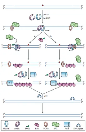
DNA mismatches can occur not only in proliferating cells but also in non-cycling cells because of base deamination or during repair attempts by error-prone DNA polymerases. The key proteins in MMR are highly conserved from bacteria to mammals: in eukaryotes there are multiple homologs of the key bacterial MutS and MutL MMR proteins (Harfe B. D. and Robertson S. J., 2000). Six human mismatch repair genes (MSH2, MSH3, MSH6, MLH1, PMS1 and PMS2) have been identified as components of this repair system that efficiently corrects single base mismatches and loops of one to three extrahelical nucleotides. MMR degrades the section of the error-containing strand (Fig. 5) and therefore provides the DNA polymerase with another chance to generate an error-free copy of the template sequence. MMR is also one of the alternative pathways involved in minimizing the toxic and mutagenic effects of the DNA 7,8-dihydro-hydroxyguanine (8-oxo-dG), a harmful DNA oxidation product causing G>T transversions and implicated in frameshift formation (Colussi C. et al., 2002). The microsatellite instability and the mutator phenotype of MMR-defective cells strongly support a crucial role of MMR in the DDR (Macpherson P. et al, 2005). Moreover, inherited MMR defects are associated with the hereditary nonpolyposis colorectal cancer (HNPCC), an autosomal dominant disorder.
The comparison of the MMR efficiency of dividing cells and differentiated cells is addressed by few studies. MMR has been reported to be active in differentiated neurons (Belloni M. et al., 1999) and as efficient as in undifferentiated cells (David P. et al., 1997; Panigrahi G. B. et al., 2005). In contrast, one report indicates that MMR proteins, namely MSH2 and MSH6, are highly expressed in mouse ESC and MMR is significantly more active in ESC than in MEF (Tichy E. D. et al., 2011).
Figure 5. The reconstituted human mismatch-repair system. The mismatch (red triangle)-bound MutSα (or MutSβ) recruits MutLα leading to the release of the sliding clamp from the site of the mismatch. (a) Clamps diffonding upstream displace RFC and load EXO1 which degrades the strand in a 5’→3’ direction. RPA stabilizes the ssDNA and the gap is filled by PCNA-bound Pol δ. DNA ligase I seals the nick completing the repair process. (b) Clamps migrating downstream encounter a PCNA molecule bound at the 3′ of the strand break. The RFC molecule that is bound at the 5′ side of the break would not be displaced by MutSα, preventing degradation in the 5′→3′ direction. The complex that contains MutSα, MutLα, EXO1, PCNA and RFC would assemble at the 3′ of the nick and mediate degradation towards the mismatch (Jiricny J., 2006).
3. DNA DAMAGE SIGNALLING
DNA damage induces an evolutionary conserved signalling pathway defined as DNA damage response (DDR) that is a network of interacting pathways coordinating repair and cell cycle progression. The DDR involves damage sensors, signal transducers and effectors. DNA damage recognition by the sensor proteins starts a cascade that results in cell cycle arrest and DNA repair and can lead to cell death depending on the type of damage.
Localization of DDR factors to DNA damage sites requires special sensor proteins that directly recognize specific DNA lesions and activate the DDR. A group of proteins, including mediator of DNA damage checkpoint protein 1 (MDC1/NFBD1), 53BP1, Mre11-Rad50-NBS1 (MRN complex) and the Rad9-Rad1-Hus1 (9-1-1) complex have been implicated as DNA damage sensors and recognize DNA strand breaks as well as region of replication stress (Ward I. et al., 2004; Wang B. et al, 2002; Stucki M. et al., 2004; Paull T.T. et al., 2005). Binding of the MRN and 9-1-1 complexes to the sites of DNA damage in chromatin activates the signal transducing kinase ataxia-telangiectasia mutated (ATM), the ATM and Rad3-related (ATR) kinase, and DNA-PK, all of which are members of the phosphoinositide 3-kinase-related kinase family. The transducer kinases respond to different types of stimuli. DNA-PK and ATM are preferentially activated in response to DSB while ATR primarily responds to SSB and stalled replication forks. ATM and ATR are conserved serine/theronine kinases characterized by a C-terminal catalytic motif containing a phosphatidylinositol 3-kinase domain. They share many targets that are phosphorylated by ATM immediately after the appearance of DSB, and later by ATR, which probably maintains these phosphorylations for extended periods of time (Shiloh Y., 2001). Once activated, ATR and ATM amplify the damage signal by phosphorylating several substrates, including key DNA damage response proteins H2AX, BRAC1, NBS1, SMC1, MDC1 and 53BP1, as well as repair proteins Ku70/80, Artemis and Rad-like proteins (Pallis A. G. et al., 2010; Freeman A. K. et al., 2010). DSB and stalled replication forks cause the phosphorylation of histone 2AX at Ser139 (H2AX for the unphosphorylated form and γH2AX for the phosphorylated form) via ATM and ATR respectively. For DSB the distribution of γH2AX within the nucleus is localised to the DSB. These γH2AX foci appear 10 min after DSB induction and over time their amount and intensity decrease due to the activity of phosphatase PP4 (Nakada S. et al., 2008; Chowdhury D., et al., 2008), testifying that DSB repair is occurring. The functional significance of γH2AX is assumed to be a signal that facilitates the repair of free DSB or DSB formed at stalled replication forks, presumably by causing the chromatin to be more accessible for DNA repair.
In order to allow time for DNA repair, ATM and ATR temporarily arrest the cell cycle by phosphorylation and activation of cell cycle checkpoints. To this aim, important phosphorylation targets of ATM and ATR are Chk1 (checkpoint kinase-1), Chk2 (checkpoint kinase-2) and p53. ATM phosphorylates Chk2 after
DSB formation at Thr68 (Zhou B. B. et al., 2000; Matsuoka S. et al., 2000), while ATR phosphorylates Chk1 at Ser345 upon stalled replication forks (Liu Q. et al., 2000; Guo Z. et al., 2000). In turn, Chk2 and Chk1 phosphorylate the transcription factor p53 at Ser20 (Shieh S. Y. et al., 2000). Phosphorylation of p53 at Ser20 does not activate the transcription factor, rather it prevents the proteasomal degradation of p53. Under unphosphorylated conditions, p53 is ubiquitinated by the ubiquitin E3 ligase MDM2 (mouse double minute 2) which targets it for proteasomal degradation. Phosphorylation of p53 by Chk1 and Chk2 prevents this process and results in stabilization of the p53 protein (Chehab N. H. et al., 1999; Chehab N. H. et al., 2000; Hirao A. et al., 2000; Unger T. et al., 1999). ATM and ATR can also phosphorylate p53 at Ser15, thereby increasing its transactivation activity (Banin S. et al., 1998; Canman C. E. et al., 1998).
Upon activation, p53 transcriptionally upregulates the cyclin-dependent kinase inhibitor p21waf1/cip1, which suppresses cyclin E/CDK2 kinase activity and prevents G1 to S phase progression (Deng C. et al., 1995). Phosphorylation of Chk2 or Chk1 by ATM and ATR respectively, leads to the phosphorylation and subsequent ubiquitination of Cdc25A, and in this way prevents the activation of CDK2, resulting in lack of DNA polymerase recruitment to replication origins and thereby blocking the transcription of genes required for S phase progression.
ATM and ATR also regulate the G2/M cell cycle block. Chk1 phosphorylates Cdc25c (cell division cycle 25 homolog C), a dual specificity tyrosine and serine/threonine phosphatase, at Ser216 causing the binding of Cdc25c to the 14-3-3σ protein (Peng C. Y. et al., 1997; Sanchez Y. et al., 1997). Within this complex, Cdc25c is transported out of the nucleus and is thereby unable to dephosphorylate/activate Cdk1, which finally results in G2/M arrest (Dalal S. N. et al., 1999).
Once the process of DNA repair has been successfully completed, cells can re-enter the cell cycle, whereas irreparable DNA damage can lead to irreversible cell cycle arrest, known as senescence, or induction of cell death.
The critical role of DDR for overall tissue integrity and function is well illustrated by the severe clinical consequences observed in both humans and mice for mutations in genes regulating this pathway (Hakem R., 2008; Kerzendorfer C. et al., 2009). Patients with mutations in the kinase ATM present blood vessels abnormalities, cerebellar degeneration, immunodeficiency and increased cancer risk (Hoeijmakers J. H., 2009) whereas ATR mutations cause developmental defects in humans and mice (Hakem R., 2008; Hoeijmakers J. H., 2009; Seita J. et al., 2010).
3.3 DNA damage response in post-mitotic cells
If accumulation of damage occurs in post-mitotic cells it can also contribute to disease progression or lead to loss of tissue homeostasis. Conditions such as neurodegeneration and cardiomyopathy may arise as a consequence of damage in terminally differentiated cells. Whereas proliferating cells adopt the canonical DDR described above, terminally differentiated cells show alterations in the response to DNA damage. This issue has been largely addressed in the muscle and nervous system also because of the availability of well characterized in vitro cell systems suitable for molecular/biochemical characterization. DNA damage induced by the anthracycline antibiotic doxorubicin results in selective decrease in the expression of muscle-specific genes in rat cardiac muscle cells providing the first evidence that DNA damage can block cell differentiation (Ito H. et al., 1990). More recently, a differentiation checkpoint activated by DNA damage was identified in skeletal muscle progenitors that coordinates the repair and the expression of differentiation-specific genes during cell cycle arrest (Puri P.L. et al., 2002). Upon exposure to genotoxic stress the myogenic differentiation is reversibly inhibited while DNA is repaired. The transient phosphorylation of MyoD by DNA damage activated c-abl that inhibits MyoD-dependent transcription of muscle genes is likely to be the key event in the coordination of repair and differentiation. The mechanism of inhibition of muscle gene transcription depends on the cell cycle phase at which myoblasts arrest in response to damage (Simonatto M. et al., 2011). This is in striking contrast with what occurs in proliferating cells exposed to genotoxic agents that differentiate due to the p53-mediated repression of the stem cell marker Nanog (Lin T. et al., 2005).
DNA damage response of post-mitotic cells differs from that of cycling cells because several genes involved in DDR are also cell cycle-related genes that undergo downregulation during terminal differentiation. An example is provided by p53 that is involved in the regulation of differentiation and development. p53 transcripts reach a maximum during differentiation of several tissues but the levels decrease strongly in cells undergo terminal differentiation. In some species the absence of p53 during development leads to dramatic defects in differentiation (Wallingford J. B. et al., 1997). Other instances are represented by myotubes that are deprived of ATR and Chk1 but retain the ability to activate the ATM-Chk2 branch upon radiation damage (Latella L. et al., 2004). In post-mitotic neurons the response to DSB is mediated by ATM (Biton S. et al., 2006; Biton S. et al., 2007). Suppression of the function of ATM attenuates both apoptosis and cell cycle re-entry triggered by some genotoxic compounds (Kruman I. I. et al., 2004). The Chk1/Cdc25A axis participates in the activation of cell cycle-mediated neuronal death (Zhang Y. et al., 2006). A recent study provides evidence that terminally differentiated astrocytes lack functional DDR signalling (Schneider L. et al., 2012). The activation of ATM and its downstream DDR factors are highly inhibited due to repression at
transcriptional level. The transcriptional control is also responsible for the strongly impairment of the axis ATM-Chk2-p53 making astrocytes extremely resistant to irradiation. DNA-PK is actually responsible for IR-induced H2AX phosphorylation in astrocytes whereas in the neural stem cells ATM appears to be the main responsible kinase within a canonical DDR.
4. CELL DEATH
If DNA lesions persist after activation of DDR and DNA repair, proliferating cells can either arrest the cell cycle progression in G1 (cellular senescence) or trigger programmed cell death pathways, such as apoptosis (self-killing) and autophagy (self-eating). Quiescent/post-mitotic cells cannot adopt the replicative senescence or the canonical cell cycle arrest-dependent apoptosis as strategies to get rid of damaged cells but they are provided of alternative strategies (e.g. autophagy).
4.1 Apoptosis
Apoptosis is considered a vital component of various processes including normal cell turnover, proper development and functioning of the immune system, hormone-dependent atrophy, embryonic development and chemical-induced cell death. Apoptosis is a well orchestrated mechanism developed by eukaryotic organisms during evolution process. It is also known as cellular self destruction or cell-suicide or programmed cell death (PCD) (Savitz S. I. et al., 1998). Apoptosis is necessary for normal development and removal of transformed cells (White E., 1996; Weil M. et al., 1996,) and plays a central role in pathogenesis of human disease when the genes controlling the apoptotic process are suppressed, overexpressed or altered by mutation (Pitchard U. et al, 1996).
The mechanisms of apoptosis are highly complex and sophisticated, involving an energy-dependent cascade of molecular events. To date, research indicates that there are two main apoptotic pathways: the extrinsic or death receptor pathway and the intrinsic or mitochondrial pathway. Both of them converge on the same execution pathway. This pathway is initiated by the cleavage of caspase-3 and results in DNA fragmentation, degradation of cytoskeletal and nuclear proteins, crosslinking of proteins, formation of apoptotic bodies, expression of ligands for phagocytic cell receptors and finally uptake by phagocytic cells.
The extrinsic signalling pathways that initiate apoptosis involve transmembrane receptor-mediated interactions. To date, the best characterized ligands and corresponding death receptors include FasL/FasR, and TNF-α/TNFR1. Upon ligand binding, cytplasmic adapter proteins are recruited which exhibit
corresponding death domains that bind with the receptors resulting in the auto-catalytic activation of procaspase-8 (Kischkel F. C. et al., 1995).
The intrinsic signaling pathways that initiate apoptosis involve non-receptor-mediated stimuli that produce intracellular signals that act directly on targets within the cell and are mitochondrial-initiated events. All of these stimuli cause changes in the inner mitochondrial membrane that results in an opening of the mitochondrial permeability transition (MPT) pore, loss of the mitochondrial transmembrane potential and release of two main groups of normally sequestered pro-apoptotic proteins from the intermembrane space into the cytosol (Saelens X. et al., 2004). The first group consists of cytochrome c, Smac/DIABLO, and the serine protease HtrA2/Omi which activate the caspase-dependent mitochondrial pathway. Cytochrome c binds and activates Apaf-1 as well as procaspase-9, forming an “apoptosome” (Chinnaiyan A. M., 1999; Hill M. M. et al., 2004). The second group of pro-apoptotic proteins, AIF, endonuclease G and CAD, are released from the mitochondria during apoptosis, translocate to the nucleus and lead to DNA fragmentation (Joza N. et al., 2001; Li L. Y. et al., 2001; Enari M. et al., 1998). The control and regulation of these apoptotic mitochondrial events occurs through members of the Bcl-2 family of proteins (Cory S. et al., 2002). To date, a total of 25 genes have been identified in the Bcl-2 family which include anti-apoptotic and pro-apoptotic members. These proteins have special significance since they can determine if the cell commits to apoptosis or aborts the process. It is thought that the main mechanism of action of the Bcl-2 family of proteins is the regulation of cytochrome c release from the mitochondria via alteration of mitochondrial membrane permeability.
p53 is considered to be a major player in the apoptotic response to genotoxin. Upon severe damage, p53 is phosphorylated at Ser46 by homeodomain-interacting protein kinase 2 (HIPK2) and subsequently acetylated on Lys382 by the CREB binding protein (CBP). This activation of p53 initiates the expression of its apoptotic target genes such as PUMA, Bax and Apaf-1. p53 also promotes the extrinsic apoptotic pathway by up-regulating Fas receptor and Fas ligand (Petak I. et al., 2000).
These major components of the apoptotic pathway have been well characterized through the use of cell-free biochemical studies and in intact cells using mitotic cell lines. However, recent evidence suggests that apoptosis is regulated very differently between mitotic and post-mitotic cells. Indeed, differently from mitotic cell, the addition of cytochrome c to cytosolic extracts or injection of cytochrome c into the cytosol, is not sufficient to trigger apoptosis in non-cycling cells, such as symphatetic neurons and cardiomyocytes, suggesting that an additional step should be required to induce apoptotic cell death. In post-mitotic cells the X-linked inhibitor of apoptosis protein (XIAP) has been identified as the critical regulator of caspase activation. In symphatetic neurons it has been characterized that the induction of apoptosis relies on both cytochrome c release and XIAP inactivation. In particular, the inhibition of
caspases by XIAP can be overcame by p53-dependent increase in Apaf-1. Consistently, p53 deficient neurons are refractory to relieve the XIAP “brake” and therefore are resistant to DNA damage (Vaughn A.E. et al., 2007). A similar mechanism has been described in myotubes which are resistant to cytosolic cytochrome c microinjection. Importantly, a key role has been attributed to Smac’s release from mitochondria to overcome XIAP and allow cytochrome c to activate caspase and lead to cell death (Smith M.I. et al., 2009).
Moreover, like neurons that are resistant to a variety of stressors, myotubes result radioresistant when compared to myoblasts. The radio-resistance has been molecularly characterized and shown to be due to the uncoupling between ATM activation and p53 phosphorylation, that is required for apoptosis induction (Latella L. et al., 2004).
The restricted apoptosis could be physiologically essential for long term tissue maintenance but it is not clear if this is a general phenomenon in post-mitotic tissues.
4. 2 Autophagy
Autophagy is a physiological mechanism, conserved from yeast to human, which occurs under basal conditions and can be stimulated by stresses such as starvation, various pathologies, or by treatment with pharmacological agents like rapamycin. In addition to its roles in maintaining normal cellular homeostasis by liberating nutrients from macromolecules and by assisting the clearance of misfolded proteins and damaged organelles, autophagy is vital in a range of physiological and pathological situations, including during early embryonic development and neonatal starvation, for the degradation of disease-causing aggregate-prone proteins and in the clearance of pathogenic bacteria. Dysfunction in the autophagy pathway has been implicated in an increasing number of human diseases, from infectious diseases to cancer and neurodegeneration (Mizushima N. et al., 2008).
During autophagy, portions of the cytoplasm, including organelles and proteins, are engulfed by a newly formed membrane, termed a phagophore or isolation membrane, to form a double-membrane vesicle, called autophagosome, which is delivered to lysosomes for hydrolytic degradation (Cecconi F. et al., 2008). More than 30 different genes regulating autophagy (ATG) have been identified in yeast, and many of these have mammalian orthologs. These genes can be grouped according to their functions at key stages of the autophagy pathway: initiation, elongation, maturation, and fusion with the lysosomes. For example, Beclin 1 is important for localization of autophagic proteins to a pre-autophagosomal structure in the initiation step, whereas the conjugation of the microtubule-associated protein light chain 3 (LC3) with the phospholipids of the vacuolar membrane is required during the elongation stage.
In different cell systems, autophagy is activated by depletion of nutrients or lack of growth factors and, according to current views, this is mediated by the mammalian target of rapamycin (mTOR). Autophagy is suppressed by mTOR, which is in turn controlled directly by the level of intracellular amino acids and indirectly by growth factors via Akt/PKB and cell energy status via AMPK (Ma X. M. et al., 2009). However, autophagy can also be induced by mTOR-independent mechanisms (Mordier S. et al., 2000).
Emerging evidence indicates that autophagy has an essential role both in cell differentiation and in the preservation of the homeostasis of cells, tissues and organisms (reviewed by Mizushima N. et al., 2010; Mariño G. et al., 2011). Testifying the essential role of autophagy in cell remodelling during erythroid differentiation, mice lacking Ulk1, an autophagic gene, show an increase in the number of reticulocytes and an impaired clearance of both mitochondria and RNA-bound ribosomes (Kundu M. et al., 2008). Moreover, in vitro and in vivo studies using Atg5-/- mice (autophagy defective) had demonstrated the requirement of autophagy during adipocyte differentiation (Baerga R. et al., 2011).
SIRT-1, a nicotinamide adenine dinucleotide (NAD+)-dependent deacetylase protein with a pleiotropic role in the maintenance of homeostasis following genotoxic stress, is one of the major player involved in the crosstalk between autophagy and cell differentiation (Fulco M. et al., 2003; Picard F. et al., 2004; Aymard E. et al., 2011).
If the analysis of several systemic and tissue-specific knockout models of ATG genes have clarified that autophagy plays an important role in mammalian development and differentiation, its involvement in the response to DNA damage is less clear. In particular, whether it functions as a survival or pro-death mechanism upon genotoxic stress is a matter of debate. However, several reports indicate that autophagy can be seen as an integral part of the DDR. A cross-talk between autophagy and a new cytoplasmic function of ATM in response to oxidative stress has been recently described (Alexander A. et al., 2010a; Alexander A. et al., 2010b). Elevated ROS levels induce ATM-dependent-TSC2 activation through the LKB1/AMPK metabolic pathway inhibiting mTORC1 and thus promoting autophagy. This route is p53-independent and ATM is activated by ROS in the cytoplasm suggesting that this pathway is distinct from the canonical activation of ATM in the nucleus upon DNA damage.
An interesting link between DDR and autophagy also emerges from a recent study in yeast (Robert T. et al., 2011). Lack of DDR and DSB repair is observed upon inhibition of histone deacetylase (HDAC) activity, resulting in hyperacetylation of proteins,. This is due to decreased association of Sae2 and Exo1 (both involved in DNA end resection during HR) with DSB ends and decreased levels of Sae2 protein. Interestingly, inhibition of mTOR activity and thus triggering of autophagy also result in decreased levels of Sae2. This model suggests that severely damaged DNA and associated machinery are removed
from the nucleus via an autophagic process regulated by the acetylation status of key repair proteins. This may be a mechanism for keeping DNA repair enzymes away from cellular DNA that is not damaged, avoiding accidental "repair" of replicating DNA.
Although the role of autophagy in the control of post-mitotic tissue integrity is broadly accepted, its involvement in the maintenance of genome stability during cell differentiation is still not elucidated.
In this study, the characterization of the response to DNA damage and the analysis of basal autophagy during muscle differentiation have been performed in order to investigate a possible involvement of the autophagic pathway in the dealing with genotoxic insults.
II. RESULTS
DNA SSBs accumulate in myotubes but do not lead to cell death
DNA repair capacity in post mitotic cells has been exploited by the use of a skeletal muscle cell differentiation in vitro system in which actively proliferating myoblasts are induced to differentiate in myotubes under appropriate cell culture conditions. A clear decrease in BER capacity of terminally differentiated muscle cells was observed and it was ascribed to the lack of DNA ligase I and the downregulation of XRCC1 affecting both the long- and the short- patch of BER. Consistently, myotubes accumulate DNA single-strand breaks (SSB) upon oxidative stress (Narciso L. et al., 2007). We carried out a further characterization of BER, focusing our analysis on the first enzymatic step of this pathway, which implies the removal of the damaged base by specific DNA glycosylases giving rise to apurinic/apyrimidinic sites rapidly converted into SSB by the action of AP endonucleases and lyases.
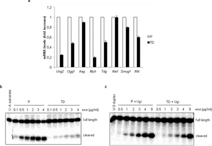
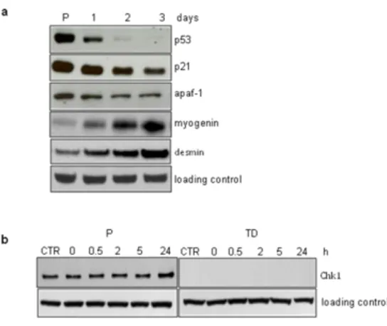
Figure 6. DNA glycosylases are downregulated in terminally differentiated muscle cells. (a) Gene expression analysis of several DNA glycosylases by RT-PCR in proliferating (P) and terminally differentiated (TD) cells. (b) UNG2 activity was measured by an in vitro functional assay. An 32P-end-labeled duplex oligonucleotide (30 mer) which contains a single U:A mismatch was incubated with different concentrations of whole cell extracts (wce) from proliferating (P) and terminally differentiated (TD) cells. (c) SMUG1 activity was measured by an in vitro functional assay. For this activity a 32P-end-labeled duplex oligonucleotide (30 mer) containing a single U:G mismatch was used as substrate and incubated at different concentrations of whole cell extracts (wce) from proliferating (P) and terminally differentiated (TD) cells. For the SMUG 1 activity assay Ugi, a specific inhibitor of UNG2, was added to the reactions. Full length and cleaved products were analysed by polyacrylamide denaturating gel electrophoresis and quantified by electronic autoradiography (Instant Imager). Error bars indicate S.D.
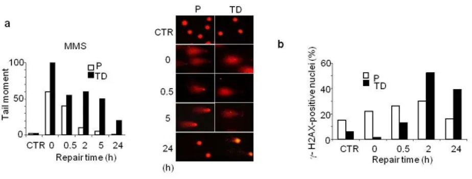
We measured the gene expression levels of different DNA glycosylases and found that Ung2 (uracil-DNA glycosylase2) and Myh (human mutY homolog) were strongly downregulated in post-mitotic muscle cells compared to the proliferating counterpart (Fig. 6a). This was in agreement with the involvement of these two enzymes in the removal of lesions formed during S-phase. The activity of UNG2, responsible for uracil removal from U:A mismatch, was also impaired in myotubes as demonstrated by a functional assay with a specific substrate (Fig. 6b), further confirming a clear down-regulation of this cell cycle-associated glycosylase in terminally differentiated cells. The presence of uracil in the genome can be due not only to replication errors, but also to spontaneous deamination of cytosine leading to U:G mismatch. The importance of ensuring uracil removal is testified by the maintenance of functionality of SMUG1 (an uracil DNA glycosylase which removes uracil from ssDNA during transcription) in non-cycling cells such as myotubes (Fig. 6c). In agreement with BER impairment in terminally differentiated cells, the exposure to methyl methanesulfonate (MMS), an alkylating agent that induces SSB as intermediates during the BER process, led to DNA lesions accumulation (Fig. 7a) which was mirrored by phosphorylation of H2AX (Fig.7b).
Figure 7. Terminally differentiated muscle cells accumulate DNA SSB after MMS exposure. (a) Left: Myoblasts (P) and myotubes (TD) were exposed to MMS (1mM, 30 min) and DNA SSB were analyzed by the comet assay at different repair times. The average of the tail moment, calculated by multiplying the total intensity of the comet tail by the migration distance from the centre of the comet head, was used as measure of DNA damage. At least 100 cells per experimental point is shown. Right: Microphotographs of proliferating (P) and terminally differentiated (TD) cells subjected to the comet assay and stained with ethidium bromide. Representative untreated control cells (CTR) and after different post-treatment times are shown. Experiments were repeated twice and one representative experiment is shown. (b) DNA damage induction and repair as detected by γH2AX foci formation after exposure to MMS (3mM, 30 min) in proliferating (P) and terminally differentiated (TD) cells.
These data strongly suggest that in postmitotic cells which are definitely impaired in replication ability, the phosphorylation of H2AX is induced by BER
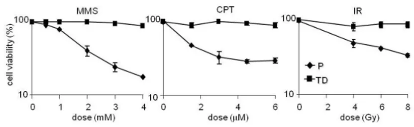
intermediates per se without any replication-dependent turning into DSB. To better understand the fate of persistent SSB in the genome of post-mitotic muscle cells, proliferating and terminally differentiated cells were exposed to different DNA damaging agents, such as MMS and camptothecin (CPT), a Topoisomerase1 (TOP1)-linked SSB inducer (Pommier Y. et al., 2010). Cell survival was measured by counting metabolically active cells. At doses that killed over 50% of the myoblasts, myotubes were resistant to the toxic effects of DNA SSB (Fig. 8).
Myotubes were also resistant to H2O2 (data non shown) but the comparison with myoblasts was hampered by the induction of differentiation upon H2O2 exposure (Kuster G. M. et al., 2010). Terminally differentiated muscle cells are therefore not only resistant to IR (Fig. 8; Latella L. et al., 2004) but also to SSB-inducing agents.
Figure 8. Myotubes are resistant to the toxic effects of SSB inducers and IR. Proliferating (P) and terminally differentiated (TD) cells were exposed to different doses of MMS, CPT (30 min treatment and 3 h respectively) and IR and cell survival was measured by counting metabolically active cells as measured by CCK-8 72 h after treatment. Error bars indicate S.D.
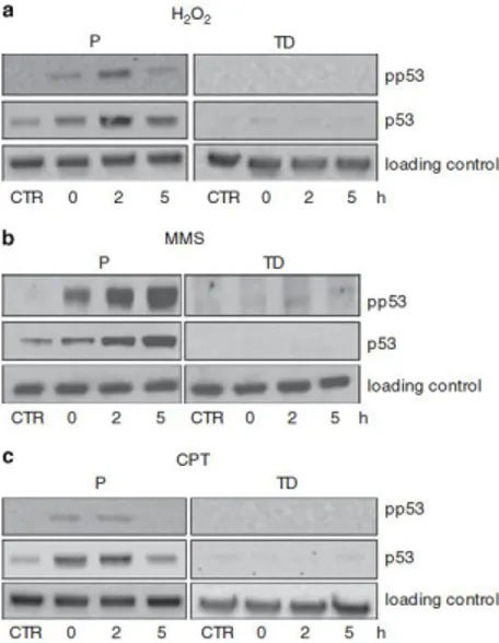
γ-H2AX response is activated in myotubes upon SSB induction and ATM is the main kinase involved
Oxidative (Narciso L. et al., 2007) and alkylation (Fig. 7b) damage is able to activate the γH2AX response in post-mitotic muscle cells. H2AX can be phosphorylated by ATM, DNA-dependent protein kinase (DNA-PK) or ATR. In myotubes ATR is missing but either ATM or DNA-PK could play this role. To identify the candidate kinase, myotubes were pre-treated with either an ATM (KU55933) or a DNA-PK kinase (NU7441) inhibitor and the induction of phosphorylated H2AX was monitored in response to H2O2 or MMS by both immunofluorescence and western blotting analysis (Fig. 9).
Interestingly, whereas the H2O2-induced H2AX phosphorylation was fully suppressed by the ATM inhibitor, it was not affected by NU7441 (Fig. 9a). Notably, when this strategy was used after exposure to MMS, ATM resulted to