Vol.:(0123456789)
1 3
Journal of Thrombosis and Thrombolysis https://doi.org/10.1007/s11239-020-02067-x
Clinical worsening despite intravenous thrombolysis in acute ischemic
stroke secondary to carotid plaque rupture
Fabrizio Sallustio1,2 · Domenico Samà1 · Alfredo Paolo Mascolo1 · Federico Marrama1 · Mauro Fresilli3 ·
Marina Diomedi1
© Springer Science+Business Media, LLC, part of Springer Nature 2020
Abstract
First-line therapy of acute ischemic stroke is intravenous thrombolysis (IVT) irrespective of etiology. We report on a patient with acute ischemic stroke secondary to carotid plaque rupture who experienced plaque thrombosis and marked clinical worsening despite IVT. While the latter is the gold standard therapy optimal platelets inhibition should be guaranteed to allow a safe as possible carotid intervention. Hereby we discuss all available strategies to be considered in order to better individualized treatment decision-making.
Case report
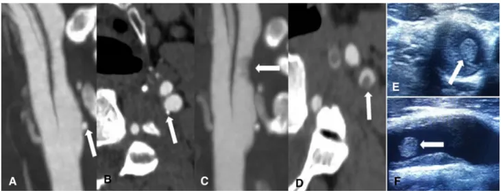
A 70-year-old right handed man presented with sudden onset of right arm weakness. Vitals were normal. Past history was unremarkable except for carotid atherosclerosis. Neuro-logical examination revealed a National Institute of Health Stroke Scale (NIHSS) of 3 (disabling and worsening right arm weakness). Laboratory tests including coagulation test showed a LDL-cholesterol of 139 mg/dl without any other abnormality. CT scan of the head ruled out any intracranial bleeding or early ischemic changes and CT-angiography (CTA) of extra and intracranial arteries revealed an eccentric plaque with surface ulcer causing medium-grade stenosis in the first segment of the left internal carotid artery (ICA) and patency of the remaining examined arteries. Full dose intravenous recombinant tissue plasminogen activator (rtPA) was started 1 h and 30 min after symptom onset. Neuro-logical status remained unchanged until 6 h after the end of treatment when severe aphasia and severe right hemiparesis occurred (NIHSS = 15). A new CTA depicted lack of central
contrast filling in the left ICA extending distally for 1–2 cm suggestive of floating thrombus with patent intracranial ves-sels, confirmed by ultrasound duplex sonography (Fig. 1). Stenting was considered at high risk of embolization and intracranial hemorrhage. 24 h after intravenous thrombolysis (IVT) CT scan of the head ruled out hemorrhagic transfor-mation and 125 mg acetylsalicylic acid and 4000 IU low-molecular weighted heparin daily for prevention of deep vein thrombosis were started. MR-diffusion weighted imaging showed a plenty of recent very small ischemic lesions in the frontal and a larger one in the parietal cortex of the left side. After clinical improvement, on day 4 carotid endarterectomy (CEA) was performed without periprocedural complications and on day 7 the patient was transferred to the rehabilitation unit with an NIHSS of 3.
Discussion
Unstable carotid plaque as the cause of acute stroke repre-sents a therapeutic dilemma. IVT remains the only unavoid-able and undelayunavoid-able option because of its efficacy in the first 4, 5 h of stroke onset [1]. In the presented case rtPA did not result in effective platelets aggregation inhibition as demonstrated by clinical worsening with appearance of detectable thrombus on CTA. Further, rtPA caused procras-tination of antiplatelets and CEA resulting in increased risk of recurrence.
Immediate deposition of activated platelets occur at the site of endothelial disruption [2]. The entity of this
* Fabrizio Sallustio [email protected]
1 Comprehensive Stroke Center, Department of Systems
Medicine, University of Tor Vergata, Viale Oxford 81, 00133 Rome, Italy
2 Neurorehabilitation Unit, Santa Lucia Foundation, Via
Ardeatina 306/354, 00142 Rome, Italy
3 Division of Surgery, University of Tor Vergata, Viale Oxford
F. Sallustio et al.
1 3
phenomenon depends on thrombogenicity of the damaged surface (i.e. content of thrombin within the mural throm-bus), the ability of the endogenous fibrinolytic system to limit the thrombus formation and the severity of stenosis [3]. Thus acute thrombosis could be theoretically counteracted by platelet inhibition and fibrinolysis. In our case, plate-let inhibitors had to be procrastinated for 24 h after IVT despite evidence of superimposed thrombosis. In a model of coronary thrombosis with endothelial disruption platelet receptor GIIb/IIIa inhibitor eptifibatide (Ep) in combination with rtPA was able to make a recanalized artery resistant to platelet recruitment, new thrombus formation and reocclu-sion. The same study demonstrated that Ep alone and rtPA alone were not able to neutralize the thrombogenicity of a damaged vessel wall with evidence of residual erythrocyte or platelet-rich mural thrombi respectively [4]. A single-arm prospective open-label study called the combined approach to lysis utilizing Ep and recombinant tissue-type plasmino-gen activator in acute ischemic stroke (CLEAR) full dose regimen showed a comparable safety of this approach with historical rates of symptomatic intracranial hemorrhage with rtPA alone [5]. We described a case of acute ischemic stroke secondary to unstable carotid plaque with superimposed giant floating thrombus successfully treated with intravenous unfractionated heparin (UFH) and delayed CEA [6]. In the present case intravenous UFH to maintain partial thrombo-plastin time at 1.5–2 times control was considered unsafe.
Carotid artery stenting (CAS) could be another valid option in cases without floating thrombus since comparable outcomes between CAS and CEA in skilled centers have been reported [7].
We conclude that in case of minor stroke secondary to plaque rupture, even without clear evidence of floating
thrombus, strategies (both pharmacological and interven-tional) other than IVT should be kept in mind as part of the therapeutic armamentarium in the concept of a more personalized medicine.
References
1. Powers WJ et al (2018) Guidelines for the early management of patients with acute ischemic stroke: a guideline for healthcare professionals from the American Heart Association/American Stroke Association. Stroke 49:e46–e110
2. Friedman M (1971) The coronary thrombus: its origin and fate. Hum Pathol 2:81–128
3. Fuster V et al (1992) The pathogenesis of coronary artery disease and the acute coronary syndromes. N Engl J Med 326:242–250 4. Rubenstein MH et al (2004) Short-term intravenous eptifibatide
infusion combined with reduced dose recombinant tissue plas-minogen activator inhibits platelet recruitment at sites of coro-nary artery injury. J Am Coll Cardiol 43:287–294. https ://doi. org/10.1016/j.jacc.2003.08.039
5. Adeoye O et al (2015) Combined approach to lysis utilizing eptifi-batide and recombinant tissue-type plasminogen activator in acute ischemic stroke-full dose regimen stroke trial. Stroke 46:2529– 2533. https ://doi.org/10.1161/STROK EAHA.115.01026 0
6. Sallustio F et al (2011) Floating carotid thrombus treated by intra-venous heparin and endarterectomy. J Vasc Surg 53:489–491.
https ://doi.org/10.1016/j.jvs.2010.08.014
7. Rocco A et al (2018) Carotid artery stent placement and carotid endarterectomy: a challenge for urgent treatment after stroke-early and 12-month outcomes in a comprehensive stroke center. J Vasc Interv Radiol 29:1254–1261. https ://doi.org/10.1016/j. jvir.2018.03.025
Publisher’s Note Springer Nature remains neutral with regard to
jurisdictional claims in published maps and institutional affiliations.
Fig. 1 CT angiography showing plaque ulceration (white arrow) of left internal carotid artery in longitudinal and axial views soon after patient admission (a, b) and plaque thrombosis (white arrow) 6 h
after intravenous thrombolysis in longitudinal and axial views (c,
d). The same findings of (c, d) were detected on ultrasound duplex