BIOS – Research Doctorate School in BIOmolecular Sciences PhD Course in Experimental and Molecular Oncology University of Pisa
Research Doctorate School in Biomedical and Pharmaceutical sciences
PhD Course in Biomedical and Pharmaceutical sciences Université de Liège
XXII cycle (2007 – 2009)
PhD Thesis
IDENTIFICATION OF NEW POTENTIALLY ACCESSIBLE
BIOMARKERS SUITABLE FOR THE DEVELOPMENT OF
THE TARGETED THERAPY OF GLIOBLASTOMA
MULTIFORME
Candidate: Davide Musmeci
Tutor, University of Pisa: Prof. Generoso Bevilacqua Tutor, University of Liège: Prof. Vincent Castronovo
Table of contents
Summary………..2 Acknowledgments..………..4 Preface…………....………..5 Section I – Introduction …..………..6 1. Cancer disease………..4 1.1 - Pathogenesis of cancer……….71.2 - Molecular Basis of Cancer Progression………...9
1.3 - Morphological basis of cancer progression………10
1.4 - Cancer therapy and its limitations………..11
2. Malignant Brain Tumors………13
2.1 - Epidemiology………13
2.2 - Malignant gliomas……….15
2.2.a – Astrocytomas………...17
2.2.b - Oligodendrogliomas and mixed oligoastrocytomas………18
2.2.c – Ependymomas……….18
2.3 - Clinical aspect ………...19
2.4 - Molecular pathology of malignant astrocytoma progression……….19
2.5 - Pathological markers of glioma………..22
2.5.a - 1p/19q loss.………..23
2.5.b - MGMT methylation status………...23
2.5.c - EGFR pathway alterations………...24
2.6 - Glioblastoma: a cancer without hope ………..……..25
2.7 – de-novo and secondary glioblastoma………..…...26
2.8 - Therapeutic strategies available: benefits and pitfalls………28
3. Targeted Tumor Therapy………....31
3.1- General aspect ………31
3.2 - Ligands for targeting applications………..33
3.2.a – Antibodies………...33
3.2.b – Peptides………..34
3.2.c - Small organic molecules……….35
3.3 - Effectors for antibody-based tumor therapy………..35 3.3.a – Immunoglobulins………36 3.3.b - Chemotherapeutic drugs………..36 3.3.c – Toxins………..36 3.3.d – Radionuclides………..37 3.3.e – Cytokines……….37 3.3.f - Photosensitizers ………...38
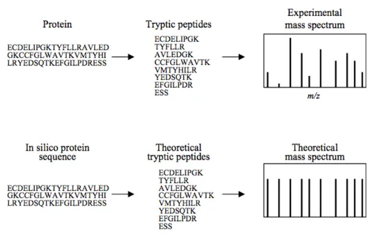
4. Proteomics approach for cancer biomarker discovery………...39
4.1 – General aspect………39
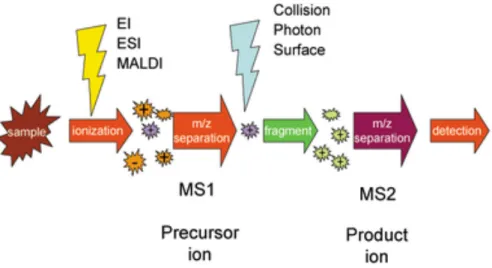
4.2 - Mass Spectrometry (MS) ………...41
4.3 - How does mass spectrometers work? ………43
4.4 - Building up a proteomic experiment………..45
4.4.a - Sample preparation……….45
4.4.b - Protein separation………46
4.4.c - Peptide and Protein Identification………46
4.5 - Electrospray Ionization………..47
Section II – Aim of the work……….………..50
Section III – Material & Methods……….……….51
5. Material and Methods ……….52
5.1 - Tissue harvesting ………..52
5.1.a - Human specimens………52
5.1.b - Cell culture and mouse model of GBM ………..52
5.1.c - Chorioallantoic membrane (CAM) assay ………53
5.2 - Tissue biotinilation and protein extraction ………53
5.3 - Mass Spectrometry analysis ………..54
5.4 - Spectra deconvolution and data processing ………...54
5.5 - Determination of protein origin ……….55
5.6 – Immunohistochemistry ……….56
5.7 - Intravenous injection of chicken embryo ………..56
5.8 - Immunofluorescence ………....58
6. Results originating using the murine model of glioblastoma………...60
6.1 - Sample processing, mass spectrometry and data analysis ………60
6.2 - Determination of mouse origin……….…….65
6.3 – Immunohistochemistry……….65
7. Results originating by human glioblastoma samples and U87-derived tumors…………...68
7.1 - Tissue processing………..68
7.2 - Validation phase ………...72
7.3 - Immunohistochemistry of CD44 and Collagen VI alpha-1 ……….72
7.4 - in-vivo validation of human CD44H and Collagen VI alpha-1………74
Section V – Discussion……….………77
Section VI – Conclusion and Perspectives……….………83
6.1 – Conclusions……….……….….84
6.2 – Perspectives……….……….……….85
Summary
Background: Glioblastoma multiforme (GBM) is the most aggressive, highly invasive and neurologically destructive among malignant brain tumors. None of the treatments currently used is effective, making GBM lethal within 12 months from diagnosis. Despite major evolution in the understanding of the molecular mechanisms involved during GBM development and progression, patients are yet in need of a successful treatment. Nowadays, one of the most promising approaches aimed to cure cancer consists on the antibody-based therapy. The systemic delivery of specific antibodies, coupled with highly cytotoxic drugs, directed towards the tumor site is considered as a promising way of treatment. Unfortunately, the bottleneck of such approach consists in the identification of antigens accessible by the blood stream.
Aim of the work: This work aims to identify and validate in-vivo new biomarkers produced either by the tumor itself or by the surrounding stroma in response to tumoral demand, which are reachable by a systemic administered compound and could be used as therapeutic targets in human GBM. Material and methods: Human GBM specimens, two different human glioblastoma cell lines (U373 and T98G) grafted in nude mice and U87-derived tumors grown in egg choriallantoic membrane (CAM) were biotinilated ex-vivo. Labeled proteins were successively isolated using affinity chromatography based on streptavidin beads. Proteins were eluted and digested with trypsin and the resulting peptides were analyzed using the 2D nano-LC MS/MS technique. Protein identification was carried out using the Mascot® search engine (Matrix Sciences, Boston, MA, USA) and the Swisprot® protein database (Swiss Institute for Bioinformatics, Basel, Switzerland). For the purpose of validation U87-derived tumors were grown on CAM. Intravenous administration of specific monoclonal antibodies into the CAM vessels was carried out in order to validate in-vivo the accessibility of the target.
Results and conclusion: expression profiles for each experimental model were determined using the Mud-PIT technique. About 30 to 35% of the total proteome were found to be accessible (membrane associated, extracellular and secreted proteins). Among the proteins identified, the study highlighted the hyaluronan receptor CD44 and tenascin-C (onco-fetal Tenascin) already known to be overexpressed in human GBM along with other new potential targets such as sparc-like 1, prosaposin and collagen 6 α1. The validation phase carried out using immunohistochemical analysis confirmed the overexpression of the proteins in high-grade gliomas. Additionally, in-vivo
experiment with the systemic administration of the monoclonal anti-human CD44 and COL6α1 antibodies into CAM vessels resulted in a site-specific tumor accumulation of the antibodies suggesting these proteins as readily accessible target for the treatment of GBM. Taken together, these results demonstrate the potential of the biotinilation technique in searching for potential accessible biomarkers. The study pointed at the usefulness of the CAM system as an alternative model of biomarker validation in comparison to the more cost and labor intense mouse model. Further investigations focusing on the development of antibody-based treatment of tumor bearing animals are the next step to envision before proposing these targets for clinical trials on humans.
ACKNOWLEDGEMENTS – RIGRAZIAMENTI – REMERCIEMENTS
Ringrazio calorosamente il Professor Generoso Bevilacqua per la sua disponibilità, la sua simpatia, la sua professionalità, il suo affetto e il costante aiuto fornitomi durante questi quattro anni di duro lavoro. Grazie per aver sempre creduto in me e per avermi dato la possibilità di fare un’esperienza di vita unica, che mi ha dato non solo la possibilità di crescere professionalmente ma anche di maturare come uomo.
Je remercie le Professeur Vincent Castronovo, qui m’a offert la possibilité de poursuivre mes recherches au sein de son laboratoire.
Je remercie aussi toute l’équipe du laboratoire de recherche sur le métastases et tous les gens rencontrés au CHU pendant ces années, pour leur amitié et leur suggestions très importants pour ma réalisation professionnelle.
Thanks to Dr. Andrei Turtoi for his aid in developing my research projects.
Un très gros merci à Vincent et Yannick, collègues avec lesquelles j’ai partagé une grande partie de ma vie en Belgique et sans lesquelles aujourd’hui je ne parlerais pas aussi bien le français.
Sul fronte italiano, un particolare ringraziamento è di dovere al Dott. Paolo Viacava, che mi ha iniziato in questo cammino verso la ricerca scientifica, al Professor Giuseppe Naccarato e al Dott. Giovanni Fanelli per la loro continua disponibilità, competenza, e affezione mostrate nei miei confronti.
Grazie ai miei nonni, Filippo, Pina e Carmelina, parte integrante della mia famiglia, per la loro continua e costante presenza nella mia vita nonostante la notevole distanza che ci separa.
Grazie anche ai miei fratelli Simona e Roberto, cosi come a tutti quegli amici che, anche senza alcun legame di sangue, sono diventati parte integrante della mia famiglia sostenendomi e spronandomi nei momenti di bisogno, guadagnando cosi un posto fisso nel mio cuore. Vi voglio bene Vincenzo, Anna Paola, Massimo e Sara, con la neo arrivata Olivia.
Un merci particulier à Sarah, pour être arrivée au bon moment. Ad maiora semper!
Infine, un grazie non basterebbe per i miei genitori Giuseppe e Lucia, ai quali quindi dedico questa tesi. Vi sono riconoscente per avermi dato la possibilità di arrivare fino a questo punto, sostenendo sempre le mie scelte con amore e passione, in ogni momento della mia vita.
Preface
Thorough the centuries cancer became one of the major health problem of all developed country, only second to cardiovascular disease in terms of death rate. A comprehensive estimation of the incidence and mortality rates from cancer can be extracted from the GLOBOCAN project of International Agency for Cancer Research (http://globocan.iarc.fr/). Over a population of 6.7 milliards peoples, in 2008, more than 12 millions new cancer cases were estimated. That led to the death of more than 7 million peoples all over the world. In Europe, cancer caused approximately 1.7 millions death in 2008, which means 4.700 death per day. In other words, approximately 196 people are dying for cancer each hour, in all European territory. Moreover, as the incidence of cancer is higher in elderly people, probably due to the accumulation of mutation within lifetime, it is expected that the current demographic shift to higher age in developed countries will further increase this major health burden of 22% in 2015.
High-grade gliomas are the most common primary brain tumor in adults. Among them glioblastoma multiforme (GBM) is the most aggressive. The current therapy for these tumors is based on maximum safe and secure surgical resection of the neoplastic mass followed by concurrent local radiation and adjuvant chemotherapy with Temozolomide. However, the ability of GBM cells to migrate through the narrow brain parenchyma, sometimes for relatively long distances, makes them elusive target for effective surgical removal and give to patients a life expectancy that rarely exceeds 12 months, which drops down to 3-4 months in case of no treatment. These data clearly indicate that major efforts are needed in cancer research, and especially in the filed of GBM, in order to fight this lethal disease.
This thesis is focused on the development of a methodological and technological approach able to identify new biomarkers potentially useful as target for new molecular drugs. The research activity necessary to the generation of this work was carried out at the Metastasis Research Laboratory (MRL) of the University of Liege, Belgium, in collaboration with the Division of Surgical, Molecular and Ultrastructural Pathology of the University of Pisa, Italy.
Section I - Introduction
1. Cancer Disease
1.1 - Pathogenesis of cancerNeoplasia or neoplasm or tumor literally means the process of "new growth”. A tumor can be benign or malignant. Malignancy is the ability of a cancer cell population to invade the surrounding stroma and to give origin to metastasis. Cancer is the common term used for all malignant neoplasms. A neoplasm originates as the result of hereditary or somatic alteration in genes that control crucial biological process. These genetic changes allow excessive and unregulated cell proliferation that further becomes autonomous and independent of physiologic growth stimuli. In this way, the entire population of cells within a neoplastic mass usually arises from a single cell that has incurred genetic changes, hence tumors are said to be clonal. All neoplasms, benign or malignant, are made up of two basic components: i) proliferating neoplastic cells that constitute their parenchyma and ii) supportive stroma, made up of extracellular matrix tissue and a huge network of blood vessels. Although parenchymal cells represent the proliferating "cutting edge" of neoplasms, and so determine their behavior and pathologic consequences, the growth and the evolution of tumors are critically dependent on the stroma supply.
As previously said, nonlethal genetic alterations are the key of the carcinogenesis process. These genetic damages, more precisely called mutations, generally hit tumor promoting and/or tumor suppressor genes. The number of mutations needed to produce the initiation of a neoplastic event may vary from one to several. An example that could better elucidate this theory is represented by the chronic myeloid leukemia (CML) where a chromosomal translocation that involves a piece of chromosome 22 being lost is reported. This was first observed by Nowell and Hungerford, who named this small chromosome the Philadelphia chromosome. Only later it was shown that this alteration was resulting from the reciprocal translocation between chromosomes 9 and 22, which produces the chimeric protein called Bcr/Abl, a constitutively active tyrosine kinase promoting cell proliferation. Therefore, CML appears to be triggered by this one-hit event, and this is probably the reason why the drug Gleevec, which targets the Bcr/Abl kinase, is effective as a single agent in CML. However, progression to a full-blown invasive metastatic cancer requires almost always multiple hits. This is the case of most adult solid cancers such as colon, lung, breast and prostate. A model of cancer progression is well represented by colon cancer, for which at least five hits appear to be required to produce an invasive carcinoma (Figure 1).
Because of genetic instability, a characteristic of most solid cancers, many genetic alterations are frequently accumulated during cancer progression. Thus, in contrast to single genetic defect cancers such as CML, the prospect of finding effective single therapeutic agents is unlikely for most solid
tumors. Multiple aberrant cell signaling pathways will need to be inhibited to achieve effective chemotherapeutic regimens. However, if there are identifiable time intervals between the multiple hits that lead to cancer, perhaps detectable by early screening for surrogate markers of progression, there may be a window of opportunity for preventive agents [Raymond W.R. 2007].
Figure 1: Progression from normal epithelium through adenoma to colorectal carcinoma is characterized
by accumulated abnormalities of particular genes. [ Davies RJ 2005]
1.2 - Molecular Basis of Cancer Progression
Heritable genetic mutations, passed down to the progeny of cancer cells, lie at the heart of carcinogenesis. Such mutations may be acquired by the action of environmental agents, such as chemicals, radiation, or viruses, or they may be inherited in the germ line. The term "environmental," used in this context, involves any acquired defect caused by exogenous agents or endogenous products of cell metabolism. Not all mutations, however, are "environmentally" induced. Some may be spontaneous and stochastic.
Two classes of regulatory genes are the principal targets of genetic damage: i) the growth-promoting protooncogenes and ii) the growth-inhibiting tumor suppressor genes. Mutations on protooncogenes are considered dominant because they transform cells despite a normal allele is preserved. Protooncogenes can be activated by increasing the synthesis of their corresponding protein, in normal form, or by alteration of the corresponding protein function, through gene mutation. In contrast, both normal alleles of the tumor suppressor genes must be damaged for transformation to occur. The loss of function of tumor suppressor gene caused by damage of a single allele is called haploinsufficiency. In addition to these two classes of genes, DNA repair genes affect indirectly cell proliferation or survival by influencing the ability of the organism to repair nonlethal damage in other genes, including protooncogenes, tumor suppressor genes, and genes that regulate apoptosis. Hence, disabilities in the DNA repair genes predispose to genome mutations accumulation and lead to neoplastic progression.
At the phenotypic level a malignant neoplasm has several attributes, such as uncontrolled and excessive growth, local invasiveness, and the ability to form distant metastases. These characteristics are acquired in a stepwise fashion during a phenomenon called tumor progression. For such reasons, the process of carcinogenesis is considered as a multistep process both at the phenotypic and the genetic levels.
Three are the main step characterizing tumor progression: initiation, promotion, and progression. Initiation can occur after a single, brief exposure to a potent initiating agent. The actual initiation events leading to transformation into a dormant tumor cell appear to occur within one mitotic cycle. Furthermore, initiation appears to be irreversible; the promoting agent can be given for up to a year later and a high percentage of tumors will still be obtained. Thus, the initiation phase only requires a small amount of time, it is irreversible, and it must be heritable because the initiated cell conveys the malignant alteration to its daughter cells. All these properties are consistent with the idea that the initiation event involves a genetic mutation, although other ‘‘epigenetic’’ explanations are possible. The promotion phase, by contrast, is a slow, gradual process and requires a more prolonged exposure to the promoting agent. Tumor promotion represents a cell proliferation phase
that propagates the initiated damage and leads to the growing of an altered clone of cells. Promotion occupies the greater part of the latent period of carcinogenesis; it is at least partially reversible and can be arrested by certain anti-carcinogenic agents. Finally, the progression stage of carcinogenesis is an extension of the tumor promotion stage and results from it in the sense that the cell proliferation caused by promoting agents allows the cellular damage inflicted by initiation, to be propagated, and the initiated cells to expand clonally. During the process of cancer progression loss of growth control and escape from host defense mechanisms become predominant phenotypic traits. At the molecular level, progression results from accumulation of genetic lesions that in some instances are favored by defects in DNA repair genes. Genetic instability, indeed, is the hallmark of the progression phase of carcinogenesis and leads to chromosomal translocations and aneuploidy that are frequently seen in cancer cells. Such alterations in the genome of the neoplastic cell during the progression phase lead to the increased growth rate, invasiveness, and metastatic capability of advanced cancer. Evidences for multistage induction of malignant tumors have been observed for mammary gland, thyroid, lung, and urinary bladder and in cell culture systems, thus it seems to be a general phenomenon.
[Robbins & Cotran 2005]
1.3 - Morphological basis of cancer progression
Malignant tumors range from well differentiated to undifferentiated. Malignant tumors composed of undifferentiated cells are said to be anaplastic. Lack of differentiation, or anaplasia, is considered a hallmark of malignant transformation. Anaplasia literally means "with no shape", the absence of any differentiative character. However, an increasing amount of literature suggests that most cancers do not represent "reverse differentiation" of mature normal cells but, in fact, arise from stem cells (known also as cancer initiating cells) that are present in all specialized tissues. As said before, cancer progression is the consequence of an uncontrolled cell proliferation from one hand and of the accumulation of genetic alterations on the other hand. The increase of these two parameters causes a series of modifications in the shape of tumor cells and in the architecture of the tissue to which these cells belong, namely:
- atypia: nuclei become larger and darker, with abnormal shape, with a prominent nucleolus. These changes are respectively due to the fact that there is an increase in DNA quantity, modifications of the nuclear proteins, a higher protein synthesis.
- pleomorphism, polymetrism and polychromatism, due to the fact the atypical characters can be of different entity in the different tumor cell clones.
- presence and number of mitosis: sign of entity of the proliferation activity of the neoplastic cell population.
These characters, reflecting the degree of severity of the disease, are very useful for grading a malignant tumor in scales that can be different according to the type of neoplasm, that in any case represent an important prognostic parameter. For instance, as explained later, their morphology allows gliomas to be grouped in four classes, with an increase in their malignant behavior moving from low to high grades.
[Robbins & Cotran 2005].
1.4 - Cancer therapy and its limitations
Classical anti-cancer therapy, including chemotherapy and irradiation, often suffers from poor selectivity and, thus, from severe toxic side effects for healthy tissues. Many therapeutic protocols for solid cancer count on maximal surgical resection of the neoplastic lesion combined with successive irradiation and/or chemotherapy, aimed to slow down the fast proliferating cancer cells. However, cell proliferation occurs also in healthy organs such as the spinal cord, mucosa, hair follicles, bone marrow, and during pregnancy. Since the vast majority of tumor cells have to be killed to achieve the complete remission of the patient, the application of high doses of drugs is required, at the expenses of severe toxic side effects. Furthermore, most chemotherapeutic agents do not preferentially accumulate at the tumor site. The dose of drug that reaches the tumor may be as little as 5-10% of the dose that accumulates in normal organs. The high interstitial pressure and the irregular vasculature of the tumor account, in part, for this difficult uptake of drugs by tumor cells. On the top of that, the activity of multidrug resistance proteins, present on the plasma membrane of cancer cells, may further decrease drug uptake [Baguley BC. 2010]. In conclusion, the majority of pharmacological approaches for the treatment of solid tumor is not sufficiently selective, limiting the dose escalation and the possibility for the patient to be cured.
Fortunately, many molecular pathways and biological characteristics of different tumor entities have been unraveled during the last decades. This knowledge could now be used to generate specific tumor therapies, either by directly targeting of the proteins involved in the neoplastic process, or by delivering targeted drugs to the tumor site.
In the 19th century, Paul Ehrlich was the first to envision antibodies as the “magic bullets” that would specifically trace and kill tumor cells. However, the introduction of antibodies in basic cancer research was possible only after 1975, when Köhler and Milstein described the possibility to generate murine monoclonal antibodies (mAb) [Köhler G. 1992]. Their development as therapeutic
agents, however, was hampered by the immune response of the patients, which readily eliminated murine antibodies. To address this problem, the first chimeric antibodies were generated in late 1980 [Riechmann L. 1988; Liu AY. 1987] and already in the 1997 the first therapeutic antibody, Rituximab, was approved by the US Food and Drug Administration (FDA) for the treatment of B-cell non-Hodgkin’s lymphoma [Grillo-Lopez AJ. 2002]. Since that time, mAb-based therapies have become a key strategy in tumor therapy with the succeeding generation of several mAbs such as Trastuzumab, targeting the HER2/neu and used for metastatic breast cancer, Bevacizumab and Cetuximab, targeting respectively VEGF and EGFR, used in the treatment of colon cancer [Scharma D. 2006]. Successively, cytotoxic drugs, cytokines, toxins or radionuclides have been conjugated to such mAbs and these constructs have been evaluated in preclinical and clinical trials. More detailed information on the mechanism of the targeted therapy will be given later on this thesis.
2. Malignant Brain Tumors
2.1 - EpidemiologyThe term brain tumor includes all tumors occurring in the central nervous system (CNS). Brain tumors account for 1.4% of all new cancer cases and they are responsible for more than the 2% of all cancer-related deaths by year [Ali-Osman F. 2005]. Approximately 175,000 new cases of brain cancer are described in the world each year with an incidence going from 4 to 10/100.000 inhabitants, according to the histological type, patient’s age, sex and country of residence. In developed country the incidence of brain tumor reaches 10/100,000 [Béhin A. 2002; Stewart BW. 2005]. Malignant brain tumors are currently classified on the basis of their histology and location. However, this classification is often complicated by the potential ability of any cell of the central nervous system to undergo malignant transformation, resulting in a mixture of cell types usually seen in many brain tumors. The main class of brain tumors is represented by malignant gliomas of astrocitic, oligodendroglial and ependymal origin, which account for more than 70% of all brain tumors. Other histological types are represented by: medulloblastomas, accounting for the 25% of child tumors; meningiomas and schwannomas, usually benign neoplasm; gangliomas, easily curable tumor using a combination of chemo- and radiotherapy; chordomas.
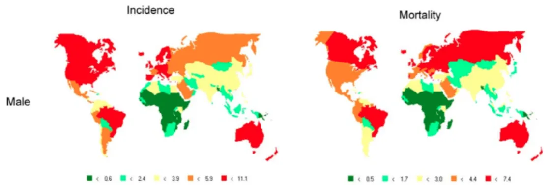
The age-adjusted incidence rates of brain tumor generally tend to be highest in developed rather than in developing countries. In Western Europe, North America and Australia there are 6 to 10 men per 100,000 inhabitants newly affected by brain tumor each year. Slightly lower is the situation for women, with 4 to 10 new case per 100,000 inhabitants (Figure 2). Even within Europe, incidence rates differ between countries (Figure 3). The lower incidence in developing countries may be partially due to a process of under-estimation, but different life-style and ethnic variances in susceptibility to develop brain tumors cannot be excluded. Generally, Caucasians are more affected than people of African or Asian descent, and this difference has been observed in children as well [Farlay J. 2000]. As the situation in Italy is concerned, brain tumors account for a yearly average of 10.5 new brain cancer per 100,000 males and 8.2 per 100,000 women. In 2002 a total of 2,414 and 1,873 new brain cancer cases were diagnosed in males and females respectively, leading to a total of 1,733 and 1,541 death, respectively.
Figure 2: Global incidence and mortality rates of male individuals affected by nervous system tumor,
adjusted to the World Standard Population (all ages; per 100,000 persons per year) [Farlay J. 2000].
Figure 3: Incidence and mortality rates (per 100,000 persons per year) of nervous system tumors in Europe, adjusted to the European Standard Population [Farlay J. 2000].
However, in the all period spanning from 1998 to 2002 brain tumors represented only 1.3% of all neoplasms diagnosed among males and 1.4% among women. They were responsible of 1.9% and 2.1% of all cancer death among males and females, respectively. As shown in Graphs 1, brain cancer incidence and mortality picks lie approximately around 70 and 80 years old, respectively for men and woman. However, it is also relatively frequent already among young patients (0-44 years); in this age range it represents the 6th most frequent tumor among males (5.5% of all cancers) and the 9th among females (2.7% of all cancers). The cumulative risk (0-74 years) of developing a brain cancer was reported as 7.2‰ (1 case by 139 men) and 5.3‰ among females (1 case by 188 women), while the cumulative risk of dying from this cancer was 4.4‰ and 3.0‰ among males and females, respectively.
Graph 1: Incidence and mortality rates of brain cancer in men and woman, within different age group
[Brain cancer, tumors in Italy 2006].
Incidence rates vary across Italy with a ratio between highest and lowest values of about 2.5 points. However, even thought incidence rates for brain cancer are increasing over the time, mortality shows a slightly decreasing tendency, probably as a results of better treatment modality used over the years [Brain cancer, tumors in Italy 2006]. Graph 2 represents in fact the situation as from the 1988. A slight decrease of mortality is observable over a period of 22 years.
Graph 2: Incidence and mortality rates of brain cancer through different periods [Brain cancer, tumors in Italy 2006]
2.2 - Malignant gliomas
Originating from neuroepithelial cells, malignant glial tumors are the most common primary tumors of the human nervous system and include the most aggressive primary brain tumors in
However, as for other human cancers, malignant gliomas take origin from genetic alterations occurring on protooncogenes and tumor suppressor genes and lead glial cells to transformation. In brain tumors, protooncogenes activation occurs almost entirely by gene amplification, resulting in an increased number of specific genes within a cell, and in a corresponding increased expression of the gene’s encoded protein. 1985 marked the first report of a specific gene alteration in a CNS tumor: the epidermal growth factor receptor (EGFR) gene amplification in GBM. Since that time, neuro-oncology research has revealed many genetic abnormalities that indicate consistent genotype-phenotype associations. On the other side, tumor suppressor genes typically associated to malignant gliomas are TP53, NF2, and VHL, with their respective cancer syndromes: Li-Fraumeni (TP53), neurofibromatosis type 2 (NF2), and von Hippel-Lindau (VHL) disease. Moreover, several studies have also reported a wide variety of environmental risk factors, including diet, smoking, alcohol, occupation exposures, radiation, infections, allergies, head trauma, and family history that are being intensively investigated for their role in glioma development.
Malignant gliomas have a peak of incidence lying around age 60 years, and they represent one of the greatest challenges in oncology. Even thought major improvements in cancer research and treatment have been made during the last decades, malignant gliomas continue to have the reputation of highly lethal diseases with very austere prognosis. This is mainly due to the unchanged poor prognosis of GBM. Traditional treatments, which rely on nonspecific, cytotoxic approaches that generally act through DNA damage, have a marginal impact on patient survival and allow GBM to become lethal within approximately 12 months from onset [Mirimamanoff RO. 2006; Stupp R. 2005; Polin RS 2005]. Only 3% of patients affected by GBM survive up to 2 years [Krex D. 2007]. Moreover, the bony limited space and the very sensitive and non-regenerative neural tissue set critical limitation to surgical treatments in the head area. The blood-brain barrier and the blood-cerebrospinal fluid barrier make the delivery of anticancer drugs to the tumor site very challenging. Molecules that are able to cross these barriers must be electrically neutral, small and lipid-soluble. Unfortunately, this is not the case for most of the chemotherapeutic available nowadays.
On the histological basis malignant gliomas are classified as: astrocytomas, oligondedrogliomas, mixed oligoastrocytomas and ependymomas.
2.2.a - Astrocytomas
The majority of brain cancers are represented by astrocytomas, tumors arising from star-shaped glial cells called astrocytes. They can develop in patients of any age preferentially in the main part of the brain, the cerebrum, and rarely spreading outside, affecting other organs. The World Health Organization (WHO) classify these neoplasms in different subtypes according to their histological features (e.g. nuclear atypia, mitotic index, microvascular proliferation index, and necrosis) and on the basis of their invasiveness and/or malignancy.
In order of increasing anaplasia, astrocytomas are named as:
- pilocytic astrocytoma (WHO grade I): is the most common pediatric brain tumor (approximately 700 children per year are affected by this tumor) rarely undergoing malignant transformation. However, even though it is the most benign among astrocytomas, depending on its location, it can interfere with vital sensory functions and frequently recur after apparent complete resection. On histological basis, pilocytic astrocytomas appear as spindle shaped with numerous collections of reddish astrocytic fibers called Rosenthal fibers.
- diffuse astrocytoma (WHO grade II): it account for 25% of all gliomas and it is usually an infiltrative tumors. Despite its relative lack of aggressive histological features, the low-grade astrocytoma in adults is fatal in the great majority of patients. Grade II astrocytoma presents low cellularity with mainly oval pale nuclei, indistinct cytoplasmic borders and fibrillary processes with only mild pleomorphism.
- anaplastic astrocytoma (WHO grade III): is a highly malignant and infiltrative glioma with an increased tendency to dedifferentiate, giving rise to secondary glioblastoma. Histologically it is characterized by high cellularity, pleomorphism of the tumor cell with basophilic nuclei.
- glioblastoma multiforme (WHO grade IV): is the most lethal tumor of the central nervous system typically affecting adults. This type of glioma has a very poor prognosis, due in part to the rapid growth and infiltration of the brain parenchyma. GBM is a highly cellular tumor with pleomorphic, basophilic nuclei with indistinct cytoplasmic borders or plump pink cytoplasm and a delicate fibrillary background. Elevated mitotic index, necrosis, and capillary endothelial proliferation are common features of such tumors.
Figure 4: Histology of GBM. Pseudopalissade srurrounding necrotic zones along with hypercellularity,
hyperchromatism and pleomorfism are typical features of these neoplasms.
2.2.b - Oligodendrogliomas and mixed oligoastrocytomas
These tumors represent a minor proportion of all primary brain tumors and are usually rare neoplasm arising from cells producing the lipid coat of the nerve cells axons, the oligodendrocytes. In the case of oligoastrocytomas, as known as mixed gliomas, the tumor shares characteristics of oligodendrocytes and astrocytes. They tend to develop preferentially in the temporal and frontal lobe of the brain and are most common in middle-aged adults. Although clinically less aggressive than astrocytomas, and with a slower growth rate, these tumors are able to cross into the cerebral spinal fluid (CSF) giving extra cranial metastasis and making surgical removal very challenging. They are both classified as low (WHO grade II) and high grade (WHO grade III).
2.2.c - Ependymomas
These tumors develop from ependymal cells, which line the ventricles of the brain and the central canal of the spinal cord. Ependymomas may spread from the brain to the spinal cord via the cerebrospinal fluid causing notable swelling of the ventricle or hydrocephalus. Ependymomas account for 4–6% of all brain tumors and occur mainly up to the age of 20 years old. In children, 30% of ependymomas appear before the age of 3 yr and
are more aggressive than in adults. Nearly 90% of pediatric ependymomas are intracranial: they occur in supratentorial or posterior fossa locations, and only 10% are intraspinal. In contrast to the earlier mentioned astrocytomas, low-grade ependymomas (grade I/II) develop metastases along the neuroaxis.
[Ali-Osman F. 2005]
2.3 - Clinical aspect
Clinical features of malignant gliomas are represented by headache, focal or generalized seizures with progressive neurological deficits, which are more or less important depending on the tumor location. Severe nausea and changes in mental status are usually present as well. All these symptoms typically occur as result of the rapid proliferation of tumor cells that generate a compression of the normal brain parenchyma. Edema and/or hemorrhage are also consequence of the process of infiltration, growth and expansion of the tumor mass within the brain, which is typical of this neoplasms.
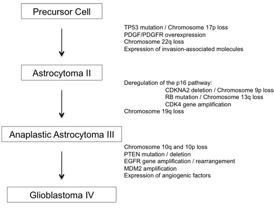
2.4 - Molecular pathology of malignant astrocytoma progression
WHO grade II astrocytomas are best characterized by inactivating mutations of the TP53 tumor suppressor gene on chromosome 17p, as well as overexpression of the platelet-derived growth factor (PDGF) α chain, and the PDGFα-receptor. Interestingly, loss of chromosome 17p in the region of the TP53 gene is closely correlated with PDGFα-receptor overexpression, suggesting that TP53 mutations may have an oncogenic effect only in the presence of PDGFα-receptor overexpression.
Allelic loss of chromosome 17p and TP53 mutations have been observed in at least one-third of adult astrocytomas, regardless of tumor grade. An integral role for p53 in the early stages of astrocytoma tumorigenesis is further evidenced by so-called secondary glioblastomas; it has been demonstrated that grade IV lesions with homogeneous TP53 mutations evolve clonally from subpopulations of similarly mutated cells present in the initial, grade II astrocytic tumors. Functional studies have recapitulated a role for p53 inactivation in the early stages of astrocytoma formation. For example, cortical astrocytes from mice that lack a functional p53 become immortalized when grown in vitro and rapidly acquire a transformed phenotype. In addition, although cortical astrocytes from mice with one copy of a functional p53 behave in a manner comparable to wild type astrocytes, subsequent loss of the one functional copy makes these cells
immortal and transformation can follow. Interestingly, those cells without functional p53 become markedly aneuploid, confirming prior reports that p53 loss results in genomic instability and that human astrocytomas with mutant TP53 are often aneuploid. Thus, the abrogation of astrocytic p53 function appears to facilitate conditions leading to neoplastic transformation, setting the stage for subsequent malignant progression. The transition from WHO grade II astrocytoma to WHO grade III anaplastic astrocytoma is accompanied by several molecular abnormalities. Studies suggest that most of these alterations converge on one critical cell-cycle regulatory complex that includes the p16, retinoblastoma (Rb), cyclin-dependent kinase 4 (cdk4), cdk6, and cyclin D1 proteins. Individual members of this pathway are altered in up to 50% of anaplastic astrocytomas and in the vast majority, if not all, GBM. Loss of chromosome 9p primarily affects the region of the cyclin-dependent kinase inhibitor 2A (CDKN2A) gene and occurs in approx 50% of anaplastic astrocytomas and glioblastomas. The CDKN2A gene encodes for the p16 and p14alternate open reading frame (ARF) proteins, and expression of these proteins is most commonly altered by homozygous deletion of the CDKN2A gene, although point mutations and hypermethylation of CDKN2A have also been found to alter p16 and p14ARF expression. Chromosome 13q loss occurs in one-third to one-half of high-grade astrocytomas, with the RB gene preferentially targeted by losses and inactivating mutations. Analyses of the loss of chromosome 13q, RB gene mutations, and Rb protein expression suggest that the RB gene is inactivated in approximately 20% of anaplastic astrocytomas and 35% of GBMs. Interestingly, RB and CDKN2A aberrations are inversely correlated in gliomas, rarely occurring together in the same tumor.
Located on chromosome 12q13-14, CDK4 is amplified in approx 15% of anaplastic astrocytomas and GBMs. This amplification frequency may be higher among gliomas without CDKN2A loss, perhaps reaching 50% of GBMs with intact p16 expression. CDK4 amplification and cyclin D1 overexpression appear to represent alternative events to CDKN2A deletions in GBMs because these genetic changes only rarely occur in the same tumor. Within this pathway, CDK6 amplification also occurs, although not as commonly as CDK4 amplification.
Further progression to GBM is characterized by the loss of chromosome 10; although occurring in anaplastic astrocytomas, this alteration can be found in 60–95% of GBMs. At least two tumor suppressor loci are implicated on the long arm of chromosome 10, as well as one potential locus on the short arm. The phosphatase and tensin homolog (PTEN)/ mutated in multiple advanced cancers 1 (MMAC1)/ TGFβ-regulated and epithelial cell-enriched phosphatase 1 (TEP-1) gene at 10q23.3 is one example of a tumor suppressor gene that has been studied, with PTEN mutations identified in approx 20% of GBMs. Moreover, introduction of wild-type PTEN into glioma cells with mutant PTEN leads to growth suppression. Nonetheless, given the remarkably high frequency of
chromosome 10 loss in GBMs, glioma tumor suppressor genes other than PTEN likely reside on this chromosome.
The epidermal growth factor receptor (EGFR) gene is the most frequently amplified oncogene in astrocytic tumors and is characteristic of so-called de novo GBMs. Although EGFR is amplified in few anaplastic astrocytomas, approx 40% of GBMs display this amplification. EGFR amplification in GBMs is almost always accompanied by loss of genetic material on chromosome 10 and these tumors often exhibit CDKN2A deletions. GBMs with EGFR gene amplification display overexpression of EGFR at both the messenger ribonucleic acid (mRNA) and protein levels, stressing the importance of this growth signal pathway to GBMs. Approximately two-thirds of the tumors having EGFR amplification undergo intra-gene deletion rearrangements that result in the overexpression of mutant EGF receptors. The most common EGFR mutant, EGFR-vIII, is known to have constitutive, ligand-independent tyrosine kinase activity, as well as an extended half-life that stimulates cell proliferation and enhances the tumorigenicity of human glioma cells in nude mice. Furthermore, the activity of this mutant has been shown to promote tumor angiogenesis, as well as to confer tumor resistance to apoptosis by increasing Bcl-XL expression. EGFR amplification and TP53 mutations appear to be mutually exclusive genetic aberrations in GBMs. One-third of GBMs have TP53/chromosome 17p alterations, one-third display EGFR gene amplification, and one-third have neither change. Experimental data supports this distinction by showing that cells lacking functional p53 are not transformed when cultured in the presence of epidermal growth factor (EGF) but are transformed in the presence of other growth factors; GBMs with TP53 mutations may therefore not be expected to acquire EGFR gene amplification if activation of the EGF-EGFR pathway does not produce a increased growth advantage in such cells. A number of additional molecular alterations occur in astrocytic gliomas for which little functional information is known. Less common genomic alterations associated with low-grade astrocytomas include loss of chromosome 22q, suggesting the presence of a chromosome 22q glioma tumor suppressor gene, and gains of chromosome 7q. In anaplastic astrocytomas and GBMs, allelic loss on 19q is quite common, being observed in up to 40% of these tumors and suggestive of a putative tumor suppressor gene.
Figure 5: Molecular genetic alterations characteristic of different grade of astrocytomas
2.5 - Pathological markers of glioma
According to the first theory attempting to explain glioma formation, it was thought that central nervous system cancer cells arise from glial cells and thus resemble their precursors in varying degrees. Thus, as envisaged, mature neurons do not give rise to tumors, while glial cells surrounding them, such as astrocytes and oligodendrocytes, that remain proliferative, would be subject to tumorigenic stimulus. This resemblance helps histopatological diagnosis and could serve to predict the potential behavior of the neoplasm.
Another theory, which is newly coming into light, is based on recent findings that suggest that there may actually exist cancer stem cells (CSC) that are responsible for several tumor types, including brain tumors. These stem cells are portrayed as very primitive cells, resembling embryonic cells and neural stem cells that are still capable of potent mitotic activity and self-renewal. This theory gives a good explanation for the recurrence of tumors and why some tumors seem to be out of reach for even advanced therapies [Singh SK. 2004].
Until recently, treatment decisions regarding malignant gliomas began with establishing diagnosis by ordinary pathology only, which during the last 100 years provided knowledge about the biology and the clinical behavior of different neoplasm. During the last 10 years, however, data from histopathological examination of tumors tissue have been supplemented by an increased use of
molecular markers for tissue diagnosis. Knowledge about the molecular biology of cancer continues to increase and currently some molecular signatures, used to classify tumors rather than to predict response to therapy, are available to clinicians. According to the molecular biology of this tumors, the principal markers used in the case of malignant gliomas are 1p/19q co-deletion, methylation of the O-6 methyl-guanine-DNA methyltransferase (MGMT) gene promoter, and alteration in the epidermal growth factor receptor (EGFR) pathway.
2.5.a - 1p/19q loss
After the empirical discovery of favorable responses to chemotherapy in a high portion of recurrent anaplastic oligodendrogliomas (WHO grade III), Cairncross et al reported that such tumors carried a loss of the short arm of chromosome 1 (1p) [Cairncross JG. 1998]. Moreover, tumors carrying co-deletions of 1p and the long arm or chromosome 19 (19q) had substantially improved survival time. This finding have been investigated and then confirmed several times over the past 10 years and the correlation have been extended to new current therapy regimen such as temozolomide and radiotherapy [Brandes AA. 2006; Kouwenhoven MC. 2006; Bauman GS. 2000]. These data suggest this marker as a useful indicator of tumor vulnerability to a broad range of therapeutic options. The frequency of 1p/19q co-deletion has been estimated to 80-90% in grade II and 50-70% in grade III oligodendrogliomas. Even though no specific tumorigenic genes have been identified, 1p and 19q loss inversely correlates with TP53 mutations, 10q deletions, and amplification of EGFR. Deletions involving 1p and 19q are uncommon in glioblastomas, but, in those cases where it has been reported, these deletions seem to predict shortened survival, possibly indicating true genomic instability [Esteller M. 2000].
2.5.b - MGMT methylation status
The orally administered alkylating drug Temozolomide, the current agent used as standard care drug for GBM since 2005, acts methylating the O6 position of the guanine nucleotide, resulting in cell death. The constitutively expressed DNA repair enzyme MGMT will transfer the methyl group from the O6 position of the modified guanine to a cysteine residue of the enzyme itself, mitigating the cytotoxic effects of the drug in normal cell. Approximately 50% of glioblastomas have decreased concentration of MGMT, making these tumors more susceptible to the effects of temozolomide [Esteller M. 2000]. Hegi and
colleagues studied 106 patients (46 with methylated tumors, 60 with un-methylated tumor) treated with temozolomide and reported a 2 years survival of 46% of patient carrying
MGMT gene methylation versus only 23% of those patients with un-methylated tumor. The
primary mechanism by which MGMT expression is reduced in glioblastoma, in fact, seems to be methylation of the MGMT gene promoter, a common way of gene silencing [Hegi ME. 2005; Martinez R. 2007]. Several studies suggested this correlation true also in pediatric glioblastomas and other low-grade gliomas [Pollack IF. 2006; Everhard S. 2006].
2.5.c - EGFR pathway alterations
Molecular targeted therapies have been recently successful in chronic myeloid leukemia by the use of Imatinib to inhibit the BCR-ABL tyrosin-kynase fusion protein, and the use of Trastuzumab in the treatment of HER-2/neu positive breast carcinoma [Druker BJ. 2006; Schnadig ID. 2006; Lohrisch C. 2001]. Approximately 60% of glioblastomas present an up-regulation of EGFR mediated signaling, driven generally by an EGFR gene amplification. Additionally, in about half of the glioblastomas over-expressing EGFR, the expression of the mutant form EGFR variant III also is found. This variant lacks of the ligand-binding domain and therefore constitutively activates the downstream cascade.
The prognostic use of EGFR overexpression and its mutations is still not clear, but it might be useful for the identification of a subgroup of tumor with more aggressive behavior [Jeuken J. 2009]. The presence of such alterations might raise the possibility to use EGFR-targeted drugs in patients affected by glioblastoma; a similar situation to the non-small-cell lung cancer with activating EGFR mutations that showed remarkable response to Erlotinib and Gefitinib. However, two studies published in 2005 sought to clarify whether assessment of EGFR status was useful in aiding the decision of using such small-molecule kinase inhibitors. Results from both studies reported that tumor responding to these drugs had an intact PTEN-AKT pathway, which is unfortunately not the case of GBM [Mellinghoff IK. 2005; Haas-Kogan DA. 2005]. Subsequent studied, in fact, reported no major responses or benefit in patients with GBM being treated with these drugs [Rich JN. 2004a; Kesari S. 2005].
2.6 - Glioblastoma: a cancer without hope
As already said GBM is the worst among brain cancer occurring in U.S. mainly in 60 years old men, with an incidence rate of 13.000 cases by year [Schwartzbaum JA. 2006]. Typical symptoms of GBM depend on which part of the brain is affected. Headaches, nausea and vomiting, cranial nerve disorders and seizures are typical results of increased intracranial pressure. GBM does not metastasize via blood stream but has a great tendency to invade within the host brain parenchyma. Indeed, the aggressive and invasive nature of GBM has been described as the principal reason of the severe prognosis of GBM. Even though, chemotherapy with Temozolomide resulted as safe and feasible treatment of GBM, greatly accountable for the 1-year patient survival there are no effective treatments targeting tumor-infiltrating cells, migrating throughout brain parenchyma [Hoelzinger DB. 2007]. Despite the usage of the most modern tools of the operating theatre, as online magnetic resonance imaging or computer-assisted navigation systems, neurosurgeons can only control the rapidly proliferating tumor mass by repetitive resections, reducing in this way the increasing intracranial pressure, caused by tumor growth, and thereby delaying imminent death of the patient. Additionally, malignant brain tumors are often located adjacent to or within functionally important areas of the brain where the use of aggressive surgery would turn into severe neurological damages for the patient. Accordingly, it appears obvious that the general principle of tumor surgery, which requires safety margin resection around the tumor mass of 1–2 cm, cannot be respected in neurosurgery. Furthermore, the blood-brain barrier and blood-cerebrospinal fluid barrier make the delivery of anticancer treatment to the tumor site a very challenging task. The first is formed of tight junctions between endothelial cells of capillaries, and the second has tightly bound choroid epithelial cells accompanied with an active organic acid transport system which impede crossing of molecules. Interstitial pressure in the brain slows down the molecular transfer to cells. Most molecules are not capable of penetrating these boundaries. Generally, molecules that are able to pass these barriers must be electrically neutral, lipid-soluble and small; unfortunately many, otherwise potent, chemotherapeutic drugs do not fit in this category. Even if the cytotoxic agent meets these criteria, it is extremely hard to reach therapeutic drug concentration and to maintain it within the proximity of the tumor site.
Moreover, neural tissue is very sensitive to radiation therapy resulting often in normal neural tissue death as well as a higher risk for radiation-induced mutations. These particular limitations demand the use of only carefully planned treatments only for those tumors that are known to respond. Considering all of this, it appears obvious that efficient treatments are based on comprehensive knowledge on tumor biology.
It has been already emphasized that histopathological examination may not be accurate enough and that genetic and molecular pathologic analysis of tumors are being used to complete diagnosis. These analyses constitute also the basis for targeted treatments, attempting to affect only those cells that express certain cancer-cell-specific features. This would minimize the damages to surrounding cells and reach a greater proportion of cancerous cells. However, it is still difficult to find such targets because GBM pathogenesis, like in the rest of malignant brain tumors, has been shown to be dependant on several tumorigenic pathways.
Many gene therapies and targeted molecular therapies have major limitations because their target products are often subject to mutation, making the treatment unstable. In addition, even specific features in certain cancer cells can be resulting from changes in very different sites along the molecular pathways. So far, it seems that future attempts to develop more effective treatments rely on accurate diagnosis, tools to estimate prognosis and development of combined therapies; an equation that is more complicated than its several factors taken alone.
2.7 – de-novo and secondary glioblastoma
Primary brain tumor present a set of characteristics common to all cancer: cell proliferation in absence of growth stimuli, avoidance of apoptosis and no limits to replication, escape from external growth-suppressive forces and immune response, new blood vessel formation, and extreme ability to invade normal brain tissue. These features are the results of a series of mutation occurring in the cells during their dedifferentiation, leading to tumor progression. At least two specific molecular pathways, by which high-grade gliomas develop, can be distinguished. Most GBMs are diagnosed without any antecedent lower-grade tumor being detected; they consist of the so called
de-novo or primary GBMs. However, a smaller group of high-grade astrocytomas is known as secondary GBMs, arising as the result of the natural recurrence and progression of previously
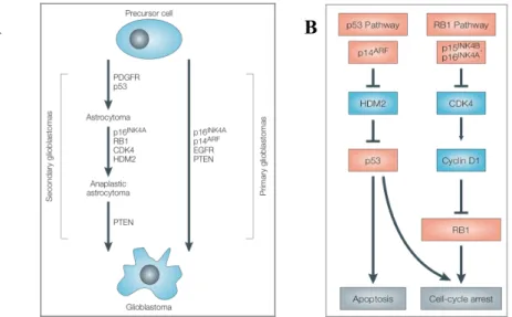
occurred lower-grade tumor. As showed by figure 6A, these two variants of GBM are determined by different sets of molecular changes. It has been reported that cancers with dysfunction in p53 pathway show resistance to radiotherapy. Indeed, high-grade gliomas are characterized by an elevated genomic instability, which is tolerated, without any induction of apoptosis, because of the disruption of the normal p53 pathway.
Figure 6: A) Molecular pathways to Glioblastoma formation - GBM can arise from the malignant progression of
lower grade astrocytomas (grade II or grade III), or more commonly de-novo as primary tumor. B) Different genetic pathways are implicated in the formation of primary or secondary GBM, each of them has a crucial role in cellular transformation [Rich JN. 2004b].
Low-grade astrocytomas and secondary GBMs often show mutation or loss of p53 protein, while in primary GBMs the protein is present but mutations and amplifications are commonly seen in its are the modulating factors. For instance, HDM2 (human double-minute 2) inhibits p53 function through inhibitory binding of p53 itself or facilitating its ubiquitinilation and successive degradation, via E3-ligase. Yet, HDM2 is amplified in a minority of astrocytomas and GBMs. p14ARF negatively regulates the HDM2 activity, thus p53 degradation. However, p14ARF gene is located on the chromosome 9p21 that indeed is frequently deleted or methylated in most of GBMs. Mutations in the pRB pathway are responsible for the unrestricted cellular proliferation of these tumors. Defects in the cell-cycle components are uncommon in low-grade gliomas, but as they progress in grade of maliganncy, tumors rapidly acquire molecular alterations that disrupt the pRB cascade. Specifically, loss of RB1 or amplification of CDK4 occurs in secondary gliomas, whereas deletions, or suppression by promotor methylation, of CDK inhibitors (CDKIs), p16INK4A and p15INK4B occur in primary GBMs. This kind of alterations, interesting the pRB pathway are found in parallel to the above discussed mutations of the p53 pathway in the majority of malignant gliomas (Figure 6B). Several growth factor pathways are also involved in the phenotype of malignant gliomas. These include epidermal growth factor (EGF), platelet-derived growth factor (PDGF),
insulin-like growth factor-1 (IGF1), hepatocyte growth factor/scatter factor (HGF/SF), vascular endothelial growth factor (VEGF) and transforming growth factor-β (TGF-β) [Rich JN. 2004b]. The mechanisms by which these pathway get activated are very important, because many interventions have been proposed and designed to target receptor activity at certain points (Table 1).
Target Agent Drug Class Development stage
in glioma
EGFR Geftinib TKI II
Erlotinib TKI II AEE788 TKI I TP-38 Ligand-toxin conjugate Peptide vaccine II II
PDGFR Imatinib mesylate TKI I/II
SU6668 TKI I
MLN518/608 TKI Preclinical
VGFR PTK787/ZK222584 TKI I
SU5416 TKI I
Table 1: Several clinical trials have been undertaken for adult and pediatric patients with recurrent
malignant gliomas with target agents. Some of these agents are listed in the table above with their target and the stage of development [Rich JN. 2004b].
2.8 - Therapeutic strategies available: benefits and pitfalls
Generally, the first step in the treatment of brain tumors is surgery. With today’s modern techniques, surgery is generally safe for most of the patients. The goals of surgery are: i) to remove as much tumor as possible; ii) to reduce the symptoms caused by the pressure that the tumor mass put on the normal brain parenchyma; and iii) to obtain tumor tissue for diagnosis and treatment planning. In some circumstances, such as certain medical conditions or concerns about the location of the tumor, a biopsy may be done in place of surgery. The tissue obtained during the biopsy is then used to make the diagnosis.
Surgical intervention to remove a brain tumor is carried out by making an opening in the skull over the tumor, what is known as a craniotomy. Brain mapping and functional MRIs help the neurosurgeon to determine vital region of the brain so as to avoid damages in these area during surgery. The surgeon can use stereotactic computerized equipment and image-guided techniques as
navigational tools, like a GPS (global positioning system) system. Those tools help to guide the neurosurgeon's access into some of the difficult or deep areas of the brain. Lasers may be also used during surgery to vaporize tumor cells. Ultrasonic aspirators are tools that break up and suction the tumor out of brain tissue. High-powered microscopes help the neurosurgeon to better see the tumor. Because the tentacle-like cells of astrocytomas grow into the surrounding tissue, these tumors cannot be totally removed.
Radiation and chemotherapy are then used to treat the remaining tumor. In adults, radiation therapy usually follows surgery. There are different types of radiation that may be given using various doses and schedules. Conventional fractionated external beam radiation is the "standard" radiation given 5 days a week for 5 or 6 weeks. A “fractionated” radiation could also be given in small doses at a time over several weeks. External beam radiation is actually the same radiation a patient is subject during a simple chest x-ray. Most forms of local radiation treat the tumor and its surrounding area. However, a sort of "local radiation" may be used to boost conventional radiation. Monoclonal antibodies may be capable of carrying radiation or drugs to the tumor site. Many of these radiation techniques are under investigation and are offered in organized testing plans called clinical trials.
Two main classes of chemotherapy drugs used in the therapy of brain tumors can be
described: cytotoxic and cytostatic drugs. Cytotoxic drugs are designed to destroy tumor cells. They work by making tumor cells unable to reproduce themselves. Bis-chloronitrosoure, lomustine, procarbazine, cisplatin, temozolomide, and irinotecan are examples of cytotoxic drugs. Of this group, temozolomide is commonly used, along with radiation therapy, as a primary treatment for newly diagnosed glioblastomas. Cytostatic drugs are used to alter tumor behavior. These drugs work by changing the tissue in and around the tumor. There are several different types of cytostatic drugs. For example, angiogenesis inhibitors are cytostatic drugs that stop the growth of new blood vessels around a tumor. An example of angiogenesis inhibitors is well represented by the anti-VEFG-A bevacizumab. Differentiating agents, such as phenylacetate, are cytostatic drugs that make malignant cells look and act like normal cells. Sometimes, cytotoxic and cytostatic drugs can be combined in the attempt to obtain a synergic action. Researchers are also developing new ways of delivering drugs to the tumor. The convection-enhanced delivery (CED), for example, uses a pump to slowly “flow” chemotherapy drug or biologic substances into the tumor site. Another method uses a biodegradable wafer that is positioned into the tumor cavity after surgery that slowly releases a chemotherapy drug within the remaining tumor tissue. Other approach uses micro particles, which release drugs into the tumor at a pre-determined rate. Chemotherapy may be used in infants and very young children to delay radiation therapy until the age of three or four. Clinical trials are underway to evaluate the most effective ways of treating these tumors in infants and children.
Several drugs are also used to alleviate symptoms of brain tumors. Among them steroids are used to decrease swelling (edema) around the tumor. Anti-epileptic drugs control seizures. Anti-emetics prevent vomiting and help control nausea. Drugs to help treating fatigue or depression may be helpful as well.
Yet, the scientific community continues to look for new drugs to treat glioblastoma and other malignant brain tumor, and many drugs are under investigation. Some of them are new drugs, some are drugs proven useful in treating other types of tumors in the body, and still others are standard brain tumor drugs administered differently. However, because they belong to the chemotherapy drug class, they can affect normal cells and patients can expect side-effects from treatments such as hair loss or lack of appetite. For this reason the investigation towards more specific targeting drugs, able to accumulate in the tumor area only and to spare normal cells from their toxic effects, are desperately needed.
3. Targeted Tumor Therapy
3.1- General aspect
As previously discussed, a promising avenue towards more selective, better anti-cancer drugs relies on the targeted delivery of bioactive molecules (drugs, cytokines, pro-coagulant factors, photo/radio-sensitizers, radionuclides, etc.) to the tumor area by means of binding molecules (e.g., monoclonal antibodies) specific to tumor-associated antigens. In this way, the selective accumulation of drugs at the tumor site would spare normal tissues and lead to an increased therapeutic drug dose at the tumor site. This will bring to a higher treatment efficacy and less side effects for the patient. The same strategy, using ligands coupled to imaging agents (e.g., fluorophores), can also be useful for the diagnostic localization of tumor lesions by imaging analysis.
Even though the Paul Ehrlich’s “magic bullet” concept was envisioned at the end of the 19th century, several technologies had to be developed before the selective delivery of therapeutic drugs to the tumor site could become a reality. Recent advances in protein engineering have made possible the generation of high-affinity human mAbs against virtually any biomolecular target. Furthermore, new technologies are becoming available for the generation of high-affinity binding peptides [Collins J. 2001], aptameres [Brody EN. 2000] and synthetic organic molecules [Erlanson DA. 2003; Melkko S. 2004] that may be used as ligands for the development of targeted anti-cancer strategies. To note, there is a fundamental difference between therapeutic strategies aiming to the inhibition of the biological function associated to a molecular target and those strategies meant to delivery anti-cancer agents to the tumor lesion. Indeed, the term “target therapy” encompasses a wide variety of different approach, which can be mainly divided into direct and indirect approaches.
Direct approaches target tumor-associated or tumor-specific proteins to alter their signaling pathway either by mAbs binding to the relevant antigens or by small-molecule drugs that interfere with these proteins (approach also known as molecular targeting). These techniques are being already widely used in cancer treatment, especially in the case of vascular tumor targeting. The existing anatomical and physiological differences in the endothelium and surrounding stroma of tumors, compared to normal tissue, provides the basis for the development of vascular tumor targeting strategies. Tumor vasculature is often disorganized and tortuous, and presents different shunts (vascular tract in which blood passes directly from an arteriole to a venule) that make blood flow inconstant. The blood lacks of oxygen and nutrient resulting in a highly hypoxic and acidic microenvironment, which is under oxidative stress [Brown NS. 2001]. Endothelial cells respond


![Figure 8: Schematic representation of a classic 2-D PAGE experiement [ Veenstra TD. 2006 ]](https://thumb-eu.123doks.com/thumbv2/123dokorg/7544962.108620/46.892.264.632.139.618/figure-schematic-representation-classic-page-experiement-veenstra-td.webp)