Ph.D. in Molecular Bio-pathology
Molecular mechanisms determining aromatase
overexpression and inducing rat tumor Leydig cell
proliferation: involvement of IGF-I and COX-2
Supervisor
Prof. Marcello Maggiolini
Coordinator
Prof. Giovanna De Benedictis
Candidate
Adele Chimento
I
Introduction………...……..….. 4 1. Endocrinology of the male reproductive system………..…………. 5 1.1 The testis: general structure………...………...………… 5
1.2 Testicular function and its regulation …………..…....….…………... 1.3 Steroid production .…..………….………..……….
6 10 2. Estrogen regulation of testicular function.…....……….……… 13 2.1 The aromatase gene: structure and regulation ……….…………... 14 2.2 Estrogen receptors (ERs) …..……….….………... 16 2.3 Distribution of ERs and aromatase in the male reproductive system . 2.3.1 ERs and aromatase in rodent testis …..……….……… 2.3.2 ERs and aromatase in human testis ………....………….. 2.4 Role of estrogens in animal male reproduction…………...….……… 2.5 Role of estrogens in human male reproduction……..…...….……… 2.6 Effects of excess estrogens on male reproduction…..……….. 2.6.1 Exposure to excess estrogens in animals……….………. 2.6.2 Aromatase overexpression in rodents………...……… 2.6.3 Exposure to excess estrogens in humans…….………. 2.6.4 Aromatase overexpression in humans……….…….
19 20 24 26 28 29 29 30 31 32 3. Testicular cancer ………...…… 33 3.1 Introduction……….
3.2 Leydig cell hyperplasia and cancer …..…………..…….…………... 3.3 Relationship between estrogens and Leydig tumors………... 4. The IGF System……….………. 4.1 Introduction………….……… 4.2 Effect of IGF-I on testicular function……….……….
33 34 36 39 39 42
II
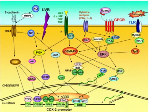
5.1 Structure, function and regulation of COX……… 5.2 The cyclooxygenase-2 (COX-2) in diseases and cancer…………....
45 49
Specific aim………...… 52
Materials and Methods………... 53
Cell cultures, animals and human samples...………...…...…… 54
Aromatase activity assay…...………...…………... 54
Radioimmuno assay (RIA)....……….……...……… 54
Chromatin immunoprecipitation (ChIP)………..………. 55
Real time RT-PCR ...………...……… 56
Western blot analysis ...………..………...……… Immunohistochemical analysis... 58 59 Cell proliferation assay ...…………...……….………...……… Non radioactive in vitro assay for PKA activity in cell lysates………. RNA interference………... Data analysis and statistical methods ...………...……. 60 60 61 61 Results……….. 62
Estradiol induces Leydig cell tumor proliferation through an autocrine mechanism ... 63
Aromatase overexpression is determined by constitutive activation of transcription factors SF-1 and CREB ... 69
IGF-I is produced by R2C cells and induces aromatase expression through PI3K- and PKC-mediated activation of SF-1 ... 70
IGF-I induces aromatase expression and activity in R2C cells ... 74 Changes in IGF-I pathway activation status lead to changes in SF-1 binding
III
COX-2 is highly expressed in tumor Leydig samples and is necessary for aromatase expression………. COX-2 inhibitor NS398 decreases pCREB and aromatase expression………. PGE2 activated pathway regulates aromatase expression………. PKA inhibitor H89 decreases aromatase expression and activity as a consequence of reduced pCREB activation……….. Inhibition of PGE2 dependent pathway decrease estradiol production and
consequently tumor Leydig cell proliferation………... 80 82 85 87 91 Discussion ... 95 References... . 104 Scientific Publications
IGF-I regulating aromatase expression through SF-1, supports estrogen dependent tumor Leydig cell proliferation.
- 1 -
Summary
Several studies on both humans and rodents indicate that prenatal or postnatal exposure to estrogens migth have a central role in the mechanism leading to male reproductive tract malformations as well as testicular tumors (1;2). While the effects of estrogen on mammary gland tumorogenesis is well known, the role of aromatase overexpression and in
situ estrogen production in testicular tumorogenesis is not clearly defined. In this study we
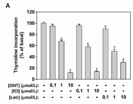
have investigated the molecular mechanisms causing aromatase overexpression and the effect of estradiol (E2) overproduction on Leydig cell tumor proliferation. Our hypotesis is that constitutive E2 production stimulates Leydig tumor cell proliferation acting on cell cycle regulators. Moreover, among several potential factors inducing aromatase, we investigated the role of IGF-I, produced locally in the testis, and of COX-2 overexpressed in other estrogen-dependent tumors. We used rat R2C Leydig tumor cells and testicular samples from Fischer rats with a developed Leydig tumor (FRTT). Both experimental models express high levels of aromatase and Estrogen Receptor alpha (ERα). Treatment with exogenous E2 induced proliferation of R2C cells and upregulation of cell cycle regulators cyclin D1 and E, that were blocked by addition of antiestrogens. These observations leaded us to suppose an E2/ERα dependent mechanism for Leydig cell tumor proliferation.
Aromatase expression in rat Leydig cells is driven by the PII promoter regulated mainly by three CRE sites and one SF1 site. Determining the molecular mechanism responsible for aromatase overexpression, we found that total and phosphorylated levels of transcription factors CREB and SF-1 were higher in tumor samples. Moreover, we found that R2C cells produce also high levels of IGF-I that increased aromatase mRNA, protein and activity as a consequence of increased total and phosphorylated SF-1 levels. Binding of IGF-I to its receptor causes receptor autophosphorylation and the activation of an intrinsic tyrosine kinase that actson various substrates, leading to activation of multiple signalingpathways including the PI3K/AKT and MAPK cascades. In addition,it has been shown that IGF-I
- 2 -
can activate the PLC/PKC pathway (3). Specific inhibitors for IGF-I receptor, Protein Kinase C and Phosphoinositol-3-kinase determined a reduction in SF1 and consequently in aromatase expression and activity. The same inhibitors were also able to inhibit the IGF-1 dependent-SF-IGF-1 recruitment to the aromatase PII promoter. These results indicate that in Leydig tumor cells one of mechanism determining aromatase overexpression is an enhanced IGF-I signaling potentiating SF-1 action.
Determining the molecular mechanism responsible for constitutive CREB phosphorylation, we investigate the role of COX-2 (cyclooxygenase-2) an enzyme involved in prostaglandins (PGs) synthesis that has not been detected in the human normal testis, but it is expressed in testicular biopsies of men with cancer. We show that COX-2 is expressed in rat Leydig tumor samples while is not detectable in normal testis. COX-2 specific inhibitor NS398 (5-50 uM) is able to reduce dose-dependently aromatase mRNA, protein expression, activity and tumor Leydig cell proliferation. NS398 significantly decreases CREB activation reproducing the same effect of H89 (a PKA inhibitor) on both aromatase and CREB. The drop in estrogen production determines a decrease in tumor Leydig cell proliferation. Moreover, the addition of increasing amounts of PGE2 were able to increase phosphorylation but not synthesis of CREB which consequently increased aromatase expression. Next, in order to evaluate PGE2 receptor (EP) subtype(s) responsible for induction of aromatase expression and activity, we used the selective inhibitors: SC19220, AH6809, AH23848 for EP1, EP2/EP4 and EP4 respectively. Our data demonstrate that only the AH23848 was able to determine a decrease in CREB phosphorylation and again in aromatase expression. These findings led us to suppose that, in tumor Leydig cells, COX-2-derived PGE2, through an autocrine mechanism, activates PKA which is responsible of CREB activation.
In summary, our results give a contribution to clarify two molecular mechanisms determining aromatase overexpression in Leydig cell tumor. The first one involves some of pathways (PI3-K and PKC) activated the IGF-I determining SF-1 production and enhanced P450 mRNA transcription. The other one involves the production of PGE2 (induced by COX-2 overexpression), which is responsible of PKA activation and CREB
- 3 -
phosphorylation. It remains to elucidate the molecular mechanisms determining IGF-I and COX-2 overexpression in tumoral Leydig cells.
However, the observations that COX-2 and IGF-I pathway inhibitors are able to decrease E2 production and to block Leydig cell tumor proliferation, open new perspectives on trapeutic approach of Leydigioma in the human.
- 4 -
- 5 -
1. Endocrinology of the male reproductive system
1.1 The testis: general structure
The human male reproductive system includes the hypothalamic-pituitary-gonadals axis, the epididymis, vas deferens, seminal vesicles, prostate and the urethra. The testis is composed primarily of seminiferous tubules packed closely together and interstitial cells (4). The seminiferous tubules are composed by Sertoli cells that support germ cells during their maturation into spermatozoa. Sertoli cells create a blood-estis barrier, and separate the germinal epithelium into basal and adluminal compartments. They are responsible for the physical support of the germ cells, in addition to providing nutrients and growth factors. The major cell in the interstitial space outside the seminiferous tubule is the Leydig cell, which produces testosterone, a necessary component for germ cell maturation. Male fertility requires the production by the testes of large numbers of normal spermatozoa through a complex process of spermatogenesis. The germ cells are sequentially organised into several layers signifying the respective mitotic or meiotic processes and spermatid development. Each seminiferous tubule is surrounded by mesenchymal cells. Among these are the peritubular myoid cells whose contractile elements generate peristaltic waves along the tubules, but do not present a tight diffusion barrier. Vascular smooth muscle cells, macrophages and endothelial cell types are also located in the interstitial space of the testis. The physiological role of macrophages has long been underestimated. In the rat, the number of macrophages is one quarter of the number of Leydig cells and the presence of macrophages is crucial for (re)population of Leydig cells during development and after experimental depletion (5;6). Immune cells, known to secrete a number of growth factors and cytokines, are part of the intratesticular communication pathways (7).
- 6 - 1.2 Testicular function and its regulation
Testes are components of both the reproductive system (being gonads) and the endocrine system (being endocrine glands). The respective functions of the testicles are:
1. producing sperm (spermatozoa); 2. producing male sex hormones.
These two functions occur in separate compartments within the testis: 1. the seminiferous tubules produce sperm and 2. the interstitial cells (i.e., Leydig cells) synthesize androgens (Fig. 1).
Figure 1. Schematic representation of functions of the testis.
Both functions of the testis, sperm-forming and endocrine, are under control of gonadotropic hormones produced by the anterior pituitary: luteinizing hormone (LH) and follicle-stimulating hormone (FSH). Synthesis and release of both FSH and LH is
- 7 -
regulated by a single gonadotropin releasing hormone (GnRH) also referred to as LHRH, a decapeptide produced by specialized neurons in the hypothalamus. Pulsatile GnRH production signals gonadotroph cells in the anterior pituitary to produce follicle-stimulating hormone (FSH) and luteinizing hormone (LH) that then act on the testis to regulate spermatogenic potential. LH binds to receptors on the surface of Leydig cells in the testis and stimulates the production of testosterone, a steroid hormone that diffuses into the seminiferous tubules. Within the seminiferous tubules only Sertoli cells possess receptors for testosterone and FSH and thus these cells are the major targets of the ultimate hormonal signals that regulate spermatogenesis.
Serum testosterone and inhibin ( Sertoli-cell product) downregulate LH and FSH secretion via negative feedback loop. Testosterone also decreases the responsiveness of the pituitary to GnRH (Fig. 2).
Figure 2. Hypothalamic-Pituitary-Testicular axis.
LH, through specific receptors found on the surface of Leydig cells, controls the production and secretion of testosterone (8;9).
- 8 -
Thus, pituitary gonadotropins are the chief regulators of testicular function. LH stimulates androgen production by Leydig cells after binding to LHR and FSH acts through its receptors in Sertoli cells (FSHR) to regulate spermatogenesis (Fig. 3).
Figure 3. Endocrine regulation of the testis. PMC, peritubular myoid cell; CRE, cAMPresponsive elements, ARE, androgen-responsive elements; ERE, estrogen-responsive elements; (by Akingbemi BT 2005).
- 9 -
The interaction of LH with its receptor initiates signalling through GTP binding proteins determining the cyclic AMP production (10). Signal transduction occurs through the protein kinase A pathway as its principal signal transduction mechanism. Some data suggests that intracellular calcium concentration can be induced by the action of LH by activating phospholipases in the lipoxygenase pathway (11). In addition, the changes in calcium can also regulate adenylate cyclase through the protein kinase C pathway.
Within the seminiferous tubules only Sertoli cells possess receptors for testosterone and FSH and thus these cells are the major targets of the ultimate hormonal signals that regulate spermatogenesis. FSH binding to its receptor is known to activate at least 5 signaling pathways in Sertoli cells: cAMP-PKA pathway, MAP kinase pathway, Phosphatidylinositol 3-kinase (PI3-K) pathway, Calcium pathway, Phospholipase A2 (PLA2) pathway. Initially FSH binding to the FSH receptor causes receptor coupled G proteins to activate adenylate cyclase (AC) and increase intracellular cAMP levels. Multiple factors can be activated by cAMP in Sertoli cells including PKA that can phosphorylate a number of proteins in the cell and also regulate the expression and activity of numerous transcription factors including CREB. During puberty, FSH activates the MAP kinase cascade and ERK kinase in Sertoli cells most likely via cAMP interactions with guanine nucleotide exchange factors (GEFs) and activation of Ras-like G proteins. ERK is capable of activating transcription factors including SRF, c-jun and CREB. In granulosa cells, FSH also activates the p38 MAP kinase. FSH and cAMP also likely act through GEFs to activate PI3-K and then phosphoinositide dependant protein kinase (PDK1) and PKB in Sertoli cells. Studies of granulosa cells identified Forkhead transcription factor (Forkhead), SGK (glucocorticoid-induced kinase) and GSK-3 (glycogen synthase kinase-3) as additional downstream targets of the PI3-K pathway. FSH also mediates the induction of PLA2 and the subsequent release of arachadonic acid (AA) and the activation of eicosanoids such as PGE2 that may act as intracellular or extracellular signaling agents (12). However, gonadal steroids, i.e., androgen and estrogen, and other agents that bind or prevent binding to steroid hormone receptors (androgen receptor AR, ERα, and ERβ), which are present in Sertoli cells, germ cells and Leydig cells also regulate
- 10 -
testicular function (9). The pathway mediated by adenosine-3',5'-cyclic monophosphate (cAMP) appears to be the primary intracellular signaling pathway in all testicular cells. However, several growth factors e.g., insulin like growth factor-1 (IGF-1) and epidermal growth factor (EGF), acting via their receptors, IGF-1R and EGF-R, possibly modulate AR and ER-mediated pathways. Thus, testicular function is regulated by interactions between several signaling pathways, some acting locally, e.g., AR and ER-mediated pathways, and others indirectly by modulating hypothalamus-pituitary function. Hormonal activation of transcriptional gene activity results in changes in cell differentiation and function.
1.3 Steroid production
Testosterone is the major androgen secreted by the testis from its site of production within the Leydig cells. In addition to testosterone, through the actions of the enzyme 5α-reductase, dihydrotestosterone is produced by the testis in smaller amounts. The testis also contributes approximately 25% of the total daily production of 17β-estradiol through the local action of the enzyme aromatase which converts androgenic substrates to this estrogen (13). The remainder of the circulating estradiol is produced by the adrenal and peripheral tissues through the actions of aromatase. Cholesterol represents the major substrate for androgen production by the Leydig cells and is derived by an uptake mechanism involving the binding of circulating low density lipoprotein to specific receptors on Leydig cells which, following internalisation provides a significant source of cholesterol (14). In addition, the Leydig cells are able to undertake de novo synthesis of cholesterol from acetate and relative contributions of these two sources is partly dependent on species and the state of stimulation of the Leydig cells.
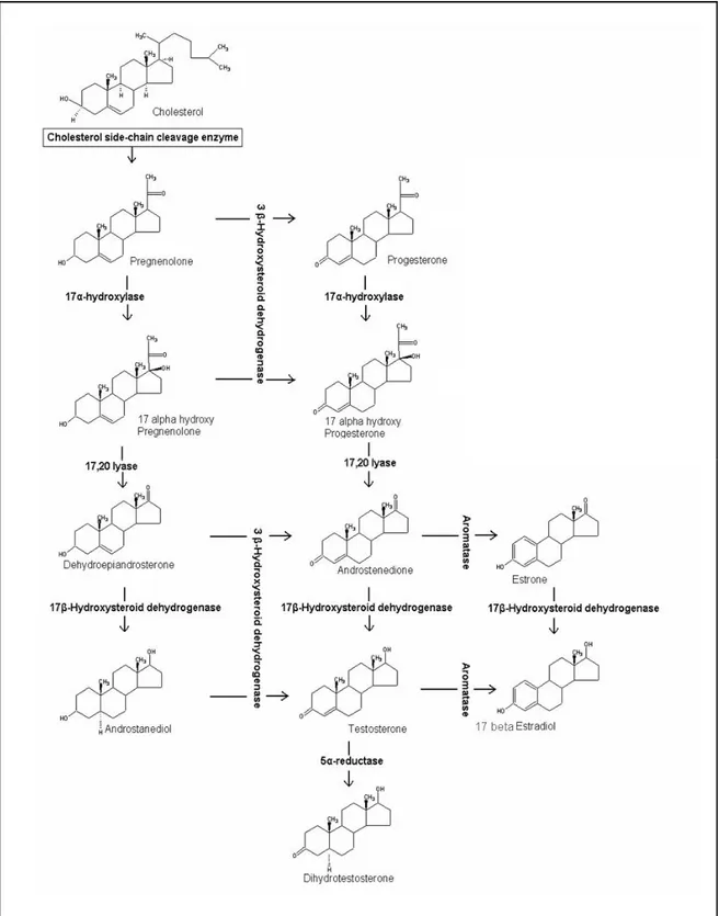
The conversion of cholesterol to testosterone involves a number of steps that are catalyzed by enzymes, predominantly belonging to cytochrome P450 family. The mobilization of cellular sources of cholesterol is achieved through the action of cholesterol ester hydrolase and subsequently, this is converted to pregnenolone by the enzyme cholesterol side-chain cleavage termed cytochrome P450SCC (15) (Fig. 4).
- 11 -
- 12 -
The conversion of cholesterol to pregnenolone is a key step at which regulation of androgen production within the Leydig cells occurs. Availability of cholesterol substrate can be rate-limiting and the intracellular trafficking of cholesterol across mitochondrial membranes is dependent on the steroidogenic acute regulatory protein (STAR) (16-18). The role of this protein has been well demonstrated in patients with mutations in the gene encoding STAR in the disorder termed congenital lipoid adrenal hyperplasia wherein the mitochondria from the adrenals and gonads of these patients are unable to convert cholesterol to pregnenolone (19). Further, the results of studies involving targeted disruption of the mouse gene encoding STAR support the data derived from human studies (20). Pregnenolone may progress to testosterone production through two pathways. It can be converted to progesterone through the enzyme 3βhydroxysteroid dehydrogenase (the D4 pathway) or can be hydroxylated at the 17α position by the enzyme 17-alfahydroxylase to form 17α-hydroxypregnenolone (the D5 pathway). The relative importance of these two pathways vary with the species and the physiological status of the male (21). The further conversion of 17α-hydroxypregnenolone through the D5 pathway involves the formation of the C19 steroid dehydroepiandrosterone catalyzed by the enzyme 17,20 lyase and both steps appear to be catalyzed by a single microsomal enzyme cytochrome P450 c17 encoded by a single copy gene on chromosome 10 (22;23). The conversion of dehydroepiandrosterone to androstenediol is mediated by a microsomal enzyme 17β-hydroxysteroid dehydrogenase encoded by a single gene (24;25). The conversion of
substrates from the D5 to the D4 pathway are catalyzed by the enzyme 3β-hydroxysteroid dehydrogenase (26). In the D4 pathway 17α-hydroxyprogesterone proceeds through the action of cytochrome P450 c17 to androstenedione and testosterone. Testosterone can be converted to a dihydrotestosterone by the enzyme 5α-reductase (27) or can be metabolised to 17β-estradiol by the enzyme aromatase (13;28).
- 13 -
2. Estrogen regulation of testicular function
Evidence supporting a role for estrogen in male reproductive tract development and function has been collected from rodents and humans. These studies fall into three categories: i) localization of aromatase and the target protein for estrogen (ER-alpha and ER-beta) in tissues of the reproductive tract; ii) analysis of testicular phenotypes in transgenic mice deficient in aromatase, ER-alpha and/ or ER-beta gene; and, iii) investigation of the effects of environmental chemicals on male reproduction. Estrogen is thought to have a regulatory role in the testis because estrogen biosynthesis occurs in testicular cells and the absence of ERs caused adverse effects on spermatogenesis and steroidogenesis (29). All of these topics will be individually discussed in this section of the introduction. In males, estrogens derive from circulating androgens. Aromatization of the C19 androgens, testosterone and androstenedione, to form estradiol and estrone, respectively, is the key step in estrogen biosynthesis, which is under the control of the aromatase enzyme (Fig. 5).
- 14 - 2.1 The aromatase gene: structure and regulation
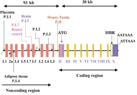
Aromatase is composed of two proteins: a ubiquitous NADPH-cytochrome P450 reductase and a cytochrome P450 aromatase (P450arom), which contains the heme and the steroid-binding pocket. In humans, P450arom is the product of a single gene located in region q21.1 of chromosome 15 and called cyp19, which belongs to the cytochrome P450 gene family. The cyp19 gene is more than 123 kb in length with a coding region of 9 exons (II-X) and 9 nontranslated exons I (30) (Fig. 6).
Figure 6. Schematic presentation of the human aromatase gene. P = promoter; (by Carreau S 2007).
Expression of the cyp19 gene is regulated by tissue-specific promoters producing alternate 5'-untranslated exons I that are then spliced onto a common 3'- splice acceptor site in exon II, upstream of the translation starting site (31-33). Therefore, there is generation of cyp19 variants with different 5’untranslated regions giving rise to different mRNAs; however, the coding sequences are identical and give rise in humans to a single protein composed of 503 amino acids with a molecular mass of 55 kDa. It is of note that P450arom is encoded by a single cyp19 gene in most species except for pigs in which three distinct genes encode
- 15 -
three aromatase isoenzymes (34) and for fish in which two cyp19 genes (specifically expressed in the brain and gonads) have been identified (35). Different mechanisms of regulation of Cyp19 gene expression have been described for various tissues. The synthesis of different aromatase isoforms between species and tissues may involve distinct aromatase genes and/or the function of different promoter elements (36). In human adipose tissue, the primary promoter I.4 lies about 15 kb upstream of the start site of translation (37;38) and is a TATA-less promoter driven by glucocorticoids and class I cytokines e.g.
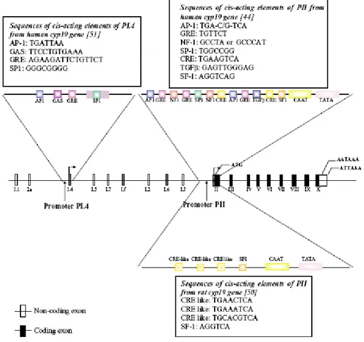
IL-6 and TNFα (33). The region of PII proximal to the translation start site regulates P450arom expression in mammalian gonads (37;39) as well as in Leydig cell tumors (40). Numerous functional motifs have been identified in P.II (33) (Fig. 7).
Figure 7. Structure of the human Cyp19 gene showing the various untranslated first exons and their corresponding promoters. The region around promoter PI.4 and PII from human and PII from rat are expanded to show the identified response elements. Sequences of these are shown in boxes; (by Carreau S 2004).
- 16 -
In the testis, FSH and LH act by increasing concentrations of intracellular cyclic AMP to induce expression of P450arom. Promoter PII activity is therefore regulated by cyclic AMP and requires the transcription factors cAMP response element binding protein (CREB), cAMP response element modulator (CREM) and steroidogenic factor-1 (SF-1). SF-1 belongs to the nuclear orphan receptor superfamily and regulates steroidogenic gene transcription (e.g. P450arom via its interaction with numerous coactivators including CREB binding protein, DAX-1, SOX-9, WT1).
It has been shown that the level of P450arom mRNA is increased in Leydig cells of mice deficient for DAX-1 (41). In addition, it is shown that liver receptor homologue-1 (LRH-1), an SF-1 homologue, wich is present in leydig cells and germ cells, but not in sertoli cells, increases the P450arom gene expression in a mouse leydig cell line (42).
Moreover, it is now clear that not only P.II drives the aromatase gene in rat testis but two additional promoters, P.I.f (brain promoter) and a new one that we called P.I.Tr (testis rat;(43), are involved. It is also demonstrated that the nutritional status of fetuses (44) and aging (Hamden K, Silandre D, Delalande C, El Feki A, Carreau S, unpublished results) can modulate aromatase gene expression in male rats.
2.2 The Estrogen Receptors (ERs)
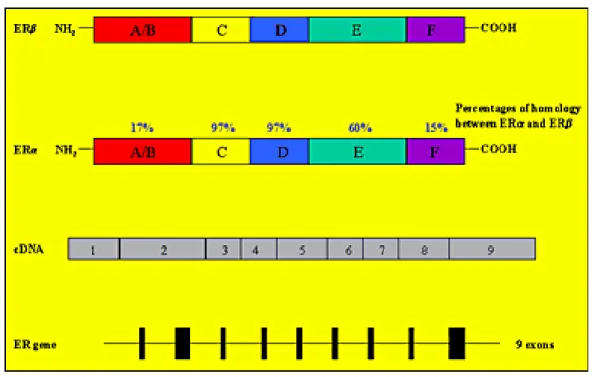
Estrogen actions are mediated by binding to specific nuclear estrogen receptors (ERs), which are ligand-inducible transcription factors regulating the expression of target genes after hormone binding. Two subtypes of ERs have been described: estrogen receptor α (ERα) and the more recently discovered estrogen receptor β (ERβ). The human gene encoding for ERα is located on the long arm of chromosome 6, while the gene encoding for ERβ is located on band q22-24 of chromosome 14.
- 17 -
The two ER (α and β) proteins have a high degree of homology at the amino acid level (Fig. 8).
Figure 8. ERs gene and its products (by Akingbemi BT 2005) .
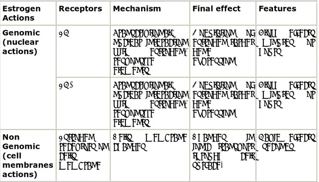
While it is clear that estrogens regulate transcription via a nuclear interaction after binding their receptors, a non-genomic action of estrogens has been recently demonstrated, suggesting a different molecular mechanism accounts for some estrogen actions. In vitro studies showed a very short latency time between the administration of estrogens and the appearance of biological effects. These actions are thought to be mediated through cell-surface receptors, which are not believed to act via a transcriptional mechanism (45). The different types of estrogen action are summarized in Table 1.
- 18 -
Table 1. Estrogen actions and related biomolecular pathways and mechanisms.
Estrogen
Actions Receptors Mechanism Final effect Features
ERα Transcriptional: nuclear interaction with estrogen-responsive elements Modulation of estrogen target gene expression. Slow effects (minutes or hours) Genomic (nuclear actions) ERβ Transcriptional: nuclear interaction with estrogen-responsive elements Modulation of estrogen target gene expression. Slow effects (minutes or hours) Non Genomic (cell membranes actions) Estrogen receptors on cells membrane Cells membrane
changes Changes in ionic transport through cell surface.
Rapid effects (seconds)
ERs are members of the steroid/thyroid hormone super family of nuclear receptors, which share a common structural architecture, and consist of three independent but interacting functional domains: the N-terminal or A/B domains, the C or DNA-binding domain, and the D/E/F or ligand-binding domain (Fig. 8). Binding of a ligand to the ER causes a series of downstream events, including receptor dimerization, receptor-DNA interactions mediated by EREs present in the promoter region of target genes, recruitment of and interaction with transcription factors, and the formation of a preinitiation complex.
Ligand- receptor interactions ultimately cause changes in target gene expression (46). The N-terminal domain of nuclear receptors encodes an activation function called AF-1, which mediates protein-protein interactions to induce transcriptional activity. It is thought that this domain is highly active in ERα-mediated stimulation of reporter gene expression from a variety of ERE-constructs but its activity in the ERβ is limited (47). On the other hand, the C-terminal or ligand-binding domain contains the AF-2 interacting surface that mediates ligand binding and receptor dimerization to stimulate transcriptional activity (48).
- 19 -
Thus, AF-1 and AF-2 are both involved in mediating the transcriptional activation functions of ERs. Although there is a high degree of homology in the DNAbinding domains of ERα and ERβ (about 95%), only a partial homology exists in the ligand-binding domain (~60%) (49). Differences in ligand ligand-binding, in association with other factors, have the effect of altering the pattern of ER-mediated transcriptional activity. For example, some agonists bind both ER subtypes with the same affinity while others preferentially bind to ERα or ERβ (50-52). There is general agreement that ERs function as dimers, and co-expression of ERα and ERβ in the same cell causes the formation of homodimers (ERα/ERα and ERβ/ERβ) or heterodimers (ERα/ERβ), which affect ligand-specificity. The interactions between ERs and EREs are complicated by other factors, including the ability of ERβ to modulate ERα transcriptional activity and recruitment of several protein co-activators and repressors by both ER subtypes. Therefore, the relative amounts of ERα and ERβ in a given tissue are key determinants of cellular responses to estrogen and other ER agonists and antagonists (53). Moreover, ER and other steroid
receptors have the ability to mediate biological effects through non-transcriptional mechanisms mediated by protein-protein interactions occurring between ERs and growth factors e.g., IGF-1 and EGF (54). Furthermore, there is growing evidence for the presence of a small pool of ERs localized to the plasma membrane. For example, BSA-conjugated E2, which is unable to gain entry into the cytosol and acts at the plasma membrane, decreased testicular androgen production in vitro (55). Membrane ER is thought to signal mainly by coupling to GTP-activating proteins and through pathways involving second messengers (e.g., calcium) and kinase cascades (56). The integration of several pathways implies that estrogen action in any particular tissue and organ is the result of activities mediated by genomic and non-genomic pathways although the physiological significance of specific pathways in the testis remains to be elucidated (57) .
2.3 Distribution of ERs and aromatase in the male reproductive system
ERs and the aromatase enzyme are widely expressed in the male reproductive tract in both animals and humans, implying that estrogen biosynthesis occurs in the male reproductive
- 20 -
tract and that both locally produced and circulating estrogens may interact with ERs in an intracrine/paracrine and/or endocrine fashion (45). The concept of a key estrogen action in the male reproductive tract is strongly supported by the fact that male reproductive structures are able to produce and respond to estrogens (58).
2.3.1 ERs and aromatase in rodent testis
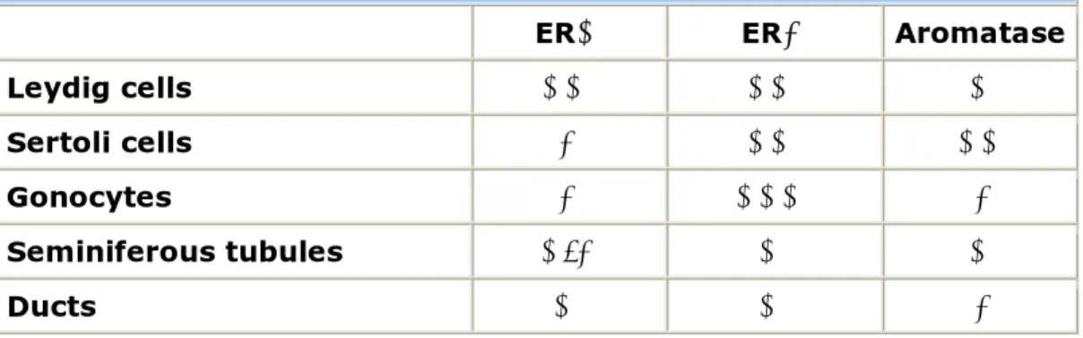
Aromatase and ERs are found at a very early stage of development in the rodent testis, thus suggesting a role for estrogens in influencing testicular development (59-61). ERα is expressed by Leydig cells in the rodent fetal testis at a developmental stage in which the androgen receptor is not yet expressed. The developing efferent ductules and epididymis also express ERα in the fetal rodent. By contrast, it is unclear whether ERα is present within the seminiferous tubules of the fetal testis, with variable results having been reported (60). ERα is abundant in the developing efferent ductules, which are the first male reproductive structures to express ERs during fetal development (62). ERβ is also found early in testis development in the gonocytes, Sertoli cells and Leydig cells, with the gonocytes showing the highest expression suggesting a role for estrogens in their maturation. In addition, ERβ is expressed by rat Wolffian ducts, the structures from which the efferent ductules and epididymis arise (60). Aromatase is expressed in both Leydig and Sertoli cells in the rodent fetal testis, but not in gonocytes and immature structures of seminal tract. ERs and aromatase distribution in the fetal testes is summarized in Table 2.
Table 2. ERs and Aromatase distribution in the rodent fetal testis.
ERα ERβ Aromatase
Leydig cells ++ ++ +
Sertoli cells - ++ ++
Gonocytes - +++ -
Seminiferous tubules +/- + +
- 21 -
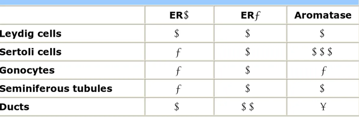
The finding of both aromatase and ERs in the developing fetal testis imply a possible involvement of estrogens in the process of differentiation and maturation of developing rodent testis from an early stage of morphogenesis (59;63). In the postnatal immature rodent testis ERα expression does not occur in the seminiferous epithelium, remaining confined to the Leydig cells, rete testis, efferent ductules and epididymis (Table 3). In the neonatal rodent testis, ERβ is widely expressed by the rat seminiferous epithelium (Sertoli cells and germ cells) as well as by Leydig cells, efferent ductules and epididymis. At this stage ERβ seems to be the only ER in germ cells and is found in pachytene spermatocytes, round spermatids, and perhaps in elongated spermatids of rats and humans (58) (Table 3).
Table 3. ERs and Aromatase distribution in postnatal immature rodent testis.
ERα ERβ Aromatase
Leydig cells + + +
Sertoli cells - + +++
Gonocytes - + -
Seminiferous tubules - + +
Ducts + ++ (?)
Aromatase is expressed by the dividing Sertoli cells and is stimulated by FSH, with the levels of aromatase declining with age. Fetal Leydig cells also have the ability to produce estrogens in response to LH, but aromatase in this cell type is expressed to a lesser degree than during neonatal life. Interestingly the neonatal testis continues to show a greater degree of aromatase expression in the Sertoli cells than in the Leydig cells (the latter only express aromatase to a greater extent in the adult rat testis when they become one of the major sources of estrogens under the influence of LH) (Table 3). Germ cells in immature rats do not yet express aromatase. ERα is expressed in the Leydig cells of both adult rats and mice (64) but not in Sertoli cells. ERα expression in adult rodent germ cells remains to
- 22 -
be confirmed, with its presence in pachytene spermatocytes and round spermatids being suggested by one study yet its absence demonstrated by others such that the prevailing view is that ERα is absent in germ cells. Studies on the precise cellular localization of ERs expression, however, are mainly based on immunocytochemistry, using different antibodies, and led to contradictory results. Whereas, it is generally agreed that both subtypes are expressed by the epithelial cells of the efferent ductules and epididymis, data concerning testicular expression differ between species, possibly due to different specificity characteristics of the antisera used. Knowledge of the distribution of ERα is of great importance in understanding estrogen action on the male reproductive tract. ERα is highly expressed in the proximal reproductive ducts (rete testis, efferent ductules, proximal epididymis) and its expression progressively decreases distally (corpus and cauda of the epidydimis, vas deferens). The highest degree of ERα expression is seen in the efferent ductules of the rat (65) and accounts for one of the most well-documented estrogenic actions on male reproductive system, that of fluid reabsorption from the efferent ductules. It has to be remarked that the concentration of ERα in the male reproductive tract is opposite to that of ERβ, which is more concentrated in the distal tract (Table 4).
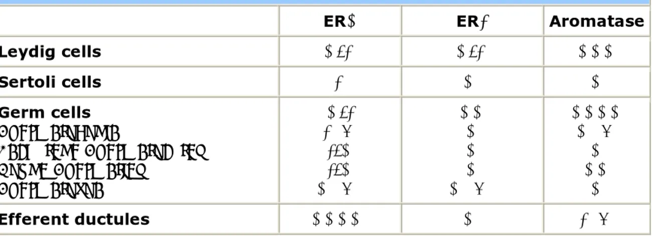
Table 4. ERs and Aromatase distribution in the adult rodent testis.
ERα ERβ Aromatase
Leydig cells +/- +/- +++ Sertoli cells - + + Germ cells Spermatogonia Pachytene Spermatocytes Round Spermatids Spermatozoa +/- - (?) -/+ -/+ + (?) ++ + + + + (?) ++++ + (?) + ++ + Efferent ductules ++++ + - (?)
- 23 -
ERβ is expressed in Leydig, Sertoli and germ cells in adult rodents (66;67)and has also been detected in primate germ cells (68). There is now considerable evidence that germ cells contain both ERβ and aromatase (68). It should be noted that there are some controversies in terms of ERβ localization, with immunohistochemical studies showing some discrepancies, possibly due to methodological differences. It seems that the regulation of gonocyte multiplication, which is under the influence of growth factors and estradiol, may occur through the involvement of ERβ (69). By adulthood, rodent Leydig cells show higher aromatase activity compared to every other age and in comparison to Sertoli cells (70). Aromatase is also expressed at high levels in germ cells throughout all stages of maturation, and its expression appears to increase as the germ cell becomes a mature spermatid (Fig. 9).
Figure 9. Aromatase and estrogen receptors (ER) in adult male rat gonad. Aromatase has been demonstrated in terms of mRNA (RT-PCR), protein (Western blots) and enzyme activity (measurements of estradiol output in culture media) in the various testicular cells. ER: estrogen receptors localization; (by Carreau S 2003, 2005).
- 24 -
Aromatase mRNA and activity, in fact, are found in germ cells from the pachytene spermatocyte stage in both rats and mice, and during their subsequent maturation into round spermatids(61;70;71). Aromatase seems to be present in higher levels in mature spermatids of the rat than in earlier germ cells (61;71;72). It is of interest that aromatase mRNA expression and enzyme activity is higher in germ cells when compared with Leydig cells, suggesting that germ cells may be a major source of estrogen in adult rodents. When fully developed spermatids are released from the epithelium, aromatase remaining in the residual body is subsequently phagocyted by the Sertoli cell. Some aromatase activity remains in the cytoplasmic droplet that remains attached to the flagellum as the sperm make its way through the epididymis, suggesting that mature spermatozoa are able to synthesize their own estrogen as they traverse the efferent ducts (73;74). The ability to synthesize estrogen gradually decreases as the droplet slowly moves to the end of the tail during epididymal transit until it's finally lost. The demonstration of aromatase in sperm is important as it suggests that the sperm itself could control the levels of estrogen present in the luminal fluid, directly modulating functions such as the reabsorption of fluid from the efferent ductules (65).
2.3.2 ERs and aromatase in the human testis
Both ERs have been found in human testis and reproductive tract. In the male fetus ERβ expression is higher than ERα, the latter being absent or expressed at very low levels. In the human fetus ERβ immunoreactivity has been shown in the seminiferous epithelium (Sertoli cells and a few germ cells) and in the epididymis suggesting a role for ERβ in the prenatal development and function of male reproductive structures (75). In adult men ERα was expressed only in Leydig cells, while ERβ has been documented in both Leydig and Sertoli cells and in the efferent ducts (76). The presence of ERs in the human epididymis is still debated, even though recently ERα has been detected in the nuclei of epithelial cells of the caput of the epididymis (77). Both ERα and β have been detected in human pachytene spermatocytes and round spermatids with in situ hybridization (78-80). These latter studies have been contradicted by more recent studies showing strong expression of ERβ in human
- 25 -
testis but failing to find evidence for ERα using immunohistochemistry (81) and RT PCR (82), suggesting that ERβ is the primary mediator of estrogen action in the human testis. Of particular interest is the demonstration of differential expression of wild type ERβ (ERβ1) and a novel human variant form of ERβ, arising from alternate splicing (ERβcx, or ERβ2), in the human testis (83). ERβ2, which may act as a dominant negative inhibitor of ER action, was highest in spermatogonia and Sertoli cells in adult men, suggesting that these cells may be "protected" from estrogen action by the expression of this variant. However wild type ERβ1 was highest in pachytene spermatocytes and round spermatids, which have been proposed to be estrogen sensitive, yet was low in less mature germ cells (81). As previously suggested by Durkee et al. (84), ERs are present in human sperm. In particular it has recently been documented by Luconi et al. (85) that the sperm membrane contains an estrogen receptor-related protein able to bind steroid hormones that may act through a calcium-calmodulin dependent pathway and thus perhaps accounts for a well documented rapid non-genomic action. Aromatase expression in the human testis is present in both somatic and germ cells from pachytene spermatocytes through elongated spermatids (80;86). Aromatase is also expressed in both human Leydig and Sertoli cells (71). Recently, the presence of aromatase has been demonstrated not only in immature germ cells, but also in mature human spermatozoa (87). In contrast to rodents, aromatase expression in human gametes is not lost during transit through the genital tracts since P450 aromatase was demonstrated in ejaculated human spermatozoa at three different functional levels: mRNA expression, protein and activity (87). Thus ejaculated human spermatozoa continue to express P450 aromatase and contain active aromatase, and thus sperm have to be considered a potential site of estrogen biosynthesis. These evidences support the concept that human spermatozoa should be considered a mobile endocrine unit since they are able to synthesize and to respond to estrogens. Again, the presence of functionally aromatase in human spermatozoa permits the conversion of androgens into estrogens throughout the whole transit of reproductive tract, an event that constantly provides free estrogens in the seminal fluid able to act on the cells of the reproductive ducts.
- 26 - 2.4 Role of estrogens in animal male reproduction
In animals, a previously unsuspected physiological role of estrogens in testicular function was revealed by the creation of the ERα knockout (αERKO) mouse. Adult, sexually mature, male αERKO mice are infertile even though the development of the male reproductive tract is largely unaffected (69). Adult testicular histology shows an atrophic and degenerating seminiferous epithelium, together with dilated tubules and a dilation of the rete testis (88). The disruption of spermatogenesis is progressive as the testicular histology is normal at ten days of age but starts to degenerate at twenty-thirty days. By about 40-60 days the tubules are markedly dilated with a corresponding significant increase in testicular volume while the seminiferous epithelium becomes atrophic. A severe impairment in tubule fluid absorption in the efferent ducts was demonstrated to be the cause of infertility in αERKO male mice, and this defect is partially mimicked also by the administration of an anti-estrogen in wild-type mice (65). In the male genital tract the highest concentration of ERα is found in the efferent ducts (89) and the estrogen-dependent fluid reabsorption in this site probably results from estrogen interaction with the ERα that seems regulate the expression of the Na(+)/H(+) exchanger-3 (NHE3). In fact, the disruption of ERα or the use of antiestrogens result in decreased expression of NHE3 mRNA, as well as in a decrease of other proteins involved in water reabsorption, such as aquaporin I (90;91). The lack of fluid reabsorption in the efferent ductules of αERKO male mice and the consequent dilatation of these ductules induces a retroactive progressive swelling of the seminiferous tubules. The seminiferous tubule damage results from the increased fluid pressure and severely impaired spermatogenesis coupled with testicular atrophy as seen at the age of 150 days (65). In addition, reproductive hormones profiles are abnormal in αERKO male mice as serum LH is significantly increased with a consequent elevated serum testosterone and Leydig cells hyperplasia, but FSH remains in the normal range (69). It is also worth noting that detailed investigations into the development of efferent ductules in αERKO male mice suggest that a congenital absence of ERα leads to developmental abnormalities in this tissue (92). The recent production of both aromatase
- 27 -
knockout (ArKO) (93) and ERβ knockout (βERKO) (94) mice supports the idea that in mice estrogen actions on the male reproductive tract are more complex than previously suggested on the basis of the αERKO mice. In fact, unlike αERKO mice, male ArKO mice are initially fully fertile (93), but fertility decreases with advancing age (95), and, conversely, βERKO mice are fully fertile and apparently reproductively normal in adulthood (94). From seven months of age male ArKO mice are not able to sire any litters. Again histology of the testes of one-year-old ArKO mice shows a disruption of spermatogenesis at the early spermatid without significant (95). The late onset of the alterated phenotype in male ArKO mice is attribuitable to estrogenic substance present in their diet and which are capable of agonistic effect on spermatogenesis (96). Despite the phenotype of αERKO male mice, the mechanism involved in the development of infertility is different in ArKO male mice, since the early arrest of spermatogenesis suggests a failure of germ cell differentiation probably caused by the lack of estrogen action at the level of the seminiferous epithelium rather than a problem referable to impaired fluid reabsorption (59). Recent findings from studies in which human germ cells were treated with estrogen in vitro suggest that estradiol may serve as a survival factor for round spermatids and that lack of estradiol may promote apoptosis with a resulting failure in elongated spermatid differentiation (79). Recently studies in mice deficient in both ER α and β (αβERKO mice) showed a male phenotype very close to that of αERKO mice with infertility and dilated seminiferous tubules (69). These findings, together with the observation that βERKO male mice are fully fertile (94), lead to the hypothesis that estrogen activity in the male reproductive tract differs with regard to both the type of estrogen receptor involved in the pathway of estrogenic action and the site of action through the male reproductive tract. Importantly, results from mice lacking functional ERs or aromatase point to an important role for estrogen in the maintenance of mating behaviour in male mice, and that infertility in αERKO, αβERKO and ArKO mice are at least in part due to reductions in various aspects of mating behavior from an early age. The above studies support the concept that a functional ERα, but not ERβ, is needed for the development and maintenance of a normal fertility in male mice (69). Clearly, further studies are needed to fully understand the
- 28 -
precise role of estrogens and their receptors in the establishment and maintenance of male fertility, and the importance of intracrine and paracrine pathways for these effects.
2.5 Role of estrogens in human male reproduction
The demonstration of abundant ERs in human efferent ducts and aromatase activity in human sperm, speaks in favor of the involvement of estrogens in male reproductive function. On the other hand, data from human subjects with congenital estrogen deficiency have provided conflicting and somewhat confusing results. The only man with estrogen resistance discovered up till now, a human equivalent of the ERKO mouse, had normal testicular volumes and a normal sperm count but with slightly reduced motility (97). The four adult men affected by congenital aromatase deficiency showed a variable degree of impaired spermatogenesis (98-101). The patient described by Carani et al., showed both a severely reduced sperm count and an impairment of sperm viability with germ cell arrest at the level of primary spermatocytes (63). A more recent patient had complete germ cell arrest on testicular biopsy but a semen analysis was not performed according to patient's religious views (98;99). Data concerning the patient described by Morishima et al. are lacking since sperm counts were not analyzed (100). It should be remarked that a clear cause-effect relationship between infertility and aromatase deficiency is not demonstrable in the patient studied by Carani et al., since one of his brothers was infertile despite the absence of mutations in the aromatase gene, suggesting an alternate common cause for their infertility (102). Recently a new patient with aromatase deficiency has been described
to have impaired fertility (101), confirming a possible association between congenital estrogen deficiency and infertility. The variable degree of fertility impairment in men with congenital deficiency of estrogen action or synthesis deficiency does not permit a firm conclusion about whether these features are a consequence of a lack of estrogen action or are only epiphenomena, even though a possible role of estrogen on human spermatogenesis is suggested by rodent studies. Recently, the administration of aromatase inhibitors to infertile men with an impaired testosterone to estradiol ratio resulted in an improvement of fertility rate (103), although in the absence of a placebo or control group, these findings
- 29 -
need to be interpreted with great caution. Clearly our knowledge of a role for estrogen in human male reproduction is far from complete. The exposure to the excess of environmental estrogens has been proposed as a possible cause of impaired fertility.
2.6 Effects of excess estrogen on male reproduction
2.6.1 Exposure to excess estrogens in animals
In order to evaluate the effect of estrogen excess on the reproductive tract, several studies have been performed in various animal species treated with diethylstilbestrol, a synthetic estrogenic compound. In male mice, the critical period for Műllerian duct formation is day 13 post-coitus. Prenatal exposure of fetal male mice to DES caused a delay in Műllerian duct formation by approximately two days as well as incomplete Műllerian duct regression with a female-like differentiation of the non-regressed caudal part (104). An increase in the expression of anti-Műllerian-Hormone (AMH) mRNA in male mice fetuses exposed to DES has also been demonstrated. This increase was not accompanied by a regression of the ducts. This data was interpreted to suggest that the asynchrony in the timing of Műllerian duct formation, with respect to the critical period of Műllerian duct regression, led to the persistence of Műllerian duct remnants at birth in male mice. Moreover DES exposure did not impair embryonal genetic development, but increased ERs number, and slightly prolonged the gestation time (cesarean sections were performed to rescue the litter and revealed no difference in size of fetuses from control and DES treated mothers). The timing of DES exposure is crucial to the induction of abnormalities of Műllerian duct development and regression (104). Many studies in rodents suggest that inappropriate exposure to estrogen in utero and during the neonatal period impairs testicular descent, efferent ductule function, the hypothalamic-pituitary-gonadal axis, and testicular function (58). The latter effect can be a direct consequence of exposure to excess estrogen, as well as a secondary effect due to perturbations in circulating hormones or the ability of the efferent ductules to reabsorb fluid. Some studies show that low dose estrogenic substances given during puberty can actually stimulate the onset of spermatogenesis, likely due to
- 30 -
stimulatory effects on FSH (105), highlighting the fact that the effects of excess estrogen on male fertility are often complex. The effects of excess estrogen in the neonatal period can impact upon the testis into adulthood, with permanent changes in testis function and spermatogenesis evident (106).
2.6.2 Aromatase over-expression in rodents
Recently a transgenic line of mice overexpressing aromatase enzyme (AROM+) has been developed (107;108). These mice show highly elevated serum estradiol concentrations, with a reciprocal decrease in testosterone concentrations. The AROM+ males display several of the changes observed in males perinatally exposed to estrogens, such as undescended testes, testicular interstitial cell hyperplasia, hypoandrogenism, and growth inhibition of accessory sex glands. A disruption of spermatogenesis has also been observed which could be a consequence of multiple factors, including cryptorchidism, abnormal Leydig cell function, hypoandrogenemia or hyperestrogenemia. Estrogens are thought to inhibit Leydig cell development, growth and function, resulting in the suppression of androgen production (58). The observation of numerous degenerating germ cells and the absence of spermatids within the seminiferous tubules of AROM+ mice suggest that germ cells development was arrested at the pachytene spermatocyte stage in the cryptorchid testes. Interestingly, the spermatogenic arrest occurred at a stage where P450arom is typically expressed. The spermatogenic arrest found in the AROM+ mice could be explained, at least partially, by the suppression of FSH action. The reduced serum FSH levels in AROM+ males are further evidence of the inhibiting actions of estrogens on FSH secretion in males. No significant differences in the LH concentrations were seen in AROM+ and wild type mice (107;108).
- 31 - 2.6.3 Exposure to excess estrogens in humans
The clinical use of diethylstilbestrol (DES) by pregnant women in order to prevent miscarriage resulted in an increased incidence of genital malformations in their sons (109). In these individuals the presence of Műllerian ducts remnants was found indicating that fetal exposure to DES may have an effect on sex differentiation in men, as is the case in rodents (104). Moreover a large number of structural and functional abnormalities were found, the most frequent being: epididymal cysts, meatal stenosis, hypospadias, cryptorchidism and microphallus (109). The frequency of abnormalities was dependent on the timing of estrogen exposure: in fact, men who were exposed to DES before 11th week of gestation (i.e. the time of Műllerian ducts formation) had a two fold higher rate of abnormalities than those who were exposed only later (109). This data supports the previously discussed hypothesis that the asynchrony between formation and regression of embryonal reproductive structures is determined by estrogen exposure. Various reports have demonstrated that semen quality of men exposed to DES in utero is significantly worse than in unexposed controls (110;111). However, the sperm concentrations of most of the DES exposed men were well above the limit at which subfertility occurs, and it is therefore not surprising that the fertility of these men was reported to be normal (112). The risk of testicular cancer among men exposed to DES in utero has been a controversial issue and several meta-analyses showed no increased risk (113). However more direct evidence will be necessary in order to fully understand this issue. While various studies suggest that environmental estrogens affect male fertility in animal models, the implications for human spermatogenesis are less clear (114). It has been demonstrated that male mice whose mothers have consumed a 29 ng/g dose of bisphenol A for seven days during pregnancy had a 20% lower sperm production as compared to control males (115). Various abnormalities in reproductive organs have also been described in males exposed to bisphenols (i.e. a significant decrease in the size of the epididymis and seminal vesicles and an increase in prostate gland volume), suggesting that bisphenols interfere with the normal development of the Wolffian ducts in a dose-related fashion. Exogenous estrogens
- 32 -
could interfere with the development of the genital structures if administered during early organogenesis, by leading to both an impairment of gonadotropin secretion and by creating an imbalance in the androgen to estrogen ratio, which may account for impaired androgen receptor stimulation or inhibition according to the dose, the cell type and age (1;116;117). An excess of environmental estrogens has been suggested as a possible cause of impaired fertility in humans (118). A progressive decline in sperm count has been reported in some Western countries during the past 50 years, suggesting a possible negative effect of environmental contaminants on male reproductive function (119). Data concerning the role of estrogens in male reproductive structure development remains conflicting. Animal studies suggest that exposure to estrogen excess may negatively affect the development of reproductive male organs. These effects, however, are considered to be the result of an impaired hypothalamic-pituitary function as a consequence of estrogen excess and of the concomitant androgen deficiency (1;117). Much of the knowledge on excess estrogen exposure and human fertility depends upon animal data and the validity of these concepts to humans has not been established.
2.6.4 Aromatase over-expression in humans
In 1996 a boy with aromatase excess syndrome was reported (120). His condition was presumably inherited in an autosomal dominant fashion with sex-limited expression as his father had a history of peripubertal gynecomastia, elevated serum estrogen levels and increased aromatase activity in vitro. The father was fertile and had a normal libido despite a small testicular volume (15 mL bilaterally), and a reduced testosterone level of 234 ng/dL (120). In the son, mild suppression of testicular growth and Leydig cell function probably reflected direct estrogen negative feedback on pituitary gonadotropin secretion. In general, the inhibitory effects of estrogen on reproductive function appear to be milder in males with aromatase excess syndrome than in patients receiving exogenous estrogens or with estrogen-secreting tumors, probably because serum estradiol and/or estrone levels are lower in the former (120).
- 33 -
3. Testicular cancer
3.1 Introduction
Although cancer of the testes is rare, accounting for only about 1 percent of all cancers in men of all ages and about 5 percent of all male genitourinary system cancers, it is the most common cancer in men between the ages of 15 and 35, and the second most common malignancy in men ages 35 to 39 (121-124).
Because the incidence of testicular cancer has risen markedly in the past 20 years, numerous studies are being conducted to explore possible environmental causes, including the mother's diet during her pregnancy as well as her use of diethlstilbestrol (DES) to prevent miscarriage. Researchers are also looking at the increasing presence of estrogen-mimicking pollutants in the environment. The most consistent occupational association has been the elevated rate among men in professional and white-collar occupation, which may be linked to an increased risk observed with lower levels of exercise. Other possible causes include hereditary factors, genetic anomalies, congenital defects involving the reproductive tract, testicular injury, and atrophy of the testes. Viral infections such as mumps, which cause inflammation of the testes, have not been proven to cause cancer.
Testicular cancer comprises a number of different diseases. Nearly all of the main cell types in the testis can undergo neoplastic transformation, but germ cell-derived tumors constitute the vast majority of cases of testicular neoplasms. Ninety-five percent of testicular cancers arise from sperm-forming, or germ cells and are called germinal tumors. The remaining 5 percent are nongerminal tumors. About 40 percent of germinal tumors are categorized as seminomas. Several other types of germinal tumors are referred to collectively as non-seminomas. Somatic cell tumors, known as sex cord-stromal neoplasms and Leydig cell tumors are relatively rare. However, being derived from endocrine active cells, they have endocrine manifestations.
- 34 - 3.2 Leydig cell hyperplasia and tumors
Although Leydig cells in adult men are considered to be a terminally differentiated and mitotically quiescent cell type, in various disorders of testicular function, focal or diffuse Leydig cell hyperplasia is very common. Micronodules of Leydig cells are frequently seen in certain conditions associated with severe decrease of spermatogenesis or germinal aplasia, such as the so-called Sertoli-cell-only syndrome (Del Castillo syndrome), cryptorchidism, or Klinefelter’s syndrome (125). A term “Leydig cell adenoma” is used when the size of a nodule exceeds several fold the diameter of a seminiferous tubule. It is unknown whether Leydig cell adenomas can progress further to form overt Leydig cell tumors, but even if it was the case, it is exceedingly rare. Morphological heterogeneity of hyperplastic Leydig cells is noticeable in some cases.
The mechanism of Leydig cell hyperplasia in the human male is still poorly understood. The disruption of hypothalamo-pituitary-testicular axis leading to an excessive stimulation of Leydig cells by LH can play a central role (125). However, molecular pathways remain largely unknown in the vast majority of cases. In a small subset of cases structural changes of the LH receptor (126;127) and G proteins (128;129) were detected. Constitutively activating mutations of LH receptor cause early Leydig cell hyperplasia and precocious puberty (126;130). Similarly, constitutively activating mutations of Gs-protein in Leydig cells lead into hyperplasia and endocrine hyperactivity (129;131). However, Leydig cell hyperplasia is distinct from tumors that are usually solitary, and the role of the LH receptor and G protein mutations in the tumorigenesis may be limited to few cases (127;129). Leydig cell hyperplasia and adenomas can be easily induced in rodents by administration of estrogens, gonadotropins and a wide range of chemical compounds. Whether or not humans would be similarly susceptible to environmental effects remains to be elucidated. Leydig cell tumors account for one to three percent of testicular neoplasms and occur in all age groups (131-133). Approximately 20 % are found before the age of 10, most often between five and ten years of age. Precocious puberty is the presenting symptom in these cases. Tumors produce androgens, mainly testosterone in a gonadotropin independent
- 35 -
manner, and therefore LH and FSH remain low in spite of external signs of puberty. Approximately 10 % of the boys also have gynecomastia that is caused by estrogens produced in excess due to aromatase activity. In adults, gynecomastia is found in approximately 30 % of patients (133). The excessive androgen secretion rarely causes notable effects in adults.
Leydig cell tumors are always benign in children and can be treated with surgical enucleation when the tumor is encapsulated (123), whereas in adults malignant tumors have been found in 10-15 % of patients, and inguinal orchidectomy remains the treatment of choice (132). The presence of cytologic atypia, necrosis, angiolymphatic invasion, increased mitotic activity, atypical mitotic figures, infiltrative margins, extension beyond testicular parenchyma, and DNA aneuploidy are associated with metastatic behavior in Leydig cell tumors (133;134). Malignant tumors are hormonally active only in exceptional cases. Benign tumors can be treated by orchidectomy, whereas an additional retroperitoneal lymphadenectomy should be considered when the gross or histological features suggest malignancy (134). Malignant tumors have not responded favorably to conventional chemotherapy and irradiation (134). Survival time has ranged from 2 months to 17 years (median, 2 years), and metastases have been detected as late as nine years after the diagnosis (133;134). Therefore follow-up of patients with malignant Leydig cell tumors has to be life-long. The remaining testis may be irreversibly damaged by longstanding high estrogen levels, resulting in both permanent infertility and hypoandrogenism(133-135). The most frequently encountered testicular neoplasm of the mouse and rat is the Leydig cell adenoma. Incidence rates vary in different strains with the Sprague-Dawley SD rat ranging from 1 to 5% and the F-344 rat reaching nearly, 100% (136). Early neoplasm are common in 1 yr old F-344 rats and become increasingly more frequent with age (137). Testicular neoplasia is less frequently observed in alla strains of mice with incidence ranging from 1 to 2,5%. Leydig cell tumors in rodents generally occur in older animals, but in human can arise in any age, the majority between 20 and 60 yr (138). The estimated incidence in man is 0.1-3 per million. The proliferative lesions in Leydig cells in rodents are similar and are observed as a continuous spectrum starting with smaller nodular foci of
- 36 -
hyperplasia leading to large Leydig cell adenomas that can eventually replace the entire testis. The distinction between hyperplasia and adenoma is not always clear, with size being the major factor in the diagnostic criteria, with some debate over when focal hyperplasia becomes early neoplasia and there can be little morphological difference between a hyperplastic nodule and a small Leidig cell adenoma. The major difference between the testicular tumors observed in human and rodents (particularly the rat) are the high incidence of germ cell tumors in human and their occurrence in relatively young men. In rats, germ cell tumors are extremely rare, but Leydig cell tumors can be almost 100% in incidence in certain strains (e.g., Fisher F-344) and occur most frequently in older animals.
3.3 Relationship between estrogens and Leydig tumors
The biological significance of estrogen-induced testicular tumorigenesis has been suggested by the in vivo model overexpressing aromatase transgenic mice (139). Half of these males were infertile and some of them showed larger than normal testis and Leydig cell hyperplasia/Leydig cell tumor. Furthermore, aromatase was markedly immunolocalized in the cytoplasm of interstitial cells, and its immunoreactivity appeared to be strongest in the testes with more advanced stages of neoplasia. The same transgenic animals exhibited estrogen circulating levels at least twice higher than those of control animals and the levels of aromatase mRNA in their testicular tissues were fourfold higher when compared with controls. It is worth to mention how ERα protein in testicular tissue of aromatase transgenic animals was very high with respect to the undetectable levels of control animals. So the authors suggest how an enhanced synthesis of estrogens in tumoral tissues led to an upregulation of ERα expression. Human Leydig cell tumor is a rare testicular neoplasm where estrogen involvement in tumorigenesis process has scarcely been investigated. Recently, a strong aromatase expression in tumoral tissues was revealed by immunostaining and western blotting (140). This finding agrees with a single previous report (141) showing the aromatase immunolocalization in Leydig cell tumors. Furthermore, aromatase expression in control human testicular tissue confirmed Turner’s
- 37 -
report in normal testes (142). The enhanced endogenous synthesis of estrogens by Leydig cell tumor was reflected in both patients by a dramatic increase of estrogen circulating levels, resulting more than twofold higher than those of adult normal male, and by the low testosterone levels (at the lower limit of normal range) (140). Moreover, the ratio between the free fraction levels of the two steroids is furthermore increased in the target tissues. The diminished sperm count and motility of both patients may not only be related to altered testicular tropism, parenchymal compression, and increased local temperature ipsilateral to the tumor (143) but also to the detrimental effects of high circulating estrogen levels on the counter-lateral gonad activity. In the adult normal male, 80% of the plasma estradiol originates from aromatization of testosterone and androstenedione in fat, striated muscle, and other tissues including bone and brain, while 20% in the circulation is secreted by the testis. So, it is reasonable to argue how the excessive increase of estradiol circulating levels, observed in the two patients with Leydig cell tumor, is the consequence of an enhanced rate of testicular secretion. This is confirmed by the evidence that estradiol, as well as E2/T ratio circulating levels, drops dramatically following surgical treatment, while for one of the two patients the persistence of a conspicuous bilateral gynecomastia led to bilateral mastectomia (140). Following orchidectomy, the two patients exhibited a moderate increase of sperm count and a remarkable augment of sperm motility (140). The latter event may be reconducted to the restored testosterone circulating levels likely affecting the entire male genital tract. The expression of ER isoforms in Leydig cell tumor is, to date, unknown. In fact, only a single work showed the ER immunolocalization in cryostat sections of Leydig cell tumor (24); recently, immunohistochemical and western blot analysis of tumoral tissues revealed the expression of ERα and of the two ERβ isoforms, ERβ1 and ERβ2, in neoplastic Leydig cells of both patients. So, the pattern of ERs expression in tumoral cells appears different from that of control Leydig cells, exhibiting only ERβ1 and ERβ2 as previously reported (81;83) .
There is a growing body of evidence that ERα and ERβ can be expressed together in the same cell type and independently expressed in another. Therefore, homodimers (ERα– ERα/ERβ–ERβ) or heterodimers (ERα–ERβ) can be formed (28). The binding affinity of
- 38 -
ERα–ERα/ ERα–ERβ dimers for a consensus DNA estrogen response element is reported to be higher than that of the ERβ –ERβ homodimer (29). Thus, the presence of ERα could reinforce the estradiol-induced tumor cell proliferation. Finally, has been demonstrated that neoplastic Leydig cells are potential estrogen biosynthesis sites and display a modified ER expression pattern. Therefore, it appears reasonable to suggest that the high estrogen levels, measured in the two patients, could play a role in the neoplastic transformation of Leydig cells, while the exclusive presence of ERα in tumoral cells could amplify E2 signaling contributing to the tumor cell growth and progression.