Strana è la nostra condizione qui sulla terra.
Ciascuno di noi viene per una breve visita,
senza sapere perchè,
eppure a volte presagendo uno scopo.
Dal punto di vista della vita quotidiana,
però, una cosa sappiamo per certo:
l' uomo è qui per il bene di altri uomini,
soprattutto di quelli dal cui sorriso e
dal cui benessere dipende la sua felicità.
Data included in this thesis were obtained as a part of the EU FP6 Integrated Project on Genetics of Healthy Ageing (GEHA). Permission to use these data in this thesis has been granted by the GEHA Consortium. It should be noted that future publications by the GEHA Consortium may include these results possibly with additional data and/or analyses. Should this occur, the results presented in the publications by the GEHA Consortium and not this thesis shall be regarded as definitive.
Table of contents
Sommario... I Summary... III List of abbreviations... V
1. Introduction... 1
1.1 Aging and mitochondria... 2
1.2 The human mitochondrial genome... 4
1.3 MtDNA variability... 10
1.4 Point mutations of the mtDNA control region in human aging... 14
1.5 ECHA and GEHA designs... 18
2. Aim of the work... 20
3. Analysis of mitochondrial DNA control region heteroplasmy in families of centenarians (ECHA study)... 22
4. Analysis of mitochondrial DNA control region heteroplasmy in ultra-nonagenarian sib-pairs (GEHA study)... 23
4.1 Introduction... 23
4.2 Materials and methods... 24
4.3 Results and discussions... 32
5. Concluding remarks... 50
6. References... 52 END SECTION: reprint of the published paper “The mitochondrial DNA control region shows genetically correlated levels of heteroplasmy in leukocytes of centenarians and their offspring”.
Sommario
Studi sull' insorgenza e l' accumulo di mutazioni somatiche a carico della regione di controllo del DNA mitocondriale hanno rivelato la presenza di mutazioni tessuto-specifiche in siti importanti per la replicazione e la trascrizione del DNA mitocondriale stesso (Michikawa et al., 1999; Wang et al., 2001). In particolare, una di esse, la transizione C150T, rimodellando una delle due origini di replicazione del DNA mitocondriale, risulta essere correlata alla probabilità di raggiungere età molto avanzate (Zhang et al., 2003). Inoltre, nello stesso studio, dall' analisi di concordanza dei livelli della mutazione in gemelli monozigoti e dizigoti, è stato ipotizzato un controllo genetico nucleare sull' insorgenza e l'accumulo di tale mutazione. Uno studio analogo, condotto su campioni provenienti da una diversa popolazione, ha invece riportato una bassa frequenza della transizione C150T e, di contro, un' alta incidenza della transizione T152C, sebbene non associata ad un rimodellamento dell' origine di replicazione (Iwata et al., 2007).
Alla luce di queste considerazioni, l' attività da me condotta durante il corso del Dottorato di Ricerca è stata rivolta a chiarire se l' insorgenza e l' accumulo di mutazioni somatiche a carico della regione di controllo del DNA mitocondriale possano rappresentare un fenomeno geneticamente controllato, teso a favorire la sopravvivenza in età avanzate.
A tal fine è stato condotto un primo studio in famiglie di centenari reclutate nell' ambito del progetto europeo ECHA (European Challenge for Healthy Aging). I risultati di questo studio hanno rilevato che l' accumulo di mutazioni eteroplasmiche a carico della regione di controllo del DNA mitocondriale è un fenomeno più frequente in soggetti centenari e i loro discendenti (figli e nipoti) rispetto a soggetti di controllo appartenenti alla stessa popolazione. Inoltre, i livelli di eteroplasmia risultavano essere correlati nelle coppie centenari-figli e non associati alla variabilità ereditata del DNA mitocondriale. Quindi, considerando l' aggregazione familiare del fenotipo longevità, questi risultati suggeriscono che l' insorgenza e l' accumulo di mutazioni somatiche nella regione di controllo del DNA mitocondriale siano un fenomeno geneticamente controllato (verosimilmente dal genoma nucleare) che favoriscono la longevità.
Per investigare ulteriormente tale controllo genetico abbiamo condotto un secondo studio su coppie di fratelli ultranovantenni reclutati nell' ambito del progetto europeo
GEHA (GEnetics of Healthy Aging). L' utilizzo di tali campioni ci permetteva anche di analizzare i livelli di eteroplasmia in diversi tipi cellulari (granulociti e linfo-monociti) appartenenti allo stesso soggetto, oltre che di analizzare campioni reclutati in diverse aree geografiche (nord e sud Italia, Finlandia) al fine di rilevare possibili differenze popolazione-specifiche. Questo studio è stato condotto utilizzando 2 diversi approcci molecolari: uno rivolto a rilevare la presenza e l' accumulo di mutazioni somatiche nella regione studiata (DHPLC quantitativa), l' altro rivolto allo screening specifico della mutazione C150T (metodo PARFAH).
L'analisi tramite DHPLC quantitativa ha rivelato una correlazione significativa dei livelli di eteroplasmia tra fratelli appartenenti alla stessa coppia in tutte le popolazioni analizzate. Inoltre è stato evidenziato, applicando metodi di permutazione, come la correlazione osservata fosse dovuta alla relazione di parentela tra i fratelli, come atteso in base all' ipotesi di un controllo genetico sulla presenza e l' accumulo di mutazioni eteroplasmiche a carico di questa regione.
Lo screening della mutazione C150T ha dimostrato la presenza di eventi somatici associati all' insorgenza e l' accumulo della mutazione (analisi nei granulociti e linfo-monociti appartenenti allo stesso individuo) e una forte correlazione tra fratelli della stessa coppia. Questi risultati sono in accordo con quelli riportati precedentemente da Zhang (2003), confermando la presenza di un controllo genetico nucleare sui livelli di eteroplasmia della mutazione C150T, presente a livello di diversi tipi cellulari (granulociti e linfo-monociti) e in campioni appartenenti a diverse popolazioni (Italiana e Finlandese).
Combinando le informazioni ottenute dall' applicazione dei due diversi metodi di analisi molecolare (DHPLC e PARFAH), ci è stato possibile distinguere le mutazioni a carico della posizione 150 dalle mutazioni somatiche a carico di altre posizioni nella stessa regione. L' analisi di correlazione della presenza/accumulo di tali mutazioni ha rivelato una correlazione significativa nelle coppie finlandesi ed in quelle reclutate nel nord Italia, ma non in quelle reclutate nel sud Italia, suggerendo la possibile presenza di fattori popolazione-specifici coinvolti nell' accumulo di mutazioni somatiche a carico di questa regione del DNA mitocondriale. Tali mutazioni potrebbero far parte di un rimodellamento generale (quindi non a carico della sola mutazione C150T) della regione di controllo, controllato dal nucleo, teso a ripristinare la funzionalità mitocondriale in seguito, ad esempio, ad un deterioramento età-correlato.
Summary
Studies on heteroplasmy occurring in the mitochondrial DNA Control Region (mtDNA CR) region demonstrated an age-related accumulation up to homoplasmy of the C150T somatic transition in leukocytes from Italian centenarians in comparison to younger controls. Moreover, 5' end analysis of nascent heavy mtDNA strands consistently revealed a new replication origin at position 149, substituting for that at 151, only in molecules carrying the C150T mutation. Finally, the concordance in mutation levels observed in twins indicated a possible nuclear genetic control on the contribution of somatic events leading to the accumulation of the mutation (Zhang et al., 2003).
In order to further understand the mechanisms leading to the occurrence and the accumulation of the C150T mutation and its relationship with longevity, during my PhD appointment I studied the occurrence and the accumulation in two population samples collected in the frame of two European research projects aimed at studying the biological basis of longevity.
The first sample was collected in Calabria (southern Italy) in the frame of the project European Challenge for Healthy Aging (ECHA) (http://biologia.unical.it/ECHA/). The sample was composed by 100 centenarians, 100 of their children, 100 of their nephews/nieces and of a control group made of 100 geographically matched subjects, belonging to the same age cohort of the children and the nephews/nieces of the centenarians. Through means of an original protocol performed by Denaturing High Performance Liquid Chromatography (DHPLC), we were able to quantify heteroplasmy levels in a fragment of the CR (16531-261 nt, 300bp). We found that the levels of heteroplasmy were similar in centenarians and their relatives (despite the different ages) and significantly higher than those found in controls. Taking into account that longevity runs in families of centenarians, the above results supported the hypothesis that high levels of heteroplasmy in the CR may provide a survival advantage. In addition, we also found that heteroplasmy levels were significantly correlated in parent-offspring pairs, but independent of mtDNA inherited variability. Taken together these results suggest that the occurrence/accumulation of heteroplasmy in the mtDNA CR can be a nuclear genetically controlled phenomenon that provides a survival advantage (Rose et al., 2007).
The second sample was collected in the frame of the Project Genetics of Healthy Aging (GEHA). The sample was composed by 195 ultra-nonagenarians sib-pairs (130 from Italy; 65 from Finland).
DHPLC-based analyses showed that the heteroplasmy levels were significantly correlated in sib-pairs in both populations. What is more, the correlation was nullified when the familial relationship was destroyed by permutation procedures. This finding was in agreement with a genetic control on the heteroplasmy levels in the mtDNA CR. Afterward, we applied a PCR Amplicon Restriction Fragment Analysis by High performance liquid chromatography (PARFAH) (Procaccio et al., 2006) for assessing, in the same sample analyzed by DHPLC, the mutation levels of the C150T transition. This analysis revealed a strong correlation of mutation levels between sibs. Moreover, the analysis of the C150T mutation in different leukocytic cells (granulocytes and lympho-monocytes) of the same subject confirmed that somatic event(s) are involved in such a phenomenon, because in some cases, no concordance was observed between granulocytes and lympho-monocytes. In addition, focusing our attention on the mtDNA CR heteroplasmy not due to the C150T transition, we found a significant correlation between sibs both in Finnish and northern Italians. Intriguingly, such a correlation is not observed between sibs from southern Italy. This finding suggests that population-specific factors play a role in such a phenomenon, and that population-specific somatic mutations, additional to the C150T, may occur in different populations.
On the whole, the work presented in this PhD thesis indicates that: i) mtDNA CR heteroplasmy is genetically controlled; ii) it is beneficial for longevity, as it runs in families of long lived subjects; iii) it can be somatically acquired. In addition, as for other aspects of the correlation between mtDNA variability and longevity, population-specific factors are involved.
List of abbreviations
Acetyl-CoA Acetyl-Coenzyme A
ADP Adenosine DiPhosphate
AIF Apoptosis Inducing Factor
ANT Adenine Nucleotide Translocator
Apaf-1 Apoptotic protease activating factor-1
ATP Adenosine TriPhosphate
BC Buffy Coats
bp base pair
COX Cytochrome c OXidase
CR Control Region
CSB Conserved Sequence Block
CYB CYtochrome B
Cytc Cytochrome c
DHPLC Denaturing High Performance Liquid Chromatography D-loop Displacement-loop
DNA DeoxyriboNucleic Acid
DZ DiZygotic ECHA European Challenge for Healthy Aging F Females FAD+ oxidized Flavin Adenine Dinucleotide FADH reduced Flavin Adenine Dinucleotide
FMN Flavin MonoNucleotide
GEHA GEnetics of Healthy Aging
GPx Glutathione Peroxidase
GPx Glutatione Peroxidase
GR Granulocytes
GTP Guanosine TriPhosphate
GTPase Guanosine TriPhosphatase
H1and H2 Heavy strand transcription sites
HPLC High Performance Liquid Chromatography
H-strand guanine-rich Heavy-strand of mtDNA HVRI and HVRII HyperVariable Region I and II
IAPs Inhibitors of Apoptosis
L Light strand transcription sites L- strand cytosine-rich Light-strand of mtDNA
LDH Lactate DeHydrogenase
LSP Light-Strand Promoter for transcription of the mtDNA LY LYmpho-monocytes
M Males
MnSOD Manganese SuperOxide Dismutase
mRNA messenger RNA
mtDNA mitochondrial DNA
mtPTP mitochondrial Permeability Transition Pore mtTFA mitochondrial Transcription Factor A MZ MonoZygotic NAD+ oxidized Nicotinamide Adenine Dinucleotide NADH reduced Nicotinamide Adenine Dinucleotide
OAA OxalAcetic Acid
OH1 and OH2 primary and secondary Origins of replication of the Heavy strand of mtDNA
OL Origin of replication of the Light strand of mtDNA
Opa1 Optic atrophy 1
OXPHOS OXidative PHOSforilation
PARFAH Pcr Amplicon Restriction Fragment Analysis by HPLC
PCR Polymerase Chain Reaction
r correlation coefficient
RFLP Restriction Fragment Length Polymorphism
RNA RiboNucleic Acid
ROS Reactive Oxygen Species
rRNA ribosomal RNA
SAM Sorting and Assembly Machinery
sTIMs small TIM proteins
TIM Translocase of the mitochondrial Inner Membrane TOM Translocase of the mitochondrial Outer Membrane
tRNA transfer RNA
UCP UnCoupling Protein
1. Introduction
Aging is a natural phenomenon which is characterized by a progressive decline of the normal physiological functions, an increase of morbidity and an increasing risk of death. Although aging is a generalized phenomenon, there is no doubt that a noticeable inter-individual variability exists with respect to the rate and the quality of aging. The continuous increase of life expectancy and of autonomous life expectancy which has occurred in the last decades in western societies clearly suggests that environmental conditions are essential to slow down human aging and to attain longevity. On the other hand, different studies have shown that the rate and the quality of aging are also influenced by genetic factors. Indeed, Perls and co-workers reported examples of familial clustering of longevity (Perls et al., 2000). For instance, Perls et al. (2002) investigated the familial predisposition for longevity in 444 centenarian pedigrees (age major or equal to 100 years) by comparing death rates and survival probabilities of siblings of centenarians with data from the same birth cohort. Interestingly, relative survival probability for these siblings increased markedly at older age, and was significantly higher than that people belonging to the same birth cohort. Moreover, siblings of centenarians maintained a life-long reduction in risk of death of approximately one-half, even up through very old age. The same result was confirmed in relatives (siblings and parents) of super-centenarians (age major or equal to 110 years) where a substantial survival advantage was reported in particular for siblings and mothers of super-centenarians. The familial clustering of extended survival has been confirmed also in the Dutch population by the Leiden Longevity Study, where a significantly low Standardized Mortality Ratios (SMR= observed N° of deaths/expected N° of deaths in the general population, adjusted for sex and calendar period) was found in parents and offspring of long-living subjects, compared to their spouses (Schoenmaker et al., 2006). However, although these studies supported a familial component for extreme longevity they do not further differentiate how much such a component is due to genetic versus environmental factors family members may share. On the other hand, studies on twins allowed to separate the genetic component from the environmental one in the variation of life span. These studies reported that the percentage of the variation in human life span which can be attributed to genetic
differences among individuals ranges from 22% to 33% (McGue et al., 1993; Herskind et al., 1996; Ljungquist et al., 1998). In addition, the studies on twins allowed the heritability of longevity to be estimated as 0.26 for males and 0.23 for females (Herskind et al., 1996). The remaining variation can be divided among conditioning factors that rise in the first part of life (month of birth, education, socio-economic state of parents) and the life style (Vaupel et al., 1998). Moreover, another study on twins demonstrated that having a co-twin surviving to old ages substantially and significantly increases the chance of reaching the same old age, and this chance is higher for MonoZygotic (MZ) than for DiZygotic (DZ) twins (Hjelmborg et al., 2006). These evidences support the existence of a genetic component affecting longevity in humans, especially at advanced ages.
Many studies have been undertaken to find out which genes affect by their variability the genetic component of longevity. These studies have been mainly carried out through case-control approach. However, more recently great efforts have been performed on linkage analysis in long lived sib pairs, which requests larger datasets. These studies have shown different genetic loci affecting longevity: 'cardiovascular genes' (APOE, APOC3, MTTP, ACE), 'immune system genes' (IL6), 'metabolism-related genes' (IGF1, GH1, HFE) and mitochondrial polymorphisms (Christensen et al., 2006 and references therein). The results obtained so far need to be confirmed by longitudinal studies which are currently ongoing. However, their soundness is suggested by the consistency between these results and those observed in model organisms. On the whole, these studies indicate that aging is mainly correlated to the decline of homeostatic capacity of the organism which declines with age, and makes less efficient not only the metabolic pathways for the conservation, the mobilization and the use of nutrients but also the response to external and internal stress.
1.1 Aging and mitochondria
It is generally assumed that damages accumulated after a variety of cellular stresses are the underlying cause of aging. The living organisms have developed different systems in order to protect the cells by curbing and repairing damages to the cellular systems. When the repair or replacement systems are unable to maintain the positive balance, the aging phenotype slowly becomes manifest.
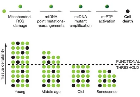
Harman (1956) was the first to propose the free radical theory of aging: aging, as well as associated degenerative diseases, could be attributed to the deleterious effects of Reactive Oxygen Species (ROS) on various cell components. The mitochondrial respiratory chain is a major site of ROS production in the cell and it has been suggested that mitochondria are prime targets of oxidative damage; consequently, the accumulation of defective mitochondria can be one of the major contributors to aging (Kirkwood, 2005). ROS damage to the mitochondria, mitochondrial DNA (mtDNA) and host cell is supposed to be one of the most important factors in determining age-related cellular decline. As shown in Fig. 1, an increased oxidative stress can activate mitochondrial transition pores (mtPTP) leading to apoptosis; this process generates, during time, the loss of cells up to exceed the tissue functional threshold. Harman (1972) proposed a refined version of the free radical theory, the "mitochondrial theory of aging": mtDNA is one of the first targets of ROS, and accumulation of mtDNA damages can be one of the phenomena which lead cells to their functional age-related decline (Fig.1).
Figure 1. Mitochondrial and cellular model of aging
Representation of the mitochondrial role in the energetic life and death of a cell. The bottom diagram represents the loss of cells in a tissue over the life. The minimum number of cells for the tissue to function normally is indicated by the dashed line (modified from Wallace, 2005).
1.2 The human mitochondrial genome The mitochondrion
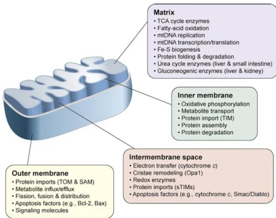
Structurally, the mitochondrion is a membrane-enclosed organelle ranging from 1–10 micrometers in size (Henze and Martin, 2003). The number of mitochondria in a cell varies widely by organism and by tissue type. Many cells have only a single mitochondrion, whereas others can contain several thousand of mitochondria. As shown in Fig.2, mitochondria contain both an inner and an outer membrane, leading to the formation of two aqueous compartments, the matrix and the inter-membrane space, where several specialized functions take place.
Figure 2. The mitochondrial sub-compartments and specialized functions
Examples of compartment-specific processes and proteins are depicted. The main function of the mitochondria is the energy conversion (fatty acid metabolism, citric acid cycle, ATP or heat production); nevertheless it is involved in a series of processes that are crucial for the cell activity (amino acid metabolism, regulation of apoptosis, regulation of cellular calcium, production of oxygen free radicals). Abbreviations: Bax, Bcl-2-associated X protein; Bcl-2, B-cell lymphoma protein 2; Opa1, Optic atrophy 1; SAM, Sorting and Assembly Machinery; sTIMs, small TIM proteins; TIM, Translocase of the mitochondrial Inner Membrane; TOM, Translocase of the mitochondrial Outer Membrane (modified from Ryan and Hoogenraad , 2007).
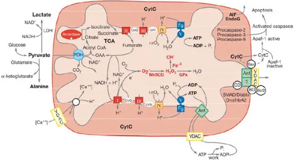
The main mitochondrial function is energy production. The process by which the mitochondrion generates energy is called OXidative PHOSphorylation (OXPHOS). The OXPHOS process, consisting in the oxidation of organic acids and fats to create a capacitor across the mitochondrial inner membrane, generates energy stored as adenosine triphosphate (ATP) molecules (Fig. 3). The OXPHOS process is efficient, but a small percentage of electrons may prematurely reduce oxygen, forming Reactive Oxygen Species (ROS), such as superoxide, as a toxic by-product of OXPHOS. This can cause oxidative stress within mitochondria and may contribute to the decline in mitochondrial function. As shown in Fig. 3, the mitochondria are also the major regulators of apoptosis, accomplished via the mitochondrial permeability transition pore (mtPTP) (Chipuk et al., 2006). The opening of mtPTP can be stimulated by: a) the mitochondrial uptake of excessive Ca2+, b) an increase of the oxidative stress, c) a decrease of the mitochondrial membrane potential, d) a decrease of the intra-mitochondrial ATP concentration (Wallace DC, 2005).
It is important to note that mitochondria should not be considered as individual organelles independent from each other, but as a dynamic network undergoing directed movements as well as fission and fusion (Ono et al., 2001). In fact, the state of the mitochondrial network morphology appears to influence a variety of other cellular functions. Mitochondrial fusion has been suggested to play an important role in mitochondrial proliferation (Chen et al., 2004) and in the propagation of signals through the organellar population (Chan et al., 2006). The mitochondrial fission and fusion machineries as well as proteins involved in mitochondrial distribution have also been shown to act in cell differentiation (Park et al., 2001), in regulating apoptotic events (Suen et al., 2008) as well as in aging (Lopez.Lluch et al., 2008).
Figure 3. Mitochondrial functions
The diagram shows the relationships of mitochondrial OXPHOS to (a) energy (ATP) production, (b) reactive oxygen species (ROS) production, and (c) initiation of apoptosis through the mitochondrial permeability transition pore (mtPTP). Abbreviations: Acetyl-CoA, acetyl-coenzyme A; ADP or ATP, adenosine di- or triphosphate; ANT, adenine nucleotide translocator; Apaf-1, apoptotic protease activating factor-1; cytc, cytochrome c; GPx, glutathione peroxidase-1; LDH, lactate dehydrogenase; MnSOD, manganese superoxide dismutase; NADH, reduced nicotinamide adenine dinucleotide; SMAD/Diablo, antagonizes inhibitors of apoptosis (IAPs); Omi/Htr A2, serine protease 24; TCA, tricarboxylic acid cycle; VDAC, voltage-dependent anion channel; I, II, III, IV, and V, OXPHOS complexes I to V (modified from Wallace, 2005).
The human mitochondrial genome, located in proximity of the inner mitochondrial membrane and organized into nucleoprotein complexes (nucleoids), is a circular DNA molecule of 16,569 base pairs which corresponds to a molecular weight of about 10 million Daltons. The normal mtDNA state is thought to be as a super coiled structure (Richter et al., 1988). In most cells it represents only the 0.5–1 % of the total DNA content (Anderson et al., 1981). The two strands of the mtDNA can be distinguished because of their different G+T content and can be separated by density in denaturing gradients, giving a Heavy or H-strand and a light or L-strand.
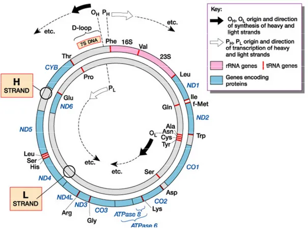
One of the main characteristics of animal mtDNA is the compact gene organization, with all the coding sequences contiguous to each other or separated by a few bases (Anderson et al., 1981). The human mtDNA contains only 37 genes (coding for 13 polypeptides, 22 tRNAs, and 2 rRNAs) and two non-coding regions (the control region
for mtDNA transcription and replication and a short segment containing the L-strand origin of replication) (Fig.4).
Figure 4. Map of the human mtDNA
Human mitochondrial genome has two strands, a guanine-rich heavy strand (H) and a cytosine-rich light (L) strand. Twenty-four encode for the translational machinery of the mtDNA (22 tRNAs, 2 rRNAs). The remaining 13 genes encode subunits of the OXPHOS system: ND1-6 and NDL4 for complex I (NADH dehydrogenase); CYB for complex III (cytochrome b); CO1-3 for complex IV (cytochrome c oxidase); ATPase 6 and 8 for complex V (ATP synthetase). MtDNA is replicated from two origins, OH for the H-strand,and OL for the L-strand (modified from Tom Strachan and Andrew Read, Human Molecular Genetics 3rd Edition, 2003).
All the 13 polypeptides are subunits of the OXPHOS machinery of the inner membrane. The remaining 76 subunits of the respiratory chain, and proteins involved in the mitochondrial activity (about 1500 genes) are encoded by the nuclear genome and must be imported into the mitochondria (Fig.5). From this perspective, to understand human cell biology and its relationship with health and diseases, it is necessary to pay attention to the intricacy of relationships among mitochondria and between mitochondrial and nuclear genomes.
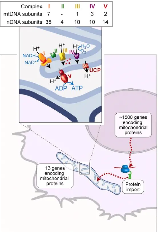
Figure 5. Mitochondrial biogenesis and the OXPHOS machinery
The protein complement of mitochondria consists of 13 subunits, encoded by mtDNA, which are synthesized in the organelle. The remaining proteins are encoded by nuclear genes and are made in the cytosol and imported into mitochondria (Calvo et al., 2006). Inset: the OXPHOS machinery of the inner membrane consists of complexes I–IV, involved in electron transfer and proton export to the intermembrane space, whereas complex V uses the proton gradient to generate ATP. Uncoupling protein (UCP) uses proton flow to generate heat. Cytochrome c (red ) is found in the intermembrane space. The total number of subunits encoded by nuclear and mtDNA are shown for each of the OXPHOS complexes (modified from Ryan and Hoogenraad , 2007).
Mitochondrial genetics
The studies in the field of mitochondrial genetics have shown a series of peculiarities and differences compared to the nuclear genome.
1) The mitochondrial genome is maternally inherited; the few mitochondria from the sperm cell that could enter the oocyte during fertilization are actively eliminated by a ubiquitin-dependent mechanism (Sutovsky et al., 2000). During oogenesis, mtDNA suffers a bottleneck phenomenon by which only a small subset of mtDNA molecules are amplified and transmitted to the offspring (Marchington et al., 1998). On this topic recent data are emerging: by studying the fate of random mtDNA mutations in the mammalian germ-line Stewart and co-workers (Stewart et al., 2008) demonstrate the importance of a purifying (negative) selection in shaping mitochondrial sequence diversity. Moreover, in the same paper, the authors propose that mtDNA in germ cells is under a different selective regime than mtDNA in somatic cells. The massive proliferation of mtDNA during oogenesis (up to about 100,000 mtDNA molecules) and the subsequent reduction of mtDNA copies (approximately 950-1,550) provide ample opportunities for functional testing of mtDNA during female germ-line development. Future research is required to unravel molecular nature and mechanism for this selection.
2) The evolution rate of mtDNA is much faster than that of the nuclear genome (Brown et al., 1979). Several reasons are invoked to explain this fact: mtDNA is less protected by proteins, it is physically associated with the mitochondrial inner membrane where damaging reactive oxygen species are generated, and it appears to have less-efficient repair mechanisms than the nucleus (Pinz & Bogenhagen, 1998).
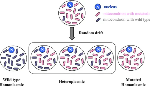
3) Cells are polyploid with respect to mtDNA: most mammalian cells contain hundreds of mitochondria and, in turn, each mitochondrion contains several (2–10) copies of mtDNA (Wiesner et al. 1992). If all mtDNA copies are identical, we have a condition known as homoplasmy, but if the mtDNA copies are not identical, we have a condition known as heteroplasmy (Fig. 6). At cell division mitochondria and their genomes are randomly distributed to daughter cells and hence, starting from a given heteroplasmic situation, different levels of heteroplasmy up to homoplasmy can arise in different cell lineages (Fig. 6).
N N NN NN NN NN N N Wild type Homoplasmic Mutated Homoplasmic Heteroplasmic Random drift N nucleus
mitocondrion with mutated mtDNA
mitocondrion with wild type mtDNA
N
N nucleus
mitocondrion with mutated mtDNA
mitocondrion with wild type mtDNA
Figure 6. Schematic representation of homoplasmy and heteroplasmy
Therefore, in a tissue, heteroplasmy can be observed at three different levels: - intercellular, in which mutant and wild-type mtDNA occur in different cells
- intracellular and inter-mitochondrial, in which mutant and wild-type mtDNAs occur in different mitochondria in the same cell
- intracellular and intra-mitochondrial, in which mutant and wild-type mtDNAs occur in the same organelle.
1.3 MtDNA variability
Features and function of mtDNA have attracted the attention of many researchers in the last decades. In fact, human mtDNA is largely studied for both inherited and somatic variability.
Inherited mtDNA variability
The inherited mtDNA variability implies both rare (mutations) and common (polymorphisms) variants. It goes through to replicative segregation (random drift of mtDNA molecules) and selection (negative or positive selection) during oogenesis. The inherited mtDNA mutations in humans often cause severe pathologies. These pathogenic mtDNA mutations fall into three categories: rearrangement mutations (insertions and deletions), polypeptide gene missense mutations, and protein synthesis (rRNA and tRNA) gene mutations. These mutations, escaping purifying selection
during the oocyte development, are inherited and, when they exceed their functional threshold, the pathologic phenotype becomes manifest. To date, about a hundred of different mtDNA inherited mutations associated with mitochondrial diseases are known (Di Mauro, Hirano, 2005). On the other hand, during human evolution, a number of non-pathogenic variants reached polymorphic frequencies. MtDNA uniparental inheritance and high mutation rate make the study of mtDNA polymorphisms a powerful tool to reconstruct genetic relationships among human populations and human evolution (Ingman et al., 2000; Finnila et al., 2001; Maca-Meyer et al., 2001; Herrnstad et al., 2002; Tanaka et al., 2004; Coble et al., 2004; Kivisild et al., 2006). In fact, clusters of haplotypes (haplogroups) have been recognised and utilized to display human radiations across the continents (Torroni et al., 1996; Finnila et al., 2001; Herrnstad et al., 2002; Silva et al., 2002; Kong et al., 2003). As a result, the mutation history of human mtDNA sequence variation can be reconstructed as a single sequential mutational tree radiating from the last common mtDNA ancestor, that existed between 150,000 and 200,000 years before present (YBP) (Fig.7). Although originally attributed to genetic drift, considerable evidences suggest that the branches of the mtDNA tree correlate with the geographic origin of indigenous human populations (Wallace et al., 2003). In fact, the major transitions in mtDNA types strongly correlate with latitude, suggesting that climatic selection played a role in shaping the present worldwide distribution of mtDNA variation. In particular, the variants affecting ATP and heat production are more frequent in cold areas where they confer an advantage (Wallace, 2007). Evidence that climatic adaptation has influenced the geographic distribution of mtDNA diversity was obtained by analyzing regional gene-specific variation (cob, cox1, atp6 genes, various nad subunit genes) (Mishmar et al., 2003). As an example, the atp6 gene was highly variable in the Arctic but was strongly conserved in tropical and temperate zone: this suggests that climatic selection may have favoured mtDNA variants that lowered the coupling efficiency of ATP production in order to increase heat production, thus permitting humans to survive in the cold northern climates (Mishmar et al., 2003).
Africa Europe Asia
Africa Europe Asia
Figure 7. Phylogenic tree of human mtDNA coding sequence variants
Haplogroups, groups of related haplotypes harbouring characteristic mtDNA sequence polymorphisms, are derived from a founding haplotype. Each haplogroup is designated by a letter with or without a subdividing letter. In this tree, the ticks around the perimeter of the circle represent the individual mtDNA sequences analyzed (1,125 sequences) to reconstruct the phylogenic tree (Ruiz-Pesini et al., 2004). The mtDNA haplogroups have proven to be geographically associated (Wallace, 2005) (modified from Wallace, 2007).
Evidences suggesting that mtDNA common variability is not neutral supported the idea of a mitochondrial paradigm for metabolic and degenerative diseases in which mtDNA common variability would play a role (Wallace, 2005). This is also in agreement with the observation that some mtDNA polymorphisms correlate with mitochondrial diseases (Torroni et al., 1997; Reynier et al., 1999) and complex traits (Wallace, 2005). Interestingly, mtDNA polymorphic variants were shown to affect also longevity phenotypes (Tanaka et al., 1998; De Benedictis et al., 1999; Ross et al., 2001; Niemi et al., 2003, Bilal et al., 2008). Multiple studies have demonstrated that in Europeans the haplogroup J is enriched in extremely old people and thus it seems to slow the aging process (De Benedictis et al., 1999; Ross et al., 2001; Niemi et al., 2003). A similar observation has been made for haplogroup D in the Japanese
population (Tanaka et al., 1998; Bilal et al., 2008). In addition, the European haplogroups J and U have been found to be protective for Parkinson’s disease (Ghezzi et al., 2005). The association of the European haplogroups J and U with longevity and neuroprotection may be explained by assuming that polymorphisms occurring in haplogroups J and U partially uncouple OXPHOS, generating heat and leaving fewer excess of electrons to generate ROS: this would be protective of neurodegenerative diseases and aging which are thought to be caused by a elevated mitochondrial ROS damage (Wallace, 2007). Of course, experimental studies are needed to verify the above hypothesis.
Somatic variability
MtDNA somatic variability is due to stochastic mutations and their replicative segregation in post-mitotic tissues. Several studies suggest a link between mtDNA mutations and aging phenotypes (Trifunovic et al., 2004 and references therein). Indeed, many studies have reported an age related accumulation of mtDNA somatic mutations. However, it is still debated whether mtDNA mutations are just markers of biological age, or they are among the causes of the age related decline. To clarify this point several studies on murine models have been carried out in the recent years. In these studies, to have direct insights on the effects of mtDNA mutations in aging, mouse models were engineered to have defects in the proofreading function of mitochondrial DNA polymerase (Pol γ). This led to the progressive, random accumulation of mtDNA mutations during the course of mitochondrial biogenesis. In these experiments the mtDNA mutator mouse displayed a completely normal phenotype at birth and in early adolescence, but subsequently it acquired many features of premature aging (Trifunivic et al., 2004). Moreover, studies in human muscle cells with atrophy have highlighted that mtDNA deletions are largely clonal and absent from phenotypically normal regions within individual muscle fibers (Fayet et al., 2002). Similar results have also been observed in buccal, epithelial and neuronal cells where age-related dysfunctions had been diagnosed (Cottrell et al., 2001; Cottrell et al., 2002; Kraytsberg et al., 2006). Although these studies do not prove causality, they provide strong evidence in support of the hypothesis that mtDNA deletions contribute to aging in mammals. Probably, senescence leads to mtDNA deletions which, on turn, exacerbate the effects of the age related decline (Wallace 2007).
Although the presence of deletions in the mtDNA of aged people has been known for a long time, the presence of point mutations has been recognised (Michikawa et al., 1999; Wang et al., 2001). In particular, Michikawa et al. (1999) found that long living subjects accumulate a number of tissue specific point mutations at critical positions of the mtDNA Control Region (CR).
1.4 Point mutations of the mtDNA control region in human aging
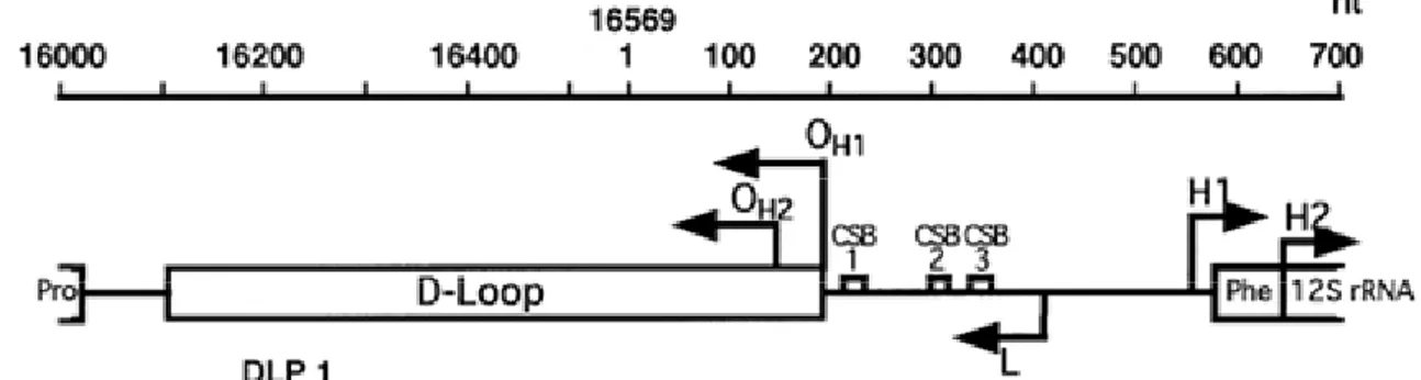
The human mtDNA CR is the main regulatory region for mtDNA transcription and replication(Fig. 8). The mtDNA CR spans about 1.1 kb between tRNAPhe and tRNAPro and contains:
1. the two initiation sites for the transcription of the H strand (H1 and H2 sites),
2. the promoter for the transcription of the L strand (L), which is also the promoter for the RNA primer of the H-strand synthesis,
3. three conserved sequence blocks (CSB),
4. the Displacement-loop (D-loop), a triple-stranded structure of 500–700 nucleotides that remains annealed to the parental L-strand, in which the primary (OH1) and secondary (OH2) initiation sites of H-strand synthesis are localized.
Figure 8. Schematic representation of the mtDNA control region
OH1 and OH1= primary and secondary origin of H-strand Synthesis. CSB1-3= conserved sequence blocks 1-3 (Walberg and Clayton, 1981). H1, H2, L: start sites for transcription of rDNA, whole H-strand and light (L)-strand, respectively (Ghivizzani et al., 1994). Pro, Phe: tRNAPro and tRNAPhe genes (modified from Attardi, 2002).
The mtDNA D-loop region is the most variable non coding portion of the human mtDNA, containing two hypervariable regions (HVR), HVRI and II (Hasegawa and Horai, 1991; Stoneking et al., 1991). Although the function for most of the CR is not well understood yet, the observation that the central domain of this region is highly evolutionarily conserved suggests that the CR is selectively constrained (Sbisà et al., 1997).
Researchers have recently focused their attention on the accumulation of mutations in the mtDNA CR. This choice is chiefly based on the consideration that this region has a greater susceptibility to mutations than other mtDNA regions (Hasegawara and Horai, 1991). If the same mechanisms operating to create variation in evolution also operate during aging, this region could represent a good model for getting insights into the mitochondrial theory of aging (Attardi, 2002). In addition, this region also contains functional elements important for mtDNA transcription and replication: mutations on this region can affect the entire mitochondrial functions influencing mtDNA replication and transcription.
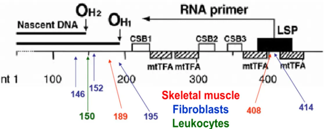
Studies on the control region have established that some specific base-substitution mutations can reach high levels in fibroblast cells derived from aged individuals (Michikawa et al., 1999) and also in skeletal muscle (Wang et al., 2001) (Fig.9). The reason why these specific mutations accumulate in mtDNA is unclear, but they are tissue-specific and occur in mtDNA control sites for replication and transcription. The localization of these aging-dependent mutations at critical control sites provides strong evidence that the mutations themselves may confer a segregation advantage to the mutant molecules. Moreover, the tissue-specificity of these point mutations and their localization at critical sites for H-strand mtDNA synthesis, suggest the involvement of cell type-specific proteins or cofactors that could provide a segregation advantage to the mutant molecules or, alternatively, could affect the susceptibility of these mtDNA sequences to oxidative damage or to replicative errors.
Skeletal muscle
Fibroblasts
Leukocytes
Skeletal muscle
Fibroblasts
Leukocytes
Figure 9. Tissue-specific aging-dependent somatic mutations identified in human mtDNA main control region
OH1 and OH2, primary and secondary origin of H-strand synthesis; LSP, promoter for transcription of L-strand and synthesis of primer for H-strand synthesis; CSB1, CSB2, and CSB3, conserved sequence blocks 1, 2, and 3. The positions of binding of mitochondrial transcription factor A (mtTFA; the densely hatched rectangle indicates a position of high-affinity binding) are shown . Blue arrows and numbers indicate the fibroblast-specific point mutations, red arrows and numbers indicate the skeletal muscle-specific point mutations, green arrow and numbers indicate the C150T transition found in leukocytes (modified from Zhang et al., 2003).
The special case of C150T mutation
One of the most important findings on the correlation between CR somatic mutations and aging came from the study of Zhang and co-workers (Zhang et al., 2003).
By a large-scale screening of the mtDNA CR in leukocytes from an Italian population, the authors observed that the homoplasmic C150T transition was more frequent in centenarians than in younger individuals. The analysis of the C150T mutation in Lympho-monocytes (LY) and Granulocytes (GR) from the same individual suggested that the mutation can be both inherited and/or somatically acquired. In addition, by the analysis of concordance in mutation levels between leukocytes of Monozygotic and Dizygotic twins, a higher concordance between MZ than DZ twins was observed. In the same study, by using fibroblast longitudinal studies, the authors showed an age-related somatic expansion of the C150T mutation. Finally, the authors found that 5' end analysis of nascent heavy mtDNA strands in fibroblasts or immortalized lymphocytes consistently revealed a new replication origin at position 149, substituting for that at 151, only in samples carrying the C150T mutation.
On the whole, the above findings led the authors to conclude that somatic events, probably under nuclear genome control, contribute to the selective accumulation of the C150T mutation in centenarians and twins (Zhang et al., 2003).
In a subsequent study, a significant greater frequency of the inherited C150T transition was found in leukocytes of very old Finnish and Japanese subjects than in leukocytes from younger individuals (Niemi et al., 2005). On the other hand, the analysis of the C150T transition in leukocytes from three groups of an Ashkenazi Jew population (female centenarians, their mixed gender offspring, and mixed gender control subjects) (Iwata et al., 2007) revealed a different situation with respect to the report of Zhang et al. (2003). In particular, the frequency of the C150T mutation was very low and, by contrast, the T152C and T195C transitions showed a fairly high frequency in all three groups of subjects. Furthermore, it was found that the heteroplasmic form of the T152C transition (presumably originated from somatic events) increases with age, but it is not associated with changes in the replication origin. This finding suggests that, if a nuclear genetic control on the accumulation of the C150T mutation (or other type of mutations in the mtDNA CR) exists, different polymorphic forms of nuclear genes and/or environmental changes may have a different effectiveness on the occurrence/accumulation of mtDNA somatic mutations. In line with these considerations, population-specificity has been already reported in relation to mtDNA inherited polymorphisms and human longevity (Dato et al., 2004). So it is arguable that the environmental changes and/or the specificity of the population gene pools can affect strongly the phenomena linked to the occurrence/accumulation of mtDNA CR somatic mutations, leading to different situations.
Taken together, these findings add intriguing clues to the relationship among mtDNA somatic mutations, aging and longevity phenotype. In addition, they underlie the relevance of the cross-talk between nucleus and mitochondria in complex traits and offer an interesting background to further investigations.
In order to study the somatic variability of mtDNA in aging and its correlation with longevity, a great advantage is provided by population samples including relatives of centenarians of different origin. This allows to verify if somatic mutations are shared by relatives of centenarians, if a genetic control of their occurrence exists, and, finally, if population-specific patterns can highlighted. For this purpose we take advantage by large population samples collected in the frame of the ECHA and GEHA projects.
1.5 ECHA and GEHA designs
The European Challenge for Healthy Aging (ECHA) and the GEnetics of Healthy Aging (GEHA) are two study designs aimed at identifying the key factors (genetic and environmental factors) involved in modulating the quality of human aging across Europe.
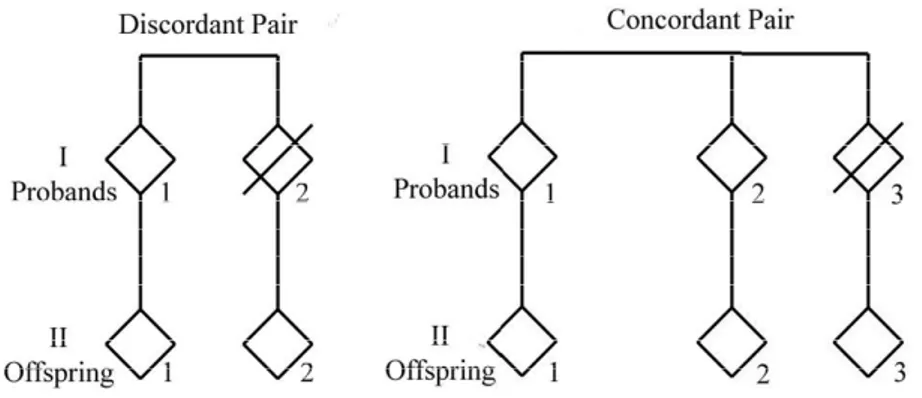
The ECHA project (http://biologia.unical.it/ECHA/) used for the first time an original study design based on the concordance/discordance between siblings of the longevity phenotype. The ECHA project started from the consideration that sib-pairs in which both sibs attained longevity (concordant pairs) are expected to share genetic and intra-familiar environmental factors favouring longevity, while sib-pairs in which a sole sib shows the longevity trait (discordant sib-pairs) are expected to share intra-familiar environmental factors but not the longevity genetic factors (De Rango et al., 2008). According to such a consideration, in the frame of the ECHA project centenarian families that were composed by a centenarian (and his/her brother/sister for the concordant families), his/her child, his/her nephew (the child of the centenarian’s sibling) were recruited (Fig.10).
Figure 10. The structure of the ECHA families
The recruited subjects came from three different European areas: southern Denmark (Denmark), Languedoc-Roussillon (France) and Calabria (Italy).
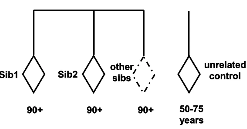
The GEHA project (http://www.geha.unibo.it/) is one of the largest and most ambitious projects ever financed in Europe: it gathers 24 European partners (coming from 11 different countries), and one Asian partner (from China). The idea is to overcome the fragmentation of the research of the genetics of aging in Europe by setting up a coherent and integrated program of research using a multidisciplinary approach (medical, genetic, demographical, bio-informatic and so on) on the study of human aging and longevity. According to many data indicating a strong familial component of longevity (Schoenmaker et al., 2006; Hjelmborg et al., 2006; Christensen et al., 2006), and according to studies demonstrating that the first degree of relatives (parents, siblings and offspring) of long lived subjects have a significant survival advantage (Perls et al., 2002; Perls et al., 2007) the GEHA consortium chose to collect an unprecedented number (2650) of trios, composed by two (or more) long living sibs (90+ years) and geographically matched younger controls (50-75 years), coming from 15 different European areas (Franceschi et al., 2007) (Fig.11).
Sib1 Sib2 other sibs
90+ 90+ 90+
unrelated control 50-75
years Sib1 Sib2 other sibs
90+ 90+ 90+
unrelated control 50-75
years Sib2 other sibs
90+ 90+ 90+
unrelated control 50-75
years
2. Aim of the work
The human mtDNA Control Region (CR) is the main regulatory region for mtDNA transcription and replication. It contains functional elements important for mtDNA transcription and replication: mutation on this region can affect the entire mitochondrial functions influencing the mtDNA replication and transcription. In addition, the mtDNA D-loop region is the most variable non coding portion of the human mtDNA. Recent studies on this region reported that there are tissue-specific age-related point mutations altering critical control sites for mtDNA transcription and replication (Michikawa et al., 1999; Wang et al., 2001). In particular, one of them, the C150T mutation, is more frequent in leukocytes of centenarians than in younger controls. The occurrence and the age-related accumulation of this mutation seems to be genetically controlled, and it seems to favour longevity (Zhang et al., 2003). Taking into account these considerations, the aim of the work carried out during my PhD appointment was:
1. to clarify whether the occurrence/accumulation of heteroplasmy in the mtDNA control region is a genetically controlled event that may favour longevity;
2. to investigate whether different blood cell types are concordant in the occurrence/accumulation of heteroplasmy;
3. to investigate the occurrence/accumulation of heteroplasmy in the mtDNA control region in different populations.
To investigate the first point, we analyzed population sample collected in the frame of the ECHA project (http://biologia.unical.it/ECHA/). The results of this study have been already published (Rose et al., 2007). In summary, the results suggest that the occurrence/accumulation of heteroplasmy in the mtDNA CR can be a genetically controlled phenomenon, likely due to the nuclear genome, that provides a survival advantage.
To further investigate such a genetic control we performed a second study on sib-pairs selected for longevity collected in the frame of the GEHA project (http://www.geha.unibo.it/). The hypothesis of our work was that if the mtDNA CR heteroplasmy is genetically determined, so that genetic factors encoded by the nuclear genome are important in its occurrence/accumulation, we expect that sib-pairs selected
for longevity will correlate for mtDNA CR heteroplasmy levels. In addition, GEHA samples allowed to analyze heteroplasmy in different blood cells (granulocytes and lympho-monocytes) and in different populations. This study is described in Chapter 4 of this thesis.
3. Analysis of mitochondrial DNA control region heteroplasmy in
families of centenarians (ECHA study)
(see END SECTION)
Abstract
Background: Studies on heteroplasmy occurring in the mitochondrial DNA (mtDNA) control region (CR) in leukocytes of centenarians and younger subjects have shown that the C150T somatic transition is over-represented in centenarians. However, whether the occurrence/accumulation of heteroplasmy is a phenotypic consequence of extreme ageing or a genetically controlled event that may favor longevity is a question that deserves further attention. To clarify this point, we set up a Denaturing High Performance Liquid Chromatography (DHPLC) protocol to quantify mtDNA CR heteroplasmy. We then analyzed heteroplasmy in leukocytes of centenarians (100 subjects), their offspring and nieces/nephews (200 subjects, age-range 65–80 years, median age 70 years), and in leukocytes of 114 control subjects sex- and age-matched with the relatives of centenarians.
Results: The centenarians and their descendants, despite the different ages, showed similar levels of heteroplasmy which were significantly higher than levels in controls. In addition we found that heteroplasmy levels were significantly correlated in parent-offspring pairs (r = 0.263; p = 0.009), but were independent of mtDNA inherited variability (haplogroup and sequence analyses).
Conclusion: Our findings suggest that the high degree of heteroplasmy observed in centenarians is genetically controlled, and that such genetic control is independent of mtDNA variability and likely due to the nuclear genome.
4. Analysis of mitochondrial DNA control region heteroplasmy in
ultra-nonagenarian sib pairs (GEHA study)
4.1 Introduction
Studies on heteroplasmy occurring in the mtDNA CR of leukocytes from centenarians and younger subjects have shown that the C150T somatic transition is over-represented in centenarians (Zhang et al., 2003). In the same paper, the concordance in mutation levels observed in twins indicated a possible nuclear genetic control on the contribution of somatic events leading to the accumulation of the mutation. A confirmation of such a genetic control came from studies in families of centenarians, showing that the heteroplasmy levels are correlated in parent-offspring pairs (Rose et al., 2007). Consequently, heteroplasmy levels showed to be significantly higher in the descendants of centenarians than in controls. Taking into account that longevity runs in families of centenarians, the above results supported the hypothesis that high levels of heteroplasmy in the CR may provide a survival advantage. On the other hand, the analysis of the C150T transition in leukocytes from an Ashkenazi Jew population revealed a low incidence of the C150T transition, but a fairly high frequency of the T152C transition (Iwata et al., 2007). This evidence suggests a population-specificity on the occurrence/accumulation of mutations in the mtDNA CR.
To further investigate the genetic control on the occurrence/accumulation of heteroplasmy in mtDNA CR, we analysed the concordance of heteroplasmy levels in GEHA sib pairs. Moreover, for testing a possible population-specificity, the correlation was analysed in samples collected in both Italy and Finland.
4.2 Materials and Methods
Population samples
A total of 195 sib-pairs (390 subjects) were analysed: 63 sib-pairs (126 subjects) were recruited in the south of Italy (Calabria), 67 sib-pairs (134 subjects) in the north of Italy (Bologna) and 65 pairs (130 subjects) in Finland (Tampere). For the Italian sib-pairs we analyzed DNAs coming from two different cell-types, granulocytes (GR) and lympho-monocytes (LY), for a total of 520 DNA samples (252 from the south of Italy, 268 from the north of Italy); for the Finnish sib-pairs we analyzed 130 DNAs coming from Buffy Coats (BC) (Table 1). The sampling was carried out in the frame of the GEHA project (Franceschi et al., 2007). It is important to notice that, taking into account the large heterogeneity of ethical rules in the different countries, the GEHA Consortium established criteria about recruitment and informed consent for privacy and confidentiality of data (Franceschi et al., 2007).
Recruitment Center Number of sib-pairs Number of subjects number of DNAs (from BC, LY and GR) Calabria (southern Italy) 63 126 (74F, 52M) 252
(126 from GR, 126 from LY) Bologna
(northern Italy) 67
134 (104F, 30M)
268
(134 from GR, 134 from LY) Tampere (Finland) 65 130 (96F, 34M) 130 (from BC) Total 195 390 650
Table 1. Samples analyzed
Abbreviations: F, females; M, males; BC, buffy coats; GR, granulocytes; LY, lympho-monocytes.
Biological samples
According to the standardization of procedure for the collection and processing of the biological material in the frame of the GEHA project, the biological samples (buffy coats, lympho-monocyte and granulocytes) were stored and shipped to the National Public Health Institute, KTL (Finland). KTL performed the centralized and automatic DNA extraction, quality control procedures and storage of the extracted DNAs. An aliquot of these DNAs was sent to us for the genetic analysis.
PCR DNA amplification
A 300 bp region of mtDNA encompassing the C150T site (region 16531-261) was amplified by AATAGCCCACACGTTCCCCTTA-3' forward primer and 5'-GCTGTGCAGACATTCAATTG-3' reverse primer (0.3 µM each) in a final volume of 50 µl, containing 80 ng DNA, 1.5 mM MgC12, 0.2 mM of each dNTP, and 2,5U of EuroTaq DNA polymerase (EuroClone). Amplification was performed in a Perkin Elmer Cetus 9600 PCR system. The amplification conditions were as follows: initial denaturation at 93°C for 30s, followed by 35 cycles at 93°C for 15s, 64°C for 20s, 72°C for 1 min. PCR products were checked by 2% agarose gel electrophoresis in TBE buffer with ethidium bromide. As a negative control we applied the same PCR protocol to DNA extracted from rho-zero cells (cells depleted of mitochondria).
Molecular cloning and DNA sequencing
The PCR products containing common (C150) and mutant (150T) sequences were cloned by using the pGEM-T Easy Vector Systems (Proniega), a system for the TA cloning of PCR products. The high-copy-number pGEM-T Easy Vector is already cut with EcoR V and provides a 3' terminal thymidine to both ends. These single 3'-T overhangs at the insertion site greatly improve the efficiency of ligation of a PCR product into the plasmids by preventing recircularization.
The PCR products with common (C150) and mutant (150T) sequences were ligated to pGEM-T Easy Vector in two reactions each containing. 5 µl of 2X Ligation Buffer, T4 DNA Ligase
• 1 µl of pGEM-T Easy Vector (50 ng) • l µl of PCR product (100 ng)
The two reactions were mixed and incubated for 1 hour at room temperature. For each reaction, a positive control, that did not contain the PCR product, was carried out. Then, Top 10 E. coli cells were transformed with the ligation mixture by electroporation. First, we prepared electrocompetent cells. To this purpose, 300 ml of L-Broth (1% Bacto-Tryptone, 0.5% Bacto-yeast extract, 0.5% NaCl) containing streptomycin (50 µg/ml) were inoculated with 6 ml of a overnight Top 10 E. coli cell culture and incubated on a rotary shaker at 37°C. The cell growth was monitored by measuring the optical density at 600 nm (OD600) every 45min-60min. When the OD600 value was equal to about 0.6 (log phase growth), the cells were removed from the shaker and placed on ice. Then the cells were centrifuged twice at 4000 rpm for 15 min at 4°C and the pellets were gently resuspended in ice-cold sterile water. Again the cells were centrifuged at 4000 rpm for 15 min at 4°C. The supernatants were removed and the pellets were resuspended in 6 ml of ice-cold 10% sterile glycerol. Then, the cells were centrifuged at 4000 rpm for 15 min at 4°C. Then pellets were suspended in 600 µl of ice-cold 10% glycerol. 20 µl aliquots of cells were prepared in pre-chilled 1.5ml eppendorf tubes.
20 µl of the electrocompetent cells and 2 µl of each ligation mixture were transferred in the 0,2 cm cuvette BIORAD and immediately electroporated at 250 kV, 25 µF, 200 ohms (Dower W.J. et al., 1988). 480 µl of liquid Luria-Bertani medium (LB) (1% Bacto-Tryptone, 0.5% Bacto-yeast extract, 1% NaCl) were added. Then the electropored cells were placed for 45 min on a rotary shaker at 37°C.100 µl of electropored cells were plated in a selective solid LB medium (1.6% agar) containing: • ampicillin (50µg/ml),
• streptomycin (50µg/ml),
• Isopropil-Thio-b-D-gal actopiranoside (IPTG) (20mM)
• 5-Bromo, 4-Chloro, 3-Indolyl β-D-galactopiranoside (XGaI) (50 mg/ml).
Then, the cells were incubated overnight at 37°C. The following day, single white colonies were picked from the solid medium, transferred in a liquid LB medium and incubated at 37°C. Finally, the plasmids containing the common (C150) and mutant (150T) sequences were prepared by using Wizard Plus SV Minipreps DNA purification system (Promega). The correct insertion of the PCR product was verified by sequence analysis.
PCR-amplified fragments were purified by QIAquick PCR purification Kit (Qiagen), and sequenced by fluorescence-based automated direct sequencing with BigDye Terminator Cycle Sequencing Ready Reaction Kit in 310 DNA sequencer (PE Applied
Biosystems). Sequencing reaction mixtures contained 4 µl of Terminator Ready Reaction Mix, 200 ng of template, 3.2 pmol of each primer in a total volume of 20 µl. Cycle sequencing was carried out for 25 cycles at 96°C for l0s, 50°C for 5s, 60'C for 4min in GeneAmp PCR system 9600. The extension products were purified by using the Centri-SepTM spin columns (Princeton Separations).
Quantitative DHPLC
Accurate heteroplasmy level determinations of the mtDNA region encompassing the C150T site were carried out by using Denaturing High Performance Liquid Chromatography (DHPLC) and direct sequencing. After PCR fragments had been denatured for 3 min at 95°C, and gradually re-annealed from 95°C to 65°C in 30 min, about 500-600ng (10-20µl) of each sample were injected onto a DNASepTM column of a Transgenomic Wave Nucleic Acid Fragment Analysis System (Transgenomic, San Jose, CA). The amplicons were eluted in 0.1 M triethylammonium acetate, pH= 7, with a linear acetonitrile gradient at a flow rate of 0.9 ml/min. Mismatches were recognized by the appearance of two or more peaks in the elution profiles. Temperature conditions, which were chosen by computer simulation (available at http://insertion.stanford.edu/melt.html) were optimized to analyse the 16531-261 region, surrounding the position 150 and other mtDNA alternative replication origins. The DHPLC peak heights were measured by using WAVEMAKER 4.0 software (Transgenomic San Jose).
In order to build a reference curve for measuring the levels of heteroplasmy in the biological samples, plasmids containing the common (C150) and the mutant (150T) sequences were mixed in different proportions (0% T with 100% C; 5% T with 95% C; 10% T with 90% C; 20% T with 80% C; 30% T with 70% C; 40% T with 60% C; 50% T with 50% C) and again submitted to PCR amplification. By this approach, artificial samples having controlled conditions of C150T heteroplasmy were created. These samples were then submitted to DHPLC and a reference curve was assembled where the ratio between the height of the heteroduplex peak (Het) and that of the total peak (both heteroduplex and homoduplex peaks in the chromatogram, see Fig.12) was reported as a function of heteroplasmy, which varied according to the proportion between the two categories of cloned plasmids (Fig.13). Peak heights for both heteroduplex and homoduplex were determined by using WAVEMAKER 4.0 software (Transgenomic
San Jose). The levels of heteroplasmy in the biological samples were then estimated on the reference curve.
[Het / Tot] = Het / (Het + Hom)
Hom mV
time (min) Het
[Het / Tot] = Het / (Het + Hom)
Hom mV time (min) Het mV time (min) Het
Figure 12. An example of DHPLC chromatogram of "artificial sample": 5% heteroplasmy level
Het = peak height of the heteroduplex peak; Hom = peak height of the homoduplex peak; Tot = Het + Hom; Het/Tot: relative height of the heteroduplex peak; x-axis: eluition time; y-axis DNA concentration measured in millivolts by optical density at 260nm. Reference curve 0 0,1 0,2 0,3 0,4 0,5 0 10 20 30 40 5 % Het He t/ T ot 0
Figure 13. Reference curve
Reference curve based on the values obtained by measuring, for each "artificial" sample, Hp/Htot. Bars indicate the standard deviations in triplicate independent
experiments. A 2nd degree of polynomial function y= β0 + β1x + β2x2 was used to fit
Quantification of the C150T mutation levels by PARFAH
Occurrence and quantification of the C150T mutation was carried out by a technique based on the use of the WAVE system for high-performance liquid chromatography (HPLC)-mediated analysis of mutation-specific restriction fragments derived from mutant PCR amplicons (Procaccio et al., 2006). Briefly, the PCR Amplicon Restriction Fragment Analysis by HPLC (PARFAH) consists of:
1. PCR amplification of the fragment under study (region 16531-261);
2. Digestion with 10U of FokI endonuclease (Biolabs) at 37°C for 3 hours of about 600-800ng (30-40µl) of PCR product by adding directly the enzyme into the PCR mix; 3. Injection of digested products (20-25µl) into the WAVE system;
4. Separation of digested fragments using the buffer gradient condition summarized in table 2.
Gradient name Time (minutes) Buffer A (%) Buffer B (%)
Loading 0 55 45 100bp (3.6 min) 0.5 50.2 49.8 225bp (3.6 min) 3.6 41.8 58.2 350bp (3.6 min) 6.8 38.2 61.8 475bp (3.6 min) 9.9 36.3 63.7 600bp (3.6 min) 13 35 65 Start clean 13.1 55 45 Stop clean 13.2 55 45 Start equilibrate 13.3 55 45 Stop equilibrate 13.4 55 45
Table 2. HPLC buffer gradient conditions used for the PARFAH method run on the Transgenomic WAVE system.
Buffer A is composed by triethyammonium acetate (TEAA) 0.1M in water and Buffer B is composed by TEAA 0.1M and 25% of acetonitrile (ACN) in water. The concentration of ACN increases over time based on this method. As the ACN concentration increases bridging capabilities of the triethyammonium (+) ions in the cartridge decrease, and the DNA fragments are released from the cartridge depending on their molecular weight.
FokI cleaves the 300bp wild-type (150C) fragment into two fragments (128bp and 172bp), but does not cleave the mutant (150T) fragment (300bp). WAVE system is equipped with an UV detector that can quantify the relative abundance of each fragment (128, 172 or 300bp) by the WAVEMAKER 4.0 software. In heteroplasmic samples the relative proportion of mutant and wild-type mtDNA were calculated by measuring the peak area percentage of the mutant fragments (Procaccio et al., 2006).
In order to check the sensitivity of the PARFAH method, we screened samples with known heteroplasmy levels obtained by mixing cloned sequences. By analyzing the samples with known heteroplasmy levels (0% of the 150T, 1% of the 150T, 2.5% of the 150T, 5% of the 150T, 95% of the 150T, 97.5% of the 150T, 99% of the 150T, 100% of the 150T) in three independent experiments, we found that the method can reliably detect up to the 2.5% of heteroplasmy.
Statistical analyses
The two-tailed Pearson test was used to perform correlation analyses (SPSS 14 software, SPSS Inc., Chicago, IL, USA). The linear correlation coefficient (r) measures the strength and the direction of a linear relationship between two variables; while the p-value measures the probability of getting a correlation as large as the observed value by random chance, when the true correlation is zero. The p-value is computed by transforming the correlation to create a t statistic having n-2 degrees of freedom, where n is the number of data pairs.
Permutation procedures were used to verify if the observed correlation in mtDNA CR heteroplasmy in sib pairs was due to the kinship between siblings or to their concordant age. Permutations were performed by implementing algorithms in MATLAB 6.1 software (MathWorks Inc.) as described in the following.
Heteroplasmy in Sib1 Heteroplasmy in Sib2 a a' b b' c c' d d' . . . . . . k j
Table 3. Dataset format used to perform correlation analyses and permutation procedures.
The letters indicate the levels of heteroplasmy in sib-pairs.
On these datasets we performed three numerical experiments.
Procedure 1. The first experiment consisted in computing the correlation coefficient r for 10,000 synthetic datasets obtained in the following way: the first column (heteroplasmy level in the sib1) is kept fixed, and the second column (heteroplasmy level in the sib2) is randomly permuted.
Procedure 2. The second experiment consisted in considering the n-1 data-sets obtained by shifting of one step, two steps, …, n-1 steps the second column of Table 3. For example the dataset corresponding to one shift was obtained by pairing a with b’; b with c’… k with a’. For each dataset the correlation coefficient and the respective p-value were computed.
Procedure 3. A third numerical experiment is based on permutations consisted in computing r for 10,000 data-sets obtained in the following way: the first and the second columns are pooled, the 2n dimensional array is randomly permuted, the resulting array is splitted into two n-dimensional arrays that are paired.
4.3 Results and discussions
MtDNA CR heteroplasmy in GEHA sib pairs
We analyzed the levels of heteroplasmy in 63 sib-pairs from southern Italy (Calabria) and 67 sib-pairs from northern Italy (Bologna). For each subject DNA was extracted from two fractions of leukocytic cells, lympho-monocytes (LY) and granulocytes (GR), previously separated. Thus, a total of 520 DNA samples was analyzed.
The mtDNA region under study (nt 16531-261, 300bp) was PCR amplified from each DNA sample, checked by electrophoresis and submitted to DHPLC. Then, the reference curve assembled by using cloned sequences with known levels of heteroplasmy (Fig.13, Materials and Methods) was used to quantify heteroplasmy. The results obtained are summarized in Fig.14, that shows the distribution of heteroplasmy levels in GR and LY from all the subjects collected in Bologna and Calabria.
We observe that the heteroplasmy levels in all the sample groups are not normally distributed as the majority of samples presents low levels of heteroplasmy (Fig.14).