Journal Papers………...
Selected Refereed International Conferences………...
Abstract...1
CHAPTER 1. Introduction
1.1 Research Motivation: Tissue Engineering...…...10
1.1.2 The challenge...11
1.1.3 The cells sources...11
1.1.4 Culture conditions...12
1.1.5 Three-dimensional interactions...12
11.2 Objective and Scope of this Thesis..………...13
CHAPTER 2. Background
2.1 Introduction ...………...18
2.2 Organ model used in this study ….………...………18
2.2.1 Human hippocampus...19
2.2.2 Clinical disease……….20
2.2.3 Cellular adhesion………..22
2.3.1 Introduction...28
2.3.2 Membrane processes...29
2.3.3 Membrane properties………30
2.3.4. Membrane preparation………34
2.3.5. Characterization of membrane……….39
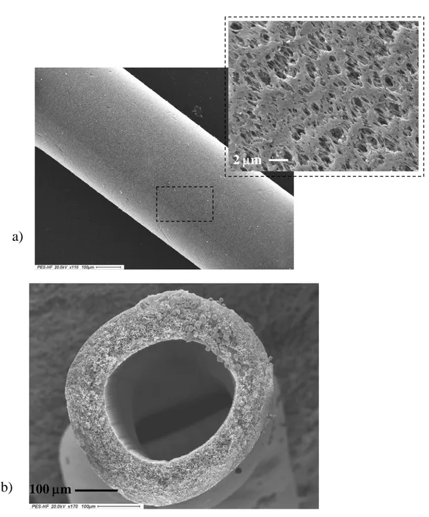
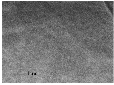
2.3.5.1 Scanning electron microscopy……….41
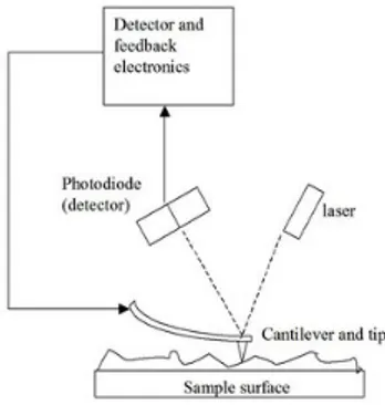
2.3.5.2 Atomic force microscopy………..41
2.3.5.3 Permeability method ………...42
2.4 Cell interaction with polymers………44
2.4.1Effect of the polymer chemistry on cell behaviour……….45
2.4.2 Biodegradable polymers………45
2.4.3 Synthetic polymers with adsorbed proteins………...46
2.4.4 Hybrid polymers with immobilized functional groups………..46
2.4.5 Influence of surface morphology on cell behaviour………..48
2.4.6 Use of patterned surfaces to control cell behaviour……….48
2.4.7 Cell interactions with three-dimensional polymer scaffolds and gel.49
CAPITOLO 3. Membrane bio-hybrid system in neuro-biotechnology
3.1 Abstract ...59
3.2 Introduction………...……...60
3.3
Membrane properties in a bio-hybrid system………....62
3.4 Membrane preparation features...………....65
3.5
Hippocampus and plasticity: functional and molecular aspects...66
3.6
Neuronal cell isolation and culture on membranes...68
3.7
Selective assay of neurodegeneration: microtubule-associated protein 2...70
3.8
Concluding remarks
……...………....72
References………...73
List of Symbol………...…...76
Figures………...….77
CAPITOLO 4. Influence of membrane surface properties on the growth of
neuronal cells isolated from Hippocampus
4.1 Abstract...85
4.2 Introduction……….…..85
4.3.5
Neuronal morphology features of the different membranes…………..92
4.3.6 Sample preparation for SEM ……...………..…. ..92
4.37 Biochemical assays...92
4.5 Results………...94
4.5 Discussion……….…96
4.6 Conclusions………...99
References……….…101
List of Symbol...105
Figures………... 107
Table………..120
CHAPTER 5.
Distinct Gaba
Aα receptor subunits exert early neurogenic
activities on hippocampal cells
5.1 Abstract...121
5.2 Introduction………...……..121
5.3 Materials and Method...123
5.3.1
Cell culture………...….. 123
5.3.6 Metabolic assays………...126
5.3.7 In situ hybridization analysis………..…127
5.3.8 Statistical analysis………...…128
5.4 Results……….129
5.4.1 Morphometric evaluations of neuronal processes from HIP culture..129
5.4.2 Role of GABA
Aα
subunits on neuronal metabolism and morphological
features ………...129
5.4.3 Effects of
α
2,5subunits on Gluergic receptor mRNA expression
levels………..131
5.5 Discussion………...131
References………...137
List of Symbol...144
Figures………..145
CHAPTER 6. Comparison of different hollow fibre membrane for the
developing of hippocampus cells
6.1 Abstract...150
6.2 Introduction………..151
6.3.5
Neuronal morphology on HFMs...…………156
6.3.6 Sample preparation for SEM ……...………. 156
6.37 Biochemical assays...156
6.4 Results………..157
6.5 Discussion……….…159
6.6 Conclusions………...161
References...163
List of Symbol...165
Figures………....167
Table………....176
CHAPTER 7. Developing a biodegradable membrane for the growth of
neuronal network
7.1 Abstract...177
7.2 Introduction………...177
7.3 Materials and Method...179
7.3.5 Neuronal morphology CS membranes………...181
7.3.6 Sample preparation for SEM………....182
7.3.6 Biochemical Assay………182
7.4 Results...182
7.5 Discussion……….…184
7.6 Conclusions………...186
References...187
Figures...189
CHAPTER 8. Conclusions and Future Research
8.1 Conclusions………197
B
B
o
o
o
o
k
k
C
C
h
h
a
a
p
p
t
t
e
e
r
r
1. L.. De Bartolo, M. Rende, G. Giusi, S. Morelli, A. Piscioneri, M. Canonaco, E. Drioli. “Membrane bio-hybrid systems: a valuable tool for the study of
neuronal activities”. In: M. Canonaco and R.M. Facciolo (Eds.) Evolutionary
Molecular Strategies and Plasticity 2007; Research Signpost, pp.379-396.
J
J
o
o
u
u
r
r
n
n
a
a
l
l
p
p
a
a
p
p
e
e
r
r
s
s
1. L. De Bartolo, M. Rende S. Morelli, G. Giusi, S. Salerno, A. Piscioneri, A. Gordano, A. Di Vito, M. Canonaco, E. Drioli. “Influence of membrane
surface properties on the growht of neuronal cells isolated from hippocampus”. Journal of Membrane Science. 2008; 325: 139-149.
2. S. Morelli, S. Salerno, A. Piscioneri, M. Rende, C. Campana, E. Drioli, L. De Bartolo. Membranes in regenerative medicine and tissue engineering. In: L. Giorno and E. Drioli (Eds.) Membrane Operations; Wiley VCH, accepted 3. M. Rende, S. Morelli, G. Giusi, S. Salerno, A. Piscioneri, A. Gordano,
M.Canonaco, E. Drioli. Effect of Membrane Surfaces on Hippocampal
Neuronal Cell Differentiation. Tissue Engineering 2008; 725-726.
1. L. De Bartolo, M. Rende, S. Morelli, G. Giusi, A. Piscioneri, F. Tasselli, S. Salerno, M. Canonaco and E. Drioli. Morphological and Functional Features
of Neurons Isolated from Hippocampus on Different Membrane Surfaces. The
International Congress on Membranes and Membrane Processes, ICOM 2008, July 12-18, 2008, Honolulu, Hawaii.
2. M. Rende, S. Morelli, G. Giusi, S. Salerno, A. Piscioneri, M. Canonaco, E. Drioli and L.De Bartolo. Effect of membrane surface on hippocampal neuronal
cell differentiation.Tissue Engineering and Regenerarive Medicine International Society,TERMIS-EU, June 22-26, 2008, Porto, Portugal.
3. L. De Bartolo, M. Rende, G. Giusi, S. Morelli, A. Piscioneri, M. Canonaco and E. Drioli. The 6th International Membrane Science and Technology Conference, IMSTEC 07, 5-9 November 2007, Sydney (Australia).
4. L. De Bartolo, M. Rende, G. Giusi, S. Morelli, A. Piscioneri, M. Canonaco and E. Drioli Neuronal Membrane Bio-Hybrid System. 21th European Conference on Biomaterials, September 09-12th, 2007, Brighton, UK
neuronal cells culture
L’obiettivo di questo lavoro di tesi è stato lo sviluppo di un sistema bioidrido a membrana, come modello di ricostruzione in vitro di tessuti neuronali per lo studio dei meccanismi di autogenerazione su differenti substrati.
Le membrane polimeriche semipermeabili, per le loro caratteristiche di separazione, immunoprotezione e di matrice artificiale possono essere adoperate per la ricostruzione dei tessuti e organi in vitro. La disponibilità di membrane in diverse configurazioni e in diverso materiale polimerico ha reso sempre più attraente e interessante il loro impiego nello sviluppo di nuovi tessuti nel settore biomedicale e diagnostico. Oggigiorno, le membrane possono essere usate per lo sviluppo di organi bioartificiali e per la ricostruzione di nuovi tessuti. Nonostante il crescente miglioramento ed i continui progressi delle tecnologie biomediche, la sostituzione di organi danneggiati da traumi e/o malattie rappresenta un problema cruciale per la moderna medicina. Le terapie attualmente in uso non solo sono estremamente costose, ma spesso non sono in grado di soddisfare pienamente gli scopi per i quali vengono applicate; il trapianto d’organi è severamente limitato dall’insufficienza dei donatori e dai problemi di compatibilità. Il superamento di queste difficoltà sembra sia possibile grazie ai continui sviluppi ottenuti in un settore di ricerca, emerso di recente nel campo delle scienze dei biomateriali, l’ingegneria tissutale o
tissue engineering. In tale ambito, sono state messe a punto tecniche che permettono di
coltivare in laboratorio linee cellulari e tessuti con le caratteristiche del ricevente [De Bartolo et al., 2007]. In vivo, le cellule sono supportate da una matrice extracellulare che influenza la loro morfologia, proliferazione e differenziazione nonché la loro funzione metabolica. Quando le cellule sono coltivate in vitro, un simile supporto meccanico e chimico deve essere fornito dall’ambiente di coltura, per questo motivo è molto importante conoscere le proprietà di membrana (chimico-fisiche, strutturali e di trasporto), che possono influenzare le funzioni che la membrana svolge e soprattutto la compatibilità del materiale polimerico a contatto con le cellule e i fluidi del corpo. Le cellule ed i tessuti
ottenuti in vitro sono poi innestati nel paziente, ripristinando le funzionalità compromesse, senza dover ricorrere al trapianto di elementi biologici prelevati da donatori estranei. I tessuti ingegnerizzati, in caso di successo, si integrano con quelli del paziente, apportando in tal modo un contributo specifico e duraturo alla cura dello stato patologico, senza richiedere debilitanti e costosi trattamenti farmacologici. In questa logica, la tecnologia è interessata allo sviluppo e alla produzione di nuovi sistemi per la coltura in vitro di cellule su larga scala, con particolare attenzione al controllo dei fenomeni e delle condizioni operative che regolano il trasporto dei nutrienti e dei prodotti di scarto del metabolismo cellulare. A tale proposito, numerosi studi sono stati condotti negli ultimi anni riguardo la possibilità di sviluppare sistemi artificiali basati sull’utilizzo di biomateriali, scaffolds e cellule, che consentano la sostituzione di tessuto nervoso danneggiato o permettano di ripristinarne l’organizzazione strutturale e anatomica e, conseguentemente, le capacità funzionali [Lee et al., 2003; Schmalenberg et al., 2005]. Tra i biomateriali impiegati nel campo della tissue engineering, le membrane polimeriche semipermeabili sembrano fornire il supporto meccanico e chimico necessario a garantire la regolazione del processo di crescita e di differenziamento delle cellule neuronali in sistemi bioibridi: le interazioni cellula-substrato inducono risposte cellulari specifiche, consentendo ai neuroni di assumere un definito orientamento nello spazio e la formazione in vitro di un ricco network di connessioni sinaptiche. Un sistema bioibrido a membrana adoperante cellule neuronali potrebbe, dunque, costituire un valido strumento nel campo dell’ingegneria tissutale, per consentire lo studio dei meccanismi molecolari alla base di malattie neurodegenerative e nello sviluppo di bio-molecole da impiegare nella terapia farmacologia, per consentire di ripristinare le funzionalità danneggiate [Simonin et al., 2006].
Lo stato dell’arte riguardante la coltura di cellule neuronali su matrici sintetiche, racchiude per la maggior parte lavori che riguardano la coltura di cellule tumorali, come linee immortalizzate. Tuttavia queste cellule hanno un metabolismo che risulta essere diverso dalle cellule primarie; di conseguenza le informazioni che si ottengono non possono essere trasportate completamente alla situazione in vivo.
Sono molti i casi in cui, per l’espansione in vitro di cellule neuronali, vengono utilizzate altre cellule di sostegno (glia, astrociti), allo scopo di supportare la crescita delle cellule di interesse (cocoltura). La cocoltura risultante può avere molti vantaggi rispetto alla coltura pura, in quando le cellule neuronali, ricevono naturalmente le sostanze necessarie per la crescita dalle cellule che in condizioni naturali fungono da sostegno.
Dallo studio di lavori su neuroni ippocampali, è possibile evidenziare che nella maggiorparte dei casi, sono stati valutati gli effetti del substrato sulle cellule, per pochi giorni di coltura (4-5 giorni); al contrario, è proprio dal 4° al 8° giorno di coltura che le cellule neuronali ippocampali, dopo la prima fase di adesione, iniziano a crescere aumentando i prolungamenti assonici e dendritici ed esplorando così l’ambiente circostante. L’adesione è importante nelle prime ore di coltura, perché indica la presenza di una buona superficie di attacco, cioè di una buona compatibilità del supporto, ma la differenziazione e vitalità a lungo termine può essere valutata solo allungando il periodo di osservazione della coltura oltre il 4° giorno. Durante la prima fase di crescita la cellula neuronale, essendo totipotente, riesce ad adattarsi all’ambiente circostante differenziandosi, grazie anche alla presenza di molecole importanti per la crescita nel mezzo di coltura. Solo successivamente alla formazione del network, si vengono a formare degli scambi molecolari con l’ambiente circostante, importanti per il mantenimento della vitalità e funzionalità cellulare a lungo termine.
L’ippocampo è un particolare zona del cervello che ha funzione di apprendimento e di memoria, risulta essere associata alla motivazione, al controllo delle emozioni, alla memoria e, oltretutto gioca un importante ruolo nel controllo delle risposte dell'organismo allo stress. Questa caratteristica plasticità è molto interessante soprattutto in caso di patologie come l’Alzahimer o in altre malattie degenerative, oppure nel coma.
Lesioni provocate all’ippocampo danno luogo a disturbi della memoria e dell’apprendimento piuttosto intensi. Sono stati testati su cavie, diversi farmaci che migliorano le funzioni cognitive in caso di danni cerebrali, ma i risultati sono modesti e a volte i farmaci risultano nocivi. Attualmente esistono evidenze sperimentali che i fattori ambientali giocano un ruolo fondamentale nel recupero delle capacità cognitive.
Essendo il numero di cellule neuronali stabilito alla nascita, e non variabile in quando i neuroni non hanno capacità di dividersi e proliferare, nasce l’esigenza di studiare i meccanismi di autogenerazione dei tessuti allo scopo di superare queste patologie e disturbi post-traumatici molto diffusi nella popolazione.
L’obiettivo di questo lavoro di tesi sperimentale è stato rivolto alla possibilità di realizzare sistemi bioibridi a membrana per la coltura di neuroni, isolati da una regione cerebrale coinvolta in importanti funzioni neurofisiologiche, quali l’apprendimento e la memoria: l’ippocampo. Numerosi lavori riportano l’utilizzo di tale regione cerebrale per gli studi neurobiologici e funzionali, valutando non solo stadi di sviluppo critici in vivo ed in vitro [Fukata et al., 2002], ma anche i meccanismi cellulari che sono alla base di svariate patologie neurodegenerative quali l’epilessia [Ullal et al., 2005]. Inoltre i neuroni ippocampali hanno una forma ben definita, facilmente monitorabili in vitro anche per lunghi periodi di coltura [Bunker and Goslin, 1998]. Il modello animale prescelto è stato l’ibernante facoltativo Mesocricetus auratus, ampiamente utilizzato negli studi neurodegenerativi, in quanto alcuni stadi di ibernazione ed in particolare il risveglio, rappresenta condizioni simil-ischemiche cui l’animale risponde con strategie di adattamento e plasticità sinaptica [Canonaco et al., 2005, 2008]. In tali sistemi la capacità delle membrane di fornire un adeguato microambiente alle cellule in coltura, dipende dalle proprietà morfologiche e chimico-fisiche della superficie di membrana e dalle proprietà di trasporto.
In una prima fase è stata sviluppata una nuova membrana piana, preparata mediante inversione di fase, a partire da un blend polimerico di polyetheretherketone (PEEK-WC) modificato e poliuretano (PU). Questa membrana offre il vantaggio di combinare le proprietà di entrambi i polimeri (biocompatibilità, resistenza termica e meccanica ed elasticità) con l’elevata proprietà di permeabilità, selettività ed una ben definita geometria dei pori della membrana. La membrana di PEEK-WC-PU è stata usata per la coltura di neuroni ippocampali in un sistema bioibrido allo scopo di rigenerare il tessuto in vitro. Per dimostrare la validità del sistema sperimentale, sono state determinate il grado di interazione e differenziazione cellulare e l’attività metabolica (cosumo di glucosio
produzione di lattato). I risultati preliminari mostrano la capacità di adattamento delle cellule neuronali ippocampali in coltura nel sistema bioibrido a membrana. Simile al comportamento delle cellule neuronali sulla polilisina, su questo tipo di membrana le cellule aderiscono e si differenziamento dando origine ad un complesso neuronal network. Come ulteriore conferma dell’avvenuto differziamento cellulare è stato utilizzato un marcatore cellulare specifico (MAP2), allo scopo di dimostrare l’inaterata struttura del citoscheletro delle cellule neuronali. I risultai di questo studio incoraggiano lo sviluppo di sistemi bioibridi a membrana per la coltura di neuroni ippocampali allo scopo di rimodellare e rigenerare in vitro tessuti in un microambiente controllato.
Le proprietà morfologiche del substrato possono svolgere un ruolo importante nell’interazione cellulare e soprattutto nel caso di cellule neuronali possono guidare topograficamente lo sviluppo di processi assonici e dendritici. A tale scopo in una seconda fase, è stato studiato l’effetto delle proprietà morfologiche della superficie di membrana, sul differenziamento delle cellule neuronali.
Membrane commerciali sia microporose di polietersulfone (PES) e poliestere (PE) e sia dense di fluorocarbone (FC) sono state caratterizzate allo scopo di definire le loro proprietà morfologiche (e.g., dimensione media dei pori, porosità, rugosità, distribuzione della dimensione dei pori) e le loro proprietà chimico-fisiche (e.g. bagnabilità, adsorbimento di acqua).
Le membrane sono state modificate con coating di polilisina allo scopo di favorire l’adesione cellulare e di creare superfici con gli stessi gruppi funzionali interagenti con le cellule ed investigare solo l’affetto della morfologia di superficie sul differenziamento cellulare. Le membrane modificate sono state caratterizzate per valutare l’uniformità del coating, la rugosità della superficie e le proprietà di trasporto. Il coating ha modificato le proprietà chimico-fisiche in termini di bagnabilità, uniformando le differenze superficiali delle membrane native. Le membrane così modificate presentavano una rugosità si superficie compresa tra 6-200 nm. La modifica di superficie ha ridotto la permeanza idraulica delle membrane di poliestere e di polietereterchetone rispettivamente del 60% e del 25%, mentre per le membrane di polisulfone è rimasta invariata.
Lo sviluppo ed il differenziamento delle cellule in coltura sono stati valutati in termini qualitativi, attraverso l’osservazione dei cambiamenti morfologici dei neuroni isolati e della formazione di un caratteristico network di prolungamenti assonici e dendritici sia sulle membrane di PEEK-WC che su membrane commerciali, quali PE, PES, FC. E’ stata valutata quindi la localizzazione e la distribuzione di marcatori strutturali come la β-tubulina, una proteina associata al citoscheletro, presente nel soma e in tutti i prolungamenti neuronali. Per favorire la visualizzazione dei prolungamenti assoni è stata scelta una proteina specifica di tale prolungamento e precisamente la Growth-Associated
Protein-43 (GAP43).
Il metabolismo delle cellule è stato valutato in termini di consumo di glucosio e produzione di lattato, presenti nel mezzo di coltura. Inoltre è stata valutata la secrezione di fattori neurotrofici come il Brain Derived Neurotrophic Factor (BDNF), al fine di analizzare l’attività neuronale di sintesi di specifici fattori differenziativi durante lo sviluppo in vitro. E’ stato in tal modo investigato il mantenimento a lungo termine della vitalità e della funzionalità dei neuroni per un periodo di tempo di 12 giorni di coltura.
La rugosità della superficie di membrana ha influenzato notevolmente l’adesione cellulare e la formazione del network meuronale sulle membrane di FC (Ra= 6nm) e di PES (Ra = 50 nm). Le cellule formano prolungamenti assonici e dendritici che si ramificano e si connettono con i prolungamenti in un network molto complesso. Al contrario in membrane più rugose come quelle di PEEK-WC (Ra= 199,2) le cellule tendono ad aggregarsi ed a formare prolungamenti che si sviluppano nei pori della membrana. Le osservazioni microscopiche sono state confermate da analisi quantitative della lunghezza assonica e del metabolismo cellulare. Sulle membrane con rugosità compresa tra 6 nm e 50 nm i prolungamenti assonici sono significativamente più lunghi rispetto alle membrane con una rugosità maggiore. Le membrane con una rugosità compresa tra 6-50 nm favoriscono la formazione di strutture neuronali polarizzate e la produzione di neurotrofine specifiche come il BDNF, che ha un ruolo nella sopravvivenza e maturazione di specifiche popolazioni neuronali nonché nella trasmissione sinaptica.
Il sistema bioibrido costituito da membrane i FC e neuroni ippocampali è stato adoperato per studiare l’azione modulatrice delle sub unità α dei recettori del acido γ-Aminobutyric type A (GABA), sull’organizzazione dei processi neuronali. A tale scopo sono stati adoperati molecole che svolgono un’azione altamente agonista sulle subunità α2 e
molecole che svolgono un'azione antagonista sulle subunità α5. Queste subunità grazie alla
loro localizzazione sinaptica ed extrasinaptica, sono critiche per le funzioni immunogeniche associative, da cui si evince che, l’interazione tra il sistema GABAergico e il sistema Gluergico può costituire un elemento potenziale che è cruciale per regolare lo sviluppo assonale e dendritico.
Successivamente sono state sviluppate membrane a fibre cave (HFMs) di PEEK-WC e di Polyacrilonitrile (PAN) allo scopo di sviluppare un sistema tridimensionale altamente integrato. Le membrane in configurazione a fibra cava sono vantaggiose rispetto alle piane poiché offrono: i) Un’ampia superficie di adesione e di scambio di nutrienti ed un volume molto piccolo; ii) una compartimentalizzazione all’inerno e all’esterno della fibra e quindi separazione del compartimento cellulare da quello ex cellulare; iii) un’adeguata perfusione evitando fenomeni di shear stress.
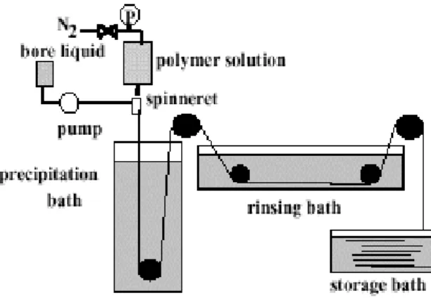
Le HFMs sono state preparate mediante dry-wet spinning e successivamente caratterizzate allo scopo di conoscerne le proprietà chimico fisiche e di trasporto.
Le proprietà di permeabilità delle membrane sono particolarmente importanti per le fibre cave per lo scambio molecolare tra il compartimento cellulare e l’ambiente esterno. Quindi è stato determinato il cut-off delle fibre di PEEK-WC e di PAN che sono rispettivamente all’incirca di 78000 Da e 81000 Da. Le membrane modificate mediante coating con PLL sono state utilizzate nella coltura a lungo termine di neuroni ippocampali.
La capacità di adesione e accrescimento delle cellule è stata valutata per 12 giorni di coltura. Sulle fibre di PEEK-WC è stata osservata la formazione di un network neuronale piuttosto complesso ed omogeneamente distribuito in tutta la superficie esterna della fibra mentre sulle membrane di PAN le cellule hanno formato in diverse zone della superficie di membrana dei distretti cellulari dai quali si sviluppano i prolungamenti neuronali. Su entrambe le fibre, le cellule sono state funzionalmente attive per 12 giorni. Le fibre di PAN
hanno sviluppato un maggior consumo di glucosio e produzione di lattato rispetto alle fibre di PEEK-WC. Il costrutto tissutale sviluppato ha mantenuto la sua funzionalità per 12 giorni di coltura. Le membrane in configurazione a fibra cava hanno consentito lo sviluppo di un sistema tridimensionale che da un punto di vista microarchitettonico consente di dirigere e guidare la rigenerazione neuronale in un sistema modello in vitro.
Nell’ultima fase di questo lavoro è stato sviluppato un sistema bioibrido adoperando una membrana biodegradabile di chitosano.
I polimeri biodegradabili sono materiali che si decompongono ma i cui prodotti di degradazione persistono a lungo nell’organismo ospitante. Spesso questo termine include le sotto classi di materiali assorbibili, riassorbibili, bioassorbibili e biodegradabili.
Molti scenari di applicazione, compresa l’ingegneria tessutale, necessitano per l’utilizzo in
vivo di matrici polimeriche biodegradabili o di strutture che siano minimamente soggette a
interazioni con il tessuto in via di sviluppo. Lo sviluppo di queste diverse funzioni solitamente richiede una microstruttura di sostegno porosa, con caratteristiche di porosità proprie dell’applicazione specifica.
Il chitosano è un polisaccaride biosintetico che è ottenuto dalla deacilazione della chitina, che è un polisaccaride naturale prodotto da un’enorme numero di organismi viventi. Il chitosano è l’unico polimero pseudonaturale caricato positivamente e questo lo rende importante per molte applicazioni biomedicali.
L’adesione e la crescita delle cellule su questa membrana è stata valutata mediante osservazione della coltura nel tempo (16g) ed analisi dell’attività metabolica (cosumo di glucosio, produzione di lattato) e di sintesi (BDNF) dei neuroni ippocampali in coltura. Le cellule neuronali ippocampali in coltura sulle membrane di chitosano aderiscono e si differenziano senza necessità di modifica della superficie di membrana attraverso l’utilizzo di biomolecole come la polilisina (PLL). La morfologia della cellula sulle membrane di chitosano del tutto simile alle cellule in coltura sul substrato di controllo (costituita da PSCD rivestito di PLL), non sono presenti differenze significative nella grandezza del soma e nella lunghezza dei neuriti. L’attività metabolica delle cellule, in termini di consumo di glucosio e produzione di lattato, viene mantenuta ad alti livelli per tutto il
periodo di coltura; esse inoltre esibiscono una maggiore capacità di sintesi della neurotrofina BDNF, conseguente ad un miglior grado di connessione sinaptica tra le cellule.
I risultati incoraggianti rappresentano un punto di partenza per lo sviluppo successivo di sistemi biobridi a membrane e biodegradabili in impianti tissutali e nella riparazione di tessuti. I risultati ottenuti rappresentano un primo approccio nella realizzazione di sistemi bioibridi che consentono una coltura di tipo tridimensionale e quindi paragonabile a quella
in vivo. Tali sistemi, adoperanti neuroni ippocampali, possono essere utilizzati nello studio, in vitro, del comportamento morfologico e funzionale di popolazioni neuronali
danneggiate in alcune delle più comuni malattie neurodegenerative, come il morbo di Alzheimer e l’epilessia [Ullal et al., 2005; Cotel et al., 2008].
1.1 Research motivation: Tissue Engineering
In the last years the big progress of cellular’s biology and biotechnology, has led to the development of news technologies for cells culture and in vitro regeneration of tissues and organs, a new field of the biomedical science knows as “Tissue engineering”. [1]
The goal of tissue engineering is the design and construction in the laboratory of living and functional components that can be used for the regeneration of malfunctioning tissues.
Although considered as a relatively new field, the first documented report of tissue engineering emerged in 1933, when tumor cells were entrapped in a polymer membrane and implanted into a pig. Tissue engineering is an interdisciplinary field that brings together the principles of life sciences and medicine with those of engineering and has three basic components: cells, supports and signals. Its development over the past decade has been the result of a variety of factors: increased knowledge and availability of cells, the advent of new biomaterials as potential templates for tissue growth, improvements in a bioreactor design and increased understanding of healing processes have all contributed. However, although tissue engineering research is evolving rapidly, there has been a hiatus in the commercial development and, hence, clinical application of engineered products.
However, progress continues and the number of people currently benefiting from tissues engineering is set to expand exponentially in the coming years [2].
1.1.2 The challenge
The challenge for tissue engineering is optimise the isolation, proliferation and differentiation of cells, and design of biohybrid systems that are able to support and coordinate the growth of three-dimensional tissues in the laboratory.
One idealistic strategy would be harvest stem cells from a patient, expand them in cell culture, and seed them in biohybrid systems. Stem cells can become many types of mature cells, via a process called differentiation, when given the specific biological stimuli. The biohybrid systems then act as template and stimulus for proliferation and differentiation of cells into the specific cells that will generate specific new tissue. The tissue can either be grown on a structure that will completely disappear (resorb) as the new tissue grows, so that only the new tissue will be implanted, or biocomposite of structure and new tissue can be implanted. The tissue engineered construct must be able to survive and restore normal function, e.g. biochemistry and both mechanical and structural integrity, and integrate with the surrounding tissues. Using cells from the same patient eliminates the problem of immunorejection that can occur with transplants from donors.
1.1.3 The cells sources
Probably the single most important element in the success of tissue engineering is the ability to generate appropriate numbers of cells (too many cells can be just as detrimental as too few) and the capacity for those cells differentiate from, and maintain, the correct phenotype and perform specific biological functions. For example, cells must produce an extracellular matrix in the correct organisation, secrete cytokines and other signalling molecules, and interact with neighbouring cells/tissues. Immediately, this raises a number of potential problems, not least of which is obtaining appropriate cell numbers to promote repair.
and are cultured either as small pieces of tissues or single cells following isolation from the tissue by digestion enzyme of such as trypsin and collagenase. The main disadvantage of primary cultures is that they become senescents, lose their ability to multiply and may lose some phenotypic characteristics with time. The main advantage of primary cultures is that they retain many of their original characteristics in their limited life span. Continuous cells lines can be maintained in culture either for a limited number of cell divisions or indefinitely. Many of these cell lines are derived from cancerous tissues, while some of these cell lines are transformed into immortal cell using viral oncogenes. These continuous cell lines have the advantage of unlimited availability but have the disadvantage of preserving few of the original cellular characteristics. Cells derived from a source must attach to substrate in order to grow, while cells derived from blood grow in suspension. Cells in suspension have a round shape, and cells attached to substrate show different morphologies depending on their tissue of origin.
Once the cells attach to substrate, they start to divide and multiply to form a complete layer (commonly known as confluent layer) covering substrate.
1.1.4 Culture condition
Cells in culture require carbohydrates, salts, aminoacids, vitamins, fatty acids and proteins to survive in vitro. The basal medium contains the essential inorganic salts, aminoacids, vitamins, fatty acids and some proteins. It also contains phenol red and bicarbonate based buffering system and a combination of different antibiotics or anti fungal agents can also be used in cell culture to avoid contamination.
As indicated above, by manipulating the culture conditions, it is possible to control the available differentiation pathways and selectively generate cultures. Such manipulations include stimulation of cells with particular cytokines, growth factors, aminoacids, other protein, drug and co-colture with target cell/tissue type. Utilising these approaches is possible to know the different effects products[2].
1.1.5 Three-dimensional interactions
The great diversity in the structure and function of various tissues and organs, is a primary reason for the highly multisciplinary nature of tissue engineering. Tissues and organs consist of specialised living cells arranged within a complex structural and functional framework know as extracellular matrix (ECM). The normal function of most cells and tissues depends on spatial interaction with neighbouring cells and with a substratum or a matrix. Cell-cell and cell-ECM interactions are coordinated by members of several families of membrane spanning proteins, called adhesion molecules. These are fundamental to cell adhesion, helping to define 3-D cellular organization and also to participate directly in cell signalling, controlling cell recruitment, growth, differentiation, immune recognition and modulation of inflammation. Consequently, the function of the ECM and 3-D cell interactions is an important aspect of generating viable constructs for in vitro tissue regeneration. A number of natural and synthetic materials have been used to produce 3D structure to function as an artificial ECM. Materials should be non toxic, have good biocompatibility, be biodegradable and be capable of interacting specifically with the cell type of interest. Work with such materials has shown how materials can also be made to be bioactive through adsorption with biomolecules and that such modifications can enable specific recruitment and adhesion of specific cell types [2].
1.2 Objective and scope of this Thesis
Restitution of brain function following trauma or disease is a challenge for neurobiologist and neurobiotechnologist, because the central nervous system (CNS) cannot regenerate on its own. Attempts to replace lost or dysfunctional neurons by means of tissue transplantation or peripheral nerve grafting have been intensely investigated for over a century. These approaches have evolved as a powerful tool for restoring function in damaged or diseased regions of the CNS and also for addressing basic issues related to development function and plasticity in the CNS. As an alternative to conventional technologies, a new approach is being investigated which uses cell engineering derived
biomaterials to influence function and differentiation of cultured cells. Neuronal tissue engineering is an emerging field which derives from the combination of various disciplines that interact in order to study the development and regeneration of neuronal tissue. Neurodegenerative processes are becoming a major health concern especially since they are the cause of many neuronal disorders.
The goal of this work is to achieve a biohybrid system constituted by membranes and hippocampal neurons for in vitro regeneration of neuronal tissue.
The neuronal tissue engineering construct, is a novel approach to study at molecular level neurodegenerative disorders, pathogenic state as well as the development of new therapeutic molecules. Polymeric semi-permeable membranes furtherer characteristics of selectivity, biostability and biocompatibility appear to be promising biomaterials for in
vitro cell growth. In fact membranes provide mechanical and chemical supports for the
anchorage-dependent cells. They allow compartmentalization of cells providing a selective transport of molecules avoiding the passage of other species.
The use of polymeric semi-permeable membranes with different physico-chemical and transport properties is an appealing approach in the tissue and bio-artificial organ engineering field, since these bio-membranes share specific features such as the selective transport of molecules, resistance and protection and for the in vitro simulation of human brain functions substitute.
In a first phase, a novel flat membrane was prepared from a polymeric blend of modified polyetheretherketone (PEEK-WC) or poly (oxa-1,4-phenylene-oxo-1,4-phenylene-oxa-1,4-phenylene-3,3(isobenzofurane-1,3-dihydro-1-oxo)diyl-1,4-phenylene) and polyurethane (PU) by inverse phase techniques. This membrane offers the advantage of combining the properties of both polymers (biocompatibility, thermal and mechanical resistance, elasticity) with those of membranes such as permeability, selectivity and well-defined geometry. This membrane is used for culture of hippocampal cells in a bio-hybrid system. For typical neuronal tissue-engineered constructs, the properties of both cell (morphology, viability functions) and material (physico-chemical, morphological and transport properties) components are very important [3].
Subsequently the effect of morphological properties of membranes in terms of pore size, porosity and roughness on the adhesion and differentiation of hippocampal neurons was investigated. To this purpose, membranes with different surface properties were used. In particular commercial microporous membranes such as polyester (PE), polyethersulfone (PES), and dense membrane as fluorocarbon (FC) membranes. This lather membrane is interesting for its permeability properties to oxygen, carbon dioxide and aqueous vapour. Microporous membranes were also developed from polyetheretherketone (PEEK-WC). These membranes used as substrates for cell adhesion are coated with poly-L-lysine (PLL) in order to have the same functional groups interacting with cells.
The behaviour of neurons isolated from the hippocampus on membranes with different surface properties was investigated in terms of cells growth parameters, and metabolic functions. Neuronal cells response to the different membrane surface was evaluated by analysing their morphology and neurite outgrowth as well as their specific metabolic functions as the neurotrophin secretion.
The developed bihoybrid system was used to evaluate the morphogenic role of γ-aminobutyric acid type A (GABAA) neuroreceptors. Of the most common 19 GABAA
receptor subunits, α subunit appears to be the main component largely responsible for inhibitory interneuronal functions; canonical elements that are mostly GABAergic in nature. The neurogenic role of GABAA α2 and α5 subunits on the receptors of the Gluergic
neuronal system, which are evolved in developmental, synthetic plasticity and excitotoxicity events, was investigated by using highly specific α2 agonist molecule,
funitrazepan and α5 selective inverse agonist ,RY-080.
Moreover, the optimization of transport, physico-chemical and structural properties of the membrane as well as fluid dynamics of cellular microenvironments could affect cell-membrane interactions and the functional maintenance of hippocampal cells.
Membranes in hollow fiber configuration are particularly useful to enhance the surface area for the adhesion of cells in a small volume. Hollow fiber membranes allow the compartmentalization of cells in the shell or in the lumen of the fiber that communicate
In bio-hybrid systems using hollow fiber membranes made of modified Polyetheretherketone (PEEK-WC) and Polyacrylonitrile (PAN) were developed.
Since an efficient transport of metabolites and nutrients is required for in vitro maintenance of cell viability and functions, the hydraulic permeance and the mass transfer of metabolites through the membranes were evaluated.
The viability of hippocampal cells was tested by assessing the cell adhesion to the hollow fiber surface. The functional behavior was investigated in terms of glucose consumption, lactate production and synthesis of brain-derived neurotrophic factor (BDNF) [4]. The developed biohybrid system was used.
Finally, biologically derived a chitosan membrane, was proposed as a potential biomaterial for supporting both adhesion and differentiation of neurons in vivo. Chitosan is a biosynthetic polysaccharide that is the deacetylated derivative of chitin, a naturally occurring polysaccharide that can be extracted from crustacean exoskeletons or generated via fungal fermentation process. This polysaccharide was used for prepare a flat membrane with phase inverse technique. Chitosan membrane has been utilized for the long term culture of hippocampal cells.
The chitosan films showed a significant enhancement of neurite outgrowth, reflecting the dependence of neuronal cell affinity to the amine content in the polysaccharide, an important relation that is important for neuronal tissue engineering applications.
This biodegradable polymer should be applicable to those tissue engineering products for which tissue repair or remodelling is the goal.
REFERENCES
1. Griffith LG, Naughton G.Tissue Engineering-current challenges and expanding
opportunities. Science 2002; 295:1009-14.
2. L.Hench and J.R. Jones. Biomaterials, artificial organs and tissue
engineering.Woodhead Publishing Limited.Cambridge England. 2004: 18:193-200.
3. M.E. Manwaring, R. Biran, P.A. Tresco, Characterisation of rat meningeal
cultures on materials of differing surface chemistry. Biomaterials 22 (2001) 3155
4. De Bartolo L, Rende M, Giusi G, Morelli S, Piscioneri A, Canonaco M, Drioli E.
Membrane bio-hybrid systems: a valuable tool for the study of neuronal activities. In:
Evolutionary Molecular Strategies and Plasticity (Canonaco M and Facciolo RM, eds), 2007; pp379-396. India: Research Signpost.
2.1 Introduction
One approach to tissue engineering is to create an in vitro environment that embodies the biochemical and mechanical signals that regulate tissue development and maintenance in vivo. The in vitro tissue engineering system is composed of three major components:
(1) Metabolically active cells able to express their differentiated phenotype,
(2) Polymeric materials that provide a three-dimensional (3D) structure for cell attachment and tissue growth,
(3) Bioreactor culture vessels that provide an in vitro environment in which cell-polymer constructs can develop into functional tissues.
These constructs can potentially be used in vitro, for controlled studies of tissue growth and function, or in vivo for tissue repair.
In this chapter, want focus on these different contributes cells and polymeric membrane; both are based on engineered constructs.
2.2 Organ model used in this study
Cells types used for this study was obtained from hippocampus of 1-2 day-old neonatal
mesocricetus auratus hamster (golden hamsters are hibernating rodents used as research
animals); the mesocricetus auratus is an optional hibernator rodent, whose brain it is equipped of the ability to put into effect various neuroprotective mechanisms like the hypothermia, drastic reduction of the metabolism and increased defended anti-oxidant during the various stages of the process of hibernation [1]. This rodent can constitute therefore a useful model in the understanding of the molecular events to the basis of neurodegenerative pathologies, like the ischemia that, in hibernator, they can be manifested at the moment of the awakening [2] [3].
The Hippocampus is a part of the forebrain, located in the medial temporal lobe. It belongs to the limbic system and plays major roles in short term memory and spatial navigation.
Humans and other mammals have two hippocampi, one in each side of the brain. In rodents, where it has been studied most extensively, the hippocampus is shaped something like a banana [4].
Fig.1 Bilateral section of mesocricetus auratus brain
In humans it has a curved and convoluted shape that reminded early anatomists of a seahorse. They are two parts: hippocampus major (or horn Ammon) and hippocampus minor, especially associated with memory.
The hippocampus has been the object of much experimental work, and theories about the nature of its function have multiplied. One possibility which has attracted attention from behaviourists is that it may be involved in memory. However, at present no clearly formulated results can be stated. This is consequence partly of the anatomical complexity of the region and partly of the semantic difficulties surrounding the words “memory” and “learning”.
2.2.1 Human hippocampus
The human hippocampus can be divided into three distinct fields, which we have labelled CA3, CA2, CA1 according to the nomenclature of Lorente de No’ (1934). Field CA3 borders the hilus of dentate gyrus, where it terminates in a complex fashion. At its border end, it borders field CA2. Cassell and Seress have estimated that there are about
2.1*106neurons in the CA3 region and about 0.22*106 pyramidal cells in CA2. These regions of hippocampus contain pyramidal cells and a number of interneurons. Field CA1 is unquestionably the most complex subdivision of the human hippocampus. It appears to be populated by a far more heterogeneous group of neuronal elements than the other hippocampal fields. There are many connections between these fields that are important for the perfect features of this forebrain’s part.
Fig.2 Cito-architecture of human hippocampus
2.2.2 Clinical disease
Various clinical conditions result in morphological alterations of the human hippocampal formation. While the causative factors are not yet know for most of these disease states, it is clear that each of the different hippocampal cytoarchitectonic fields is more or less vulnerable to damage. In ischemia and temporal lobe epilepsy, for example, field CA1 of the hippocampus suffers the greatest neuronal cell loss. In other neuropathological conditions while in the Alzheimer’s disease, the hippocampus is one of the first regions of the brain to suffer damage; memory problems and disorientation appear among the first symptoms. Among the many conditions that produce pathological changes in the
hippocampal formation Alzheimer’s disease is probably the most devastating. Myriad studies have come to clear conclusion that this disorder is distinctly different from an elaboration of normal aging. Alzheimer’s disease is associated with four neuropathological correlates in the hippocampal formation: neuronal cell loss, neurofibrillary tangles, neuritic plaques, and granulovacuolar degeneration. The researches have conducted systematic analyses of cell loss in the hippocampal formation, and they estimate that there is decrease of some 56% in number of hippocampal pyramidal cells in the CA1 fields. As result of the cell loss and other pathological sequelae of Alzheimer’s disease, the hippocampal formations becomes, in essence, functionally disconnected from its major afferent and efferent interactions. Given the important role the hippocampal formation is known to play in certain forms of memory, it is likely that a major portion of the problems the memory function observed Alzheimer’s disease is attributable to damage of the hippocampal formation.
Damage to the hippocampus can also result from oxygen starvation (anoxia), encephalitis or medial temporal lobe epilepsy. Temporal lobe or complex partial epilepsy is another neurological disorder in which the hippocampal formation is severely affected. This most common form of epilepsy is generated from a dramatic loss of neurons in CA1 region during the hippocampal formation. In approximately two-thirds of cases of temporal lobe epilepsy, the hippocampal formation is only structure that shows pathological modifications, but in many cases cell loss in the CA3 portion. In some cases, also the organization of cell layer and metabolic alteration (hypoxia, anoxia, ecc.) influence the functions of neuronal cells, which are cause of this disease.
Multiple lines of research have recently converged on a critical role for the hippocampus in episodic memory, choice the ability to remember specific personal experiences (Tulving, 1983). Humans with selective hippocampal damage exhibit deficits in episodic memory, sometimes with relative sparing of the ability to acquire general factual knowledge, or semantic memory.
Patients with extensive hippocampal damage, like Ischemia, Schizophrenia, and other disorders, demonstrate anterograde memory impairment apparently resulting from the
hippocampal damage, may experience amnesia, that is, inability to form or retain new memories [5].
Know the tissues formation and the physiological and pharmacological developing of neuronal network, in vitro, can contribute to understand the causative factors of this pathological disease.
2.2.3 Cellular adhesion
A major goal of tissue engineering is to employ the principles of rational design to recreate appropriate signals to cells that promote biological processes leading to production of new tissues or repair of damaged ones. A key modulator of cell behaviour is the extracellular matrix (ECM) that provides individual cells with architectural cues of time and space, modulates bioavailability of soluble growth and differentiation factors, and organizes multicellular tissue development.
In vivo, cells are supported from one extracellular matrix that it influences the
morphology, proliferation and metabolic functions. Moreover cells sense and response to a variety of signals that include those that is soluble such as growth factors, differentiation factors, cytokines, and ion gradients. In addition, cell behaviour and phenotype is governed by responses to other types of signals that include mechanical forces, electrical stimuli, and various physical cues. Immobilized protein matrices that generally are fixed in space also regulate cell function. The general term that has come to denote the complex mixture of proteins on outside of cells that governs their behaviour is the ECM. Evolution has provided cells with surface receptor to ECM components that enable them to recognize and decipher the signals that they encounter from the ECM and which influence cell growth, division and differentiation. [6]
For descriptive purposes, cell adhesion is classified into categories of cell-substratum and cell-cell attachment. Cell-cell interactions may occur between like cells (homotypic events) or between dissimilar cells (heterotypic events).
There exists in tissues an exquisite balance between the anabolic process of ECM production and the catabolic process of ECM turnover. Introduction of foreign materials
inevitably disrupts this natural homeostasis. A goal of tissue engineering is to successfully introduce the materials that will, through stimulation of anabolic processes, lead to ECM production and acceptance of engineered material. Achievement of this goal requires a through understanding of the structural relationships among molecules in the ECM, their molecular interactions directing cell adhesion events, their biosynthesis and turnover, and their natural functions.
2.2.3.1 Basement membrane and focal adhesion
Basements membranes, also called basal lamina, are sheets of highly organized ECM that are associated with the basal o different type of cells. They establish cell polarity, influence cell metabolism, induce cell differentiation, direct cell migration, and organize membrane receptors. The basement membrane exists as a complex meshwork comprised of the proteins collagen IV, laminin, proteoglycans. The spatial relationships among these molecules were examined is like as “mesh-work. The basements membrane provides an anchor for cells, which adhere to it using specific surface receptors called integrins [6].
Fig 3 Extracellular matrix structure [Colonna S. et al., 2006]
Focal adhesions are the sites of cell attachment to substrata and then recognized as interfaces with the cytoskeleton [7]. Focal adhesion belongs to the contractile class of
matrix contacts, distinct from protrusive contacts or those that provide mechanical support [8]. The process of anchoring and spreading of cells on substrata occur as multistep processes that involve, among other things, clustering of surface receptors at sites of focal adhesions. Protein complexes link the cytoplasmic tails of surface receptors to the cytoskeleton, facilitating cell adhesion and transmitting signals through the intracellular network that ultimately signal to the nucleus to inform the cell that it has attached. This form of signaling has come to be called “outside in“signalling and is a key component of engineering functional interfaces between materials and living cells[9]. During the attachment of cells to substrata, cells surface receptors recognize ECM components through a specific and reversible process. The initial attraction and attachment often involves nonintegrin adhesion events that recognition the glycosaminoglycans. Once cells adhere, higher affinity interactions such as those involving integrin receptor are stabilized during the spreading phase of adhesion. It is this quality that ensures reversibility of integrin-mediated cell adhesion and allows cells both attach and detach from biological substrata at sites of focal adhesions.
Receptor clustering during adhesion triggers cascades of events involving protein phosphorylation and dephosphorylation that carry intracellular signals through the cell from surface to nucleus, ultimately leading to changes in gene transcription. Changes that occur within cells and that feedback to modulate the activity of surface receptor have come to be called “inside out” signals and modulate activity of both integrin and nonintegrin receptors in focal adhesions.
2.2.3.2 Molecules of ECM
Protein and carbohydrate components of secreted ECM are self-associated through a different type of interactions, including charge properties, ion and metal bridging, hydrophobic domains, redox interactions, and covalent bonding. The structures that form may produce either two-dimensional networks such as the meshwork of the basement membrane, or three-dimensional structures in space such as that of territorial matrix. While most of the assembly is thought to occur extracellularly, there is evidence that
some degree of assembly may be initiated during biosynthesis and take place within intracellular secretory vesicles creating a sort of ”pre-fabricated” scaffold to promote rapid assembly once secretion into the extracellular compartment occurs [10]. In this model system, it has been proposed that distinct intracellular vesicles form and are directionally released during exocytosis; these have been termed “basal laminar vesicles” and “apical vesicles”. The ECM molecules have different roles outside the cells. The collagens form what can be thought of a “functional aggregates” with noncollagenous molecules to form macrostructures including fibrils, basement membranes, filaments, canals, and sheets. The fibronectin is a glycoprotein that binds cell surfaces as well as various other molecules including collagen, heparin sulphate proteoglycans, and fibrin. Fibronectin is involved in different functions including cell migration, wound haling, cell proliferation, blood coagulation, and maintenance of cell cytoskeleton. Fibronectin served as prototype for the development of the RGD-peptides now widely used in modification of biomaterials for the purpose of tissue engineering [11].
Laminin is common ECM component found in basement membranes and used as a substratum for cell migration by many cell types. It has a clear role in cell migration and tissue morphogenesis during embryonic development [12]. It is a favourite ECM-based substrate for cells in the neural system [13].
Proteoglycanes and Glycosaminoglycans play different functions. Perlecan is secreted entirely into the matrix, Syndecan posses a transmembrane domain and remains as an integral component of the plasma membrane, and Glypican is lipid-liked [14].
The properties of ECM molecules make them ideal for cell and tissue engineering. Their multifunctional nature makes them ideal for promotion of cell specific adhesion via integrins and other surface receptors. These properties, if well understood, allow the ECM to serve as a rich source for mining and future development of novel tissue engineering applications.
2.2.3.3 Molecules adhesion in neuronal cells
Neurons develop in vitro through outgrowth of axon’s cone. During this phase of outgrowth, neurons meet numerous microenvironments, some regions contained adhesive molecules and other regions not optimal for the growth. The axon’s cone has some receptors (integrines) that they recognize the proteins in the substrate and direct the neuron outgrowth along this path. That can be verified in vitro: a portion of nervous tissues in culture on a suitable substrate, it does not extend dendrites and axons on Petri dish; but if the plastic dish comes covered with fibronectin it is possible to observe the increase of dendrites axons extensions. There is an optimal correlation between the outgrowth of axons and the presence of laminin on the substrate, on the contrary, the proteoglycans, inhibits the axonal growth [15]. There is a reason why the glycoprotein laminin have a role in the migration and the outgrowth of the axon. The integrines of the axon’s cone have a receptor capable to recognize the sequence RDG and YISRG of the protein laminin and a glycotransferase that recognize particular carbohydrate lateral chains of the molecule of the laminin. [16].
2.2.4 Neurons in culture
Primary hippocampal neuronal cells obtained from 1, 2 days year’s old mesocricetus
auratus, can be plated at low density on plastic Petri dish. By following individual cells
plated at low density with time, we can identify characteristic morphological changes that occur in the development of hippocampal neurons in culture. We can divide the events of development of hippocampal neurons in culture into five stages.
In stage 1, shortly after have attached to the substratum, they become surrounded by flattened, motile lamellipodia. In stage 2, with time, the lamellipodia condense at several discrete sites along the cell’s circumference; from these sites short, “minor” processes emerge. Minor processes, typically four to six in number, are roughly equal in length (20-30 μm), giving the cells a symmetrical appearance. They are highly dynamic, extending and retracting for short distances over a period of 12 to 24h. Cells at this stage appear to be unpolarized, and cell’s processes will become the axon. At stage 3, polarity first, becomes evident when one of the minor processes begins to elongate continuously without retraction until it becomes much longer than the other processes. At this stage, the axon can be distinguished from the other processes on ultrastructural and immunocytochemical grounds. In stage 3 neurons develop a single axon and several minor processes; occasional cells with a multiple axons also are observed. A small number of non neuronal cells also are present. Throughout stage 3, the axon continues to grow at rapid rate, but the remaining minor processes undergo little net elongation. In stage 4, after 2 to 4 days in culture, the remaining minor processes begin to elongate and to acquire the taper and branching pattern characteristic of dendrites. It is at this stage that presynaptic specializations first form on the cell body and dendrites and the microtubule organization typical of mature dendrites first arises.
With subsequent development, the density of the axonal network increases, dendritic arbors become more elaborate and highly branched, synaptic contacts develop in a large numbers, dendritic spines appear, and spontaneous electrical activity propagates throughout the neuronal network. These aspects of development reflect the continuation of processes begun at earlier stages of development. However, in contrast to the events of stages 1 to 4, which appear to be largely endogenously determined, many of these later aspects of neuronal maturation are highly dependent on cell interactions. To emphasize this important difference, we refer to this later period of development as stage 5. Cells in this stage increase the size of cell body and the diameter of dendritic processes that can be identified readily as they emerge from the cell body because of their large diameter, but the thinner, distal portions of dendrites become lost within the neuronal network, as does the
cell’s axon. In addition, axons frequently course along the dendrites and, by light microscopy, such axons cannot be distinguished from the underlying dendrites. These features can be resolved by intracellular injection of fluorescent dye such as immunocytochemical markers. The development of immunocytochemical markers that selectively stain axons or dendrites has been a particular important advance for identifying processes in cell culture and for studying their development. The presence of the machinery for protein synthesis in dendrites, but not in axons, offers an alternative and complementary method for distinguishing dendrites from axons and assessing their differentiation.
2.3 Polymeric membranes
2.3.1 Introduction
Membranes have gained an important place in tissue engineering and are used in a broad range of applications. The key property that is exploited is the ability of membrane to control the permeation rate of chemical species through the membrane. The membrane may discriminate between the two type’s molecules which differ for different size, shape or chemical structure ecc. in the separation process. The basic principle of any separation process is that a certain amount of energy is required to accomplish the separation. Hence, two substances A and B will mix spontaneously when the free enthalpy of the product is smaller than the sum of the free enthalpies of the pure substances. The minimum amount of energy (Wmin), necessary to accomplish complete separation is at least equal to or larger then the free enthalpy of mixing [17]:
Wmin ≥ ΔGm = ΔHm-TΔSm
In practice, the energy requirement for separation will be many times greater than this value Wmin. Different types of separation processes exist and each requires a different
processes can be applied, with a consequently energy saving respect to the traditional methods.
Membrane technology is an emerging technology and because of its multisciplinary character it can be used in a large number of separation processes. The benefits of membrane technology can be summarised as follows:
- separation can be carried out continuously - energy consumption is generally low
- membrane processes can easily be combined with other separation processes (hybrid processing)
- separation can be carried out under mild conditions - up-scaling is easy
- membrane properties are variable and can be adjusted - no additives are required
But drawbacks should be mentioned:
- concentration polarisation/ membrane fouling - low membrane lifetime
- low selectivity or flux
- up-scaling factor is more or less linear.
Recently news membrane technologies have reduced these disadvantages, creating a very qualitatively process in many applications [18].
2.3.2 Membrane processes
Every membrane separation processes is characterised by the use of membrane to accomplish a particular separation. The membrane has the ability to transport one component more readily then other because of difference in physical and/or chemical properties between the membrane and the permeating components. Transport through the membrane takes place as result of driving force acting on the components in the feed. In many cases the permeation rate through the membrane is proportional to the driving force,
i.e. the flux- force relationship can be described by a linear phenomenological equation. Proportionality between the flux (J) and the driving force is given by
J = - A dX/ dx
where A is called the phenomenological coefficient and (dX/dx) is the driving force, expressed as the gradient of X (temperature, concentration, pressure) along a coordinate x perpendicular to the transport barrier.
For a pure component permeating through a membrane, it is possible to employ linear relations to describe transport. However, when two or more components permeate simultaneously, such relations cannot be generally employed since coupling phenomena may occur in the fluxes and forces. These coupling phenomena can be described in terms of the formalism of non equilibrium thermodynamics. Other than the driving force, the membrane itself is the principal factor determining the selectivity and flux. In fact the nature of the membrane (its structure and material) determines the type of application, ranging from the separation of microscopic particles to the separation of molecules of an identical size or shape. The product obtained is determined by the applied pressure and the membrane resistance (or permeability).
2.3.3 Membrane properties
Synthetic membranes can be made from a large number of different materials, and can be divided further into organic (polymeric) and inorganic membranes. I would concentrate the attention on the organic membrane, which have been the subject of this study.
Polymers are high molecular weight components built up from a number of basic units, the monomers. The number of structural units linked together to form the long chain molecule is defined as the degree of polymerisation. A polymer chain has an infinite number of different conformations; with increasing of segments the physical, chemical and mechanical properties of the polymer change. When the segment is constituted by a repetition of a single monomer, this polymer is called homopolymer, when the repeating
units are different in the copolymers. In the copolymers, two or plus monomers are coupled together in various ways resulting in a number of different structures.
A polymer used in this study is the Polyester (PE), is a category of polymers which contain the ester functional group in their main chain (Fig.4).
PE membrane is made from a thin, microporous, hydrophilic polyester material film with a high degree of solvent resistance. It is ideal for use in blood assays or general filtration where chemically aggressive solvents may be used.
Fig. 4 Chemical structure of Polyester
A polymer used in many applications is the polytetrafluoroethylene (PTFE) (Fig.5). In the 1960's, Dr. Roy Plunkett, a Dupont chemist, was working on a project to find alternate materials for cooling purposes. He had stored some tetrafluorethylene gas (similar to freon) in a container and left it overnight. When he returned in the morning, the gas was gone and in its place was a white wax-like substance. Teflon had been discovered. PTFE is an extraordinary highly inert material, which exhibits excellent thermal and chemical stability. Some say that it is the most useful material known to man. This is because it has a combination of properties that are very hard to find.
]
n[
Fig. 5 Chemical structure of Polytetrafluoroethylene
PTFE is composed of carbon and fluorine. Carbon-fluorine and carbon-carbon bonds are among the strongest in single bond organic chemistry. This accounts for many of its properties. Because of the strong bonds, much thermal energy must be used to break down the material.
Because of his hydrophobic nature, is used in a variety of healthcare and industrial applications, as food & beverage, electronics and pharmaceutical industries.
A very important class of polymers are the polysulfones (PSf) and polyethersulfones (PES) (Fig.6).
Fig. 6 Chemical structure of Polyethersulfones
The PES posses very good chemical and thermal stability; are widely used as basic materials for ultrafiltration membranes and support materials for composite membranes. Polyethersulfones membranes are also used in the medical sector and in the food sector (membrane technology), because is a low protein binding membrane.
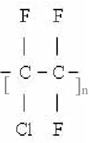
A very important class of polymers are the vinyl polymers, which are obtained by polymerisation of vinyl compounds H2C = CHR, where the side group –R is different for
different polymers. The position of the side group R has very important influence on the polymer properties. Between some important vinyl polymers, i would like to quote, in particular the polyacrylonitrile (PAN) with a -CN group, used for made of hollow fiber membrane (Fig.7) [17].
Fig. 7 Chemical structure of Polyacrylonitrile
PAN is a versatile polymeric material commonly used for very few products. In 1893 acrylonitrile was prepared by Moureu, the monomer found use as a copolymer with styrene, and especially as a terpolymer with styrene and butadiene (ABS). Homopolymers of polyacrylonitrile have been uses as fibers and for made asymmetric semipermeable membrane for dialysis and/or ultrafiltration.
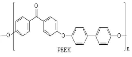
The polyetheretherketone (PEEK) is a new chemically and thermally resistant polymer, but with a slow solubility a room temperature. The chemical structure is given in Fig.8.
![Fig 3 Extracellular matrix structure [Colonna S. et al., 2006]](https://thumb-eu.123doks.com/thumbv2/123dokorg/2878491.10139/36.892.285.531.730.934/fig-extracellular-matrix-structure-colonna-al.webp)