REVIEW ARTICLE
Systematic review of extracellular vesicle-based treatments for lung injury: are
EVs a potential therapy for COVID-19?
Kasra Khalaj a,b, Rebeca Lopes Figueira a,b, Lina Antounians a,b, Giuseppe Lauriti c,d and Augusto Zani a,b aDevelopmental and Stem Cell Biology Program, Peter Gilgan Centre for Research and Learning, The Hospital for Sick Children, Toronto, Ontario, Canada; bDivision of General and Thoracic Surgery, The Hospital for Sick Children, Toronto, Ontario, Canada; cDepartment of Pediatric Surgery, Spirito Santo Hospital, Pescara, Italy; dDepartment of Medicine and Aging Sciences, G. D’Annunzio University, Chieti- Pescara, Italy
ABSTRACT
Severe COVID-19 infection results in bilateral interstitial pneumonia, often leading to acute respiratory distress syndrome (ARDS) and pulmonary fibrosis in survivors. Most patients with severe COVID-19 infections who died had developed ARDS. Currently, ARDS is treated with supportive measures, but regenerative medicine approaches including extracellular vesicle (EV)-based therapies have shown promise. Herein, we aimed to analyse whether EV-based therapies could be effective in treating severe pulmonary conditions that affect COVID-19 patients and to understand their relevance for an eventual therapeutic application to human patients. Using a defined search strategy, we conducted a systematic review of the literature and found 39 articles (2014–2020) that reported effects of EVs, mainly derived from stem cells, in lung injury models (one large animal study, none in human). EV treatment resulted in: (1) attenuation of inflammation (reduction of pro-inflammatory cytokines and neutrophil infiltration, M2 macrophage polarization); (2) regeneration of alveolar epithelium (decreased apoptosis and stimulation of surfactant production); (3) repair of microvascular perme-ability (increased endothelial cell junction proteins); (4) prevention of fibrosis (reduced fibrin produc-tion). These effects were mediated by the release of EV cargo and identified factors including miRs- 126, −30b-3p, −145, −27a-3p, syndecan-1, hepatocyte growth factor and angiopoietin-1. This review indicates that EV-based therapies hold great potential for COVID-19 related lung injuries as they target multiple pathways and enhance tissue regeneration. However, before translating EV therapies into human clinical trials, efforts should be directed at developing good manufacturing practice solutions for EVs and testing optimal dosage and administration route in large animal models.
ARTICLE HISTORY
Received 24 April 2020 Revised 4 June 2020 Accepted 2 July 2020
KEYWORDS
Exosome; acute lung injury; ali; sars-CoV-2; coronavirus; regenerative medicine; pandemic; cell-free; microRNA; miRNA
Introduction
On 11 March 2020, the World Health Organization declared the coronavirus disease (COVID-19) outbreak a pandemic [1]. COVID-19 is a new disease in humans that is caused by the severe acute respiratory syndrome (SARS) CoV-2 virus, a positive-sense single-stranded RNA virus [2]. This viral outbreak started at the end of 2019 in Wuhan, China, and has quickly been spreading across the world [3,4]. In humans, transmission of this new coronavirus occurs primarily via respiratory dro-plets and often results in a respiratory tract infection. Although we are still learning about the epidemiology of this viral illness, COVID-19 seems to affect patients of different age and sex with varying degrees of viru-lence [5,6]. Some individuals are either asymptomatic or have a mild disease with fever, cough and fatigue, whereas others develop bilateral interstitial pneumonia
with abnormal findings on chest computed tomogra-phy [7–9]. Moreover, some subjects have a severe form of COVID-19 that rapidly progresses to acute respira-tory distress syndrome (ARDS) and might result in sepsis and multiple organ failure [3,4]. ARDS is a life- threatening condition characterized by an acute onset (within a week of a known insult) of respiratory failure not fully explained by cardiac function or volume over-load, with diffuse opacities on lung imaging, and severe hypoxaemia requiring mechanical ventilation, as sum-marized in the 2012 Berlin definition [10,11]. In ARDS, lungs have diffused alveolar and endothelial cell damage with severe inflammation, increased vascular permeability and poor pulmonary oxygenation (Figure 1). Further, pulmonary interstitial fibrosis caused by excessive fibrin deposition is observed at autopsy, as CONTACT Augusto Zani [email protected] Division of General and Thoracic Surgery, The Hospital for Sick Children, Toronto, ON M5G 1X8, Canada
Supplemental data for this article can be accessed here JOURNAL OF EXTRACELLULAR VESICLES
2020, VOL. 9, 1795365
https://doi.org/10.1080/20013078.2020.1795365
© 2020 The Author(s). Published by Informa UK Limited, trading as Taylor & Francis Group on behalf of The International Society for Extracellular Vesicles.
This is an Open Access article distributed under the terms of the Creative Commons Attribution-NonCommercial License (http://creativecommons.org/licenses/by-nc/4.0/), which permits unrestricted non-commercial use, distribution, and reproduction in any medium, provided the original work is properly cited.
well as in ARDS survivors with healing pneumonia (Figure 1) [12]. In COVID-19 patients, ARDS appears to be even more severe. According to the first available data, 1 in 4 patients with COVID-19 (26%) develops ARDS, which has been shown to be a negative prognostic factor for survival, as more than 90% of non-survivors developed ARDS [3]. Moreover, it has been anticipated that even if the virus is fully eradicated, a proportion of COVID-19 survivors will develop pulmonary fibrosis [13]. This would be in line with similar coronavirus outbreaks, such as the Middle East Respiratory Syndrome (MERS), where a third of survivors had lung fibrosis after hospital discharge [14].
During this global pandemic, we are witnessing an incredible amount of efforts directed at stopping the viral spread, with public health measures such as social distancing, as well as a rush to develop a new vaccine or an effective antiviral drug to combat COVID-19. Whilst awaiting these more direct anti- viral measures, efforts are being aimed at testing new strategies that would attenuate the overactive response of the infected lungs and prevent or treat the compli-cations of the viral pneumonia. At present, there is no effective treatment for ARDS beyond supportive mea-sures. A regenerative medicine approach with the use of mesenchymal stromal cells (MSCs) has shown
promise as it targets multiple pathways [15,16]. However, the use of cell-based therapies still has hur-dles to pass, including the potential cell variability, the large-scale production and the reconstitution limita-tions of cryopreserved cells [17]. Extracellular vesicles (EVs) could be an alternative treatment strategy in this context as they have several advantages: EVs are cell-free, immunologically innocuous, not teratogenic and contain no adventitious agents [18,19]. Moreover, EVs derived from various sources have been used to modulate the inflammatory response in sepsis and attenuate multiple organ failure [20,21]. In this regard, EVs could be addressing the plea for a multi-targeted therapy that has been made on the principle that a single drug or antiviral therapy would unlikely be able to improve the most severe forms of COVID-19 [22]. With these promising features and the urgent need for an effective therapy for COVID-19 patients, a phase I clinical trial has recently been registered in China (NCT04276987) for the investigation of MSC- EVs as a therapy for ARDS secondary to COVID-19 [23]. While this reflects the increasing interest in testing the therapeutic potential of EVs, it remains unclear whether we are ready to use EVs as a treatment to battle the COVID-19 pandemic. Figure 1. Restorative effects of extracellular vesicle (EV) therapies in acute respiratory distress syndrome (ARDS). Compared to the normal alveolus (A), ARDS (B) is characterized by increased pulmonary inflammation [1], increased immune cell recruitment including neutrophils and macrophages [2], increased alveolar epithelial cell apoptosis [3], inactivated surfactant from degradation of alveolar surfactant layer and alveolar wall collapse [4], as well as increased endothelial cell permeability and gap junction formation [5], fibrin deposition [6] and increased platelet formation. EV treatments (C) can ameliorate the majority of these ARDS features by resolving inflammation and angiogenesis [1], altering local immune cell recruitment [2], decreasing apoptosis in alveolar epithelial cells [3], stimulating surfactant proteins leading to re-expansion of the alveolus [4], restoring endothelial junction proteins and decreasing endothelial barrier permeability [5], and reducing fibrin levels [6].
The aim of this systematic review was to analyse whether EV-based therapies could be effective in treating severe pulmonary conditions that affect COVID-19 patients, namely severe pneumonia, ARDS, acute lung injury (ALI) and pulmonary fibro-sis, and to understand their relevance for an eventual therapeutic application to human patients. This review will inform the international scientific com-munity not only about the potential efficacy of EVs on the injured lung, but also about the optimal EV source, administration route, dosage and other fac-tors that are critical to translate laboratory findings from the bench to the bedside.
Methods
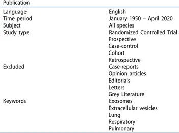
A systematic review of the literature was conducted to search for all articles reporting the experimental or clinical use of any type of EVs as a treatment for lung conditions related to COVID-19, i.e. pneumonia, ARDS, ALI and pulmonary fibrosis. This review was performed following the guidelines outlined in the Cochrane Handbook for Systematic Reviews of Interventions [24], and the Preferred Reporting Items for Systematic Reviews and Meta-analysis (PRISMA) statement [25]. This review was registered on the international prospec-tive register of systematic reviews PROSPERO (registra-tion #CRD42020176266) (Na(registra-tional institute for Health Research) [26]. The systematic review was conducted using a defined search strategy by four investigators (KK, RF, LA and GL) using electronic databases (PubMed/MEDLINE, Scopus, Cochrane Collaboration and Web of Science) (Table 1).
Inclusion/exclusion criteria
MeSH headings and terms used were “Exosomes OR Extracellular vesicles” and “Lung OR Respiratory OR Pulmonary” (Supplemental file 1). Studies published from 1950 until April 24th, 2020 were included. Reference lists were searched to identify relevant cross- references. Case reports, opinion articles, editorials and letters were excluded. All grey literature publications (i.e. reports, theses, conference proceedings, bibliographies, commercial documentations and official documents not published commercially) were excluded, except for abstracts published in the online accessible books of the International Society for Extracellular Vesicles annual meetings (ISEV; www.isev.org) held between 2016 and 2019 (Table 1). All studies that reported at least one outcome of interest (pneumonia, ARDS, ALI, or pulmonary fibrosis) were included in the analysis. After the selection of potential eligible papers using the title and the abstract, three reviewers (KK, RF and LA) independently retrieved the full-text articles to assess the final eligibility. Any disagreement over the eligibility of a specific study was resolved through the discussion with a fourth author (AZ). For the definition and classi-fication of EVs, we followed the minimal information for studies of extracellular vesicles (MISEV2018) guide-lines [27]. Synthetic-made nanoparticles were excluded by manual curation from the analysis as they did not meet the ISEV EV definition (Figure 2).
Table 1. Inclusion criteria of the systematic review. Publication
Language English
Time period January 1950 – April 2020
Subject All species
Study type Randomized Controlled Trial Prospective Case-control Cohort Retrospective Excluded Case-reports Opinion articles Editorials Letters Grey Literature Keywords Exosomes Extracellular vesicles Lung Respiratory Pulmonary
Figure 2. Diagram of workflow in the systematic review according to the PRISMA statement.
Outcome measures
We included the type of experimental model, EV source, EV separation and characterization techniques, details of EV administration including dosage and route, and EV biological effects. Additional evaluation of EV pathways/ mechanisms of action was performed. Further analysis was conducted on comparative studies which were clas-sified as having two or more different comparative EV groups, including but not limited to different sources of EVs or derivation/modification strategies on the same in vitro or in vivo model of administration.
Quality assessment
The risk of bias for each study was evaluated in dupli-cate (RF and GL) using the Systematic Review Centre for Laboratory Animal Experimentation (SYRCLE) risk of bias tool [28]. The Confidence in the Evidence from Reviews of Qualitative research (CERQual) tool was also utilized to assess evidence quality of each outcome in the systematic review [29]. The aim of this tool is to evaluate the extent to which the results of animal studies can be generalized to clinical trials or applica-tion, as determined by the results of evidence quality. Differences between the two reviewers (RF and GL) were resolved through consensus and discussion with a third author (AZ). Moreover, the present study was assessed in duplicate by two investigators (RF and GL) using A Measurement Tool to Assess Systematic Reviews, AMSTAR 2 [30]. A PRISMA figure following PRISMA checklist criteria was created [25].
Results
Of the 4,925 titles and abstracts screened, 233 articles were reviewed, 90 were assessed for eligibility and 39 were included in the final analysis (Figure 2) [31–69]. The selected studies were published between 2014 and 2020 and included in vitro, ex vivo, and in vivo studies (Table 2). None of the studies selected was conducted on human subjects. The most frequently used source of EVs was from MSCs derived from the bone-marrow or the umbilical cord of animal or human origin. EVs were also isolated from other stem cell sources such as adipose tissue, urine (urine-derived induced pluri-potent stem cells) and menstrual blood (endometrial stem cells). Other sources of EVs included: fibroblasts, blood (serum and whole blood), placenta, lung spher-oids, pulmonary endothelial cells and endothelial pro-genitor cells, primary adipose tissue, amnion epithelial cells, neutrophils and Staphylococcus aureus. The majority of the in vivo studies were performed in
mouse models, with only one reporting data of EV therapy in a large animal model (pigs) [59]. The selected studies addressed the effects of EVs as a therapy for ALI/ARDS and pneumonia, as well as for prevention or treatment of pulmonary fibrosis. To model ALI/ARDS, most studies used administration of either lipopolysaccharide (LPS), bleomycin or Escherichia coli, in isolation or in association with mechanical ventilation injury, trauma, influenza virus injection or carbonyl dichloride inhalation.
Overall, the effects of the different EV therapies on the experimental models were reported to recover lung injury, improve respiratory function and increase animal survival rate (Table 2). In ALI/ARDS, EV administration led to reduction of inflammation, alveolar epithelial regeneration and repair of the pul-monary endothelium [39,60,62] The EV modulation of the inflammatory response was primarily achieved by reduction of pro-inflammatory cytokines and neu-trophil infiltration, enhancement of anti- inflammatory cytokines and macrophage polarization to the M2 reparative phenotype. Specifically, EV administration resulted in the downregulation of interleukin (IL)-1β, tumour necrosis factor-α (TNF- α), IL-6 [37,47,62], macrophage inflammatory protein 2 (MIP-2), neutrophil chemokine KC [36,47], and in the upregulation of IL-10 (anti-inflammatory) [35,45]. Moreover, EVs re-established alveolar epithelial cell homoeostasis, with prevention of apoptosis [40], increased epithelial cell migration [39] and stimulated surfactant production by upregulation of surfactant protein C expression that resulted in resolution of alveolar wall collapse [42]. EVs were also reported to restore endothelial cell junction protein expression including vascular endothelial cadherin and occludin [60], and endothelial cell-cell adhesion factors via beta-catenin [34] and intercellular adhesion molecule 1 (ICAM-1) pathways [62,64]. Further, EVs also pre-served the alveolar-capillary barrier [37] and enhanced pulmonary endothelial cell proliferation
[60] (Table 2). In studies modelling pneumonia, EVs
induced adaptive immunity and reduced bacterial load [67]. Studies that tested EVs on models of pul-monary fibrosis reported that EV treatment reduced collagen deposition and density, halted fibrosis pro-gression by inhibiting myofibroblast differentiation, and improved the Ashcroft score, a standard scoring system for pulmonary fibrosis [32,43,50,65].
Most studies reported that EVs exert their effects via a number of different mechanisms. Many studies included experimental conditioned medium groups, which demonstrated that EVs were responsible for the effects observed. MicroRNAs (miRNAs) were the 4 K. KHALAJ ET AL.
Table 2. Articles reporting the effects of EV therapy on lung injury models.
Study Source of EVs Model Sample EV effects EV treatment outcome summary
Wang et al. 2020 [31] Adipose MSCs ALI: in vivo mouse (LPS) Lung tissue BAL
↓TNF-α, ↓IL-1β, ↓IL-6, ↑IL-10,
↓iNOS, ↓NF-κB Reduction of inflammation (decreased pro-inflammatory cytokines, increased anti-inflammatory cytokines, and reduced neutrophils in alveolar fluid), pulmonary endothelial barrier permeability, and alveolar septal thickness
ALI: in vitro mouse BMDMs (LPS)
BMDMs ↓TNF-α, ↓IL-1β, ↓iNOS, ↑YM-1, ↑MRC-1, ↑miR-27a-3p Dinh et al. 2020 [32] Lung spheroid cells Fibrosis: in vivo mouse (BLM) Lung tissue ↑AQP5, ↑vWF, ↓αSMA,
↓SMAD3, ↓Hydroxyproline Promotion of alveolar repair (increased aquaporin), attenuation of vascular injury and reduction of collagen deposition
Gao et al. 2020 [33] Adipose MSCs ALI: in vivo rat (PM2.5) Lung tissue BAL
↓TNF-α, ↓ROS Reduction of inflammation (decreased pro-inflammatory cytokines and oxidative stress), alveolar epithelial apoptosis and necrosis, and alveolar wall oedema and collapse
ALI: in vitro rat AEC2 (PM2.5) AEC2 ↓Apoptosis Yu et al. 2020 [34] Adipose tissue, Adipose MSCs,
Serum
ALI: in vivo mouse (Ventilator- induced lung injury)
Lung tissue BAL
↓IL-6, ↓TRPV4, ↑β-Catenin,
↑VE-Cadherin ↓MPO Reduction of inflammation (decreased pro-inflammatory cytokines) and pulmonary endothelial barrier permeability ALI: in vitro PMVECs (Cyclic
stretching)
PMVECs ↓TNF-α, ↓IL-6, ↓TRPV4, ↑β-Catenin, ↑VE-Cadherin Huang et al. 2019 [35] Adipose MSCs ALI: in vivo mouse (LPS) Lung tissue
BAL
↓IL-1β and ↑IL-10 Reduction of inflammation (lower neutrophil and macrophage recruitment in alveolar fluid) and alveolar wall thickness
ALI: in vitro mouse BMDMs (LPS)
BMDMs ↓IL-6, ↓IL-1β, ↓TNF-α, ↓iNOS, ↑TGF-β1, ↑YM-1 Silva et al. 2019 [36] Bone marrow MSCs ARDS: in vivo mouse (LPS) Lung tissue ↓TNF-α, ↓IL-6, ↓KC, ↓VEGF,
↓TGF- β Reduction of inflammation (lower neutrophils and macrophages in alveolar fluid) and alveolar wall collapse ARDS: in vitro mouse alveolar
macrophages (LPS)
Serum ↓iNOS, ↓IL-1β, ↓IL-6, ↑Arginase, ↑TGF-β Zhang et al. 2019 [37] PMVECs with high levels of
Syndecan-1 (SDC1)
ALI: in vivo mouse (LPS) Lung tissue ↓IL-6, ↓IL-1β, ↓TNF-α Reduction of inflammation (decreased pro-inflammatory cytokines), preservation of pulmonary endothelial function, and decrease in alveolar wall thickness ALI: in vitro mouse PMVECs
(LPS)
PMVECs ↓F-actin, ↓MLC, ↓MYPT1, ↓NF-κB
Hao et al. 2019 [38] Bone marrow MSCs ALI: in vivo mouse (E. coli) BAL ↓MIP-2, ↓TNF-α, ↑LTB4 Antimicrobial effect (increased monocyte phagocytosis and decreased bacterial levels) and reduction of inflammation (decreased leukocytes and neutrophils in alveolar fluid) ALI: in vitro mouse RAW267.4
cells (LPS)
RAW267.4 ↓MRP1-protein, ↑miR-145 Kim et al. 2019 [39] Placental chorionic and
decidual MSCs
ALI: in vitro human BEAS-2B and THP-1 cells (LPS)
THP-1 BEAS-2B
↓TNF-α Reduction of inflammation (decreased oxidative stress and pro-inflammatory cytokines) and restoration of bronchiolar epithelial cell migration and proliferation
Yi et al. 2019 [40] Bone marrow MSCs ALI: in vivo mouse (LPS) Lung tissue ↓MPO, ↓IL-1β, ↓TNF-α, ↓IL-6,
↑KGF, ↑IL-10, ↓SAA3 Reduction of inflammation (decreased pro-inflammatory cytokines), alveolar epithelial apoptosis, and lung interstitial vessel and alveolar septal thickness ALI: in vitro mouse AEC2 (LPS) BAL
AEC2
↓SAA3, ↓NF-κB, ↓ERK1/2, ↓p38MAPK, ↓MEK1/2, ↓JNK, ↑miR-30b-3p
Chen et al. 2019 [41] Umbilical cord MSCs ALI: in vivo rat (BLM) Lung tissue BAL
↓TNF-α, ↓IL-6, ↑HGF, ↑c-Met,
↑Akt, ↑mTOR Reduction of inflammation (reduced leukocytes and neutrophils in alveolar fluid and pro-inflammatory cytokines), alveolar epithelial apoptosis, alveolar wall thickness, pulmonary hyaline membrane, and collagen deposition
ALI: in vitro rat AEC2 and rat PMVECs (BLM)
AEC2 PMVECs
↑c-Met, ↑Akt, ↑mTOR, ↑HGF Xu et al. 2019 [42] Bone marrow MSCs ALI: in vivo rat (Phosgene-
induced)
Lung tissue BAL Plasma
↓MMP-9 ↑SP-C ↓TNF-α, ↓IL-1β,
↓IL-6, ↑IL- 10 Restoration of respiratory function (increased peak of inspiratory and expiratory flow, decreased lung resistance), reduction of inflammation (decreased oedema, pro- inflammatory cytokines), promotion of alveolar epithelial surfactant synthesis
Mansouri et al. 2019 [43] Bone marrow MSCs Fibrosis: in vivo mouse (BLM) Lung tissue BAL
↓Arg-1, ↓CCL2 Reduction of inflammation (decreased pro-inflammatory cytokine and Arg-1), alveolar epithelial apoptosis, septal thickness, and pulmonary collagen deposition
JOURNAL
OF
EXTRACELLULAR
VESICLES
Table 2. (Continued).
Study Source of EVs Model Sample EV effects EV treatment outcome summary
Loy et al. 2019 [44] Umbilical cord MSCs ALI: in vitro human AECs (H5N1)
AECs No particular mechanism studied
Restoration of alveolar fluid clearance and reduction of alveolar protein permeability
Li et al. 2019 [45] Bone marrow MSCs ALI: in vivo rat (Traumatic) Lung tissue BAL
↓P2X7, ↓MDA, ↓H2O2, ↑GSH, ↑SOD, ↓TNF-α, ↓IL-6, ↓IL-8, ↑IL-10
Reduction of inflammation (decreased oedema and pro- inflammatory cytokines), capillary hyperaemia, and alveolar wall thickness
Varkouhi et al. 2019 [46] γ–primed umbilical cord MSCs ALI: in vivo rat (E. coli) Lung tissue BAL
↑eNOS, ↓TNF-α Increase of animal survival, reduction of inflammation (decreased pro-inflammatory cytokine, increase of macrophage bacterial phagocytosis), restoration of alveolar epithelial surfactant synthesis and pulmonary endothelial cell permeability, and reduction of alveolar wall thickness
Zhou et al. 2019 [47] Umbilical cord EPC (rich in miR-126)
ALI: in vivo mouse (LPS) Lung tissue BAL
↓TNF-α, ↓IL-6, ↓IL-1β, ↓IFN-γ, ↓MIP-1, ↓MIP-2, ↓MIG, ↓IP- 10, ↓MPO
Reduction of inflammation (reduced pro-inflammatory cytokines and neutrophils in alveolar space), alveolar wall thickness, and hyaline membrane formation
ALI: in vitro human AECs (LPS) AECs ↑Claudin1, ↑Claudin4, ↑Occludin Park et al. 2019 [48] Bone marrow MSCs ALI: ex vivo perfused human
lung (E. coli)
Lung tissue BAL
TNF-α (no difference) Antimicrobial effects (increased macrophages phagocytosis and decreased bacterial levels), restoration of alveolar epithelial surfactant synthesis and alveolar fluid clearance, reduction of pulmonary endothelial permeability and alveolar wall thickness
Royce et al. 2019 [49] Amnion epithelial cells Fibrosis: in vivo mouse (BLM) Lung tissue ↓TGF- β Reduction of tissue inflammation and myofibroblast accumulation
Sun et al. 2019 [50] Menstrual blood-derived endometrial stem cells
Fibrosis: in vivo mouse (BLM) Lung tissue ↓Hydroxyproline, ↓MDA, ↑Let-7 Reduction of inflammation (decreased inflammasome), DNA damage (decreased ROS) and collagen deposition Fibrosis: in vitro mouse AECs
(BLM)
AECs ↓ROS, ↓LOX1, ↓NLRP3, ↓Hydroxyproline, ↓MDA, ↑Let-7
Liu et al. 2019 [51] Umbilical cord MSCs ALI: in vivo rat (Burn) Lung tissue Serum
↓TNF-α, ↓IL-1β, ↓IL-6, ↑IL- 10, ↓MDA, ↓MPO, ↑SOD, ↓TLR4, ↓p-p65, ↑miR-451
Reduction of inflammation (lower pro-inflammatory cytokines and decreased immune cell recruitment) Sun et al. 2018 [52] Whole blood Fibrosis: in vivo mouse (Zeocin) Lung tissue ↓Hydroxyproline Reduction of inflammation (decreased immune cell
recruitment), alveolar wall thickness and collagen deposition
Bandeira et al. 2018 [53] Adipose MSCs Fibrosis/Silicosis: in vivo mouse (Silica)
Lung tissue ↓TGF-β, ↓TNF-α, ↓IL-1β Reduction of inflammation (decreased pro-inflammatory cytokines and macrophages) and collagen deposition Tan et al. 2018 [54] Amnion epithelial cells Fibrosis: in vivo mouse (BLM) Lung tissue ↑CTNNB1, ↑BMP4, ↑BMPR1,
↑FOXM1, ↑LEF1, ↑NFATC1, ↑PGK1, ↑PTN, ↑SCA1, ↑WLS, ↓cMYC
Prevention and reduction of inflammation (lower CD4 + T cells and pulmonary interstitial macrophages) and collagen deposition
Hu et al. 2018 [55] Bone marrow MSCs ALI: in vitro human LMVECs (Cytomix)
LMVECs ↑VE-cadherin, ↑ZO-1, ↑p-myosin light chain 2, ↑Ang1, ↑S1PK
Decrease of microvascular permeability
Zhang et al. 2018 [56] Pulmonary microvascular endothelial cells
ALI: in vivo mouse (LPS) Lung tissue ↓TNF-α, ↓IL-1β, ↓IL-6 Reduction of inflammation (lower pro-inflammatory cytokines) and restoration of pulmonary endothelial function (rearranged cytoskeleton and decreased microvascular permeability)
BAL ↓TNF-α, ↓IL-2, ↓IL-3, ↓IL-6, ↓GM-CSF, ↓CCL-2, ↓MCP-5, ↓CCL-5
Shah et al. 2018 [57] Bone marrow MSCs ARDS: in vivo mouse (LPS) Lung tissue ↑Runx1p66/p52, ↑TβRI/Alk5 Reduction of perivascular area and interstitial thickness Wu et al. 2018 [58] Endothelial progenitor cells ALI: in vivo rat (LPS) Lung tissue ↑RAF, ↑ERK, ↓SPRED-1, ↓MDA,
↑miR-126 Enhancement of gas exchange (improved PaOinflammation (reduced oedema and neutrophil in alveolar 2), reduction of space), regeneration of pulmonary endothelial cells and reduction of alveolar wall thickness
6
K.
KHALAJ
ET
Table 2. (Continued).
Study Source of EVs Model Sample EV effects EV treatment outcome summary
Khatri et al. 2018 [59] Bone marrow MSCs ALI: in vivo pig (Influenza) Lung tissue BAL Nasal swabs
↓TNF-α, ↓CXCL10, ↑ IL-10 Reduction of inflammation (decreased pro-inflammatory cytokines and chemokines), lung endothelial cell apoptosis, alveolar wall thickness and collapse, and inhibition of influenza virus replication
ALI: in vitro pig LECs (Influenza) LECs ↓Apoptosis Wang et al. 2017 [60] Bone marrow MSCs ALI: in vitro mouse PMVECs
(LPS)
PMVECs ↑VE-cadherin, ↑ZO-1, ↓IL-6,
↑IL-10 Reduction of inflammation (decreased pro-inflammatory cytokines) and pulmonary endothelial permeability Morrison et al. 2017 [61] Bone marrow MSCs ARDS: in vivo mouse (LPS) BAL ↓TNF-α Reduction of inflammation (lower pro-inflammatory cytokine
and neutrophils in alveolar fluid) Gao et al. 2017 [62] Neutrophils loaded with
piceatannol
ALI: in vivo mouse (LPS) Lung tissue BAL Plasma
↓TNF-α, ↓IL-6, ↓MPO Increase in animal survival, reduction of inflammation (lower pro-inflammatory cytokines, leukocytes and neutrophils in alveolar fluid) and pulmonary endothelial permeability ALI: in vitro human HUVECs
(LPS)
HUVECs ↓ICAM-1, ↓IκBα, ↓p65
Tang et al. 2017 [63] Bone marrow MSCs ALI: in vivo mouse (LPS) Lung tissue BAL ↓MIP-2 Reduction of inflammation (decreased pro-inflammatory cytokines, leukocytes and neutrophils in alveolar fluid), pulmonary endothelial permeability, and alveolar wall thickness
ALI: in vitro human LMVECs and mouse RAW264.7 cells (LPS)
LMVECs RAW264.7
↓TNF-α, ↑IL-10 Ju et al. 2017 [64] Urine-derived pluripotent stem
cells
ALI: in vitro human MVECs (LPS)
MVECs ↓ICAM-1, ↓MPO Reduction of inflammation (attenuated neutrophils adhesion and blocked inflammatory activation in the pulmonary endothelium)
Shentu et al. 2017 [65] Bone marrow MSCs Fibrosis: in vitro human LFCs LFCs ↓αSMA, ↓Col IA1, ↓Col III A1 Reduction of myofibroblast accumulation and collagen deposition
Li et al. 2015 [66] Bone marrow MSCs ALI: in vivo mouse (E. coli) BAL ↓MIP-2 Reduction of inflammation (decreased pro-inflammatory cytokines, leukocytes and neutrophils and increased anti- inflammatory cytokines in the alveolar fluid)
ALI: in vitro mouse RAW264.7 cells (E. coli)
RAW264.7 ↓TNF-α, ↑IL-10 Choi et al. 2015 [67] S. aureus Pneumonia: in vivo mouse
(Staphylococcus aureus)
Lung tissue Blood
↓IL-1β, ↓IL-6, ↑IL-17, ↑IL-4,
↑IFN-γ Increase in animal survival, reduction of inflammation (decreased pro-inflammatory cytokines) and induction of adaptative immunity (increased T cell response) Monsel et al. 2015 [68] Bone marrow MSCs Pneumonia: in vivo mouse
(E. coli)
Lung tissue BAL
↓MIP-2, ↑KGF, ↓TNF-α Increase in animal survival and antimicrobial effect (increased monocyte phagocytosis), reduction of inflammation (lower pro-inflammatory cytokines and neutrophil in alveolar space), pulmonary endothelial permeability, and alveolar wall thickness
Pneumonia: in vitro human AEC2 (LPS and Cytomix)
AEC2
Zhu et al. 2014 [69] Bone marrow MSCs ARDS: in vivo mouse (E. coli)
Lung tissue BAL
↓MIP-2 Reduction of inflammation (lower pro-inflammatory cytokines, leukocytes and neutrophils in alveolar fluid) and interstitial thickness
ARDS: in vitro mouse RAW264.7 cells (E. coli)
RAW264.7 ↑IL-10, ↓TNF-α, ↓MIP-2 Abbreviations:
AEC2: Alveolar epithelial cells type 2; AECs: Alveolar epithelial cells; ALI: Acute lung injury; ANG1: Angiopoietin 1; AQP: Aquaporin; ARDS: Acute respiratory distress syndrome; BAL: Bronchioalveolar lavage; BEAS-2B: Bronchial epithelial cells; BLM: Bleomycin; BMDMs: Bone marrow-derived macrophages; BMP: Bone morphogenetic protein; BMPR1: Bone morphogenetic protein receptor 1; COL: Collagen; CTNNB1: Catenin beta 1; CXCL: Chemokine (C-X-C motif) ligand; EPC: Endothelial progenitor cell; ERK1/2: Extracellular regulated kinase ½; EVs: Extracellular vesicles; FOXM: Forkhead Box M1; GM-CSF: Granulocyte macrophage colony stimulating factor; HGF: Hepatocyte growth factor; HUVECs: Human umbilical vein endothelial cells; ICAM-1: Intercellular adhesion molecule-1; IFN: Interferon; IL: Interleukin; IP: Interferon gamma-induced protein; IкBα: Nuclear factor of kappa light polypeptide gene enhancer in B-cells inhibitor, alpha; JNK: c-Jun N-terminal kinases; KC: Keratinocyte chemoattractant; KGF: Keratinocyte growth factor; LECs: Lymphatic endothelial cells; LEF1: Lymphoid enhancer-binding factor-1; LFCs: Lung fibroblast cells; LMVEC: Lung microvascular endothelial cell; LOX1: Lectin-like oxidized LDL receptor-1; LPS: Lipopolysaccharide; LTB4: Leukotriene B4; MCP: Monocyte chemotactic protein; MDA: Malondialdehyde; MEK1/2: P-dual specificity mitogen-activated protein kinase ½; MIG: Monokine induced by gamma interferon; MIP: Macrophage induced protein; MIR: MicroRNA; MMP-9: Matrix metalloproteinase-9; MPO: Myeloperoxidase; MRC-1: Mannose Receptor C-Type 1; MRP1: Multidrug resistance associated protein 1; MSC: Mesenchymal stem/stromal cell; MVECs: Microvascular endothelial cells; NFATC1: Nuclear factor of activated T-cells, cytoplasmic 1; NF-κB: Nuclear factor kappa-light-chain- enhancer of activated B cells; NLRP3: NLR Family pyrin domain containing 3; NOS: Nitric oxide synthase (iNOS – inducible, eNOS – endothelial); P38MAPK: p38 mitogen-activated protein kinase; PGK1: Phosphoglycerate kinase 1; PM2.5: fine particulate matter; PMVECs: Pulmonary microvascular endothelial cells; PTN: Pleiotrophin; RAW267.4: Monocyte/macrophage lineage; ROS: Reactive oxygen species; S1PK: Sphingosine kinase; SAA3: Serum amyloid A3; SCA1: Stem cell antigen-1; SMA: Smooth muscle actin; SMAD3: Mothers against decapentaplegic homolog 3; SOD: Superoxide dismutase; SP-C: Surfactant protein-C; SPRED-1: Sprouty-related, EVH1 domain-containing protein 1; TGF: Transforming growth factor; THP-1: Human monocyte cell line; TLR: Toll-like receptor; TNF: Tumour necrosis factor; TRPV4: Transient receptor potential vanilloid 4; TβRI: Transforming growth factor-beta receptor I; VE-Cadherin: Vascular endothelial cadherin; VEGF: Vascular endothelial growth factor; VWF: Von Willebrand factor; WLS: Wnt Ligand Secretion Mediator; YM-1: Chitinase 3-like 3, a macrophage protein; ZO-1: Tight junction protein
JOURNAL
OF
EXTRACELLULAR
VESICLES
molecules contained in the EV cargo that were most often investigated. More than one study reported that miR-126 was involved in the attenuation of inflamma-tion via different pathways. Wu et al. demonstrated EV modulation in endothelial cells via sprouty-related EVH1 domain-containing protein 1 axis, which tar-gets the RAF/ERK signalling pathway [58], and Zhou et al. showed that EVs increase expression of tight junction proteins via the phosphoinositide-3-kinase regulatory subunit 2 (PIK3R2), as well as high mobi-lity group box 1 (HMGB1), and vascular endothelial growth factor-α (VEGFα) [47]. Interestingly, miR-126 has been reported to suppress inflammation in several conditions in other organs, is highly expressed in the lung of normal patients, and downregulated in the airway cells of patients with lung conditions, such as
cystic fibrosis [70–73]. Yi and colleagues reported that EVs containing increased miR-30b-3p levels could increase alveolar epithelial cell proliferation and diminish apoptosis via decreased serum amyloid A3, a positive regulator of the inflammatory response [40]. This study is in line with another report showing that miR-30b-3p levels are decreased both in children with pneumonia and in mice with LPS-induced ALI [74]. Further, MSC-EVs suppressed multidrug resistance- associated protein 1 through transfer of miR-145, which positively signalled through the leukotriene (LTB4/BLT1) signalling pathway [38], and reduced TNF-α via increased M2 reparative macrophage polar-ization from miR-27a-3p transfer [31]. Both miR-145 and −27a-3p have been independently reported to modulate lung fibrosis by targeting myofibroblast
Table 3. Articles comparing the effects of different EV populations.
Study EV populations EV treatment outcome summary
Dinh et al. 2020 [32] LSC-EVs vs MSC-EVs Both EV populations ameliorated lung fibrosis. LSC-EVs promoted more alveolar repair via increased aquaporins and had reduced SMAD3 levels compared to MSC-EVs
Yu et al. 2019 [34] Mouse AT-EVs vs. S-EVs vs. ADSC-EVs
All EV populations restored adherens junction protein expression and attenuated inflammation. AT-EVs and ADSC-EVs were more efficient in suppressing endothelial inflammation than S-EVs
Huang et al. 2019 [35] Human adipose MSC-EVs from young (25 years old) vs. old (72 years old) donors
Young (but not old) MSC-EVs alleviated ALI and altered macrophage phenotypes
Zhang et al. 2019 [37] Mouse PMVECs with high vs. low syndecan-1
SDC1-high-EVs (but not SDC1-low-EVs) ameliorated lung inflammation by reducing pro- inflammatory cytokines
Kim et al. 2019 [39] Human DMSC23-EVs vs. CMSC29-EVs
Both EV populations reduced pro-inflammatory cytokines and increased the migration of human bronchial epithelial cells
Yi et al. 2019 [40] Mouse MSC-EVs with high vs. low miR-30b-3p
Only MSC-EVs with high miR-30b-3p levels increased cell proliferation and reduced cell apoptosis and pro-inflammatory cytokines
Varkouhi et al. 2019 [46] Human interferon-γ-primed MSC-EVs vs. naïve MSC-EVs
Both EV populations enhanced survival and bacterial phagocytosis in THP-1 cells and reduced the alveolar wall thickness. Only IFN-γ-primed MSC-EVs decreased pro- inflammatory cytokines
Zhou et al. 2019 [47] Human EPC-EVs vs. NIH3T3- EVs
EPC-EVs (but not NIH3T3-EVs) reduced pro-inflammatory cytokine and chemokine production and lung tissue injury and restored alveolar barrier
Tan et al. 2018 [54] Human AEC-EVs vs. HLF-EVs AEC-EVs and HLF-EVs similarly reduced neutrophil infiltration and interstitial macrophages. AEC-EVs were more effective in attenuating lung fibrosis in aged mice compared to HLF-EVs
Wang et al. 2017 [60] Mouse HGF knockdown MSC- EVs vs. MSC-EVs
MSCs-EVs (but not HGF knockdown MSC-EVs) reduced pro-inflammatory cytokines and restored endothelial permeability regulation
Gao et al. 2017 [62] Human NS-EVs vs. NC-EVs NS-EVs and NC-EVs similarly reduced pro-inflammatory cytokines and leukocytes and restored alveolar epithelial function
Tang et al. 2017 [63] Human Ang-1 knockdown MSC-EVs vs. MSC-EVs
MSC-EVs (but not Ang-1 knockdown MSC-EVs) reduced lung inflammation and pulmonary capillary permeability
Li et al. 2015 [66] Human MSCIPC-30-EVs vs. MSCIPC-60-EVs vs. MSCIPC- 90-EVs
MSCIPC-60-EVs were more effective at reducing pro-inflammatory cytokines and increasing anti-inflammatory cytokines than MSCIPC-30-EVs and MSCIPC-90-EVs Monsel et al. 2015 [68] Human MSC-EVs vs. NHLF-EVs MSC-EVs (but not NHLF-EVs) increased survival rate and decreased pro-inflammatory
cytokines
Zhu et al. 2014 [69] Human MSC-EVs vs. NHLF-EVs MSC-EVs (but not NHLF-EVs) reduced inflammation and protein permeability and decreased lung injury
Abbreviations
ADSC-EVs: Adipose-derived stem cell extracellular vesicles; AEC-EVs: Amnion epithelial cell extracellular vesicles; ALI: Acute lung injury; AT-EVs: Adipose
tissue extracellular vesicles; BEAS-2B: Bronchial epithelial cell line; CMSC29-EVs: Chorionic-derived mesenchymal stem cell extracellular vesicles; DMSC23-
EVs: Decidual-derived mesenchymal stem cell extracellular vesicles; EPC-EVs: Endothelial progenitor cell extracellular vesicles; EVs: Extracellular vesicles; HGF: Hepatocyte growth factor; HLF-EVs: Human lung fibroblast extracellular vesicles; LSC-EVs: Lung spheroid cell extracellular vesicles; MSC-EVs:
Mesenchymal stem/stromal cell extracellular vesicles; MSCIPC-30-EVs: MSCs subjected to Ischaemic Pre-Conditioning for 30 minutes-derived extracellular vesicles; MSCIPC-60-EVs: MSCs subjected to Ischaemic Pre-Conditioning for 60 minutes-derived extracellular vesicles; MSCIPC-90-EVs: MSCs subjected to Ischaemic Pre-Conditioning for 90 minutes-derived extracellular vesicles; NC-EVs: Nitrogen cavitation generated extracellular vesicles; NHLF-EVs: Normal human lung fibroblasts; NIH3T3-EVs: fibroblast-derived extracellular vesicles; NS-EVs: Naturally secreted extracellular vesicles; PMVEC-EVs: Pulmonary microvascular endothelial cell extracellular vesicles; SDC1: Gene for encoding syndecan-1; S-EVs: Serum-derived extracellular vesicles
Table 4. Details of EVs used as a therapy in lung injury models.
Study EV source EV separation EV characterization EV dosage
Wang et al. 2020 [31] Adipose MSCs Pre-clearing cells/debris, UC (118,000 g for 16 h at 4°C)
NTA; DLS; TEM; Flow Cytometry (CD63); WB (CD40, CD44, CD63, CD81, CD105; GM130, Calnexin)
1 dose; 100 μg/mL of EVs added to culture medium, or 50 µg/ 0.05 ml of EVs, administered via intratracheal injection (in vivo)
Dinh et al. 2020 [32] Lung spheroid cells Ultrafiltration, Centrifugation (5000 g for 10–15 min)
NTA; TEM; WB (CD63, CD81, TSG101) 1 dose; 10 × 109 particles per kg of body weight, administered intranasally (aerosolized) Gao et al. 2020 [33] Adipose MSCs Ultracentrifugation (details unspecified) NTA; TEM; WB (CD63, TSG101, Alix,
GM130)
1 dose; 1 × 10⁹ EVs added to culture medium, or 2.5 ~ 2.8 × 10¹⁰ EVs in 20 μL PBS, administered via intratracheal injection (in vivo)
Yu et al. 2020 [34] Adipose tissue, Adipose MSCs, Serum
Pre-clearing cells/debris, TEIR NTA; TEM; WB (CD63, HSP70, TSG101) 1–2 doses; 0, 25, 50, and 100 μg/ml of EVs added to culture medium or injected intravenously 1 h before and immediately after mechanical ventilation Huang et al. 2019 [35] Adipose MSCs Pre-clearing cells/debris, UC (118,000 g for
16 h at 4°C)
NTA; TEM; WB (CD63, CD81, CD105, CD44, GM130, Calnexin)
1 dose; 100 μg/200 µl of EVs, administered via tail vein injection
Silva et al. 2019 [36] Bone marrow MSCs Pre-clearing cells/debris, UC (100,000 g for 1 h at 4°C, twice)
NTA; SEM 1 dose; EVs from 105 cells, administered via jugular vein injection
Zhang et al. 2019 [37] PMVECs Pre-clearing cells/debris, UC (100,000 g for 1 h at 4°C, twice)
NTA; TEM; WB (CD9, CD63, CD81) 2 doses; 3 μg/g of EVs, administered via tail vein injection Hao et al. 2019 [38] Bone marrow MSCs Pre-clearing cells/debris, UC (100,000 g for
1 h at 4°C, twice)
NTA; SEM; Flow Cytometry (CD9, CD44) 1–4 doses; 3, 6, and 12 × 108 particles added to culture medium, or 90 µl of EV per mouse (1x10¹⁰ particles), administered intravenously (in vivo)
Kim et al. 2019 [39] Placental chorionic or decidual MSCs
Pre-clearing cells/debris, UC (100,000 g for 1 h at 4°C, twice)
NTA; TRPS; SEM; TEM; AFM 1–2 doses; ranging 6 × 10⁵–1.5 × 10⁷ particles per ml, added to culture medium
Yi et al. 2019 [40] Bone marrow MSCs Pre-clearing cells/debris, centrifugation (10,000 g for 1 h at 4°C, twice)
NTA; Flow Cytometry (CD63) 1 dose; 1 μg/100 μL or 100 μg/200 μL, administered via intravenous injection (caudal veins)
Chen et al. 2019 [41] Umbilical cord MSCs Pre-clearing cells/debris, UC (100,000 g for 1 h at 4°C, twice)
SEM; Flow Cytometry (CD34, CD44, CD45, CD73, CD105)
1 dose; 4 mg/kg, administered via intratracheal injection Xu et al. 2019 [42] Bone marrow MSCs Pre-clearing cells/debris, UC (100,000 g for
1 h at 4°C, twice)
NTA; TEM; WB (CD9, CD63, CD81) 1 dose; EVs from 5 × 10⁶ cells, administered via intratracheal injection
Mansouri et al. 2019 [43] Bone marrow MSCs Pre-clearing cells/debris, UC (100,000 g for 3.5 h at 4°C)
NTA, TEM, WB (Alix, CD63, CD9, Flot-1) 1 dose; diluted in PBS to correspond to 5 × 10⁶ cell equivalent, administered via intravenous injection Loy et al. 2019 [44] Umbilical cord MSCs or bone
marrow MSCs
Pre-clearing with filtration; ExoEasy Maxi Kit, miRCURY Exosome Isolation Kit
Not specified 1 dose; not specified concentration of EVs, administered via intravenous injection
Li et al. 2019 [45] Bone marrow MSCs Pre-clearing cells/debris, centrifugation (12,000 g for 1 h at 4°C, twice)
NTA; TEM; WB (CD9, CD63, CD81) 2 doses per day for 7 days; 25 μg of EVs, administered via tail vein injection
Varkouhi et al. 2019 [46] γ–primed umbilical cord MSCs
Pre-clearing cells/debris, UC (100,000 g for 1.5 h at 4°C)
TEM; Flow Cytometry for size 1 dose; 10 × 108 EVs/kg, administered via intravenous injection
Zhou et al. 2019 [47] Umbilical cord EPCs Pre-clearing cells/debris, TEIR NTA; WB (CD9, CD63, CD81) 1 dose; 70 μg of EVs, administered via intratracheal injection Park et al. 2019 [48] Bone marrow MSCs Pre-clearing cells/debris, UC (100,000 g for
1 h at 4°C, twice)
NTA; SEM; Flow Cytometry (CD9, CD44); 1 dose; 200 μl – 400 μl of EVs, administered via intravenous injection
Royce et al. 2019 [49] Amnion epithelial cells Not specified Not specified 1 dose; 5–25 μg of EVs, administered intranasally Sun et al. 2019 [50] Menstrual blood-derived
endometrial stem cells
TEIR NTA; TEM; WB (TSG101, CD9, CD63, Calnexin)
1 dose; 0.5 mg/kg of EVs, administered via tail vein injection Liu et al. 2019 [51] Umbilical cord MSCs Pre-clearing cells/debris, ExoQuick NTA; TEM; WB (CD9, CD63) 1 dose; 800 μg of EVs, added to culture medium, or
administered via tail vein injection (in vivo) Sun et al. 2018 [52] Whole blood Sonication, Ultrafiltration (9,000 g for
15 min)
TEM; AFM 2 doses; 2.5 mg/kg and 0.25 mg/kg of EVs, administered intranasally (nasal drip)
Bandeira et al. 2018 [53] Adipose MSCs Pre-clearing cells/debris, UC (100,000 g for 2 h)
TEM; NTA, WB (CD63, CD81, LAMP-1, CD9) 1 dose; 50 µl of EVs, administered via intratracheal injection Tan et al. 2018 [54] Amnion epithelial cells Centrifugation (details unspecified) NTA; TEM; WB and Flow cytometry (CD9,
CD81, Alix, HLA-G)
1 dose; not specified concentration of EVs, added to culture medium JOURNAL OF EXTRACELLULAR VESICLES 9
Table 4. (Continued).
Study EV source EV separation EV characterization EV dosage
Hu et al. 2018 [55] Bone marrow MSCs Pre-clearing cells/debris, UC (100,000 g for 1 h at 4°C, twice)
NTA; SEM; Flow Cytometry (CD9, CD44) 1–2 doses; 30 µl or 60 µl of EVs, added to culture medium Zhang et al. 2018 [56] Serum TEIR NTA; TEM; WB (CD63, Flot-1, TSG101) 1 dose; 10 μl – 500 μl of EVs, administered via intratracheal
injection Shah et al. 2018 [57] Bone marrow MSCs Pre-clearing cells/debris, UC (28,000 g for
2 h at 4°C, twice)
NTA; Flow Cytometry for size 2 doses; 2.9 × 10⁵ and 5.8 × 10⁵ of EVs in 100 μl PBS, administered via retroorbital injection
Wu et al. 2018 [58] Endothelial progenitor cells Pre-clearing cells/debris, UC (100,000 g for 2 h at 4°C, twice)
NTA; TEM; WB (CD63, Alix, TSG101) 1 dose; 100 μg, administered via tail vein injection Khatri et al. 2018 [59] Bone marrow MSCs Pre-clearing cells/debris, UC (25,000 g for
70 min at 4°C)
TEM; WB (CD9, CD63, CD81) 1 dose; 10 μg/mL of EVs added to culture medium, or 80 μg/ kg of EVs, administered via intratracheal injection Wang et al. 2017 [60] Bone marrow MSCs Pre-clearing cells/debris, UC (100,000 g for
1 h at 4°C, twice)
Flow Cytometry (CD29, CD34, CD44, CD45, CD105) TEM; SEM;
1 dose; unspecified concentration of EVs, added to culture medium
Morrison et al. 2017 [61] Bone marrow MSCs Pre-clearing cells/debris, UC (100,000 g for 2 h)
Flow Cytometry for size and CD44 1 dose; unspecified concentration of EVs added to different models, administered intranasally (in vivo)
Gao et al. 2017 [62] Neutrophils NS-EVs: pre-clearing cells/debris, UC (100,000 g for 1 h). NC-EVs: nitrogen cavitation chamber at a pressure of 400–500 psi for 20 min at 0°C
qNano and dynamic light scattering; cryo- TEM for NC-EVs; WB (IntegrinB2, LAMP- 1, Calnexin, COXIV)
1 dose; unspecified number of drug loaded EVs (piceatannol at 3 mg/kg), administered via intravenous injection
Tang et al. 2017 [63] Bone marrow MSCs Pre-clearing cells/debris, UC (100,000 g for 1 h at 4°C, twice)
TEM and SEM Not specified Ju et al. 2017 [64] Urine-derived pluripotent stem
cells
Pre-clearing cells/debris, UC (120,000 g for 70 min at 4°C, twice)
NTA; TRPS; TEM; WB (CD63, TSG101, Alix) 1 dose; 10 μg protein of Exo/siRNA (EV) compound or micropoly with siRNA at a final concentration of 100 nM, added to culture medium
Shentu et al. 2017 [65] Bone marrow MSCs Pre-clearing cells/debris, UC (100,000 for 1 h, twice)
NTA; TEM; WB (CD63, CD81, Calnexin) 1 dose; 10 μg of EVs added to culture medium
Li et al. 2015 [66] Bone marrow MSCs ExoQuick WB (CD63) 1 dose; 1.5 μg/g of EVs, administered via tail vein injection Choi et al. 2015 [67] S. aureus Pre-clearing cells/debris, UC (150,000 g for
3 h at 4°C)
Not specified 1 dose; 10 μg/ml of EVs added to culture medium Monsel et al. 2015 [68] Bone marrow MSCs Pre-clearing cells/debris, UC (100,000 g for
1 h at 4°C)
TEM, WB (CD44) 3 doses; EVs from 10 μL per 1 × 10⁶ cells added to culture medium, or EVs administered intratracheal (30 µl or 60 µl) or intravenously (90 µl) (in vivo)
Zhu et al. 2014 [69] Bone marrow MSCs Pre-clearing cells/debris, UC (100,000 g for 1 h at 4°C, twice)
TEM and SEM 1–2 doses; 30 µl or 60 µl of EVs, administered via intravenous or intratracheal injection
Abbreviations: AFM: Atomic force microscopy; Alix: Apoptosis-Linked Gene 2-Interacting Protein X; CD: Cluster of differentiation; COXIV: Cytochrome c oxidase Complex IV; DLS: Dynamic light scattering; EV: Extracellular vesicle; Flot-1: Flotillin 1; GM: Golgi matrix; HLA: Human leukocyte antigen; HSP: Heat shock protein; IntegrinB2: Integrin beta 2; LAMP-1: Lysosome-associated member glycoprotein 1; MSC: Mesenchymal stromal/stem cell; NTA: Nanoparticle tracking analysis; PMVEC: Pulmonary microvascular endothelial cell; SEM: Scanning electron microscopy; TEIR: Total exosome isolation reagent; TEM: Transmission electron microscopy; TRPS: Tunable resistive pulse sensing; TSG: Tumour susceptibility; UC: Ultracentrifugation; WB: Western blot
10
K.
KHALAJ
ET
differentiation [75,76]. All of the miRNAs identified in our systematic review (miRs-126, −30b-3p, −145, −27a-3p) have been reported by other groups to be present in their respective source EV cargo content [77–80].
In addition to these miRNAs, some studies showed that the EV effect on ARDS lungs was due to the release of proteins, such as syndecan-1 [37], hepatocyte growth factor [60] and angiopoietin-1 [63]. All these three proteins are known to attenuate lung injury in several models: syndecan-1 modulates the innate immune response to influenza infection via c-Met sig-nalling [81]; hepatocyte growth factor mediates epithe-lial-mesenchymal interaction for lung regeneration following lung injury [82]; lastly, angiopoietin-1 plays a role in attenuating lung permeability and inflamma-tion in experimental pneumonia and ALI [83,84]. All these three proteins have already been described to be present in their respective source cargo, as reported in Exocarta and Vesiclepedia [85,86].
To better understand EV mechanisms of action, some studies performed a comparative analysis on EV efficacy and regenerative capacity according to their source (Table 3). Different sources of MSC-EVs, namely chorionic and decidual, had the same capability in regenerating the injured lung [39] (Table 3). Huang and colleagues demonstrated that EVs derived from human adipose MSCs from a young patient (25 years old) decreased inflammation levels more efficiently than those isolated from an old patient (72 years old) [35].
We also analysed EV separation and characteriza-tion methodology with a specific reference to ISEV guidelines, as well as dosage and administration route
(Table 4). Ultracentrifugation was the most popular EV
separation technique, followed by reagent-based com-mercially available kits. Only few studies followed ISEV recommendations and additional experimental details were not reported through online databases, such as EV-track [87]. The concentrations and dosage fre-quency were not consistent across studies, even when the same source of EVs was used. On average, the models were treated with one or two doses of EVs over a 1–10-day period. In in vivo animal models, the EV administration route was either intravenous or intratracheal or a combination of both. None of the studies included in this analysis isolated EVs following good manufacturing practices (GMP).
The quality assessment using SYRCLE showed that the selected studies had a medium level of bias, as reported in the quantitative assessment (Supplemental file 2 and Supplemental file 3). The majority of the
in vivo studies presented high or unclear risk of bias due to a lack of information regarding selection method, allocation concealment, animal housing and replacement of dropout animals. Moreover, both in vivo and in vitro studies did not commonly report information regarding randomization and blinding. The present systematic review was independently assessed using AMSTAR 2 [30] and received a “high” score (Supplemental file 4). A list of excluded articles is provided in Supplemental file 5. Articles were excluded if they did not match the outcome of interest (e.g. lung injury models not relevant to COVID-19, such as bronchopulmonary dysplasia, cystic fibrosis and asthma) or the treatment criteria (e.g. EVs employed as biomarkers and not for treatment). The CERQual tool revealed that the inflammatory response and lung injury recovery in the alveolar epithelium received “high” scores, whereas the other selected parameters had lower scores (Supplemental file 6). Lastly, the PRISMA checklist was completed which included further details for the review scoring (Supplemental file 7).
Discussion
This systematic review reveals that a number of studies demonstrate that EVs of various sources are able to recover lung injury, improve respiratory function and in some cases increase animal survival. The majority of these articles were published in the last 4 years, which is indicative of how active the EV community has been in this field. Two thirds of the studies included in our review utilized MSC-EVs as a therapy for lung injury. Likely, these studies have stemmed out of the vast litera-ture on lung regeneration using stem cell-based thera-pies, which primarily reported the anti-inflammatory properties of MSCs in animal models of lung injury [17]. Moreover, MSCs have also been tested as a therapy for patients with moderate to severe ARDS in phase 1 and phase 2a clinical trials [15,16]. These trials concluded that MSCs were safe to administer to ARDS patients, but their efficacy still had to be determined in a larger phase 2b trial. With these promising human data, in the past months we have witnessed an increasing number of registered clinical trials testing MSCs in patients with COVID-19-related lung injury [88], as well as two scientific articles reporting beneficial effects of MSCs in patients [89,90]. Leng et al described that seven COVID-19 patients with pneumonia had symptom resolution 2–4 days following administration of clinical grade human MSCs [89]. Similarly, Liang et al reported the case of a 65-year-old patient, who was critically ill with COVID-19 and recovered following administration JOURNAL OF EXTRACELLULAR VESICLES 11
of human umbilical cord MSCs [90]. Although both studies lack details on cell source and characterization, enrolment criteria and patient clinical status pre- and post-treatment, it is encouraging that MSC-based thera-pies could improve the outcomes of patients with COVID-19-related lung injury. As EVs are considered to be one of the most important effectors of MSC para-crine mechanism of action [91], the results of studies employing MSCs for COVID-19-related lung injury could be useful in predicting the potential outcome of MSC-EV-based therapies in the same population.
Our review confirms that also EVs from different sources address multiple pathways and can be benefi-cial to treat lung injury. In the analysed studies, EVs had the capacity to attenuate the inflammatory response and switch it to a reparative phenotype, to arrest the injury to the alveolar epithelium and imple-ment its regeneration, to repair the lung vascular damage, and to ameliorate and prevent pulmonary fibrosis. Furthermore, of relevance to COVID-19, EVs have been reported to exert some antiviral properties, as shown in the only large animal study where MSC- EV injection inhibited influenza virus replication in pigs and virus-induced apoptosis in porcine lung epithelial cells [59]. Moreover, as COVID-19 infection is frequently complicated by multiple organ failure, it is unlikely that a single drug will be able to improve the most severe forms of COVID-19 [22], whereas EV- based treatments could yield more promising out-comes. However, many key considerations remain to be addressed in this exciting and relatively new field.
Before testing these promising EV therapies on human subjects, a number of translational issues should be carefully considered as summarized by Rohde et al [92] and as recently recommended by the ISEV and International Society for Cell & Gene Therapy (ISCT) communities [93]. Firstly, the optimal EV source should be identified. In this regard, our review shows that EVs derived from stem cells hold great promise, as they have been reported to reduce inflammation and regenerate and repair the injured lung tissues. Moreover, to keep consistency of the EV production and to maintain homogeneity of the EV cargo, several approaches could be considered, such as immortalization of EV parent cells or cell banking [92]. Additional steps include upscaling conditions, type of culture medium to use for cell growth and expansion, need for cell preconditioning and condi-tioned medium production for EV separation. Once the ideal conditioned medium is obtained, it is key to establish the optimal EV separation technique. This step is critical as previous studies have shown that different EV separation methods would produce
preparations with varying EV size, yield and protein content, which could possibly alter the EV functional potential [94,95]. In the present systematic review, the included studies used different separation techniques, with the most popular being differential ultracentrifu-gation. Disadvantages of this technique include the presence of contaminants, a pellet that can be difficult to resuspend, and aggregated EVs with lower function-ality [96]. On the other hand, utilizing other EV separation techniques such as size exclusion chromato-graphy or magnetic bead isolation might yield an EV preparation that is purer, but less effective and needing concentration [94,96]. The heterogeneity of separation techniques found in our systematic review mirrors the recognized lack of harmonization across laboratories, which inevitably impedes reproducibility and delays clinical translation [96]. Even studies that used the same separation technique, such as ultracentrifugation, employed different protocols and potentially yielded different EV preparations. Although batch-to-batch variation might be minimal if a harmonized approach is followed, quality control measures should be con-ducted along each step of the manufacturing process. Likewise, these steps should be conducted under GMP guidelines, to ensure compliance with the guidelines recommended by regulatory agencies for clinical appli-cation of pharmaceutical products. Although recom-mendations on the separation and use of GMP-grade EVs have been proposed [97–99], the process is rela-tively costly and none of the studies included in this review tested GMP-grade EVs on the lung injury mod-els reported.
An additional challenge in using EV therapies in COVID-19-infected patients is the specific nature of the disease. Factors such as the determination of the optimal route of administration, dosage and patient selection should be further considered. Although EV therapies have been reported to be efficacious with both intravenous and intratracheal routes, both have advantages and disadvantages. The intratracheal route has the benefit of delivering the EV treatment topically, without the possible risk of diluting the EVs and their cargo throughout the body. However, if the EV treat-ment is delivered in an aerosolized form, there is a risk that EVs would localize primarily to the proximal air-ways without sufficiently reaching the lower airair-ways, which are more affected. Moreover, disconnecting the ventilator to administer the EV aerosol would be chal-lenging for COVID-19 patients, as they are severely hypoxaemic, and for their caregivers due to the poten-tial risk of viral exposure. EVs administered intratra-cheally in a liquid form could be an additional burden for the “wet” ARDS lungs, especially if the volume of 12 K. KHALAJ ET AL.
the bolus, calculated on the predicted body weight is substantial. On the other hand, the intravenous route offers the advantage of being easily reproducible in human patients, as it has already been tested for other types of regenerative medicine compounds, such as stem cells [16]. However, a potential challenge for EV intravenous administration in COVID-19 patients lies in the reported characteristic of EVs to enhance coagulability [100,101]. Recent reports have indicated that COVID-19 predisposes patients to thrombotic disease, with disseminated coagulation without bleeding, and contributes to poor outcome and prognosis [102–104]. Nonetheless, it remains unknown how EVs administered to COVID-19 patients would function in the context of a disseminated intravascular coagulation. Moreover, there is still little evidence that intravenously adminis-tered EVs would reach the inflamed lungs, as the EV ability to naturally home to specific regions of the body remains debated [105]. In order to produce EVs with organ-specific targeting ability, some groups have taken advantage of natural homing processes of EVs, i.e. EVs isolated from B cell specifically bind to folli-cular dendritic cells [106], and enhanced their ability to load therapeutic cargo through alterations in their bio-genesis [107,108]. Conversely, groups have demon-strated that EV uptake and biodistribution is multidirectional between various cell types and do not display cell-specific homing mechanism [109–111]. Nonetheless, patients with severe COVID-19 infection often suffer from multiple organ failure and sepsis [20], and in this context EVs administered intravenously might be beneficial for the recovery of the other organs in addition to the lung. Moreover, it has become evi-dent that patients with COVID-19 may develop myo-cardial dysfunction and damage, microvascular dysfunction, plaque instability and myocardial infarc-tion [112]. Stem cell-derived EVs have been reported to exert cardioprotective effects and promote formation of new cardiomyocytes in the ischaemic heart [113]. Therefore, intravenous EV administration could have beneficial effects on the cardiovascular system of patients with COVID-19.
With regards to dosage, in the studies herein analysed, the majority of groups extrapolated their EV doses on the basis of cell equivalents described in stem cell-based therapies. Although this might not be the right approach as EVs have a different potency than their parental cells [68], it is reasonable as there has not yet been any trial testing EVs in human patients. Phase 1 trials should be designed to address the safety of EV treatment, specifically focusing on EV pharmacokinetic and pharmacodynamic
characteristics. To study the safety of EVs as a drug, it is critical to establish the therapeutic index, i.e. the amount of EVs that causes the ther-apeutic effect without causing toxicity.
Lastly, patient selection is crucial and should be exam-ined closely. Clinical parameters and possibly available biomarkers may be helpful to identify the inclusion and exclusion criteria. Ideally, patients should be enrolled in carefully designed clinical trials, where phase 1 would inform on the safety of EVs (escalating doses), phase 2 would test EV efficacy and side effects (therapeutic doses), and phase 3 would determine EV efficacy, effectiveness and safety [114]. Following this systematic approach, an EV-based therapy might eventually be approved by national regulatory authorities for use in the general popu-lation. Alternatively, especially during a crisis period like this pandemic, patients could be offered an EV therapy outside of a trial through compassionate or named patient use. Compassionate use is restricted to patients who are not responding to conventional treatment strategies and have developed an immediate life-threatening clinical sta-tus, who do not meet the inclusion criteria for a trial if one is available, and whose caregivers agree that the benefit justifies the potential risks of the treatment [115]. Named patient use refers to the treatment of a single individual with an unauthorized product, under the direct responsi-bility of their physician [116]. We anticipate that during these unprecedented times, every effort is made by ethical boards and regulatory agencies to streamline and expedite the process [117]. However, the high quality of research should be maintained by respecting regulatory standards.
We acknowledge that the results of this systematic review are limited by the quality of the studies reported in the literature. As a result, we were not able to compare the findings obtained in different studies through a meta- analysis due to the heterogeneity in the models, EV sources and doses, as well as outcome measures. Moreover, only approximately half of the studies investi-gated the EV mechanism of action, by addressing EV specificity or cargo content. Nonetheless, it is encoura-ging to see that in a relatively short period of time, several groups have demonstrated the efficacy of EV therapies in lung injury. Moreover, in the quality assessment of the selected articles using CERQual tool, the two parameters that were mostly associated with the recovery of COVID- 19 related lung injury, namely inflammation and alveolar epithelial damage, received high scores. As CERQual is a tool to evaluate preclinical studies and assess their potential for clinical translation [118], our rating is indi-cative of the high potential to transfer the findings from the selected animal studies to clinical application.
In conclusion, EV-based therapies hold great potential as a novel treatment for COVID-19-related lung injury, JOURNAL OF EXTRACELLULAR VESICLES 13