Contents lists available atScienceDirect
Brain Research
journal homepage:www.elsevier.com/locate/brainres
Research report
Methamphetamine persistently increases alpha-synuclein and suppresses
gene promoter methylation within striatal neurons
Francesca Biagioni
a,1, Rosangela Ferese
a,1, Fiona Limanaqi
b, Michele Madonna
a, Paola Lenzi
b,
Stefano Gambardella
a, Francesco Fornai
a,b,⁎aI.R.C.C.S. I.N.M. Neuromed, via Atinense 18, 86077 Pozzilli (IS), Italy
bDepartment of Translational Research and New Technologies in Medicine and Surgery, University of Pisa, via Roma 55, 56126 Pisa, Italy
H I G H L I G H T S
•
Methamphetamine persistently reduces nigrostriatal DA innervation.•
Methamphetamine persistently increases striatal alpha synuclein.•
Methamphetamine does not alter synuclein gene sequence and copy number.•
Methamphetamine depresses methylation within alpha synuclein gene promoter.•
Methamphetamine-induced toxicity and epigenetic stimulation of SNCA do not overlap. A R T I C L E I N F O Keywords: Methamphetamine Alpha-synuclein Hypomethylation SNCA promoterEpigenetics of drug abuse Transmission electron microscopy
A B S T R A C T
Methamphetamine (Meth) produces a variety of epigenetic effects in the brain, which are seminal to establish long-lasting alterations in neuronal activity. However, most epigenetic changes were described by measuring the rough amount of either histone acetylation and methylation or direct DNA methylation, without focusing on a specific DNA sequence. This point is key to comprehend Meth-induced phenotypic changes, brain plasticity, addiction and neurodegeneration. In this research paper we analyze the persistence of Meth-induced striatal synucleinopathy at a prolonged time interval of Meth withdrawal. At the same time, Meth-induced alterations, specifically within alpha-synuclein gene (SNCA) or its promoter, were evaluated. We found that exposure to high and/or prolonged doses of Meth, apart from producing nigro-striatal toxicity, determines a long-lasting increase in striatal alpha-synuclein levels. This is consistent along immune-blotting, immune-histochemistry, and electron microscopy. This was neither associated with an increase of SNCA copy number nor with alterations within SNCA sequence. However, we documented persistently demethylation within SNCA promoter, which matches the increase in alpha-synuclein protein. The amount of the native protein, which was measured stoichiome-trically within striatal neurons, surpasses the increase reported following SNCA multiplications. Demethylation was remarkable (ten-fold of controls) and steady, even at prolonged time intervals being tested so far (up to 21 days of Meth withdrawal). Similarly alpha-synuclein protein assayed stoichiometrically steadily increased roughly ten-fold of controls. Meth-induced increase of alpha-synuclein was also described within limbic areas. These findings are discussed in the light of Meth-induced epigenetic changes, Meth-induced phenotype altera-tions, and Meth-induced neurodegeneration.
1. Introduction
Methamphetamine (Meth) administration leads to experimental
parkinsonism in rodents (Wagner et al., 1979, 1980a,b; Ricaurte et al., 1980, 1982; Wagner et al., 1983; Ricaurte et al., 1984; Seiden, 1985; Cadet et al., 2010; Smith et al., 2012; Kousik et al., 2014; McConnell
https://doi.org/10.1016/j.brainres.2019.05.035
Received 30 April 2019; Received in revised form 24 May 2019; Accepted 27 May 2019
⁎Corresponding author at: I.R.C.C.S Neuromed, via Atinense 18, 86077 Pozzilli (IS), Italy, and Department of Translational Research and New Technologies in Medicine and Surgery, University of Pisa, via Roma 55, 56126 Pisa, Italy.
E-mail addresses:[email protected](F. Biagioni),[email protected](R. Ferese),[email protected](F. Limanaqi),
[email protected](M. Madonna),[email protected](P. Lenzi),[email protected](S. Gambardella),[email protected], [email protected](F. Fornai).
1These authors equally contributed to the present manuscript.
Available online 28 May 2019
0006-8993/ © 2019 The Authors. Published by Elsevier B.V. This is an open access article under the CC BY-NC-ND license (http://creativecommons.org/licenses/BY-NC-ND/4.0/).
et al., 2015) and primates (Seiden et al., 1976; Seiden 1985; Preston et al., 1985; Woolverton et al., 1989; Villemagne et al., 1998, 1999; Kowall et al., 2000; Aubert et al., 2005), while in humans long-lasting Meth intake may increase the risk to develop Parkinson’s disease (PD) (Volkow et al., 2001; Callaghan et al, 2012).
PD is included in a disease group named synucleinopathies (Galvin et al., 1999; McCormack and Di Monte, 2009). In fact, apart from rare PD phenotypes (Doherty et al., 2013) the occurrence of alpha-synuclein (α-syn) aggregates is a hallmark of such a disorder (Baba et al., 1998; Spillantini et al., 1998; Campbell et al., 2000; Iseki et al., 2000; Irvine et al., 2008; Goedert et al., 2010; Lee et al., 2010; Wong and Krainc, 2017; Melki, 2018). This occurs in sporadic and mostly genetic PD. In turn, mutations in the gene coding for α-syn (SNCA) are associated with genetic, dominantly inherited PD (Nussbaum and Polymeropoulos, 1997; Gasser, 1998; Spacey and Wood, 1999; Hardy et al., 2006; Gasser, 2007; Mizuno et al., 2008; Singleton et al., 2013; Koros et al., 2017), which suggests a detrimental gain of function of α-syn. In line with this, SNCA multiplications lead to familial PD (Singleton et al., 2003, 2013; Farrer et al., 2004; Ferese et al., 2015), where α-syn is overexpressed but its amino-acid sequence is intact and PD phenotype varies, being often severe (Kiely et al., 2015).
Such evidence strengthens the concept that a gain of function of α-syn (either due to altered conformation or an excess amount of nor-mally conformed protein) drives molecular events leading to brain al-terations. These data suggest that environmental factors, including drugs, when able to increase persistently the expression of SNCA may foster the occurrence of a synucleinopathy.
In fact, a previous study demonstrated that repeated Meth admin-istrations lead to increased expression of α-syn in the substantia nigra, suggesting that environmental factors, including drugs, may predispose to PD (Fornai et al., 2005a). In fact, in sporadic PD patients, an epi-genetic activation of intron1 in the SNCA promoter region may take place (Matsumoto et al., 2010).
Meth administration produces a powerful DA release, which affects post-synaptic neurons. In fact, high doses of Meth produce striatum-driven motor stereotypies, since striatal neurons are affected indeed by Meth administration (Zhu et al., 2006; Lazzeri et al., 2007; Cadet et al., 2013; Limanaqi et al., 2018). At the same time, due to a powerful connection between nigro-striatal TH positive and intrinsic striatal neurons, joined with a high propensity of α-syn to propagate along anatomically connected regions (Desplats et al., 2009; Olanow and Prusiner, 2009), it is likely that Meth-induced α-syn overexpression may recruit the post-synaptic site. Remarkably, the spreading of α-syn is fostered by overexpression of α-syn itself within target neurons (Thakur et al., 2017). Again, D1-like DA receptors are involved in Meth-induced accumulation of α-syn within striatal neurons (Lazzeri et al., 2007). This is demonstrated by preventing Meth-induced synucleino-pathy by pre-treating mice with D1-like DA receptor antagonists or reproducing α-syn accumulation by administering D1-like DA receptor agonists dose-dependently in vivo or in vitro within mouse striatum or primary striatal cell cultures, respectively (Lazzeri et al., 2007), or even by administering DA to cell lines (Matsumoto et al., 2010). More re-cently, these effects were confirmed within hippocampal neurons, which accumulate α-syn following D1-like receptor agonists (Reichenbach et al., 2015). This is consistent with a number of studies showing the recruitment of non-canonical D1-like receptor signaling in the epigenetic effects which take place both in Meth-addicted and PD brains (Aubert et al., 2005; Guigoni et al., 2005, 2007; Biagioni et al., 2009; Berthet et al., 2009, 2012; Fornai et al., 2009; Barroso-Chinea et al., 2015; Li et al., 2015). In the present study, we used various treatment protocols aimed at assessing the amount and persistence of Meth-induced increase in α-syn. In addition, the effects of Meth ad-ministration at different dosing and following different time intervals were analyzed at genetic level. Since amphetamines alter DNA stability by inducing long tails in the comet assay (Frenzilli et al., 2006, 2007), we sought to comprehend whether Meth-induced alterations of striatal
α-syn may depend on striatal DNA alterations. Therefore, we analyzed whether Meth induces DNA point mutations leading to a PARK1-like genotype or a gene multiplication (as it occurs in the PARK4 genotype). Finally, considering the amount of epigenetic effects produced by Meth (Zhu et al., 2006; Renthal and Nestler 2008; Cadet et al., 2010, 2013; Robison and Nestler 2011; Omonijo et al., 2014; Godino et al., 2015; Li et al., 2015; Massart et al., 2015; Walker and Nestler 2018; Limanaqi et al., 2018), we investigated ex vivo, within the mouse striatum, whether an activation in the promoter region of SNCA was generated by these protocols of Meth administration. Therefore, we analyzed the potential time-course and dosing-dependency of CpG islands de-methylation within SNCA promoter.
In fact, it was crucial to understand whether a reiterated Meth ex-posure, among a variety of epigenetic effects (Omonijo et al., 2014), might specifically generate an up-regulation of SNCA, persisting for weeks of Meth withdrawal. Again, since we detected indeed a long-lasting increase in striatal-syn by immune-blotting and immune-his-tochemistry (which could not reliably detect neuronal compartmenta-lization), we further applied ultrastructural morphometry of intrinsic striatal neurons. Within these cells, we could also measure in situ the stoichiometric amount of α-syn molecules by immune-gold transmis-sion electron microscopy. Experimental protocols encompass acute and prolonged Meth administrations, where the effects of Meth on striatal α-syn expression were matched with a rough assay of nigro-striatal toxicity.
2. Results
2.1. Meth reduces tyrosine-hydroxylase immune-staining in the acute time-course protocol
Immune-blot analysis (Fig. 2A, left item) shows that administration of Meth (5 mg/Kg × 5, 2 h apart) in a single day, does not modify striatal TH levels at 1 h but produces a moderate TH loss at 24 h and 7d following administration. Densitometric analysis (Fig. 2A, right item) indicates that such a dose of Meth reduces the level of the TH protein to roughly a half of Controls at 24 h and 7 d. TH immune-histochemistry substantiates the decrease of TH immune-reactivity within the dorsal striatum, at 24 h and 7 d after Meth administration. In Fig. 2B, left panel, the representative pictures show intense TH immune-staining both in Control and at 1 h after Meth. In contrast, pale TH immune-staining is visualized at 24 h and 7 d following Meth administration. When the density of TH immune-staining was quantified in the graph of Fig. 2B, right panel, the numerical values confirm almost a half re-duction of striatal TH immune-staining at 24 h and 7 d following Meth, while at 1 h after Meth the staining is similar to Controls.
2.2. Meth reduces tyrosine-hydroxylase immune-staining following both sub-acute and chronic time-course protocols
Fig. 3A, left item shows representative SDS-PAGE immune-blotting of TH from saline and Meth-administered mice according to the sub-acute (5 mg/kg × 7d) or chronic (5 mg/kg × 21 d) protocol. In both protocols, the time-course was carried out at 24 h, 7 d, and 21 d after the last Meth injection. Representative blots show a decrease in the TH spot, which is similar for all time intervals following Meth adminis-tration (24 h, 7d and 21d) according to the sub-acute time-course protocol (daily administration of Meth 5 mg/kg × 7d). In the right lanes of the blot, the effects of a prolonged (21 d) daily Meth admin-istration (chronic time-course protocol) produced a similar decrease in TH density, independently of the time interval of Meth withdrawal. This indicates that TH loss induced by Meth persists at 21 d of Meth withdrawal following a mild (5 mg/kg × 7 d) daily dose of Meth. This occurs even when such a daily dose is administered only for 7 d. In fact, as shown in the graph ofFig. 3A, prolonging Meth administration up to 21 d does not further decrease striatal TH levels detected at SDS-PAGE
immune-blotting. On the other hand, these data indicate that Meth is neurotoxic even when it is administered in single slight doses (5 mg/kg) for a week. InFig. 3B left item, representative pictures of striatal TH-immune-staining, at different time intervals, following a sub-acute Meth administration are shown. These pictures provide indirect evi-dence for a decrease in nigro-striatal DA innervation, at 24 h, 7 d and 21 d following a week of 5 mg/Kg daily Meth administration. These representative pictures are backed by values reported in the graph of
Fig. 3B. It is worth mentioning that following this protocol of Meth administration, at 7 d of withdrawal there is a further decrease of TH levels compared with the loss measured at 24 h. Remarkably, at pro-longed time intervals of Meth withdrawal (21 d) a significant recovery of striatal TH occurs compared with the TH loss which was measured both at 24 h and at 7 d. This recovery is partial since TH levels at 21 d of withdrawal remain significantly lower than Controls. Similar results were obtained for the same time intervals, following a prolonged daily
Fig. 1. Pilot dual
alpha-synuclein-immune-staining compared with classic tyrosine-hy-droxylase-immune-staining in the whole striatum. Slices from Controls (saline-treated mice) at striatal level (AP = +1.10 from Bregma) were stained to show re-presentative images of differential α-syn immune-staining depending on different primary antibody and tissue processing. In this pilot study striatal α-syn immune-staining with Sigma Aldrich antibody (AB_10746104; middle slice) was compared with a currently available BD anti-α-syn-antibody (AB_398108; right slice); the left slice reports a classic striatal TH immune-staining (Sigma Aldrich; AB_477560; left slice). Immune-staining with anti-α-syn-antibody shows a higher specificity for Sigma compared with BD anti-α-syn-anti-body. This staining is comparable with that obtained using the α90 antibody both for immune-histochemistry and immune-cyto-chemistry (Totterdell et al., 2004) and it was quite similar to total α-syn (AB_52168; 1:200 in BSA 1% PBST; Abcam, Cambridge, UK), used byNovello et al. (2018)or human anti-α-syn (AB_36615, 1:1000, Abcam, UK) as used by Mulcahi et al. (2006). Scale bar 200 µm.
Fig. 2. Methamphetamine reduces tyrosine-hydroxylase immune-reactivity in the acute time-course protocol. In panel A, immune-blot analysis shows a significant
reduction in TH protein levels at 24 h and 7d after the last Meth administration according to the acute time-course protocol (5 mg/Kg × 5, 2 h apart). No changes were detected at 1 h (left and right panel) (*p = 0.0052 vs. saline alone). In panel B, immune-histochemical analysis of TH expression confirm the data obtained from immune-blot analysis at the times analyzed. Densitometric analysis ratifies the data obtained in the striatum of Meth-treated animals compared with Controls (saline-treated mice,Fig. 1B, right panel) (**p < 0.0001 vs. saline and Meth 1 h). Scale bar 100 µm.
Meth administration lasting up to three weeks (chronic time-course protocol). This is evident both in representative pictures ofFig. 3C, left item, and from values expressed by the graph ofFig. 3C. Although the number of daily administration was prolonged up to 21 d, we failed to document a further decrease of TH levels at 7 d compared with 24 h (graph of Fig. 3C), which instead was present following a 7 d daily treatment (graph of Fig. 3B). In contrast, the recovery which we documented in the graph ofFig. 3B at 21 d of withdrawal was already present in the graph of Fig. 3C at 7 d of withdrawal. This apparent discrepancy is likely to rely on the time intervals between the first Meth injection and the sacrifice which was 28 d for the graph of Fig. 3B (when 21 d of withdrawal were considered) and, again 28 d, for the graph ofFig. 3C (when 7 d of withdrawal were considered). No addi-tional recovery was observed in the graph of Fig. 3C when Meth withdrawal was protracted for a longer time interval (up to three-fold, 21 d, compared with 7d,Fig. 3C andFig. 3B, respectively). Therefore, it seems that a 5 mg/Kg daily administration of Meth produces a max-imum of neurotoxicity in 14 days; after this time interval recovery takes place and this happens even when Meth is protracted for additional 14 days. In fact, TH staining following 21 d of daily Meth administra-tion is comparable to TH staining measured at 14 d following Meth administration. After these time intervals, TH levels increase. This is
consistent with the finding that TH levels at 14 d following the begin-ning of Meth (5 mg/Kg, daily) 7 d exposure were not decreased when adding further daily doses (up to 21). Similarly, TH levels at 28 d fol-lowing the beginning of Meth were not surpassed at 42 d (adding fur-ther 14 d of recovery time). Thus, prolonging the withdrawal period three-fold (from 7 d to 21 d) does not add a significant recovery of striatal TH, which at any rate, remains significantly lower than Con-trols.
2.3. Neither acute nor chronic protocols of Meth administration alter significantly nigral neurons
None of the experimental protocols being tested so far produces a noticeable alteration in the number of nigral neurons as reported in the representative pictures ofFig. 4. In detail, as shown inFig. 4A no evi-dent decrease in TH-positive nigral cell bodies was detected at 1 h, 24 h, or 7 d following an acute protocol of Meth administration (5 mg/ Kg × 5, 2 h apart in a single day). Similarly, no effect was detectable in Fig. 4B following a chronic protocol of Meth administration (daily doses of 5 mg/Kg × 21 d) neither at 1 d nor at 7 d or 21 d following the last Meth injection. As shown consistently by a number of studies, when the effects Meth produce a moderate decrease (roughly a half) of striatal DA
Fig. 3. Methamphetamine reduces tyrosine-hydroxylase immune-reactivity in the sub-acute and chronic time-course protocol. Immune-blot analysis demonstrates a
significant reduction in TH protein levels in the dorsal striatum in both sub-acute and chronic administration protocols (5 mg/Kg × 7 d or 5 mg/Kg × 21 d) (shown in
A) (*p < 0.0001 vs. saline). In panel B, immune-histochemistry shows a significant reduction of TH protein levels at 24 h and 7 d, and a partial recovery at 21 d after
the last Meth administration according to the sub-acute time-course protocol (*p < 0.0001 vs. saline; **p < 0.0001 vs. saline and Meth 24 h; *** p < 0.0001 vs. saline, Meth 24 h and Meth 7 d). As shown in panel C, TH levels were significantly reduced at 24 h after the last Meth administration according to the chronic time-course protocol. At 7 d and 21 d of Meth withdrawal, there is a partial recovery of TH, which in any case, remains significantly lower than Controls (saline treated-mice; *p < 0.0001 vs. saline; **p < 0.0001 vs. saline and Meth 24 h). Scale bar 100 µm.
terminals detected either as TH immune-staining (or VMA2/DAT im-mune-staining/DA levels) no noticeable decrease in nigral neurons can be observed. This was described instead when a massive striatal de-pletion of DA terminals occurs (Sonsalla et al., 1996; Ares-Santos et al., 2014; Moratalla et al., 2017; Limanaqi et al., 2018).
2.4. Meth persistently increases striatal alpha-synuclein immune-staining in sub-acute and chronic time-course protocols only
InFig. 5A, left item, representative pictures at different time in-tervals following acute Meth protocol (5 mg/Kg × 5, 2 h apart) are re-ported. In these pictures, Meth increases striatal α-syn immune-staining only transiently, at 1 h after administration, while these levels fall back below control values at 24 h and 7 d after a single day of multiple Meth injections. This is clearly expressed by numbers of the graph ofFig. 5A. These data clearly diverge from those obtained by measuring striatal TH. In fact, striatal TH was similar to Controls at 1 h, while α-syn was significantly lower than Controls at 24 h and 7 d following a multiple administration in a single day. On the other hand, α-syn increases significantly only transiently, at 1 h following multiple daily injections, while it decreases at 24 h and 7 d. In detail, at 7 d striatal α-syn was even lower than Controls suggesting that a loss of TH terminals might have reduced baseline striatal α-syn levels without providing for an increase in striatal α-syn synthesis. These data indicate a discrepancy between Meth-induced neurotoxicity and Meth-induced α-syn expres-sion. The early and transient increase in striatal α-syn protein at 1 h occurred in the absence of any significant change of striatal TH protein. On the other hand, when a frank TH decrease was evident, at 24 h, and mostly 7 d following Meth, α-syn levels were similar or lower than controls. This markedly contrasts with a steady and significant increase in striatal α-syn which was detected following a multiple (both 7 d and 21 d) daily injection protocol, as shown inFig. 5B and C. In fact, re-presentative pictures ofFig. 5B and C provide an image of a steady increase in α-syn in the dorsal striatum at each time interval. Such an increase, which occurs already at 24 h, is not modified at 7 d and 21 d following a week of daily 5 mg/kg Meth injections. Thus, a steady in-crease of α-syn is observed at different time intervals following the
sub-acute protocol. When a daily Meth administration was prolonged for three weeks (chronic protocol) the increase in α-syn was slightly dif-ferent at 24 h and 7 d compared with 21 d of Meth withdrawal (42 d following the first Meth injection). Thus, Meth-induced increase of α-syn needs multiple days of Meth injections to occur steadily, while the amount depends on the number of daily injections. This was evident when comparing α-syn levels following seven Meth injections (Fig. 5B, left item and graph) with that induced by twenty-one injections (Fig. 5C, left item and graph). A remarkable difference was noticed between multiple injections in a single day compared with multiple days of single Meth injections. In the former case, only a transient (at 1 h) increase of α-syn could be detected, which vanished at 24 h and 7 d. Remarkably, Meth-induced loss in striatal TH immune-staining and Meth-induced increase in striatal α-syn immune-staining clearly di-verge at specific time intervals. In fact, as reported inFig. 3, the loss of nigro-striatal TH following Meth is persistent along each time interval (up to 21 d) and each protocol of protracted Meth administration. Nonetheless, it is worth of noting that following a sub-acute and chronic protocol of Meth administration (Fig. 3B and C) there is a significant recovery of striatal TH levels (at 21 d following the sub-acute treatment, and at both 7 d and 21 d following the chronic treatment) which does not occur for α-syn levels (Fig. 5B and C, respectively). In addition, following the chronic protocol, α-syn levels further increase at 21 d compared with 24 h and 7 d of Meth withdrawal. These data indicate that (i) Meth-induced increase of striatal α-syn does not depend on the severity of Meth-induced nigro-striatal toxicity; (ii) Meth-induced in-crease of striatal α-syn is not reverted during Meth withdrawal, when it may augment at longer time intervals. This latter point suggests a persistent switch in the rate of α-syn synthesis and/or clearance within the striatum. Altogether results indicate that all protocols produce neurotoxicity but only when Meth administration is reiterated for sev-eral days, α-syn levels are consistently increased for a long-time in-terval of Meth withdrawal. In contrast, Meth toxicity is mitigated over the withdrawal time. This discrepancy is not surprising considering the molecular mechanisms which are known to govern Meth-induced neurotoxicity in comparison with those events which are known to produce Meth-induced plasticity. Thus, the persistent increase in α-syn
Fig. 4. Representative tyrosine-hydroxylase-immune-staining following acute and chronic methamphetamine administration in the substantia nigra. As shown in A no
evident decrease in TH positive nigral cell bodies was detected at 1 h; 24 h; or 7d following an acute protocol of Meth administration (5 mg/Kg × 5, 2 h apart in a single day). Similarly, no effect was detectable in B following a chronic protocol of Meth administration (daily doses of 5 mg/Kg × 21 d) neither at 1 d nor at 7 d or 21 d following the last Meth injection. As shown consistently by a number of studies, when the effects Meth produce a moderate decrease (roughly a half) of striatal DA terminals detected either as TH immune-staining (or VMA2/DAT immune-staining/DA levels) no noticeable decrease in nigral neurons can be observed. This was described instead when a massive striatal depletion of DA terminals occurs (Sonsalla et al., 1996; Ares-Santos et al., 2014; Moratalla et al., 2017; Limanaqi et al., 2018). Scale bar 200 µm.
following Meth might be considered within a scenario of Meth-induced epigenetic-driven plasticity. It is well known that, although Meth toxicity and epigenetics greatly rely on DA release (Schmidt et al., 1985; Stone et al., 1988; Li et al., 2015; Biagioni et al., 2009), the threshold dose which is required to produce these events is different. In fact, Meth produces epigenetic effects even when it is administered at very low doses such as those used to induce behavioral sensitization, which do not necessarily produce toxicity, while toxic events require high cumulative Meth doses. Most importantly, sensitization strongly depends on daily Meth administration over repeated days while neu-rotoxicity is quickly obtained by multiple injections in a single day. This calls for considering the hypothesis that Meth-induced long-lasting α-syn over-expression may play a role in Meth-induced sensitization. This hypothesis would be worth to be explored in a behavioral context. In any case, Meth-induced persistent α-syn overexpression represents a plastic phenomenon, which called for assessing Meth-induced persis-tent changes in the regulation of SNCA.
2.5. Genomic analysis of SNCA
In search for potential mutations induced by Meth administration in
SNCA following these experimental conditions, we carried out both a
mutation analysis and Copy Number Variation (CNVs) of SNCA in 12
mice (N = 9, Meth- and N = 3, saline-treated mice) for the acute time-course protocol and 24 mice for the sub-acute (N = 9, Meth- and N = 3, saline-treated) and chronic (N = 9, Meth- and N = 3, saline-treated) time-course protocols. We could not identify any mutations or varia-tions in the copy number of SNCA in any experimental groups (Fig. 6).
2.6. Identification of DNA regions containing CpG islands within the SNCA promoter
To date, information on CpG islands within SNCA promoter is available for rattus norvegicus genome (Jiang et al., 2014). To identify CpG islands within SNCA promoter in the mouse, online database and bioinformatics tools were applied. NCBI Gene (https://www.ncbi.nlm. nih.gov), identified a unique region containing CpG islands of the SNCA mouse promoter in Chromosome 6, spanning from 60,829,051 Mb to 60,829,238 Mb (reference genome: GRCm38/mm10). This 237 bp re-gion contains 8 CpG islands, and BLAST multiple alignment (Basic Local Alignment Search Tool, https://blast.ncbi.nlm.nih.gov/Blast.cgi) re-vealed 88% homology with the DNA sequence of rattus norvegicus containing the CpG islands reported byJiang et al. (2014). Considering these data, the 237 bp DNA region was selected for the methylation analysis (Fig. 7).
Fig. 5. Methamphetamine increases alpha-synuclein immune-reactivity in the acute, sub-acute and chronic time-course protocols. Immune-histochemical analysis
shows a remarkable increase of α-syn protein in acute time-course protocol at 1 h after the last administration of Meth. This increase is no longer evident at 24 h and 7 d (panel A, *p < 0.0001 vs. saline; **p < 0.0001 vs. saline and Meth 1 h; *** p < 0.0001 vs. saline, Meth 1 h and Meth 24 h). When administered according to sub-acute and chronic time-course protocols, Meth significantly increases α-syn immune-reactivity at any time of withdrawal analyzed (5 mg/Kg × 7 and 5 mg/ Kg × 21, panel B and C, respectively; in B *p < 0.0001 vs. saline; in C *p < 0.0001 vs. saline; **p < 0.0001 vs. saline and Meth 24 h; *** p < 0.0001 vs. saline, Meth 24 h and Meth 7 d). Scale bar 100 µm.
2.7. The increase in alpha-synuclein protein levels induced by Meth relates to Meth-induced demethylation in the SNCA promoter
2.7.1. Acute time-course protocol (Fig. 8)
The effects of Meth administration (5 mg/kg × 5, 2 h apart) on SDS-PAGE immune-blotting of striatal α-syn and demethylation of SNCA promoter were evaluated in a total of 12 mice (N = 9, Meth- and N = 3, saline-treated animals) sacrificed at 1 h, 24 h and 7d after the last Meth administration. Immune-blot analysis was very similar to previous data reported inFig. 5A showing a consistent increase in α-syn protein levels at 1 h after the last Meth injection. However, such an increase did not persist at 24 h and 7 d following such an acute protocol. This is shown by representative immune-blots ofFig. 8A, and by densitometric ana-lysis of all blots reported in the graph ofFig. 8A. The Methylated DNA Immune-precipitation (MeDIP) assay allows quantifying the DNA me-thylation level within selected DNA regions. The assay identified a marked percentage of hypomethylation within CpG islands of the
mouse SNCA promoter. When DNA from Meth-treated mice was com-pared with Controls at 1 h following Meth, the percentage of de-methylation was 92.66%, ranging from 76.6 up to 98.5 compared with Controls. In contrast, no differences in methylation levels were ob-served in Meth-treated mice at 24 h and 7d after Meth (Fig. 8B).
2.7.2. Both sub-acute and chronic time-course protocol produce a remarkable and persistent demethylation of CpG islands within SNCA promoter (Fig. 9)
The effects of a sub-acute and chronic protocol (Fig. 9A and B, re-spectively) of Meth administration were analyzed on immune-blots of striatal α-syn and MeDIP assay of striatal SNCA promoter (Fig. 9C and D). The results of the sub-acute protocol were obtained from a total of 12 mice (N = 9, Meth- and N = 3, saline-treated animals). The results of the chronic protocol of Meth administration were obtained from a total of 12 mice (N = 9, Meth- and N = 3, saline-treated animals). In both sub-acute (Fig. 9A) and chronic (Fig. 9B) time-course protocols Meth produces a steady increase in α-syn blots. Similarly, Meth pro-duces a consistent and steady demethylation of SNCA promoter fol-lowing both sub-acute (Fig. 9C) and chronic (Fig. 9D) time-course protocols. Remarkably, demethylation of SNCA promoter was still re-markable at 21 d following the last Meth administration independently of the cumulative dose of Meth (7 × 5 = 35 mg/Kg or 21 × 5 = 105 mg/Kg). This indicates the occurrence of a marked and persis-tent epigenetic alteration of SNCA following a wide range of Meth doses, which was already present after 1 week of daily Meth adminis-tration. The amount of demethylation within SNCA promoter was comparable between a week and three weeks of daily Meth treatment. In fact, in both protocols, the epigenetic effects reached almost the maximal intensity (roughly 90% of demethylation within CpG islands). In detail, a sub-acute treatment of 7 d produces marked hypo-methylation in mice sacrificed after 24 h (% hypohypo-methylation ranging between 94% and 97.2%), 7 d (% hypomethylation ranging between 98.1% and 99%), and 21d (% hypomethylation ranging between 84.8% and 85.8%). Therefore, a Meth treatment of 7 d produces a steady percentage of hypomethylation at 24 h and 7 d (average percentage 96.23% and 98.43% respectively), which persists at 21 d (average percentage 85.1%, *p < 0.0001) (Fig. 9C).
Prolonging Meth treatment for 21d reproduces a marked
Fig. 6. Evaluation of gross deletions and duplications (CNVs) within SNCA. The
plot shows the absence of deletions and duplications in mice treated with Meth 5 mg/Kg × 5, 2 h apart (METH1, METH2, METH7) sacrificed at 1 h, and four Controls (SAL1, SAL2, SAL3 and SAL4). The x-axis shows the genomic region analyzed, as reported inTable 2. The Y-axis shows the amount of DNA mea-sured through qRT-PCR. Value of 1 corresponds to a diploid genomic region; values of 0.5 and 1.5 (evidenced by the dotted lines) are considered the cut-off values for heterozygous deletion or duplication, respectively. The minimum and maximum values for the standard deviation correspond to 0.8 and 1.2, re-spectively.
Fig. 7. DNA region containing CpG islands
within SNCA promoter in the mouse. The figure shows the localization of the mouse SNCA on chromosome 6, and the insert shows the promoter region of 237 bp. Rattus norvegicus sequence is shown in blue (Jiang et al., 2014), mouse sequence in black. The underlined region is the homologous DNA region between Rattus norvegicus and mouse. This region contains 8 CpG islands shown in the black boxes. The arrows indicate the primers used for SNCA MeDIP analysis.
hypomethylation in mice sacrificed after 24 h (% hypomethylation ranging between 90% and 94.1%), 7d (% hypomethylation ranging between 87.6% and 90.3%), and 21d (% hypomethylation ranging between 71% and 88%). Similar to a 7 d Meth treatment, the 21 d chronic treatment pro-duces a steady hypomethylation at 24 h and 7 d (average percentage 91.4% and 94% respectively), which persists significantly at 21 d (average percentage 82.1%) (Fig. 9D).
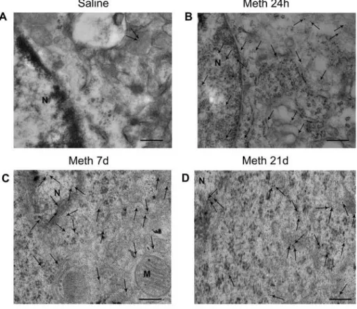
2.8. Alpha-synuclein immune-gold staining increases persistently only after reiterated daily Meth administration
The occurrence of an extensive demethylation within SNCA pro-moter, which steadily persisted at 3 weeks of Meth withdrawal, calls for a quantitative assessment of α-syn molecules within striatal cells fol-lowing both acute (a single day of multiple Meth injections) and chronic (21 days of single injections) protocols of Meth administration.
2.8.1. A single day of multiple Meth injections transiently alters alpha-synuclein immune-gold particles within striatal neurons (Fig. 10)
Fig. 10 reports representative pictures from striatal cells stained with α-syn immune-gold particles (10 nm diameter, arrows) following a
single day of multiple Meth administrations (5 mg/Kg × 5, 2 h apart). Saline-treated cell shows a few α-syn immune-gold particles. When administered according to the acute time-course protocol, Meth in-creases α-syn immune-gold particles both in the nucleus and cytosol at 1 h but not at 24 h or 7 d. In the graph ofFig. 10the number of immune-gold particles is expressed as a percentage of control. At 1 h after the last Meth injection, a robust increase in α-syn immune-gold particles occurs, which vanishes at 24 h and 7 d after Meth administration.
2.8.2. Chronic Meth administration produces a dramatic and persistent increase in alpha-synuclein immune-gold particles (Fig. 11)
Representative micrographs from striatal cells stained for α-syn immune-gold particles (diameter = 10 nm, arrows). Only a few α-syn immune-gold particles are detected in a control cell. When adminis-tered according to a chronic protocol (5 mg/Kg × 21 d), Meth induces a remarkable increase in α-syn particles both in the nucleus and cyto-plasm, which is present at 24 h following the last Meth injection and persists at 7 and even at 21 days of Meth withdrawal. In the graph of Fig. 11the persistent increase is reported as a percentage of Controls (saline-treated mice). It is remarkable that the highest number of α-syn immune-gold particles were counted at the longest time interval of Meth withdrawal.
2.9. Chronic Meth administration produces a widespread increase in brain alpha-synuclein
As shown inFig. 12A, upper line, representative hippocampal slices show a considerable α-syn immune-staining from a control mouse. This is robustly increased following a daily Meth administration for 7 d when observed at 21 d of Meth withdrawal. Remarkably, such an in-crease was enhanced at 21 d of Meth withdrawal when daily Meth administration was prolonged for 21 d. Similar findings were noticed for the piriform cortex and the ventral striatum, mostly the olfactory tubercle (Fig. 12B lower line). Other representative areas shown in Fig. 13A (isocortex);Fig. 13B (dorsal striatum); Fig. 13C (substantia
nigra) confirm the widespread nature of the increase in α-syn
immune-staining. This is the case of the sensorimotor iso-cortex (Fig. 13A) which extends just above the white matter and corpus callosum surrounding the dorsal striatum (Fig. 13B) and the substantia nigra (Fig. 13C). It is remarkable how the ventromedial aspect of the substantia nigra pars
compacta which extends its dendrites in the reticulata is much more
stained compared with the lateral part, which lends substance to dif-ferent parts of the mosaic (more the striosomes) affected by Meth (Granado et al., 2010). This representative overview, which is con-sistent along different groups of mice, witnesses for the occurrence of a Meth-induced brain alteration characterized by a spread increase of a-syn, which is reminiscent of a degenerative synucleinopathy.
3. Discussion
In the present manuscript we provide evidence that Meth adminis-tration, when prolonged for a few weeks, persistently induces nigro-striatal DA denervation, where partial recovery reaches a plateau which remains significantly lower than Controls. When a chronic adminis-tration of Meth is carried out, an increase of striatal α-syn occurs, which persists up to three weeks following Meth withdrawal. Remarkably, striatal α-syn immune-staining is further increased during the late withdrawal period, which follows a chronic protocol of daily Meth administration. This phenomenon is neither associated with mutations in the SNCA gene sequence, nor with multiplication of SNCA. In con-trast, we report a strong demethylation within a specific DNA region, which is expected to be the mouse SNCA promoter based on the homology with SNCA promoter sequence available for rattus norvegicus (Jiang et al., 2014). We identified this region based on database (https://www.ncbi.nlm.nih.gov), and bioinformatics tools which iden-tified a unique region containing CpG islands of the SNCA mouse
Fig. 8. Methamphetamine increases alpha-synuclein protein levels and
pro-motes hypomethylation of SNCA promoter in the acute time-course protocol. In panel A, immune-blot analysis demonstrates a significant increase in α-syn protein levels at 1 h. Densitometric analysis was obtained as the mean ± S.E.M calculated from three blots. The figure shows a representative gel of the mean values quantified in the graph as arbitrary units (*p < 0.0001 vs. saline; **p < 0.0001 vs. saline and Meth 1 h). In panel B, hypomethylation of SNCA promoter following acute Meth administration (5 mg/Kg × 5, 2 h apart). In this experimental condition, significant SNCA hypomethylation was observed only at 1 h of Meth withdrawal, while no changes were observed at 24 h and 7 d (*p = 0.0036; **p = 0.05 vs. Meth 1 h).
promoter in Chromosome 6, spanning from 60,829,051 Mb to 60,829,238 Mb (reference genome: GRCm38/mm10). This region con-tains 8 CpG islands. When BLAST multiple alignment (https://blast. ncbi.nlm.nih.gov/Blast.cgi) was applied, this region revealed 88% homology with the DNA sequence of the SNCA promoter in rattus
nor-vegicus reported byJiang et al. (2014). The amount of demethylation of the SNCA promoter proceeded even at 21 d of Meth withdrawal. In line with this, the amount of α-syn immune-gold particles increased over time even when Meth administration was withdrawn. In fact, α-syn immune-gold particles reach the highest level at the longest time in-terval of Meth withdrawal tested so far (up to 21 d). It is important to notice that such a persistent and always increasing α-syn accumulation is independent of Meth toxicity (as measured by a loss of striatal TH immune-staining or a decrease in the blotted striatal TH protein). In fact, when striatal TH levels partially recover, the accumulation of striatal α-syn proceeds, continuing even beyond the time interval when TH levels reach a plateau. The occurrence of such a specific epigenetic
alteration concomitant to and following chronic Meth administration appears as a plastic effect, which needs a reiterated drug administration to take place. In fact, following a classic protocol of Meth toxicity based on multiple Meth injections (5 mg/Kg, 2 h apart, in a single day), de-spite a significant striatal TH loss is documented at 24 h and 7 d, neither lasting changes in α-syn levels nor persistent demethylation in the
SNCA promoter are detected. Altogether this evidence witnesses for an
independency between induced nigro-striatal toxicity and Meth-induced α-syn overexpression, being the latter effect the consequence of a long-lasting and progressive epigenetic effect.
Increase of α-syn in the brain is commonly considered to be detri-mental and it characterizes a number of neuropathological conditions which form a group of neurological disorders named synucleinopathies. For instance, the mutation in the gene coding for α-syn (SNCA) pro-duces a genetic parkinsonism (PARK1), which is believed to derive from a gain of function of α-syn variants. A specific point mutation in the
SNCA (p.G51D) instead, shifts the PD phenotype towards another
Fig. 9. Methamphetamine increases alpha-synuclein protein levels and promotes hypo-methylation of SNCA promoter in the sub-acute and chronic time-course
protocols. In panel A, immune-blot analysis demonstrates a significant increase in α-syn protein levels at 7 d which persists at 21 d after the last administration of Meth according to the sub-acute (5 mg/Kg × 7 d) time-course protocol (*p = 0.0016 vs. saline alone). Similar results are obtained when Meth is administered according to the chronic (5 mg/Kg × 21 d) time-course protocol as shown in panel B (*p = 0.0125 vs. saline alone). Densitometric analysis was obtained as the mean ± S.E.M calculated from three blots. The figure shows two representative gels of the mean values plotted in the graphs of panel A and B. In panel C and D, a consistent hypomethylation of SNCA promoter following a sub-acute and chronic treatment is shown (5 mg/Kg × 7 d and 5 mg/Kg × 21 d, in C and D, respectively; in C *p < 0.0001 vs. saline alone; **p < 0.0001 vs. saline and Meth 24 h; ***p < 0.0001 vs. Meth 24 h and Meth 7 d; in D *p < 0.0001 vs. saline alone; **p < 0.0001 vs. saline and Meth 7 d).
synucleinopathy named multiple system atrophy (MSA, Fares et al., 2014; Fujioka et al., 2014; Kiely et al, 2015). The α-syn protein itself in its native physiological conformation appears to be strongly detri-mental when it is expressed in excess due to SNCA multiplication (PARK4). In this latter case, the disease phenotype, which may vary, is often more severe compared with that observed in sporadic PD. This is not surprising, since as shown by Burrè et al. (2013, 2015) a wide amount of native α-syn detaches from membrane binding and tends to spontaneous misfolding. Thus, an excess of α-syn is expected to over-whelm cell aggregates. Despite SNCA mutations occur rarely in persons affected by PD, the presence of α-syn aggregates is a hallmark of almost all PD phenotypes. This suggests that α-syn is constantly recruited during the natural history of PD in spite of its sporadic or genetic transmission and independently of the specific trigger which initiates the disease process. These facts cast the hypothesis that, in the mole-cular chain of events leading to neurodegeneration a key and constant step consists in increasing α-syn levels or reducing its proper metabo-lism. As a proof of concept in 2005 we firstly tested such an hypothesis
to check whether an environmental stimulus known to affect the cell biology of DA neurons may also converge in increasing α-syn levels within these cells. Thus, we administered Meth, which apart from being a psychostimulant drug of abuse is a powerful neurotoxin for DA neu-rons. In line with the hypothesis, TH-immune-positive neurons of the
substantia nigra pars compacta developed intense α-syn
immune-fluor-escence at a short-time interval following an acute experimental pro-tocol of Meth administration (Fornai et al., 2005a). These neurons possess high levels of α-syn as shown by immune-blotted tissue micro-punched from substantia nigra from Meth-exposed mice compared with Controls (Fornai et al., 2005a). These findings followed up a previous paper where the occurrence of α-syn-ubiquitin and parkin-positive bodies in the substantia nigra of Meth-treated mice was demonstrated (Fornai et al., 2004). These previous studies led us to hypothesize that, within the Meth-exposed brain, molecular mechanisms bridging drug abuse and neurodegenerative disorders may take place (Iacovelli et al., 2006). Consistently, these findings were backed up byPurisai et al. (2005), who found that MPTP increases α-syn immune-staining in the
Fig. 10. Acute methamphetamine administration
increases alpha-synuclein immune-gold particles at 1 h of withdrawal. Representative micrographs from striatal cells stained for α-syn immune-gold particles (10 nm in diameter) from saline- and Meth-treated mice at 1 h, 24 h, and 7 d of withdrawal. Panel A shows a representative cell from a Control (saline-treated) mouse which contains very few cytosolic α-syn immune-gold particles (arrows). At 1 h of with-drawal, acute Meth administration (5 mg/Kg × 5, 2 h apart) robustly increases α-syn particles both in the nucleus and cytoplasm (arrows), as shown in panel B. Instead, at 24 h and 7 d of Meth with-drawal, the amount of α-syn particles (arrows) falls back nearly to control levels (panel C and D, re-spectively). The graph reports the stoichiometric count of α-syn immune-gold particles in striatal cells from Meth-treated mice expressed as a percentage of values obtained from Controls (saline-treated mice). The data indicate that, when Meth is administered according to an acute time-course protocol, it sig-nificantly increases α-syn only at short-time inter-vals (1 h) of withdrawal (*p < 0.05 with respect to saline). N = nucleus; M = mitochondria Scale bars A, C, D = 400 nm; B = 200 nm.
substantia nigra of primates, and McCormack et al. (2008) who de-monstrated that, following MPTP α-syn accumulates as a proteinase K resistant misfolded protein. These data strengthened the hypothesis that environmental factors acting on protein synthesis or metabolism may lead to synucleinopathies by triggering α-syn accumulation and struc-tural alterations. Thus, according toVance et al. (2010)exposure to environmental neuro-toxicants may produce a higher susceptibility to neurodegeneration by triggering either up-regulation and/or patholo-gical modifications of α-syn. When trying to dissect the molecular events leading to environmentally-induced α-syn accumulation, it was demonstrated that α-syn accumulation was DA-dependent. In fact, in
vivo, in DA-depleted striata, Meth was no longer able to increase α-syn
immune-staining but this was re-established by administering D1-like DA receptor agonists. In vitro evidence related such an effect to the recruitment of beta-arrestin which is considered to be key in DA-related striatal sensitization and is recruited indeed during Meth exposure (De Blasi et al., 2003; Beaulieu et al., 2005, 2008; Beaulieu and Caron,
2008; Fornai et al., 2008). It is likely that a link exists between Meth-induced DA release, DA toxicity and overexpression of α-syn. However, as hypothesized byVance et al. (2010)at that time it was puzzling to decipher in which way environmental factors increase α-syn expression and alteration. The present study provides direct evidence showing how Meth administration may increase α-syn through a profound and per-sistent effect on the SNCA promoter region. Due to previous evidence which indicated a role of DA receptors in increasing α-syn protein following Meth administration, we analyzed the effects of Meth upon
SNCA promoter in a brain area where DA storage is more abundant.
This is why we investigated such an effect at striatal level. It is well known that Meth toxicity mostly involves striatal DA terminals while neuronal cell bodies in the substantia nigra and striatum may be affected as well (Wagner et al., 1980a, b; Schmidt et al., 1985; Sonsalla et al., 1996; Hirata and Cadet, 1997; Zhu et al., 2006; Granado et al., 2010, 2011a, 2011b; Ares-Santos et al., 2012, 2014; Moratalla et al., 2017). Striatal neurons remain a pivot for Meth-induced plasticity and
Fig. 11. Chronic methamphetamine administration
produces a dramatic and persistent increase of alpha-synuclein immune-gold particles at each withdrawal time. Representative micrographs from striatal cells stained for α-syn immune-gold particles (10 nm in diameter) from saline- and Meth-treated mice at 24 h, 7 d, and 21 d of withdrawal. Panel A shows a representative cell from a saline-treated mouse which contains very few cytosolic α-syn im-mune-gold particles (arrows). When administered according to a chronic time-course protocol, Meth produces a robust increase in α-syn immune-gold particles both in the nucleus and cytoplasm (arrows) of striatal cells at 24 h of withdrawal (panel B), which persists at 7 d and 21 d of withdrawal (panel
C and D, respectively). The graph provides a
quan-tification of Meth-induced increase of α-syn im-mune-gold particles following a chronic protocol compared with Controls (saline-treated mice). (*p < 0.05 with respect to saline; **p < 0.05 with respect to Meth withdrawn at 24 h and 7 d). N = nucleus; M = mitochondria Scale bars A, B, C = 200 nm; D = 100 nm.
Fig. 12. Methamphetamine-induced increase
in limbic (hippocampal, piriform and olfac-tory) alpha-synuclein-immune-staining. As shown in A, upper line, representative hippo-campal slices show a considerable α-syn im-mune-staining from a control mouse. This is robustly increased following a daily Meth ad-ministration for 7 d when observed at 21 d of Meth withdrawal. Remarkably, such an in-crease was enhanced at 21 d of Meth with-drawal when daily Meth administration was prolonged for 21 d. Similar findings were no-ticed for the piriform cortex and the ventral striatum, mostly the olfactory tubercle (in B, lower lane). Scale bar 200 µm.
Fig. 13. Alpha-Synuclein-immune-staining of iso-cortex, dorsal striatum and substantia nigra following acute and chronic methamphetamine administration. Other
representative areas confirm the widespread nature of Meth-induced α-syn immune-staining. This is the case of the sensorimotor iso-cortex (in A, upper lane) which extends just above the white matter and corpus callosum surrounding the dorsal striatum (in B, middle lane) and the substantia nigra (in C, lower lane). This representative overview is reminiscent of a degenerative synucleinopathy. Scale bar 200 µm.
epigenetic alterations and play an important role in long-lasting beha-vioral changes produced by Meth administration.
When tested on nigral neurons, Meth-induced overexpression of α-syn was associated only with two-fold demethylation of the SNCA promoter region compared with Controls. In detail, demethylation of the SNCA promoter never reached 50% in any CpG islands (Jiang et al., 2014). The data provided here at striatal level indicate that the amount of demethylation in the SNCA promoter region surpasses 95% (20-fold control values), and mostly, provide evidence for a persistent effect which remains steady at 21 days of Meth withdrawal. Such a strong effect may be due to several issues; the method used here, which con-sists in antibody-mediated detection of methylated cytosine and re-presents the most sensitive and specific tool to quantify the amount of DNA methylation. Another crucial point consists in the amount of striatal compared with nigral DA levels. Such a difference slightly varies for animal species and strains, although from a general standpoint this ranges between 10-fold and 15-fold for striatal compared with nigral DA levels. If one assumes that DA is a pivot in the expression of α-syn (Lazzeri et al., 2007) including the quote deriving from epigenetic sti-mulation, the different effect might be explained. Again, going back to the raw data shown here, it is intriguing to note the remarkable increase in α-syn protein which was detected by immune-gold stoichiometry compared with SDS-PAGE immune-blotting. It is likely that these methods grasp differently the protein amount. In fact, according to Vance, and mostly considering delayed times of withdrawal used here, it is plausible that a great part of α-syn is present as misfolded protein aggregates which do not run in gel electrophoresis as native α-syn. This may justify the great difference between immune-gold electron micro-scopy and immune-blotting. Similarly, this consideration may apply to the lower amount detected when α-syn is not measured in situ at ul-trastructural level. Starting from our previous studies at nigral level, the present study moves the focus on the effects induced by Meth at striatal level disclosing novel avenues related to addiction and neurodegen-eration. In fact, an increase of α-syn within striatal neurons is well established in Huntington’s disease where α-syn deleteriously interacts with Huntingtin (Herrera and Outeiro, 2012; Poças et al., 2015) thus worsening the physiopathology of HD. In fact, a direct correlation was drawn between α-syn levels and the amount of neuropathology in HD (Corrochano et al., 2012a; Tomás-Zapico et al., 2012). Remarkably, as reported byCorrochano et al (2012b)α-syn levels modulate Hunting-ton's disease in mice even considering disease phenotype and age at onset. Thus, the role of α-syn in the physiopathology of medium size striatal neurons is key since its deletion mitigates HD symptoms while its overexpression worsens the disease phenotype (Corrochano et al 2012b; Tomás-Zapico et al., 2012). Considering the data reported in the present manuscript and classic studies reviewed byJakel and Maragos (2000)the role of DA in producing Meth toxicity seems to apply also to non-DA neurons since most of its epigenetic effects are grounded on DA release (Cadet et al., 2010b; Godino et al., 2015; Limanaqi et al., 2018). Finally, a role of α-syn in behavioral sensitization and addiction should be considered in the light of the present data. In fact, the remarkable increase in α-syn which we report representatively within hippocampus and other limbic regions such as the olfactory tubercle and the piriform cortex suggests that the increase in α-syn may contribute significantly to the process of behavioral sensitization. Again, some brain area where Meth produces a remarkable increase in α-syn is relatively poor of DA terminals, which lends substance to the hypothesis that DA per se is not sufficient to sustain the sensitization process induced by Meth, and other chemical species, such as oxidative endogenous compounds, are key for producing epigenetic alterations (Limanaqi et al., 2018).
In any case, apart from the strong take-home messages discussed above, the smoldering impression we seem to get from all these data is the ambiguous significance of α-syn overexpression in the brain. Intriguingly, a similar cautious approach was adopted very recently in an opinion article hypothesizing novel vistas on α-syn in the brain in health and disease (Espay et al., 2019). In this scenario, we have to
consider the chance that, despite a discrepancy exists at chronic time intervals between TH and α-syn expression, this still might reflect a transient phenomenon. In fact, prolonged exposure (over several months) to high levels of α-syn may finally trigger neurodegeneration. This is also related to the natural trend of native α-syn to misfold, which is expected to be enhanced during prolonged time intervals. This hy-pothesis remains open and deserves dedicated investigations.
4. Experimental procedures 4.1. Animals
C57 black 6/J, 8 weeks-old male mice (Charles River Calco, Mi, Italy, N = 120) were housed in small cages (N = 6 per cage; cage length = 27 cm; cage width = 21 cm; cage height = 14 cm). They were kept under controlled environmental conditions (temperature = 22 °C; humidity = 40%) with food and water ad libitum and with a 12 h light/ dark cycle. All these measures were kept constant all over the study since the effects of Meth markedly vary depending on housing condi-tions beyond temperature, such as cage size and number of animals per cage (Fornai et al., 2004). Mice were handled in accordance with the Guidelines for Animal Care and Use of the National Institutes of Health, and adequate measures were taken to minimize animal pain and dis-comfort. The experimental protocol was approved by the local Ethical Committee, and by the Ministry of Health. As a mouse strain, we chose C57/6J-Black mice based on previous literature and our previous stu-dies in different mouse strains. In fact, the inbred C57-Black mice possess a very stable DA phenotype and are mostly sensitive to DA neurotoxins. This allows increasing the statistical power to detect even small differences in each biological value under analysis. Again, amongst various C57-Black strains, we preferred the 6/J compared with 6/N where a spontaneous SNCA variant was reported (Schlüter et al., 2003). As a species, mice were preferred over rats due to a high sen-sitivity to Meth which, amongst rodents, makes them closer to primates. In addition, C57 Black mice when administered Meth doses which are toxicant for the DA system, are more resistant to 5-HT toxicity which is more evident in rats (O’Dell et al., 2012). Such an additional target of Meth toxicity might confound the significance of the present study, which is centered on DA-related disorders, DA-projecting areas and α-syn as a detrimental protein related to DA pathways, including post-synaptic target neurons.
4.2. Methamphetamine administration and experimental design
A total of 120 mice were dedicated to these experiments. A group of 48 mice was dedicated to electron microscopy, while 72 mice were sacrificed for immune-histochemistry, protein assay, and DNA analysis. These latter 72 mice were divided into different experimental groups (N = 24 each) according to three experimental protocols.
These protocols consisted in different dosing and timing of Meth or saline administration. Methamphetamine (Meth, Sigma Aldrich Saint Louis, MO, U.S.A., Authorization n° SP/096, 05.15.2016, granted by the Italian Ministry of Health, was dissolved in saline and it was injected i.p. in a volume of 200 µL; saline was administered i.p. at the same volume). In the first subgroup (N = 24) mice were sacrificed at dif-ferent time intervals (1 h, N = 6; 24 h, N = 6; 7 d N = 6) following the last Meth injection; an additional group of mice (N = 6) was adminis-tered saline (Controls). Meth was adminisadminis-tered at the dose of 5 mg/Kg (×5, 2 h apart). From now on we refer to this protocol as acute time-course protocol since Meth was administered in a single day with a short-time interval between starting Meth and sacrifice.
In the second subgroup of mice (N = 24) Meth (5 mg/Kg), was jected daily, for 7 d. These mice were sacrificed at different time in-tervals (24 h, N = 6; 7 d, N = 6; 21 d, N = 6) after the last adminis-tration; an additional group (N = 6) was administered saline (Controls). From now on we refer to this protocol as sub-acute time-course protocol
(5 × 7) since Meth was administered for 7 d.
The third subgroup of mice (N = 24) received Meth (at the dose of 5 mg/Kg) daily, for 21 d and they were sacrificed at different time in-tervals (24 h, N = 6; 7 d, N = 6; 21 d, N = 6) after the last injection; an additional group (N = 6) was administered saline (Controls). This protocol was named chronic time-course protocol (5 × 21). This cor-responds to the most chronic condition since Meth treatment lasted 21 d. In both sub-acute and chronic protocols, the longest withdrawal between the last Meth injection and sacrifice was 21 d, which allows detecting the persistence of the effects induced by Meth. One should consider that, while in the sub-acute protocol the longest time interval between the first Meth injection and sacrifice was 28 d, in the chronic protocol the longest time interval was protracted up to 42 d. Again, the cumulative dose of Meth in the sub-acute time-course protocol was 35 mg/Kg (7 × 5 mg/Kg), whilst this dose rose up to 105 mg/Kg (21 × 5 mg/Kg) in the chronic time-course protocol. These mice were sacrificed by deep chloral hydrate anesthesia and their brains were quickly removed and processed according to different procedures.
Since the critical data were obtained following acute and chronic protocols, further experiments were aimed at analyzing ultrastructural morphometry according to both acute and chronic time-course proto-cols. For these additional experiments we used 48 mice, which were sacrificed according to the acute (N = 24) or the chronic (N = 24) time-course protocol. In detail, according to the chronic protocol mice re-ceived Meth (at the dose of 5 mg/kg), once daily, for 21 d and they were sacrificed at different time intervals (24 h, N = 6; 7 d, N = 6; and 21 d, N = 6) after the last injection; an additional group (N = 6) was ad-ministered saline (Controls). Twenty-four mice were used in the acute protocol where they were administered Meth (N = 18, 5 mg/Kg × 5, 2 h apart), and they were sacrificed at 1 h (N = 6), 24 h (N = 6), and 7d (N = 6) after treatment; Control mice (N = 6) were administered saline. Each mouse from each protocol used for electron microscopy was perfused with a solution containing 2% paraformaldehyde and 0.1% glutaraldehyde under deep chloral hydrate anesthesia.
During and after each treatment mice were housed N = 6 per cage and they were kept under controlled environmental conditions (temperature = 22 °C; humidity = 40%) with food and water ad li-bitum with a 12 h light/dark cycle until sacrifice.
4.3. Post-sacrifice brain processing
In the first experimental block (N = 72), after sacrifice, the brains were quickly removed from the skull and they were divided into two hemispheres; one hemisphere was constantly used for immune-histo-chemical studies, while the other hemisphere was used either for im-mune-blot analysis, or DNA extraction for SNCA mutation detection, and methylation detection assay within SNCA promoter. In the second experimental block (N = 48) mice were perfused trans-cardially under deep chloral hydrate anesthesia. The brains were dissected and im-mersed at 4 °C, overnight, in the perfusing solution. Tissue blocks from striata at the same level of that used for other assays were post-fixed.
4.4. Immune-histochemistry
Since Meth administration may lead to nigro-striatal DA denerva-tion, we carried out in all mice immune-histochemistry and Sodium Dodecyl Sulphate-PolyAcrylamide Gel Electrophoresis (SDS-PAGE) immune-blotting for Tyrosine-Hydroxylase (TH) to assess indirectly the loss of striatal DA innervation following different protocols of Meth administration, at different time intervals after the last Meth injection. In the same hemisphere, at the same striatal level, we analyzed the effects of Meth administration on TH and α-syn immune-histochem-istry, while in the contralateral hemisphere either striatal TH and α-syn SDS-PAGE immune-blotting, or DNA analysis were carried out. For TH and α-syn immune-histochemistry the hemisphere was removed and placed in a fixing solution overnight. Mice specimens were fixed in
Carnoy’s solution (ethanol 60%, chloroform 30% and glacial acetic acid 10%), they were embedded in paraffin, and 10 µm thick tissue sections were cut and mounted on slides for immune-histochemical analysis, where TH and α-syn immune-reactivity were counted in serial sections to keep constant and comparable the level of analysis for both antigens. In detail, TH and α-syn immune-reactivity were carried out in the dorsal striatum at a level ranging between +0.62 mm and +0.74 mm from bregma according to the atlas of Franklin and Paxinos (1997). At this level, densitometric count was carried out. At the very same level we report the representative pictures for the sensorimotor iso-cortex, the ventral striatum including the olfactory tubercle, and the piriform cortex. Hippocampal staining for α-syn was reported for a rostrocaudal level ranging from −1.82 mm to −2.06 mm from bregma. When both immune-staining were carried out at nigral level, a range from −3.16 mm to −3.08 mm from bregma was scanned.
Tissue sections were de-waxed and antigen retrieval was carried out by exposing the slices to citrate buffer, pH 6.0, for 10 min. Brain slices were then incubated with 0.01% Triton X-100 (Sigma Aldrich, Milan, Italy) for 15 min and were soaked in 3% hydrogen peroxide to block endogenous peroxidase activity. Then, they were incubated overnight at 4 °C with mouse monoclonal anti-TH antibody (1:100; Sigma Aldrich; Antibody registry: AB_477560), rabbit anti- α-syn (1:100; Sigma Aldrich; Antibody registry: AB_10746104); the sections were incubated with anti-mouse or anti-rabbit biotinylated secondary antibodies (1:200; Vector Laboratories, Burlingame, CA, U.S.A.) for 1 h, at 22 °C. Peroxidase activity was revealed by using a solution containing 0.04% of 3,3′-diaminobenzidine-tetrahydrochloride (DAB; Sigma Aldrich) pH 7.6, for 3 min at 22 °C. The stained sections were dehydrated, cleared and coverslipped with Micromount (Diapath, Martinengo, BG, Italy). The specificity and quality of anti- α-syn immune-staining which was detected in brain slices was compared by using antibodies (1:100; Sigma Aldrich; Antibody registry: AB_10746104) and (1:300; BD; Antibody registry: AB_398108).
This was carried out in pilot staining in the present work and con-sidering our previous studies as well as additional references where striatal α-syn immune-staining was carried out (Schlüter et al., 2003; Fornai et al., 2005a,b; Betarbet et al., 2006): for western blotting monoclonal anti-α-syn synuclein-1 (Transduction Labs,Betarbet et al., 2006). Data provided a signal which was similar to total α-syn (AB_52168; 1:200 in BSA 1% PBST; Abcam, Cambridge, UK), used by Novello et al. (2018)or human anti-α-syn (AB_36615, 1:1000, Abcam, UK) as used byMulcahy et al. (2012). The data obtained with Sigma Aldrich were quite similar to the one we obtained with anti-α-Syn (Transduction Laboratories, Lexington, KY, USA) (Schlüter et al., 2003). The pilot studies for striatal α-syn immune-staining compared Sigma Aldrich antibodies with a presently available BD anti-α-syn antibody (AB_398108); both stainings were compared with classic striatal TH immune-staining inFig. 1, which shows a higher specificity for Sigma compared with BD anti-α-syn- antibody. This staining was also com-parable with that obtained using the α90 antibody both for immune-histochemistry and immune-cytochemistry (Totterdell et al., 2004).
4.4.1. Densitometric analysis of striatal alpha-synuclein and tyrosine-hydroxylase immune-staining
Intensity of striatal TH and α-syn immune-reactivity was semi-quantified by measuring relative optical densities. In fact, images were acquired at low magnification (2.5×) and antigen densitometry was calculated as the ratio between optical density in the dorsal striatum and that in the white matter (i.e. corpus callosum). Despite being a semi-quantitative assay, values were consistent with a very low S.E.M.
4.5. Immune-blotting
The hemisphere was placed on a wet paper placed on ice-cold Petri dish to dissect the rostral part of the dorsal striatum. Briefly, the lateral wall of the third ventricle was pulled aside, the anterior commissure