Therapeutic outcomes of transplantation of amniotic
fluid-derived stem cells in experimental ischemic stroke
Naoki Tajiri
1,
Sandra Acosta
1,
Gabriel S. Portillo-Gonzales
1,
Daniela Aguirre
1,
Stephanny Reyes
1,
Diego
Lozano
1,
Mibel Pabon
1,
Ike Dela Peña
1,
Xunming Ji
2,
Takao Yasuhara
3,
Isao Date
3,
Marianna A. Solomita
1,
Ivana Antonucci
4,
Liborio Stuppia
4,
Yuji Kaneko
1and
Cesar V. Borlongan
1*
1Center of Excellence for Aging and Brain Repair, Department of Neurosurgery and Brain Repair, University of South Florida Morsani College of Medicine,
Tampa, FL, USA
2Department of Neurosurgery, Xuanwu Hospital, Capital Medical University, Beijing, China
3Department of Neurological Surgery, Okayama University Graduate School of Medicine, Dentistry and Pharmaceutical Sciences, Okayama, Japan 4Laboratory of Molecular Genetics, DISPUTer, School of Medicine and Health Sciences, “G. d ’Annunzio” University, Chieti-Pescara, Italy
Edited by:
Thorsten Doeppner, University of Duisburg-Essen, Germany Reviewed by:
Josephine Herz, University Clinic Essen, University of
Duisburg-Essen, Germany Seongjin Yu, NHRI, Taiwan *Correspondence: Cesar V. Borlongan, Center of Excellence for Aging and Brain Repair, Department of Neurosurgery and Brain Repair, University of South Florida Morsani College of Medicine, 12901 Bruce B. Downs Blvd., Tampa, FL 33612, USA
e-mail: [email protected]
Accumulating preclinical evidence suggests the use of amnion as a source of stem cells for
investigations of basic science concepts related to developmental cell biology, but also for
stem cells’ therapeutic applications in treating human disorders. We previously reported
isolation of viable rat amniotic fluid-derived stem (AFS) cells. Subsequently, we recently
reported the therapeutic benefits of intravenous transplantation of AFS cells in a rodent
model of ischemic stroke. Parallel lines of investigations have provided safety and efficacy
of stem cell therapy for treating stroke and other neurological disorders. This review article
highlights the need for investigations of mechanisms underlying AFS cells’ therapeutic
benefits and discusses lab-to-clinic translational gating items in an effort to optimize the
clinical application of the cell transplantation for stroke.
Keywords: cerebral ischemia, neural stem/progenitor cells, regenerative medicine, neurogenesis, neurotrophic factors
WHY IS THERE A NEED FOR NOVEL TREATMENTS IN
STROKE?
Stroke, the fourthleading cause of death and the leading cause
of disability in the United States (Roger et al., 2012), has only
one FDA-approved drug, namely tissue plasminogen activator
(tPA). Due to tPA limitations and complications, which include
a limited therapeutic window (4.5 h from disease onset to tPA
administration) and adverse effects associated with delayed
treat-ment (i.e., hemorrhagic transformation), only a mere 3 percent of
ischemic stroke patients actually benefit from the tPA treatment
(Graham, 2003; Yip and Demaerschalk, 2007). This significant
unmet clinical need for stroke has prompted investigations to
increase the therapeutic window with innovative treatment
strate-gies specifically targeting the restorative phase, which begins days
to weeks post-stroke (Matsukawa et al., 2009; Yu et al., 2009;
Antonucci et al., 2011; Kaneko et al., 2011; Manuelpillai et al.,
2011).
DO STEM CELLS EXIST IN THE AMNION?
Stem cells have emerged as a prospective restorative agent for
stroke due to their ability to abrogate sub-acute and chronic
secondary cell death associated with the disease (Borlongan et al.,
1997; Nishino and Borlongan, 2000). We previously reported
the isolation of viable rat amniotic fluid-derived stem (AFS)
cells (Tajiri et al., 2012). Together with other research teams, the
grafted stem cells’ production of trophic factors and cytokines, as
well as the increase in levels of neurotrophic factors and reduced
inflammatory response in the ischemic stroke region, have been
directly attributed to the positive effects by transplantation of
AFS cells (Yu et al., 2009; Antonucci et al., 2011; Kaneko et al.,
2011; Manuelpillai et al., 2011). Furthermore, the inhibition of
apoptosis and oxidative stress, in tandem with stimulation of
angiogenesis, neurogenesis, and synaptogenesis, may be linked
as a benefit of AFS cells against stroke deficits (Yu et al., 2009;
Antonucci et al., 2011; Kaneko et al., 2011; Manuelpillai et al.,
2011). Even though stem cells can be harvested from various
sources (Borlongan et al., 2004, 2005; Hematti et al., 2004; Clavel
and Verfaillie, 2008; Burns et al., 2009; Ou et al., 2010), including
bone marrow, fetal and embryonic tissues, amnion-derived stem
cells are an appealing choice because of many logistical and ethical
advantages including the ease of isolation of the stem cells from
amnion tissue and the fluid (Yu et al., 2009; Antonucci et al., 2011;
Kaneko et al., 2011; Manuelpillai et al., 2011). Similar to
amniotic-tissue derived cells, the harvest of AFS cells poses negligible risk
of injury to the fetus. These cells are isolated from amniotic
fluid collected from amniocentesis, a pre-natal exam performed
at around 15–20 weeks of gestation. Accordingly, since AFS cells
can be isolated much earlier, compared to amniotic tissue-derived
cells, AFS cells possess properties that closely resemble embryonic
and mesenchymal cell markers, which could be more beneficial
at treating diseases. The low immunogenicity, low tumorgenicity,
high proliferative capacity and anti-inflammatory characteristics,
are phenotypic features of transplantable cells (Newman et al.,
2005) that support AFS cells to be a safe and effective donor cell
for stroke (Yu et al., 2009; Antonucci et al., 2011; Kaneko et al.,
2011; Manuelpillai et al., 2011). Another distinguishing feature
of AFS cells when compared to amniotic tissue-derived cells is
the sterility involved. AFS cells are harvested via amniocentesis
under aseptic condition, but the sterility may be compromised
when stem cells are extracted from amnion tissue during child
delivery.
AFS cells can phenotypically commit to various lineages (Prusa
and Hengstschlager, 2002; In’t Anker et al., 2003; Prusa et al.,
2003; Fauza, 2004; Tsai et al., 2004, 2006; McLaughlin et al., 2006).
A misleading concept is the term “fluid” associated with AFS
cells because cells isolated during amniocentesis are comprised of
multiple stem cells originating from extra-embryonic and
embry-onic tissues (Prusa and Hengstschlager, 2002) and their properties
differ with gestational age (Yu et al., 2009; Antonucci et al., 2011;
Kaneko et al., 2011; Manuelpillai et al., 2011). Accordingly,
phe-notypic characterization of AFS cells reveal a plethora of stem cell
subtypes, from pluripotent embryonic stem cells to multipotent
adult stem cells owing likely to the age-dependent tissue plasticity
potential (De Coppi et al., 2007; Mauro et al., 2010). Indeed, AFS
cells from second trimester amniotic fluid display the ability to
differentiate into all three germ layers and express Oct-4, Nanog,
and SSEA-4 (Roubelakis et al., 2007), which are pluripotent
embryonic stem cell markers (Yu et al., 2009; Antonucci et al.,
2011; Kaneko et al., 2011; Manuelpillai et al., 2011). Although
considered as “adult stem cells”, the doubling time for the AFS
cells population is approximately 30–36 h with a high
regen-eration capacity that can be extended for over 250 doublings
without any measurable loss of chromosomal telomere length
(De Coppi et al., 2007). Altogether, these studies support the
amnion as a potent source of stem cells for investigations of basic
science concepts related to developmental cell biology, but also for
therapeutic purposes such as AFS cell transplantation for treating
human disorders, especially stroke.
ARE AFS CELLS TRANSPLANTABLE AND DO THEY EXERT
FUNCTIONAL BENEFITS?
The following protocol allows the isolation of AFS cells from
isolated amniotic fluid samples obtained from timed pregnant
Sprague-Dawley rats at gestation age 16–18 weeks (Tajiri et al.,
2012). For each sample, 2–3 ml of amniotic fluid, corresponding
to a cell number ranging from 2 × 10
3to 2 × 10
6are centrifuged
for 10 min at 1800 rpm. The high variance in the amount of cells
which can be isolated from amniotic fluid is influenced by factors
such as child birth, genetics, and fluid cell isolation. It is expected,
however, that these discrepancies can be overcome when
technol-ogy is able to be standardized with appropriate quality control
and assurance. This will yield a more accurate and efficient
cell isolation in the near future. Pellets are then resuspended in
Iscove’s modified Dulbecco’s medium supplemented with 20%
FBS, 100 U/ml penicillin, 100
µg/ml streptomycin (Sigma), 2 mM
L-glutamine, 5 ng/ml basic fibroblast growth factor (FGF2) and
incubated at 37
◦C with 5% humidified CO
2
. After 7 days,
non-adherent cells are removed and the non-adherent cells are allowed
to grow in the same medium, which is changed every 4 days.
When the cell culture reaches confluency (about 20 days after
the primary culture), cells are treated with 0.05% trypsin and
0.02% EDTA, then counted and replaced in 25 cm
2culture flasks.
For routine AFS cell procedure, an approximate yield of about
22 million stem cells per amnion fluid aspirate is anticipated.
The viability yield of AFS cells is about 70%, which would be
an estimated 15 million stem cells per amnion fluid aspirate.
An estimated range of several million AFS cells per milliliter of
amnion fluid are therefore expected to be obtained.
This protocol allows ample AFS cells for transplantation
stud-ies. Indeed, accumulating preclinical data have demonstrated the
potential of transplantation of AFS cells for treating experimental
models of brain diseases (Yu et al., 2009; Antonucci et al., 2011;
Kaneko et al., 2011; Manuelpillai et al., 2011). For example,
AFS cells are a potentially valuable source of stem cells to treat
Parkinson’s disease because under standard neuronal induction
protocols for stem cells, AFS cells preferentially differentiate
into a dopaminergic phenotype (Pisani et al., 2005). In parallel,
transplantation of AFS cells has been examined in stroke, with
AFS transplanted ischemic stroke mice exhibiting reduced
short-term memory impairment and improved sensorimotor ability,
somatosensory functions, and motor coordination (Rehni et al.,
2007). Although this study shows the beneficial effects of AFS cell
transplantation in experimental stroke, the mechanism
underly-ing the observed therapeutic benefits remained underexplored.
Perhaps AFS cells are mediating specific neurotransmitter release
to restore cellular function in an injured brain (Broughton et al.,
2013). This may explain a reversal of disease-induced impairment
of memory following AFS cell transplantation in an ischemic
model of stroke when transfer latency time (TLT) is used as a
parameter of memory. If administration of AFS cells following
middle cerebral artery occlusion and reperfusion significantly
attenuated ischemia-reperfusion induced increase in day 7 TLT,
it is likely that the AFS cells are secreting the neurotransmitters
necessary to ameliorate memory and subsequently, cognition in
general (Rehni et al., 2007).
HOW DO AFS CELLS AFFORD REPAIR OF THE STROKE BRAIN?
Our group transplanted AFS cells in experimental stroke
ani-mals (via occlusion of the middle cerebral artery;
Borlongan
et al., 1998) using motor and cognitive tests, and subsequent
histological analysis of the brain to assess the mechanism of
action associated with AFS cell therapy for stroke. Although still
impaired compared to sham-operated animals (Acosta et al.,
2010), intravenous transplantation of AFS cells (1 million viable
cells) in adult Sprague-Dawley rats at 30 days after experimental
stroke revealed that motor and cognitive deficits were reduced at
60 days post-MCAo compared to vehicle-infused stroke animals
(Tajiri et al., 2012). Moreover, histological analyses revealed that
the AFS cell-transplanted stroke animals significantly decreased
infarct volumes by 92% compared to the vehicle-infused stroke
animals, which likely facilitated the recovery of motor and
cog-nitive functions. In addition, cell proliferation was significantly
upregulated in the neurogenic subventrical zone of the AFS
cell-transplanted stroke animals compared to the vehicle-infused
stroke animals. In tandem, immature neuronal cells also increased
in the subventrical zone of AFS cell-transplanted stroke animals
compared to the vehicle-infused stroke animals. The increased
cell proliferation and reduced neuronal loss in the
subventri-cal zone was similarly observed in the dentate gyrus, another
major neurogenic niche. That AFS cell transplantation attenuated
stroke-induced behavioral and histological deficits coincided with
increased cell proliferation and neuronal differentiation in the two
neurogenic sites, namely subventricular zone and dentate gyrus,
implicates a major role of graft-induced host tissue repair in the
brain remodeling process following stroke (Table 1).
CAN WE TRANSLATE AFS CELL GRAFT-MEDIATED
FUNCTIONAL RECOVERY IN EXPERIMENTAL STROKE
TO THE CLINIC?
Many stroke victims present with symptoms characterized by
sensorimotor and cognitive functions (Grefkes et al., 2008; Rush
et al., 2010; Lin et al., 2011). Up to now, most stroke animal
models have focused on motor impairments, disregarding the
cognitive declines that proceed after the brain offense (Borlongan,
2009; Chopp et al., 2009; STEPS, 2009). Findings on the
pathol-ogy of ischemic stroke support that secondary cell death may
extend beyond the routine cortical damage towards the
hip-pocampus, which is the key brain structure for learning and
memory consolidation (Scoville and Milner, 1957; Shors et al.,
2002; Saxe et al., 2006; Dupret et al., 2008). A decline in adult
neurogenesis is magnified during aging and correlates with
wors-ening of cognitive functions (McDonald and Wojtowicz, 2005;
Drapeau and Nora Abrous, 2008; Encinas et al., 2011).
Interestingly, the impaired neurogenesis and the cognitive decline
in the aging hippocampus (Freret et al., 2009; Dhawan et al., 2010;
Zvejniece et al., 2012) also accompany stroke (Dhawan et al., 2010;
Wattanathorn et al., 2011; Zvejniece et al., 2012).
Whereas our cognitive test results did not show any difference
in learning performance between the transplanted and
vehicle-infused stroke animals, the AFS cell-transplanted stroke animals
demonstrated a significantly improved reference memory
com-pared to the vehicle-infused stroke animals. This improvement
in the memory task directly correlates with increased
neurogen-esis in the hippocampus of AFS cell-transplanted stroke animals
relative to vehicle-infused stroke animals. In order to increase
the clinical relevance of cell therapy for stroke, especially when
evaluating cognitive deficits, is to employ a hippocampal
depen-dent task for spatial memory and reference memory (Gallagher
et al., 1993; Duva et al., 1997; Clarke et al., 1999; Anisman
and McIntyre, 2002; Broadbent et al., 2004; Clark et al., 2007;
Gillani et al., 2010; Bergado et al., 2011; Jurgens et al., 2012),
and long-term potentiation (Bliss and Collingridge, 1993; Norris
and Foster, 1999; Gusev and Alkon, 2001; Richardson et al.,
2002; Vorhees and Williams, 2006; Pisu et al., 2011; Xu et al.,
2012). Our data advanced the notion that transplanted AFS cells
selectively aid in the recovery of the reference memory, but not
task acquisition. Cognitive improvements in stem/progenitor cell
transplanted stroke animals have been linked previously with the
decline in cerebral infarct volumes (Nishino et al., 1993; Fukunaga
et al., 1999, 2003; Mimura et al., 2005), as well as reduced
secondary cell death loss in brain areas known to modulate motor
and cognitive functions (Ebrahimi et al., 1992; Pisani et al., 2005;
Takahashi et al., 2008; Tabuse et al., 2010; Miyoshi et al., 2012).
That the AFS cell transplanted stroke animals displayed improved
motor and cognitive performance, reduced the brain infarcts and
the secondary cell death, and enhanced the level of neurogenesis
provide insights on possible mechanisms of action underlying
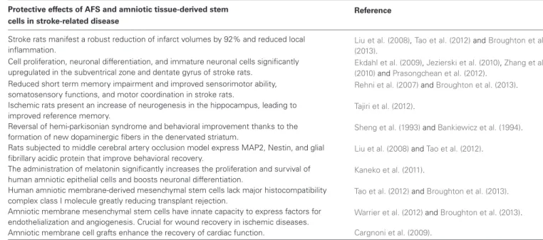
Table 1 | Effects of AFS cells and amniotic tissue-derived cell in stroke-related disease.
Protective effects of AFS and amniotic tissue-derived stem Reference cells in stroke-related disease
Stroke rats manifest a robust reduction of infarct volumes by 92% and reduced local inflammation.
Liu et al. (2008),Tao et al. (2012)andBroughton et al.
(2013). Cell proliferation, neuronal differentiation, and immature neuronal cells significantly
upregulated in the subventrical zone and dentate gyrus of stroke rats.
Ekdahl et al. (2009),Jezierski et al. (2010),Zhang et al.
(2010)andPrasongchean et al. (2012).
Reduced short term memory impairment and improved sensorimotor ability, somatosensory functions, and motor coordination in stroke rats.
Rehni et al. (2007)andBroughton et al. (2013).
Ischemic rats present an increase of neurogenesis in the hippocampus, leading to improved reference memory.
Tajiri et al. (2012).
Reversal of hemi-parkisonian syndrome and behavioral improvement thanks to the formation of new dopaminergic fibers in the denervated striatum.
Sheng et al. (1993)andBankiewicz et al. (1994).
Rats subjected to middle cerebral artery occlusion model express MAP2, Nestin, and glial fibrillary acidic protein that improve behavioral recovery.
Liu et al. (2008)andTao et al. (2012).
The administration of melatonin significantly increases the proliferation and survival of human amniotic epithelial cells and boosts neuronal differentiation.
Kaneko et al. (2011).
Human amniotic membrane-derived mesenchymal stem cells lack major histocompatibility complex class I molecule greatly reducing transplant rejection.
Tao et al. (2012)andBroughton et al. (2013).
Amniotic membrane mesenchymal stem cells have innate capacity to express factors for endothelialization and angiogenesis. Crucial for wound recovery in ischemic diseases.
Warrier et al. (2012)andBroughton et al. (2013).
Amniotic membrane cell grafts enhance the recovery of cardiac function. Cargnoni et al. (2009).
the functional benefits afforded by AFS cell transplantation. Our
observed transplant-mediated recovery of motor and cognitive
functions confirms a recent report of AFS therapeutic effects in
stroke (Rehni et al., 2007), but our study expanded the short
timeline of 7 days from the previous study to 63 days.
The transplantation of AFS cells shows extensive therapeutic
benefits such as reduction in infarct volume, enhanced cell
proliferation, increased neuronal differentiation, and improved
memory and motor skills, seen with other stem cells (Yasuhara
et al., 2006a,b, 2008; Hara et al., 2007). The exact underlying
mechanism of these benefits is still unknown and understanding
therapeutic pathways could potentially yield promising insights
into optimizing AFS cell transplantation towards the clinical trial
stage. Clinical trials that closely reflect the preclinical data on
safety and efficacy of AFS cells in animal models of stroke will
enhance the functional outcome of this cell therapy in stroke
patients. Based on preclinical data demonstrating AFS cell safety
and efficacy, patients who suffer a stroke and show significant
inflammation of the brain or who display short-term memory
loss due to the accompanying injury to the hippocampus seem
to be the best candidates for future clinical trials of AFS cells.
Moreover, AFS cell therapy trials could be extended to patients
who have suffered from cardiac stroke, as animal models show
promising improvement in cardiac functions following AFS
cell transplantation (Bollini et al., 2011). In addition, a better
understanding of the mechanism(s) involved in the functional
improvement seen in transplanted stroke animals will likely
further optimize AFS cell therapy.
When contemplating with clinical applications of AFS cells,
the route of transplantation is key to successful outcomes of
targeted stroke patient population. Intracerebral (Rehni et al.,
2007; Liu et al., 2008), intraperitoneal (Ghionzoli et al., 2010),
and intravenous (Tajiri et al., 2012) have been explored in AFS
transplantation in experimental models of cerebral ischemia. The
intravenous route of transplantation is a minimally invasive
pro-cedure and poses less risk to the patient compared to intracerebral
transplantation. A peripheral injection method may be favored
over the direct transplantation route so that the stem cells can
be administered quickly after the onset of a stroke. A limited
FDA-approved clinical trial for intravenous transplantation of
placenta/amnion-derived stem cells in sub-acute stroke patients
was terminated with no available safety and efficacy results
(ClinicalTrials.gov., 2014).
The recommendations of Stem cell Therapeutics as an
Emerg-ing Paradigm for Stroke (STEPS;
Borlongan, 2009; Chopp et al.,
2009; STEPS, 2009) are likely to facilitate the translational
research on AFS cell grafts for stroke. Such translational blueprint
for cell therapy in stroke provides important guidance in
experi-mental stroke testing and stem cell therapy, including the need for
multiple strains, both genders, and age groups to evaluate safety
and efficacy of AFS cells (Borlongan, 2009; Chopp et al., 2009;
STEPS, 2009). Equally important is the phenotypic
characteri-zation of AFS cells in order to allow replications of the results,
but also to maintain quality control and quality assurance of
the cells for transplantation therapy in the clinic. With this in
mind, another key component of translating laboratory studies
into clinical applications is to carefully and critically analyze the
type of transplantation procedure to be employed in the clinic.
The efficient isolation of AFS cells (i.e., during amniocentesis)
(Kalogiannidis et al., 2011) may prove applicable to allogenic
grafts in the stroke patients. The animal model shows the efficacy
of AFS cells 30 days after stroke, which suggests its beneficial
application to chronic stroke patients.
The collection of the AFS cells will likely require banking of
cells associated with storage costs that may not be covered by
health insurance. This will in turn require the donor to shoulder
the costs ranging to thousands of dollars. The studies conducted
so far exploring the immunogenic properties of human amniotic
cells have revealed that these cells inhibit the function of
differ-ent immune cells of the innate and adaptive immune response
(Borlongan et al., 1996; Insausti et al., 2014); however, it is also
documented that even autologous grafts have the potential to
elicit an immune response. Therefore, further investigations to
determine whether AFS cells are truly immune-free need to be
conducted. In addition, the heterogenic population of AFS cells
contains many cell phenotypes. While such variety of cell lineages
suggests differentiation capability of AFS cells to multiple cell
phenotypes and thus allowing many disease indications for AFS
cell therapy, the stability of phenotypic expression and functional
effects of AFS cells remain to be fully investigated. Altogether,
these challenges may seem to hinder translational potential of
AFS cells, but they pose as open avenues of research waiting to
be discovered towards realizing the full potential of AFS cells for
transplant therapy.
It is also important to discuss the practical issues associated
with stroke therapy in adults. The unpredictable occurrence of
a stroke event can be addressed by the ready availability of AFS
cells. However, the route of stem cell administration may be
dictated by the stroke disease phase. For example, acute stroke
will require rapid delivery of cells via minimally invasive route,
while chronic stroke may be amenable to intracerebral delivery
of the cells. In addition, the ready availability of the amnion
cells and timing of transplantation for both acute and chronic
phases of stroke will require cryopreserved cells, thereby requiring
high viability of cells following freezing and thawing storage. In
this case, a panel of cell release criteria monitoring high viability
of cells, normal karyotyping, and phenotypic stability (among
other cell quality assurance issues) need to be in place prior to
transplantation of AFS cells as these are common cell release
criteria in place for other donor stem cell types. Ultimately, the
maintenance of therapeutic efficacy due to scarce data to date
providing “efficacy” readout for cells prior to transplantation is a
challenge. A simple assay of neurotransmitter release or growth
factor secretion after thawing may need to be developed as an
efficacy release criterion which should enhance the therapeutic
outcome of AFS cell transplantation.
Transplanted AFS cells-induced cell proliferation, in tandem
with a decrease in neuronal loss, produced robust reduction in
cerebral infarcts (Tajiri et al., 2012). The subventricular zone and
dentate gyrus, two neurogenic niches in the brain, are critical to
the repair of damaged brain tissue (Ekdahl et al., 2009; Jezierski
et al., 2010; Zhang et al., 2010; Prasongchean et al., 2012). The
results imply that AFS cell transplantation may have enhanced
endogenous repair mechanisms by maximizing the potential of
these two neurogenic sites to confer a host brain remodeling
pro-cess. Intravenous AFS cell transplantation in the chronic stage of
experimental stroke decreased motor and cognitive impairments,
maintained cell proliferation, and increased cell differentiation
in the host brain (Tajiri et al., 2012). The mechanism of action
appears to involve AFS grafted cells’ capacity to solicit endogenous
stem cells for brain repair. These observations offer insights into
therapeutic pathways of brain repair, and guide the design of
clinical trials of cell therapy in stroke.
ACKNOWLEDGMENTS
Cesar V. Borlongan is funded by the National Institutes of Health
1R01NS071956-01A1, the Department of Defense
W81XWH-11-1-0634, and VA Merit Review.
REFERENCES
Acosta, S., Jernberg, J., Sanberg, C. D., Sanberg, P. R., Small, B. J., Gemma, C., et al. (2010). NT-020, a natural therapeutic approach to optimize spatial memory performance and increase neural progenitor cell proliferation and decrease inflammation in the aged rat. Rejuvenation Res. 13, 581–588. doi: 10.1089/rej. 2009.1011
Anisman, H., and McIntyre, D. C. (2002). Conceptual, spatial and cue learning in the Morris water maze in fast or slow kindling rats: attention deficit comorbidity. J. Neurosci. 22, 7809–7817.
Antonucci, I., Stuppia, L., Kaneko, Y., Yu, S., Tajiri, N., Bae, E., et al. (2011). Amniotic fluid as a rich source of mesenchymal stromal cells for transplantation therapy. Cell Transplant. 20, 789–795. doi: 10.3727/096368910x539074 Bankiewicz, K. S., Palmatier, M., Plunkett, R. J., Cummins, A., and Oldfield, E. H.
(1994). Reversal of hemiparkinsonian syndrome in nonhumans primates by amnion implantation into caudate nucleus. J. Neurosurg. 81, 869–876. doi: 10. 3171/jns.1994.81.6.0869
Bergado, J. A., Almaguer, W., Rojas, Y., Capdevila, V., and Frey, J. U. (2011). Spatial and emotional memory in aged rats: a behavioral-statistical analysis. Neuroscience 172, 256–269. doi: 10.1016/j.neuroscience.2010.10.064
Bliss, T. V., and Collingridge, G. L. (1993). A synaptic model of memory: long-term potentiation in the hippocampus. Nature 361, 31–39. doi: 10.1038/361031a0 Bollini, S., Cheung, K. K., Riegler, J., Dong, X., Smart, N., Ghionzoli, M., et al.
(2011). Amniotic fluid stem cells are cardioprotective following acute myocar-dial infarction. Stem Cells Dev. 20, 1985–1994. doi: 10.1089/scd.2010.0424 Borlongan, C. V. (2009). Cell therapy for stroke: remaining issues to address before
embarking on clinical trials. Stroke 40, S146–S148. doi: 10.1161/STROKEAHA. 108.533091
Borlongan, C. V., Evans, A., Yu, G., and Hess, D. C. (2005). Limitations of intravenous human bone marrow CD133+ cell grafts in stroke rats. Brain Res. 1048, 116–122. doi: 10.1016/j.brainres.2005.04.087
Borlongan, C. V., Hadman, M., Sanberg, C. D., and Sanberg, P. R. (2004). Central nervous system entry of peripherally injected umbilical cord blood cells is not required for neuroprotection in stroke. Stroke 35, 2385–2389. doi: 10.1161/01. str.0000141680.49960.d7
Borlongan, C. V., Hida, H., and Nishino, H. (1998). Early assessment of motor dys-functions aids in successful occlusion of the middle cerebral artery. Neuroreport 9, 3615–3621. doi: 10.1097/00001756-199811160-00012
Borlongan, C. V., Koutouzis, T. K., Jorden, J. R., Martinez, R., Rodriguez, A. I., Poulos, S. G., et al. (1997). Neural transplantation as an experimental treatment modality for cerebral ischemia. Neurosci. Biobehav. Rev. 21, 79–90. doi: 10. 1016/0149-7634(95)00063-1
Borlongan, C. V., Stahl, C. E., Cameron, D. F., Saporta, S., Freeman, T. B., Cahill, D. W., et al. (1996). CNS immunological modulation of neural graft rejection and survival. Neurol. Res. 18, 297–304.
Broadbent, N. J., Squire, L. R., and Clark, R. E. (2004). Spatial memory, recognition memory and the hippocampus. Proc. Natl. Acad. Sci. U S A 101, 14515–14520. doi: 10.1073/pnas.0406344101
Broughton, B. R., Lim, R., Arumugam, T. V., Drummond, G. R., Wallace, E. M., and Sobey, C. G. (2013). Post-stroke inflammation and the potential efficacy of novel stem cell therapies: focus on amnion epithelial cells. Front. Cell. Neurosci. 6:66. doi: 10.3389/fncel.2012.00066
Burns, T. C., Verfaillie, C. M., and Low, W. C. (2009). Stem cells for ischemic brain injury: a critical review. J. Comp. Neurol. 515, 125–144. doi: 10.1002/cne.22038 Cargnoni, A., Di Marcello, M., Campagnol, M., Nassuato, C., Albertini, A.,
and Parolini, O. (2009). Amniotic membrane patching promotes ischemic rat heart repair. Cell Transplant. 18, 1147–1159. doi: 10.3727/096368909x1248 3162196764
Chopp, M., Steinberg, G. K., Kondziolka, D., Lu, M., Bliss, T. M., Li, Y., et al. (2009). Who’s in favor of translational cell therapy for stroke: STEPS forward please? Cell Transplant. 18, 691–693. doi: 10.3727/096368909X470883
Clark, R. E., Broadbent, N. J., and Squire, L. R. (2007). The hippocampus and spatial memory: findings with a novel modification of the water maze. J. Neurosci. 27, 6647–6654. doi: 10.1523/jneurosci.0913-07.2007
Clarke, M. S., Prendergast, M. A., and Terry, A. V. Jr. (1999). Plasma membrane ordering agent pluronic F-68 (PF-68) reduces neurotransmitter uptake and release and produces learning and memory deficits in rats. Learn. Mem. 6, 634– 649. doi: 10.1101/lm.6.6.634
Clavel, C., and Verfaillie, C. M. (2008). Bone-marrow-derived cells and heart repair. Curr. Opin. Organ Transplan. 13, 36–43. doi: 10.1097/MOT.0b013e3282f428d1 ClinicalTrials.gov. (2014). A Phase 2A, Prospective, Multi-Center,
Random-ized, Double-Blind, Placebo-Controlled, Dose-Escalation Study to Evaluate the Safety of Intravenous Infusion of Human Placenta-Derived Cells (PDA001) for the Treatment of Adults Following Ischemic Stroke. Available online at: http://clinicaltrials.gov/ct2/show/NCT01310114
De Coppi, P., Bartsch, G. Jr., Siddiqui, M. M., Xu, T., Santos, C. C., Perin, L., et al. (2007). Isolation of amniotic stem cell lines with potential for therapy. Nat. Biotechnol. 25, 100–106. doi: 10.1038/nbt1274
Dhawan, J., Benveniste, H., Nawrocky, M., Smith, S. D., and Biegon, A. (2010). Transient focal ischemia results in persistent and widespread neuroinflamma-tion and loss of glutamate NMDA receptors. Neuroimage 51, 599–605. doi: 10. 1016/j.neuroimage.2010.02.073
Drapeau, E., and Nora Abrous, D. (2008). Stem cell review series: role of neuro-genesis in age-related memory disorders. Aging Cell. 7, 569–589. doi: 10.1111/j. 1474-9726.2008.00369.x
Dupret, D., Revest, J. M., Koehl, M., Ichas, F., De Giorgi, F., Costet, P., et al. (2008). Spatial relational memory requires hippocampal adult neurogenesis. PLoS One 3:e1959. doi: 10.1371/journal.pone.0001959
Duva, C. A., Floresco, S. B., Wunderlich, G. R., Lao, T. L., Pinel, J. P., and Phillips, A. G. (1997). Disruption of spatial but not object-recognition memory by neurotoxic lesions of the dorsal hippocampus in rats. Behav. Neurosci. 111, 1184–1196. doi: 10.1037/0735-7044.111.6.1184
Ebrahimi, A., Pochet, R., and Roger, M. (1992). Topographical organization of the projections from physiologically identified areas of the motor cortex to the striatum in the rat. Neurosci. Res. 14, 39–60. doi: 10.1016/s0168-0102(05) 80005-7
Ekdahl, C. T., Kokaia, Z., and Lindvall, O. (2009). Brain inflammation and adult neurogenesis: the dual role of microglia. Neuroscience 158, 1021–1029. doi: 10. 1016/j.neuroscience.2008.06.052
Encinas, J. M., Michurina, T. V., Peunova, N., Park, J. H., Tordo, J., Peterson, D. A., et al. (2011). Division-coupled astrocytic differentiation and age-related depletion of neural stem cells in the adult hippocampus. Cell Stem Cell 8, 566– 579. doi: 10.1016/j.stem.2011.03.010
Fauza, D. (2004). Amniotic fluid and placental stem cells. Best Pract. Res. Clin. Obstet. Gynaecol. 18, 877–891. doi: 10.1016/j.bpobgyn.2004.07.001
Freret, T., Bouet, V., Leconte, C., Roussel, S., Chazalviel, L., Divoux, D., et al. (2009). Behavioral deficits after distal focal cerebral ischemia in mice: useful-ness of adhesive removal test. Behav. Neurosci. 123, 224–230. doi: 10.1037/a00 14157
Fukunaga, A., Kawase, T., and Uchida, K. (2003). Functional recovery after simultaneous transplantation with neuro-epithelial stem cells and adjacent mesenchymal tissues into infarcted rat brain. Acta Neurochir. (Wien) 145, 473– 480; discussion 480–471.
Fukunaga, A., Uchida, K., Hara, K., Kuroshima, Y., and Kawase, T. (1999). Differ-entiation and angiogenesis of central nervous system stem cells implanted with mesenchyme into ischemic rat brain. Cell Transplant. 8, 435–441.
Gallagher, M., Burwell, R., and Burchinal, M. (1993). Severity of spatial learning impairment in aging: development of a learning index for performance in the Morris water maze. Behav. Neurosci. 107, 618–626. doi: 10.1037/0735-7044.107. 4.618
Ghionzoli, M., Cananzi, M., Zani, A., Rossi, C. A., Leon, F. F., Pierro, A., et al. (2010). Amniotic fluid stem cell migration after intraperitoneal injection in pup rats: implication for therapy. Pediatr. Surg. Int. 26, 79–84. doi: 10.1007/s00383-009-2504-x
Gillani, R. L., Tsai, S. Y., Wallace, D. G., O’Brien, T. E., Arhebamen, E., Tole, M., et al. (2010). Cognitive recovery in the aged rat after stroke and anti-Nogo-A immunotherapy. Behav. Brain Res. 208, 415–424. doi: 10.1016/j.bbr.2009. 12.015
Graham, G. D. (2003). Tissue plasminogen activator for acute ischemic stroke in clinical practice: a meta-analysis of safety data. Stroke 34, 2847–2850. doi: 10. 1161/01.str.0000101752.23813.c3
Grefkes, C., Nowak, D. A., Eickhoff, S. B., Dafotakis, M., Kust, J., Karbe, H., et al. (2008). Cortical connectivity after subcortical stroke assessed with functional magnetic resonance imaging. Ann. Neurol. 63, 236–246. doi: 10.1002/ana. 21228
Gusev, P. A., and Alkon, D. L. (2001). Intracellular correlates of spatial memory acquisition in hippocampal slices: long-term disinhibition of CA1 pyramidal cells. J. Neurophysiol. 86, 881–899.
Hara, K., Matsukawa, N., Yasuhara, T., Xu, L., Yu, G., Maki, M., et al. (2007). Trans-plantation of post-mitotic human neuroteratocarcinoma-overexpressing Nurr1 cells provides therapeutic benefits in experimental stroke: in vitro evidence of expedited neuronal differentiation and GDNF secretion. J. Neurosci. Res. 85, 1240–1251. doi: 10.1002/jnr.21234
Hematti, P., Hong, B. K., Ferguson, C., Adler, R., Hanawa, H., Sellers, S., et al. (2004). Distinct genomic integration of MLV and SIV vectors in primate hematopoietic stem and progenitor cells. PLoS Biol. 2:e423. doi: 10.1371/journal. pbio.0020423
Insausti, C. L., Blanquer, M., Garcia-Hernandez, A. M., Castellanos, G., and Moraleda, J. M. (2014). Amniotic membrane-derived stem cells: immunomod-ulatory properties and potential clinical application. Stem Cells Cloning 7, 53–63. doi: 10.2147/SCCAA.S58696
In’t Anker, P. S., Scherjon, S. A., Kleijburg-van der Keur, C., Noort, W. A., Claas, F. H., Willemze, R., et al. (2003). Amniotic fluid as a novel source of mesenchymal stem cells for therapeutic transplantation. Blood 102, 1548–1549. doi: 10.1182/blood-2003-04-1291
Jezierski, A., Gruslin, A., Tremblay, R., Ly, D., Smith, C., Turksen, K., et al. (2010). Probing stemness and neural commitment in human amniotic fluid cells. Stem Cell Rev. 6, 199–214. doi: 10.1007/s12015-010-9116-7
Jurgens, H. A., Amancherla, K., and Johnson, R. W. (2012). Influenza infec-tion induces neuroinflammainfec-tion, alters hippocampal neuron morphology and impairs cognition in adult mice. J. Neurosci. 32, 3958–3968. doi: 10. 1523/JNEUROSCI.6389-11.2012
Kalogiannidis, I., Prapa, S., Dagklis, T., Karkanaki, A., Petousis, S., Prapas, Y., et al. (2011). Amniocentesis-related adverse outcomes according to placental location and risk factors for fetal loss after midtrimester amniocentesis. Clin. Exp. Obstet. Gynecol. 38, 239–242.
Kaneko, Y., Hayashi, T., Yu, S., Tajiri, N., Bae, E. C., Solomita, M. A., et al. (2011). Human amniotic epithelial cells express melatonin receptor MT1, but not melatonin receptor MT2: a new perspective to neuroprotection. J. Pineal Res. 50, 272–280. doi: 10.1111/j.1600-079x.2010.00837.x
Lin, M. S., Chiu, M. J., Wu, Y. W., Huang, C. C., Chao, C. C., Chen, Y. H., et al. (2011). Neurocognitive improvement after carotid artery stenting in patients with chronic internal carotid artery occlusion and cerebral ischemia. Stroke 42, 2850–2854. doi: 10.1161/STROKEAHA.111.613133
Liu, T., Wu, J., Huang, Q., Hou, Y., Jiang, Z., Zang, S., et al. (2008). Human amniotic epithelial cells ameliorate behavioral dysfunction and reduce infarct size in the rat middle cerebral artery occlusion model. Shock 29, 603–611. doi: 10. 1097/SHK.0b013e318157e845
Manuelpillai, U., Moodley, Y., Borlongan, C. V., and Parolini, O. (2011). Amniotic membrane and amniotic cells: potential therapeutic tools to combat tissue inflammation and fibrosis? Placenta 32(Suppl. 4), S320–S325. doi: 10.1016/j. placenta.2011.04.010
Matsukawa, N., Yasuhara, T., Hara, K., Xu, L., Maki, M., Yu, G., et al. (2009). Therapeutic targets and limits of minocycline neuroprotection in experimental ischemic stroke. BMC Neurosci. 10:126. doi: 10.1186/1471-2202-10-126 Mauro, A., Turriani, M., Loannoni, A., Russo, V., Martelli, A., Di Giacinto, O., et al.
(2010). Isolation, characterization and in vitro differentiation of ovine amniotic stem cells. Vet. Res. Commun. 34(Suppl. 1), S25–S28. doi: 10.1007/s11259-010-9393-2
McDonald, H. Y., and Wojtowicz, J. M. (2005). Dynamics of neurogenesis in the dentate gyrus of adult rats. Neurosci. Lett. 385, 70–75. doi: 10.1016/j.neulet.2005. 05.022
McLaughlin, D., Tsirimonaki, E., Vallianatos, G., Sakellaridis, N., Chatzistamatiou, T., Stavropoulos-Gioka, C., et al. (2006). Stable expression of a neuronal dopaminergic progenitor phenotype in cell lines derived from human amniotic fluid cells. J. Neurosci. Res. 83, 1190–1200. doi: 10.1002/jnr.20828
Mimura, T., Dezawa, M., Kanno, H., and Yamamoto, I. (2005). Behavioral and histological evaluation of a focal cerebral infarction rat model transplanted with neurons induced from bone marrow stromal cells. J. Neuropathol. Exp. Neurol. 64, 1108–1117. doi: 10.1097/01.jnen.0000190068.03009.b5
Miyoshi, E., Wietzikoski, E. C., Bortolanza, M., Boschen, S. L., Canteras, N. S., Izquierdo, I., et al. (2012). Both the dorsal hippocampus and the dorsolateral striatum are needed for rat navigation in the Morris water maze. Behav. Brain Res. 226, 171–178. doi: 10.1016/j.bbr.2011.09.011
Newman, M. B., Misiuta, I., Willing, A. E., Zigova, T., Karl, R. C., Borlongan, C. V., et al. (2005). Tumorigenicity issues of embryonic carcinoma-derived stem cells: relevance to surgical trials using NT2 and hNT neural cells. Stem Cells Dev. 14, 29–43. doi: 10.1089/scd.2005.14.29
Nishino, H., and Borlongan, C. V. (2000). Restoration of function by neural transplantation in the ischemic brain. Prog. Brain Res. 127, 461–476. doi: 10. 1016/s0079-6123(00)27022-2
Nishino, H., Koide, K., Aihara, N., Kumazaki, M., Sakurai, T., and Nagai, H. (1993). Striatal grafts in the ischemic striatum improve pallidal GABA release and passive avoidance. Brain Res. Bull. 32, 517–520. doi: 10.1016/0361-9230(93) 90300-z
Norris, C. M., and Foster, T. C. (1999). MK-801 improves retention in aged rats: implications for altered neural plasticity in age-related memory deficits. Neurobiol. Learn. Mem. 71, 194–206. doi: 10.1006/nlme.1998.3864
Ou, Y., Yu, S., Kaneko, Y., Tajiri, N., Bae, E. C., Chheda, S. H., et al. (2010). Intra-venous infusion of GDNF gene-modified human umbilical cord blood CD34+ cells protects against cerebral ischemic injury in spontaneously hypertensive rats. Brain Res. 1366, 217–225. doi: 10.1016/j.brainres.2010.09.098
Pisani, A., Centonze, D., Bernardi, G., and Calabresi, P. (2005). Striatal synaptic plasticity: implications for motor learning and Parkinson’s disease. Mov. Disord. 20, 395–402. doi: 10.1002/mds.20394
Pisu, M. G., Dore, R., Mostallino, M. C., Loi, M., Pibiri, F., Mameli, R., et al. (2011). Down-regulation of hippocampal BDNF and Arc associated with improvement in aversive spatial memory performance in socially isolated rats. Behav. Brain Res. 222, 73–80. doi: 10.1016/j.bbr.2011.03.021
Prasongchean, W., Bagni, M., Calzarossa, C., De Coppi, P., and Ferretti, P. (2012). Amniotic fluid stem cells increase embryo survival following injury. Stem Cells Dev. 21, 675–688. doi: 10.1089/scd.2011.0281
Prusa, A. R., and Hengstschlager, M. (2002). Amniotic fluid cells and human stem cell research: a new connection. Med. Sci. Monit. 8, RA253–RA257.
Prusa, A. R., Marton, E., Rosner, M., Bernaschek, G., and Hengstschlager, M. (2003). Oct-4-expressing cells in human amniotic fluid: a new source for stem cell research? Hum. Reprod. 18, 1489–1493. doi: 10.1093/humrep/ deg279
Rehni, A. K., Singh, N., Jaggi, A. S., and Singh, M. (2007). Amniotic fluid derived stem cells ameliorate focal cerebral ischaemia-reperfusion injury induced behavioural deficits in mice. Behav. Brain Res. 183, 95–100. doi: 10.1016/j.bbr. 2007.05.028
Richardson, D. P., Byrnes, M. L., Brien, J. F., Reynolds, J. N., and Dringenberg, H. C. (2002). Impaired acquisition in the water maze and hippocampal long-term potentiation after chronic prenatal ethanol exposure in the guinea-pig. Eur. J. Neurosci. 16, 1593–1598. doi: 10.1046/j.1460-9568.2002.02214.x
Roger, V. L., Go, A. S., Lloyd-Jones, D. M., Benjamin, E. J., Berry, J. D., Borden, W. B., et al. (2012). Executive summary: heart disease and stroke statistics—2012 update: a report from the american heart association. Circulation 125, 188–197. doi: 10.1161/CIR.0b013e3182456d46
Roubelakis, M. G., Pappa, K. I., Bitsika, V., Zagoura, D., Vlahou, A., Papadaki, H. A., et al. (2007). Molecular and proteomic characterization of human mesenchymal stem cells derived from amniotic fluid: comparison to bone marrow mesenchy-mal stem cells. Stem Cells Dev. 16, 931–952. doi: 10.1089/scd.2007.0036 Rush, B. K., McNeil, R. B., Gamble, D. M., Luke, S. H., Richie, A. N., Albers, C. S.,
et al. (2010). Behavioral symptoms in long-term survivors of ischemic stroke. J. Stroke Cerebrovasc. Dis. 19, 326–332. doi: 10.1016/j.jstrokecerebrovasdis.2009. 09.009
Saxe, M. D., Battaglia, F., Wang, J. W., Malleret, G., David, D. J., Monckton, J. E., et al. (2006). Ablation of hippocampal neurogenesis impairs contextual fear conditioning and synaptic plasticity in the dentate gyrus. Proc. Natl. Acad. Sci. U S A 103, 17501–17506. doi: 10.1073/pnas.0607207103
Scoville, W. B., and Milner, B. (1957). Loss of recent memory after bilateral hip-pocampal lesions. J. Neurol. Neurosurg. Psychiatry 20, 11–21. doi: 10.1136/jnnp. 20.1.11
Sheng, J. G., McShane, L. M., Plunkett, R. J., Cummins, A. C., Oldfield, E. H., Kopin, I. J., et al. (1993). Dopaminergic neuronal sprouting and behavioral recovery in hemi-parkinsonian rats after implantation of amnion cells. Exp. Neurol. 123, 192–203. doi: 10.1006/exnr.1993.1152
Shors, T. J., Townsend, D. A., Zhao, M., Kozorovitskiy, Y., and Gould, E. (2002). Neurogenesis may relate to some but not all types of hippocampal-dependent learning. Hippocampus 12, 578–584. doi: 10.1002/hipo.10103
STEPS Participants. (2009). Stem Cell Therapies as an Emerging Paradigm in Stroke (STEPS): bridging basic and clinical science for cellular and neuro-genic factor therapy in treating stroke. Stroke 40, 510–515. doi: 10.1161/ STROKEAHA.108.526863
Tabuse, M., Yaguchi, M., Ohta, S., Kawase, T., and Toda, M. (2010). A simple behavioral test for locomotor function after brain injury in mice. J. Clin. Neurosci. 17, 1412–1416. doi: 10.1016/j.jocn.2010.01.056
Tajiri, N., Acosta, S., Glover, L. E., Bickford, P. C., Jacotte Simancas, A., Yasuhara, T., et al. (2012). Intravenous grafts of amniotic fluid-derived stem cells induce endogenous cell proliferation and attenuate behavioral deficits in ischemic stroke rats. PLoS One 7:e43779. doi: 10.1371/journal.pone.00 43779
Takahashi, K., Yasuhara, T., Shingo, T., Muraoka, K., Kameda, M., Takeuchi, A., et al. (2008). Embryonic neural stem cells transplanted in middle cerebral artery occlusion model of rats demonstrated potent therapeutic effects, compared to adult neural stem cells. Brain Res. 1234, 172–182. doi: 10.1016/j.brainres.2008. 07.086
Tao, J., Ji, F., Liu, B., Wang, F., Dong, F., and Zhu, Y. (2012). Improvement of deficits by transplantation of lentiviral vector-modified human amniotic mesenchymal cells after cerebral ischemia in rats. Brain Res. 1448, 1–10. doi: 10.1016/j. brainres.2012.01.069
Tsai, M. S., Hwang, S. M., Tsai, Y. L., Cheng, F. C., Lee, J. L., Chang, Y. G., et al. (2006). Clonal amniotic fluid-derived stem cells express characteristics of both mesenchymal and neural stem cells. Biol. Reprod. 74, 545–551. doi: 10. 1095/biolreprod.105.046029
Tsai, M. S., Lee, J. L., Chang, Y. J., and Hwang, S. M. (2004). Isolation of human multipotent mesenchymal stem cells from second-trimester amniotic fluid using a novel two-stage culture protocol. Hum. Reprod. 19, 1450–1456. doi: 10. 1093/humrep/deh279
Vorhees, C. V., and Williams, M. T. (2006). Morris water maze: procedures for assessing spatial and related forms of learning and memory. Nat. Protoc. 1, 848– 858. doi: 10.1038/nprot.2006.116
Warrier, S., Haridas, N., and Bhonde, R. (2012). Inherent propensity of amnion-derived mesenchymal stem cells towards endothelial lineage: vascularization from an avascular tissue. Placenta 33, 850–858. doi: 10.1016/j.placenta.2012. 07.001
Wattanathorn, J., Jittiwat, J., Tongun, T., Muchimapura, S., and Ingkaninan, K. (2011). Zingiber officinale mitigates brain damage and improves memory impairment in focal cerebral ischemic rat. Evid. Based Complement. Alternat. Med. 2011:429505. doi: 10.1155/2011/429505
Xu, J., Wang, Y., Li, N., Xu, L., Yang, H., and Yang, Z. (2012). L-3-n-butylphthalide improves cognitive deficits in rats with chronic cerebral ischemia. Neurophar-macology 62, 2424–2429. doi: 10.1016/j.neuropharm.2012.02.014
Yasuhara, T., Borlongan, C. V., and Date, I. (2006a). Ex vivo gene therapy: trans-plantation of neurotrophic factor-secreting cells for cerebral ischemia. Front. Biosci. 11, 760–775. doi: 10.2741/1834
Yasuhara, T., Hara, K., Maki, M., Mays, R. W., Deans, R. J., Hess, D. C., et al. (2008). Intravenous grafts recapitulate the neurorestoration afforded by intracerebrally delivered multipotent adult progenitor cells in neonatal hypoxic-ischemic rats. J. Cereb. Blood Flow Metab. 28, 1804–1810. doi: 10.1038/jcbfm.2008.68 Yasuhara, T., Matsukawa, N., Yu, G., Xu, L., Mays, R. W., Kovach, J., et al.
(2006b). Behavioral and histological characterization of intrahippocampal grafts of human bone marrow-derived multipotent progenitor cells in neona-tal rats with hypoxic-ischemic injury. Cell Transplant. 15, 231–238. doi: 10. 3727/000000006783982034
Yip, T. R., and Demaerschalk, B. M. (2007). Estimated cost savings of increased use of intravenous tissue plasminogen activator for acute ischemic stroke in Canada. Stroke 38, 1952–1955. doi: 10.1161/strokeaha.106.479477
Yu, S. J., Soncini, M., Kaneko, Y., Hess, D. C., Parolini, O., Borlongan, C. V., et al. (2009). Amnion: a potent graft source for cell therapy in stroke. Cell Transplant. 18, 111–118. doi: 10.3727/096368909788341243
Zhang, C., Chopp, M., Cui, Y., Wang, L., Zhang, R., Zhang, L., et al. (2010). Cere-brolysin enhances neurogenesis in the ischemic brain and improves functional outcome after stroke. J. Neurosci. Res. 88, 3275–3281. doi: 10.1002/jnr.22495 Zvejniece, L., Svalbe, B., Liepinsh, E., Pulks, E., and Dambrova, M. (2012). The
sensorimotor and cognitive deficits in rats following 90- and 120-min transient occlusion of the middle cerebral artery. J. Neurosci. Methods 208, 197–204. doi: 10.1016/j.jneumeth.2012.05.018
Conflict of Interest Statement: CVB holds patents and has pending patents in stem
cell biology and applications.
Received: 31 May 2014; accepted: 23 July 2014; published online: 13 August 2014. Citation: Tajiri N, Acosta S, Portillo-Gonzales GS, Aguirre D, Reyes S, Lozano D, Pabon M, Peña ID, Ji X, Yasuhara T, Date I, Solomita MA, Antonucci I, Stuppia L, Kaneko Y and Borlongan CV (2014) Therapeutic outcomes of transplantation of amniotic fluid-derived stem cells in experimental ischemic stroke. Front. Cell. Neurosci. 8:227. doi: 10.3389/fncel.2014.00227
This article was submitted to the journal Frontiers in Cellular Neuroscience. Copyright © 2014 Tajiri, Acosta, Portillo-Gonzales, Aguirre, Reyes, Lozano, Pabon, Peña, Ji, Yasuhara, Date, Solomita, Antonucci, Stuppia, Kaneko and Borlongan. This is an open-access article distributed under the terms of the Creative Commons Attribution License (CC BY). The use, distribution or reproduction in other forums is permitted, provided the original author(s) or licensor are credited and that the original publication in this journal is cited, in accordance with accepted academic practice. No use, distribution or reproduction is permitted which does not comply with these terms.