Challenging AQP4 druggability for NMO-IgG antibody binding using
molecular dynamics and molecular interaction
fields
Giuseppe Felice Mangiatordi
a, Domenico Alberga
b,c, Lydia Siragusa
d, Laura Goracci
e,
Gianluca Lattanzi
b,c, Orazio Nicolotti
a,c,⁎
a
Dipartimento di Farmacia— Scienze del Farmaco, Via Orabona, 4, Università di Bari “Aldo Moro”, Bari, Italy
b
Dipartimento Interateneo di Fisica“M. Merlin”, Università di Bari “Aldo Moro” and INFN, Via E. Orabona, 4, I-70126 Bari, Italy
c
Centro Ricerche TIRES, University of Bari“Aldo Moro”, Via Amendola 173, I-70126 Bari, Italy
d
Molecular Discovery Limited, 215 Marsh Road, Pinner, Middlesex, London HA5 5NE, UK
eDepartment of Chemistry, Biology and Biotechnology, University of Perugia, 06123 Perugia, Italy
a b s t r a c t
a r t i c l e i n f o
Article history:
Received 18 November 2014
Received in revised form 26 February 2015 Accepted 23 March 2015
Available online 31 March 2015 Keywords:
Molecular dynamics Molecular interactionfields Aquaporins
Druggability Molecular docking
Neuromyelitis optica (NMO) is a multiple sclerosis-like immunopathology disease affecting optic nerves and the spinal cord. Its pathological hallmark is the deposition of a typical immunoglobulin, called NMO-IgG, against the water channel Aquaporin-4 (AQP4). Preventing NMO-IgG binding would represent a valuable molecular strategy for a focused NMO therapy. The recent observation that aspartate in position 69 (D69) is determinant for the formation of NMO-IgG epitopes prompted us to carry out intensive Molecular Dynamics (MD) studies on a num-ber of single-point AQP4 mutants. Here, we report a domino effect originating from the point mutation at posi-tion 69: wefind that the side chain of T62 is reoriented far from its expected position leaning on the lumen of the pore. More importantly, the strength of the H-bond interaction between L53 and T56, at the basis of the loop A, is substantially weakened. These events represent important pieces of a clear-cut mechanistic rationale behind the failure of the NMO-IgG binding, while the water channel function as well as the propensity to aggre-gate into OAPs remains unaltered. The molecular interaction fields (MIF)-based analysis of cavities complemented MDfindings indicating a putative binding site comprising the same residues determining epitope reorganization. In this respect, docking studies unveiled an intriguing perspective to address the future design of small drug-like compounds against NMO. In agreement with recent experimental observations, the present study is thefirst computational attempt to elucidate NMO-IgG binding at the molecular level, as well as a first effort to-ward a less elusive AQP4 druggability.
© 2015 Elsevier B.V. All rights reserved.
1. Introduction
As a family of pore-forming membrane proteins, aquaporins (AQPs) allow the fast and selectiveflux of water and other small solutes through biological membranes[1–6]. Among the 13 homologous aqua-porins identified so far, Aquaporin-4 (AQP4) has attracted increasing interest in the last years. Indeed, it plays a critical role in deafness[7, 8], in the formation of brain edema[9,10]and is the target antigen of IgG autoantibodies in the neuroinflammatory demyelinating disease Neuromyelitis Optica (NMO)[11,12], a multiple sclerosis-like disorder that predominantly features immunopathology in the optic nerves and the spinal cord[13]. The binding of serum autoantibody (NMO-IgG) to AQP4 enhances the perivascular inflammation characteristic of NMO and induces breakdown of the blood–brain barrier. The presence
of NMO-IgG against AQP4 is presently considered a signal for the onset of NMO, being detected in about 75% of patients showing such a disabling disorder[13]. AQP4 is expressed at the plasma membrane in astrocytes, in the central nervous system, and in various peripheral organs such as skeletal muscle and kidney[14,15]. From a structural point of view, it is characterized by six transmembrane helices forming a water selective pore in the center of the molecule[16].
Two different human isoforms of AQP4 have been identified so far, namely M1 (32 kDa) and M23 (30 kDa). They share identical extracellu-lar domain residues but M1 comprises 22 more amino acids at the cytoplasmatic N terminus[17,18]. These isoforms are organized in the membrane as heterotetramers, which can, in turn, aggregate to form structures known as Orthogonal Arrays of Particles (OAPs) of different sizes[19–23]. Notably, M23 is the OAP-forming isoform while it has been observed that a homotetramer of M1 is unable to form OAPs. In other words, variously sized OAPs can be observed depending on the ratio at which the two isoforms are expressed[20]. Importantly, it has been shown that conformational changes at the level of the extracellular
⁎ Corresponding author. Tel.: +39 080 5442551. E-mail address:[email protected](O. Nicolotti).
http://dx.doi.org/10.1016/j.bbamem.2015.03.019
0005-2736/© 2015 Elsevier B.V. All rights reserved.
Contents lists available atScienceDirect
Biochimica et Biophysica Acta
loops A, C and E might be of relevance for the adoption of a correct epitope molecular conformation[24].
In this respect, a coauthored recent work[25]has shown that the mutation of an aspartate in position 69 (D69) impairs the binding between AQP4 and NMO-IgG leaving the water channel function unal-tered as well as the formation of the OAPs. Based on 20 ns MD simula-tions, we speculated that the replacement of D69 with a histidine was responsible for an increased mobility of loop A, with the consequent disruption of the conformational epitopes. Building on this hypothesis, in the present work we have extended the previous MD simulations to 100 ns-long trajectories for the AQP4 tetramer of wild type (WT) and each mutated form (MT). The longer simulation time allowed us to identify the reorientation of the side chain of T62 as a key element for epitope recognition and thus for NMO-IgG binding. In recent years molecular dynamics (MD) simulations proved effective to obtain im-portant structural and mechanistic insights into the water transport of aquaporins[26–33]. Here, we employ the same technique to obtain some insight on NMO-IgG epitope reorganization. In addition, the need to elucidate the druggability of AQP4 has prompted us to use the GRID molecular interactionsfields (MIFs)[34]. We have sampled the AQP4 surface in the search for appropriately sized cavities acting as putative binding sites for protein–ligand recognition. In particular, the AQP4 druggability has been assessed with respect to known targetable proteins. Finally, inducedfit docking simulations were successfully carried out on three compounds that are, to date, the only ones demon-strating an appreciable inhibition of NMO-IgG binding to AQP4 in NMO patient sera.
2. Materials and methods
2.1. From X-ray structure to model system preparation
The starting structure of AQP4 was obtained from the Protein Data Bank (PDB), entry 3GD8[16]. The obtained crystal wasfirst pretreated using the protein preparation module available from the Schrödinger Suite 2013[35]which enables to add missing hydrogen atoms and to determine the optimal protonation states for histidine residues. The simulation system was built as follows. A 120 × 120 Å[2]POPC (1-palmitoyl,2-oleoyl-sn-glycero-3-phosphocholine) bilayer patch was first built using the membrane plugin of VMD (Visual Molecular Dynamics)[36], with the membrane normal along the z-axis. A tetra-mer of AQP4 was then embedded in this bilayer and lipid molecules within 0.8 Å of heavy atoms of the protein were removed. To neutralize the system, 23 Na+
and 19 Cl−ions were added using the VMD's “autoionize” plugin, generating a 100 mM ionic concentration and a final system of 135,833 atoms (number computed for WT). Both mutat-ed and WT protein structures were incorporatmutat-ed into a periodic box of TIP3P water molecules[37]extended by 18 Å in each direction from all protein atoms using the“solvate” plugin of VMD. In the D69H
mutat-ed protein, two tautomeric states for histidine are possible. In thefirst (D69HSD), the hydrogen atom is bonded to the carbonδ of H69, while
in the second (D69HSE) the hydrogen atom is bonded to the carbonε
of H69. In our investigation, the tautomeric state of histidine 69 was univocally assigned on the basis of the hydrogen bond network predict-able from the X-ray of AQP0 (PDB codes 1YMG and 2B6P). Indeed, this was the rationale inspiring the mutations at position 69[25]since NMO-IgGs did not recognize chimeras made by AQP0 transmembrane domains[24]. The pretreatment of such crystals using the protein prep-aration module available from the Schrödinger Suite 2013[35]returned hydrogen bonded to carbonδ of H69. So, hereafter D69
H will indicate a mutation involving hydrogen bonded to carbonδ of H69. However, since tautomers can interconvert under physiological conditions and play very different actions, we decided to build also the D69HSE system, as a control MD simulation. The obtained data are shown in the Supporting Information (Fig. S1).
2.2. Molecular dynamics simulations
All MD simulations were performed using NAMD 2.9[38]and the CHARMM27 forcefield[39]. The full system was minimized to remove steric clashes in the initial geometry and gradually heated up to 310 K within 500 ps of MD. The SHAKE algorithm was employed to constrain all R\H bonds. Periodic boundary conditions were applied in all directions. A non-bonded cut-off of 12 Å was used, whereas the Particle-Mesh-Ewald (PME)[40]was employed to include the con-tributions of long-range interactions. All simulations were performed in an isothermal–isobaric ensemble (1 atm, 310 K) with a Nosè–Hoover Langevin barostat[41,42](oscillation period 200 fs, decay coefficient 100 fs) and a Langevin thermostat[43](damping coefficient 1 ps−1).
The time step was set to 2 fs, and coordinates were saved every 1000 steps (2 ps). For all the considered systems, the equilibration of the structure required less than 5 ns and, thus, thefirst 5 ns were removed from the analysis of the obtained 100 ns of trajectory. All simulations were performed on the FERMI supercomputer at CINECA, Italy. 2.3. Calculation of the osmotic permeability
The osmotic permeability (pf) matrix was calculated in the
frame-work of the theory proposed by Zhu et al.[44]The trajectory was subdivided in time intervals of 5 ns each, and the pfwas calculated for
each monomer and each time interval.
Thefirst step consisted in the computation of the variable dn:
dn¼ X
i∈ S tð Þ
dzi=L
where dziis the displacement of the water molecule i along the z
direc-tion within the time interval dt and S(t) is the set of water molecules inside the channel of length L at time t.
Considering n(t) as the integral of dn over time, the total diffusion coefficient Dnis given by the mean-square displacement as follows:
n2ð Þt
D E
¼ 2Dntþ C
where C is a constant.
Finally the osmotic permeability was computed as: Pf ¼ vWDn
where, vwis the average volume occupied by a single water molecule.
2.4. Identification of cavities
The FLAPsite[45–47]algorithm was used for the identification of cavities within the AQP4 crystal structure (PDB entry: 3GD8). The pro-cedure started by embedding the target protein into a grid with a spatial resolution of 1.0 Å. Two GRID[34]probes were used by the detection routine: the GRID[34]H probe (shape) to identify pocket points and the DRY probe (hydrophobic) for buriedness calculation prioritizing hydrophobic cavities. Once all pockets points were identified by H probe, the next step focused on grid points located within a distance of 4 Å from the closest protein atom, excluding protein surface points. For the remaining points, a buriedness-index was calculated. The buriedness-index was also weighted by the hydrophobicity computed using the GRID hydrophobic DRY probe. Points with a buriedness-index lower than a pre-determined threshold were discarded. The remaining points were processed by two morphological operations, namely erosion to remove small anomalies (decreasing the size of the cavity) and dilation tofill holes and connect areas (increasing the size of the cavity). By using the GRID hydrophobic probe (DRY), hydrophobic interactions were calculated for each cavity. Cavities showing favorable
hydrophobic interactions were prioritized, since they usually bind drug molecules.
2.5. Druggability assessment
We collected a reference dataset containing 43 target structures defined as ‘druggable’ by Cheng et al.[48]The cavities for each target were detected by the FLAPsite tool[45,46]. Volsurf[49,50]descriptors were used to characterize either the AQP4 cavity close to the loop A or each pocket in the reference druggable target dataset. Volume, surface, rugosity, globularity, hydrophobicity, hydrophilicity and charged resi-dues descriptors were calculated. Pocket volume and surface represent the water-excluded volume and the accessible surface respectively, traced out by the GRID OH2 probe (water probe). Rugosity and globularity represent the presence of wrinkles or creases on the pocket surface and the degree of sphericity of the pocket, respectively. Hydro-phobicity and hydrophilicity were quantified using the GRID probes DRY and OH2 respectively. The hydrophobic probe is used to mimic the pocket attractive hydrophobic interactions, while the water probe is used to mimic solvation–desolvation processes. GRID probes N1+ and O− are instead suitable probes used to detect charge–charge elec-trostatic interactions but also polar interactions generated by pocket residues. A comparative analysis of the AQP4 cavity against the well-known druggable active sites reference dataset was performed by plot-ting distributions of the corresponding chemical–physical properties. 2.6. Docking studies
Flexible receptor docking studies were performed using a multi-stage inducedfit docking protocol (IFD) available from the Schrödinger Suite 2013[35]. A cubic docking grid centered on the centroid of resi-dues T62, D69 and T56 and having a side equal to 26 Å was used. In thefirst stage, the van der Waals radii of the protein and the ligand were scaled by a factor of 0.5 and ligands were docked using the default Glide SP mode. Subsequently, prime was used to predict and optimize the selected protein side chains. The poses were scored andfiltered, andfinally ligands were redocked using the Glide SP mode and scored. 3. Results
All results shown in the present section were obtained analyzing the calculated MD trajectories (95 ns at 310 K after 5 ns of equilibration for each considered system). Beyond the wild type (WT), three different
engineered mutants were investigated: D69E, D69H and M70V. The latter
was employed as a negative control, since such mutation is known not to affect the NMO-IgG epitope unlike the other MTs[25]. A synoptic view is reported inFig. 1.
First, we report the analysis of distances of alpha carbon atoms aver-aged among the four monomers (C-alphaAV) in order to investigate the
occurrence of possible conformational changes in the nearby of the con-sidered mutations. Second, we analyze the differences between WT and MTs in terms of H-bond interactions during the simulation. Third, we show the obtained osmotic permeabilities for the WT and all the consid-ered MTs. Fourth, we describe the predicted AQP4 cavity, pointing out its putative role as a binding site.
3.1. C-alphaAVdistances
As shown inFig. 2, the AQP4 tetramer is axially-symmetric with re-spect to the z-axis passing through the center of the system. As a result, the distances of a given residue in the 4 monomers from the z-axis are expected to be almost equal. Keeping this in mind, the global effect of the mutations was evaluated by averaging data over the four mono-mers. In particular, the distance between the C-alpha of a given residue in a given monomer vs. its specular symmetric (i.e. monomer A vs. B and monomer C vs. D, seeFig. 2) was computed. A value averaged along the obtained MD trajectory was considered for both occurrences (monomer A vs. monomer B and monomer C vs. monomer D, seeFig. 2). Finally, the two obtained values were further averaged in order to have a single parameter for each residue, hereafter named C-alphaAV.
As shown inFig. 3a, C-alphaAVdistances for all MTs and the WT do
overlap over the entire protein sequence, including the extracellular loops. Interestingly, an exception is represented by few residues just upstream the mutation, in particular from residue G60 to residue L66, a region that is part of the extracellular loop A. This becomes even clearer inFig. 3b where a zoomed view is shown. The uncertainties related to C-alpha distances were calculated by the block averaging method (see the section“statistical analysis” in the supporting informa-tion for details). In particular, threonine at posiinforma-tion 62 (hereafter referred to as T62) is the residue showing the largest difference when comparing C-alphaAVof WT and MTs. In other words, the computed
C-alphaAVvalues suggest a sort of“domino effect”[51]where the
substi-tution of a single residue in position 69 induces a substantial modi fica-tion of the conformafica-tion of other residues upstream the mutafica-tion.
Focusing our attention on residue T62, the CalphaAVvalues closest to
WT (31.37 ± 0.66 Å) are exhibited by D69E (31.39 ± 1.73 Å) and M70V
(31.45 ± 0.65 Å), while a substantial difference is shown by D69H
(34.73 ± 0.47 Å), being such CalphaAVvalueN 3 Å compared to WT.
Such trend, based on averaged values, is also confirmed when the two computed C-alpha distances (monomer A vs. B and monomer C vs. D) are considered separately. Regarding residue T62, for instance, both data for WT (29.86 ± 0.54 and 32.88 ± 0.38) are lower than those ob-tained for D69H (35.22 ± 0.36 and 34.24 ± 0.30). The reported error
bars clearly demonstrate the statistical significance of the observed dif-ferences. Thesefindings are in agreement with our recent work[25]
where wefirst hypothesized that the effect of the point mutation in 69 is transferred to residues upstream the mutation, including T62. We do not report significant differences in the C-alphaAVdistances
between WT and D69E. However this does not imply that the D69E mutation has no effect, as will be explained below.
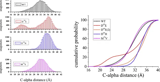
As shown inFig. 4a, data taken from the trajectory of WT display a Gaussian distribution. In particular, most of the obtained MD snapshots show C-alpha distances in a range of 30–34 Å. The distribution function computed for the D69E mutant provided a wealth of additional
informa-tion with respect to the mere analysis of the C-alphaAV. Despite the fact
that C-alphaAVvalues computed for T62 of WT and D69E are almost
equal, their corresponding distribution functions are strongly different. In fact, a bimodal distribution can be detected only for D69E thus
indicat-ing the occurrence of two dominant conformational states. Thefirst one (hereafter referred to as D69E(1)), missing in WT, discloses a peak
around 21 Å whereas the second one (D69E(2)) is shifted to larger
values (peak around 36 Å) with respect to WT. More importantly, this second and more populated state shows a nice overlap with that of D69H, the mutant showing the largest difference in terms of C-alpha
AV
compared to WT. On the other hand, M70V does not display any
appre-ciable difference compared to WT, thus confirming the indications obtained by the analysis of the sole C-alphaAV.
For the sake of completeness,Fig. 4b shows the computed cumula-tive probabilities for WT and all the considered mutants: it is clear that the curves computed for D69E(2) and D69H do overlap and strongly
diverge from WT (shift of about 4 Å). In other words, the trajectories of D69H and D69E suggest that a mutation in position 69 may turn away
T62 residues and induce a significant diversion of loops A. In this re-spect, it is worth saying that a value of distanceb 27 Å is still found in about 15% of the WT and M70V snapshots while it does not occur in
D69E(2) and D69H.
We notice also that the conformational state D69E(1) results from
the interaction, occurring at the monomer D, between K64, positively charged and belonging to loop A, and the negatively charged mutated residue, namely E69. This event is quite persistent for about 35 ns and leads to the closure of loop A and, consequently, to a strong decrease of the C-alpha distance (monomer A vs. monomer B) computed for T62. For further information regarding the D69E(1) state the interested
reader is referred to Figs. S7 and S8 in the Supporting Information.
3.2. Analysis of hydrogen bond interactions
To further investigate the diverse behaviors of WT and MTs, we conducted a deep analysis of the hydrogen bond interactions occurring in the tetramer within the simulated trajectories. Interestingly, the sole appreciable difference was observed for threonine at position 56 (T56), a residue belonging to loop A. Suchfinding supports again the hypoth-esis that this extracellular loop is under the remote control of the single point mutation at position 69. In this respect,Table 1reports the rate of
Fig. 2. a) Top view and b) lateral view of the investigated systems. Water molecules and the membrane bilayer are rendered as sticks while the AQP4 tetramer is shown in cartoon representation.
Fig. 3. a) C-alphaAVvalues (Å) computed for all residues of the investigated systems.
b) Zoomedfigure showing C-alphaAVdata and corresponding error bars (Å) computed
occurrence (%) of the hydrogen bond established between the side chain of T62 (donor) and the backbone of Leucine in position 53 (L53— acceptor) during the entire analyzed trajectories.
The values inTable 1indicate the percentage of frames in which the hydrogen bond is formed, using as thresholds a distance atom acceptor (AA)–atom donor (AD) equal to 3 Å and an angle AD–H–AA equal to 160°. In all considered MTs, such interaction occurs less frequently with respect to WT and, again, the major difference is observed in the case of D69E and D69H, being the corresponding percentages (22.72%
and 11.00% respectively) lower than WT (32.40%). On the other side, data obtained from the trajectory of M70V (27.24%) are closer to WT. Is it worth noting that such percentage was computed by assuming that the four monomers behave independently, so that for each system (i.e. WT and MTs) a trajectory equal to 95 ns × 4 was considered. Nev-ertheless, the robustness of these data is confirmed by the evidence that the observed differences also occur considering each monomer separately. Regarding D69H, for instance, the values obtained for
mono-mers A (10.62%), B (14.16%), C (10.16%) and D (9.06%) are all lower with respect to those computed for WT, being equal to 31.96% (monomer A), 39.22% (B), 20.76% (C) and 37.64% (D). For the sake of clarity, represen-tative MD snapshots are shown inFig. 5to indicate the residues engaged in hydrogen bond interactions for the WT and all MTs.
A further proof reinforcing the solidity of these data is given by the time dependent evolutions of the distance between atom acceptor (oxygen atom of the hydroxyl group of T56) and atom donor (oxygen atom of the carbonyl of the backbone of L53). Fig. S2 (see supporting in-formation) shows the results taking into account all the monomers of WT and D69H: frames showing distances compatible with the H-bond
interactions are much less frequent in all the monomers of D69H with
respect to WT and the H-bond interaction, when it occurs, persists only for a few nanoseconds. In summary, the analysis of the hydrogen
bond interactions indicates that the conformational modification of the loop A, determined by the mutation at position 69, could be the re-sult of the weakening of an interaction involving the residues T56 and L53.
3.3. Computation of the osmotic permeability
In a previous study[26], we found that two different gating mecha-nisms can exist inside the water pore of WT. One is associated with the well-known selectivityfilter (SF) on the extracellular side (residues H201 and R216); the other to the spontaneous reorientation of the im-idazole ring of a histidine from loop B (H95) enabling an H-bond inter-action with a cysteine belonging to the loop D (C178) at the cytoplasmic end (CE). Notably, two different local constriction indicators (LCIs) were defined: 1) dSF, which measures the distance between the nitrogen
hydrogen bond (HB)-acceptor atom of H201 and the closer terminal nitrogen atom of R216 at the selectivityfilter (SF); 2) dCE, which
mea-sures the distance between the nitrogen HB-acceptor atom of H95 and the sulfur atom of C178 at the CE. Based on the time evolution of the LCIs, 4 different representative segments of trajectory were selected and for each of them the total osmotic permeability was computed in order to evaluate how it can be related to the different states, namely open/open, open/closed, closed/open and closed/closed (following the scheme SF/CE).
Herein, we extend this analysis to all the investigated MTs. However, as for WT, variations at the level of SF are quite rare and even when they occur, the closed state rapidly switches back to the open one. For the sake of simplicity, the states herein considered were therefore those open/open and open/closed. The results are shown inTable 2while the time dependent evolutions of dCEfor all the considered systems
are given in Figs. S3–S6 (see Supporting Information).
In all the considered systems, the gating mechanism at the cytoplas-mic end affects heavily the permeability of the pore, with a decrease of pf, resulting from the closure of CE, in the order of about 70%.
Further-more, the values computed for all the MTs, considering both open and closed states, are in good agreement with those obtained for WT. In other words, these data suggest that although the mutation D69 can strongly affect the epitope formation, it does not have any effect in terms of AQP4 water channel properties. Remarkably, these results are in full agreement with recent experimental observations[25].
Fig. 4. Distance distribution function (left) and cumulative probability (right) of C-alpha distances computed for residue T62. D69E(2) refers to data computed for the second
conforma-tional state of D69
E.
Table 1
Occupancy (%) of the H-bond (donor–acceptor) occurring between the side chain of T62 and the main chain of L53 in the considered systems. An atom donor (AD)–atom acceptor (AA) distance equal to 3 Å and an angle AD–H–AA equal to 160° were used as thresholds to define the presence of the H-bond.
H-bond WT D69E D69H M70V
3.4. Analysis of cavities
With the aim to target the AQP4 protein with small-molecules and look for potentially active blockers, we searched for all the potential cav-ities that are present in the AQP4 crystal structure (PDB code 3GD8)[16]
with the FLAPsite algorithm. As shown inFig. 6, the sole cavity predicted is located in the neighborhood of loop A. This observation suggests that this region would be suitable for accommodating small-molecules, ther-apeutically important, to prevent antibody binding.
Further investigations of the potential druggability of this site were performed using molecular interactionfields (MIF)-based descriptors. As shown inFig. 7a, the AQP4 cavity (blue dot) falls in the most populat-ed range of volume, surface, rugosity, and globularity distributions of druggable active sites dataset made of 43 X-ray solved pharmaceutically relevant targets[48]. Furthermore, the computation of the interaction descriptors (hydrophobic, hydrophilic and charge–charge/polar inter-actions) revealed that the AQP4 cavity interaction values were again
in the same range of the reference druggable dataset, falling in the max-imum distribution range (seeFig. 7b).
4. Discussion
Collectively, our results indicate that D69 is a key element for the re-mote control of the loop A conformation of AQP4 and, more specifically, for pushing residue T62 in or out of the lumen pore. The analysis of the 95 ns-long trajectories of WT, D69E, D69H and M70V suggest that
sub-stantial modifications at the level of loop A with respect to WT can be
Fig. 5. Selected frames showing the H-bond interaction involving the side chain of T56 and the backbone of L53 in WT and M70
V and the putative interaction between T56 and E69 present in D69
E and between T56 and H69 present in D69
H.
Table 2
Osmotic permeabilities (pf) computed for WT and all MT forms of the representative
segments of the trajectory corresponding to different states (open and closed) of the CE region. All data are reported in 10−14cm3/s unit.
System Status (CE)
Open Closed WT 9.5 ± 2.1 2.5 ± 0.9 D69 E 8.7 ± 2.5 2.3 ± 0.9 D69 H 8.2 ± 3.2 2.6 ± 1.8 M70
V 11.8 ± 2.1 2.1 ± 0.9 Fig. 6. Cavity identified in the AQP4 crystal structure (PDB entry: 3GD8) by the FLAPsite algorithm.
detected only for two MTs, namely D69E and D69H. On the other side,
the analysis of the computed osmotic permeabilities does not return any appreciable difference when comparing WT and MTs. These ob-servations match recent experimentalfindings whereby the consid-ered mutants do not affect AQP4 water channel properties[25]. The mutation of D69 has instead dramatic effects on all NMO-IgG epi-topes. As experimentally observed, the NMO-IgG binding is ham-pered mainly when the aspartate at position 69 is replaced by histidine. No effects on NMO-IgG binding were instead observed for the M70V case. Actually, our MD studies confirm a substantial
overlap of the M70V mutant with WT, with respect to the conformations
assumed by loop A. The good match between theoretical end experi-mental data allows us to speculate on the features of the conformational epitope.
More specifically, it can be reasonably assumed that, in WT and M70V, NMO-IgG binding can be ensured by a speci
fic conformation of T62 that is able to protrude inside the central pore lumen in a kind of “closed conformation”, which is forbidden to D69E and D69H having a
higher propensity to equilibrate in a kind of“open conformation”, thus disrupting the conformational epitope.
Moreover, the analysis of the H-bond interactions confirms the in-volvement of the loop A. Indeed, a substantial difference in terms of H-bond occurrence was observed between WT and the MTs. This inter-action involves the hydroxyl group of T56 acting as H-bond donor and the carbonyl group of the backbone of L53 acting as H-bond acceptor. It should be noted that T56 belongs to loop A while L53 is part of the first transmembrane segment, named TM1. Furthermore, although the mutated residue is at position 69, T56 is located at the base of the loop A, having an adjacent residue belonging to a transmembrane region. As clearly shown inFig. 5, the effect of mutations at position 69 is that of rotating the sidechain of T56 with the consequent disruption of the interaction T56SIDE–L53MAINto favor the hydrogen bond engagement
with the side chain of the glutamate or of the histidine in position 69. This becomes even clearer if we consider the average distances computed from the center of mass of the side chain, hereafter termed as CoMAV,
instead of CalphaAV. In fact, considering the mutated residue D69H, we
observe an increase in the CoMAVvalueN3 Å when compared to WT
(14.74 Å vs. 11.50 Å), thus confirming a net trend for MTs to place the mutated residue towards T56.
Taken together, these results indicate that the effect of the D69 mu-tation consists in altering the loop A conformation and in particular the spatial position of T62. Interestingly, here wefirst report that such a conformational modification is the result of upstream weakening of the hydrogen bond interaction T56SIDE–L53BACKBONE. This event is, in
turn, due to the higher propensity of the side chain of the mutated residue at position 69 to attract the side chain of T56. As mentioned above, our herein presented hypothesis is supported by experimental literature[25,52]. In particular Miyazaki et al.[52]conclude not only that the“replacement of loop A drastically reduces the binding of the antibodies to human AQP4” but also that, among the other residues, “T62 and/or L64 are involved in the epitope of the monoclonal antibod-ies”. The epitope molecular features are recognition elements essential to address the design of modulators of the NMO-IgG AQP4 binding. This is a topic of extreme interest being AQP4 druggability[53]yet to demonstrate. It is well known that druggability studies are indeed im-portant to evaluate the likelihood that small drug-like molecules bind a given target[54,55]. Indeed, an obstacle in the development of ef fi-cient AQP4 blockers might be due to the extreme difficulty to identify cavities that would be large enough to accommodate possible drug-like small-molecules. In this respect, wefirst considered the chance to detect cavities in the AQP4 surface by using FLAPsite and then we assessed their suitableness for the binding of small-molecules by com-parison with a dataset of druggable active sites[48]. Using the FLAPsite algorithm[34]the entire surface of the AQP4 crystal structure (PDB entry: 3GD8) was scanned looking for suitable molecular cavities. We found only a putative binding site at the top of the channel located
close to loop A (seeFig. 6). The druggability of such potential binding site was, thus, assessed by a knowledge-based approach. Using MIF-based descriptors, the chemical–physical properties of the AQP4 cavity were compared with those of well-known druggable active sites. As shown inFig. 7, the chemical–physical properties of the AQP4 cavity are in the same range of the druggable reference dataset. More impor-tantly, the AQP4 cavity intercepted, among the distribution ranges of the size, shape and interaction descriptors, the highest populated bins. Such knowledge-based observation discloses an intriguing scenario and, to some extent, prompts us to hypothesize that AQP4 could be con-sidered as a druggable target. Interestingly, MD studies and experimen-tal evidences univocally indicate that loop A is important for NMO-IgG binding; in addition the FLPAsite algorithm suggests that a binding pocket for drug-like small-molecules is at the base of this loop. To the best of our knowledge, the sole small-molecules ever demonstrating an appreciable inhibition of NMO-IgG binding to AQP4 in NMO patient sera are arbidol, tamarixetin and berbamine (seeFig. 8). In this respect, we refer to the paper of Tradtrantip et al.[56]reporting, for these three compounds, classical docking simulations focused into a regionflanked by residues at the base of loop A (namely I57, N58 and V68). Retrospec-tively, this region discloses a substantial overlap with the AQP4 cavity identified by FLAPsite. On the basis of MD studies indicating that loop A experiences a conformational change after mutating the aspartate at position 69 (whose main effect was that of preventing NMO-IgG bind-ing without affectbind-ing water permeability), we carried out inducedfit docking (IFD) simulations enabling the ligand to determine conforma-tional changes of the residues forming the AQP4 cavity, designated as a receptor active site.
The top scoring poses obtained for arbidol, tamarixetin and berbamine are shown inFig. 8. Importantly, the lowest-energy docked conformations were found at the loop A and almost completely inside our AQP4 cavity. We notice that a very large docking box (side equal to 26 Å, by default) was used in order to take into account possible interactions with other extracellular loops. Consistently with the inves-tigation of Trandtrip et al.[56]the compounds projected toward a hy-drophobic cavity between the two transmembrane regions connected by loop A. However, here, the use of an IFD protocol allowed us to obtain several important additional clues. Arbidol and tamarixetin establish a strong H-bond interaction with D69, whose side chain changes its con-formation with respect to the crystal (see zoom inFig. 8). Along with D69, also L72 and W59 change their conformation in order to maximize a hydrophobic and an aromatic–aromatic interaction respectively. Moreover, both ligands engage an H-bond interaction with the back-bone of W59 whereas in the case of tamarixetin an additional interac-tion with N58 is also found. In this respect, IFD calculainterac-tions enrich the observations coming from MD and FLAPsite, complementing those mo-lecular analyses. Taken together, our study indicates that the identified AQP4 cavity has the desirable physico-chemical profile as well as the suitable room to bind drug-like molecules.
5. Conclusions
The present paper reports how the harmonized use of advanced bio-physical and drug discovery strategies, already proved to be valuable for other aquaporins[57], allow in approaching very challenging targets as AQP4. Supported by very recent experimental observations[25,56], we suggest that the surface of loop A and, more importantly, of the residues flanking its basis forms a druggable cavity and, thus, a valuable target for the structure-based design of compounds able to inhibit NMO-IgG binding to AQP4 in NMO patients. At present, we are screening the entire Protein Data Bank searching for reliable cross-relationships with known targetable proteins with the aim to discover new NMO-IgG bind-ing blockers. This ongobind-ing attempt might help to address future design of small-molecule modulators and to plan rational drug repurposing strategies.
Transparency Document
TheTransparency documentassociated with this article can be found, in the online version.
Acknowledgements
This work was funded under the program FIRB (Futuro in Ricerca 2012, RBFR12SJA8_003). We acknowledge the CINECA awards nos. HP10CL5BLB-hAQP4 and HP10B4VZO7-epi-NMO under the ISCRA ini-tiative for the availability of high-performance computing resources and support.
Appendix A. Supplementary data
Details on the statistical analysis performed to estimate the uncer-tainties related to our data, data from D69HSE mutated form, time
depen-dent evolutions of dCEin all the considered systems, the time-dependence
of the distance between the oxygen of the hydroxyl group of T56 (atom acceptor) and the oxygen of the carbonyl of the backbone of L53 (atom donor) in WT and D69H and all the details about the conformational
state of D69H are given in the Supporting Information. This material is available free of charge via the Internet athttp://pubs.acs.org. Supple-mentary data associated with this article can be found, in the online version, athttp://dx.doi.org/10.1016/j.bbamem.2015.03.019.
References
[1] S. Nielsen, E.A. Nagelhus, M. Amiry-Moghaddam, C. Bourque, P. Agre, O.P. Ottersen, Specialized membrane domains for water transport in glial cells: high-resolution immunogold cytochemistry of aquaporin-4 in rat brain, J. Neurosci. Off. J. Soc. Neurosci. 17 (1997) 171–180.
[2] M. Borgnia, S. Nielsen, A. Engel, P. Agre, Cellular and molecular biology of the aqua-porin water channels, Annu. Rev. Biochem. 68 (1999) 425–458.
[3] H. Sui, B.G. Han, J.K. Lee, P. Walian, B.K. Jap, Structural basis of water-specific trans-port through the AQP1 water channel, Nature 414 (2001) 872–878.
[4] R. Sachdeva, B. Singh, Insights into structural mechanisms of gating induced regula-tion of aquaporins, Prog. Biophys. Mol. Biol. 114 (2014) 69–79.
[5] B. Ilan, E. Tajkhorshid, K. Schulten, G.A. Voth, The mechanism of proton exclusion in aquaporin channels, Proteins 55 (2004) 223–228.
[6] H. Li, H. Chen, C. Steinbronn, B. Wu, E. Beitz, T. Zeuthen, G.A. Voth, Enhancement of proton conductance by mutations of the selectivityfilter of aquaporin-1, J. Mol. Biol. 407 (2011) 607–620.
[7] G.P. Nicchia, R. Ficarella, A. Rossi, I. Giangreco, O. Nicolotti, A. Carotti, F. Pisani, X. Estivill, P. Gasparini, M. Svelto, A. Frigeri, D184E mutation in aquaporin-4 gene im-pairs water permeability and links to deafness, Neuroscience 197 (2011) 80–88.
[8] J. Li, A.S. Verkman, Impaired hearing in mice lacking aquaporin-4 water channels, J. Biol. Chem. 276 (2001) 31233–31237.
[9] Z. Zador, S. Stiver, V. Wang, G.T. Manley, Role of aquaporin-4 in cerebral edema and stroke, Handb. Exp. Pharmacol. (2009) 159–170.
[10]M.C. Papadopoulos, A.S. Verkman, Aquaporin-4 and brain edema, Pediatr. Nephrol Berl. Ger. 22 (2007) 778–784.
[11] A.S. Verkman, Aquaporins in clinical medicine, Annu. Rev. Med. 63 (2012) 303–316.
[12] M.C. Papadopoulos, J.L. Bennett, A.S. Verkman, Treatment of neuromyelitis optica: state-of-the-art and emerging therapies, Nat. Rev. Neurol. 10 (2014) 493–506.
[13] V.A. Lennon, T.J. Kryzer, S.J. Pittock, A.S. Verkman, S.R. Hinson, IgG marker of optic– spinal multiple sclerosis binds to the aquaporin-4 water channel, J. Exp. Med. 202 (2005) 473–477.
[14] M. Amiry-Moghaddam, O.P. Ottersen, The molecular basis of water transport in the brain, Nat. Rev. Neurosci. 4 (2003) 991–1001.
[15] K. Yoneda, N. Yamamoto, K. Asai, K. Sobue, Y. Fujita, M. Fujita, M. Mase, K. Yamada, M. Nakanishi, T. Tada, Y. Miura, T. Kato, Regulation of aquaporin-4 expression in astrocytes, Mol. Brain Res. 89 (2001) 94–102.
[16] J.D. Ho, R. Yeh, A. Sandstrom, I. Chorny, W.E.C. Harries, R.A. Robbins, L.J.W. Miercke, R.M. Stroud, Crystal structure of human aquaporin 4 at 1.8 Å and its mechanism of conductance, Proc. Natl. Acad. Sci. 106 (2009) 7437–7442.
[17] F. Umenishi, A.S. Verkman, Isolation and functional analysis of alternative promoters in the human aquaporin-4 water channel gene, Genomics 50 (1998) 373–377.
[18]B. Yang, T. Ma, A.S. Verkman, cDNA cloning, gene organization, and chromosomal localization of a human mercurial insensitive water channel evidence for distinct transcriptional units, J. Biol. Chem. 270 (1995) 22907–22913.
[19]M. Tajima, J.M. Crane, A.S. Verkman, Aquaporin-4 (AQP4) associations and array dynamics probed by photobleaching and single-molecule analysis of green fluores-cent protein-AQP4 chimeras, J. Biol. Chem. 285 (2010) 8163–8170.
[20]B.-J. Jin, A. Rossi, A.S. Verkman, Model of aquaporin-4 supramolecular assembly in orthogonal arrays based on heterotetrameric association of M1–M23 isoforms, Biophys. J. 100 (2011) 2936–2945.
[21] R. Iorio, J.P. Fryer, S.R. Hinson, P. Fallier-Becker, H. Wolburg, S.J. Pittock, V.A. Lennon, Astrocytic autoantibody of neuromyelitis optica (NMO-IgG) binds to aquaporin-4 extracellular loops, monomers, tetramers and high order arrays, J. Autoimmun. 40 (2013) 21–27.
[22] A. Rossi, F. Baumgart, A.N. van Hoek, A.S. Verkman, Post-golgi supramolecular assembly of aquaporin-4 in orthogonal arrays, Traffic Cph. Den. 13 (2012) 43–53.
[23] A. Rossi, T.J. Moritz, J. Ratelade, A.S. Verkman, Super-resolution imaging of aquaporin-4 orthogonal arrays of particles in cell membranes, J. Cell Sci. 125 (2012) 4405–4412.
[24] F. Pisani, M. Mastrototaro, A. Rossi, G.P. Nicchia, C. Tortorella, M. Ruggieri, M. Trojano, A. Frigeri, M. Svelto, Identification of two major conformational aquaporin-4 epitopes for neuromyelitis optica autoantibody binding, J. Biol. Chem. 286 (2011) 9216–9224.
[25] F. Pisani, M.G. Mola, L. Simone, S. Rosito, D. Alberga, G.F. Mangiatordi, G. Lattanzi, O. Nicolotti, A. Frigeri, M. Svelto, G.P. Nicchia, Identification of a point mutation impairing the binding between aquaporin-4 and the neuromyelitis optica autoanti-bodies, J. Biol. Chem. 289 (2014) 30578–30589.
[26] D. Alberga, O. Nicolotti, G. Lattanzi, G.P. Nicchia, A. Frigeri, F. Pisani, V. Benfenati, G.F. Mangiatordi, A new gating site in human aquaporin-4: insights from molecular dynamics simulations, Biochim. Biophys. Acta 1838 (2014) 3052–3060.
[27] L. Janosi, M. Ceccarelli, The gating mechanism of the human aquaporin 5 revealed by molecular dynamics simulations, PLoS One 8 (2013) e59897.
[28] M. Hashido, A. Kidera, M. Ikeguchi, Water transport in aquaporins: osmotic permeabil-ity matrix analysis of molecular dynamics simulations, Biophys. J. 93 (2007) 373–385.
[29]Y. Cui, D.A. Bastien, Water transport in human aquaporin-4: molecular dynamics (MD) simulations, Biochem. Biophys. Res. Commun. 412 (2011) 654–659.
[30] Y. Wang, E. Tajkhorshid, Nitric oxide conduction by the brain aquaporin AQP4, Proteins 78 (2010) 661–670.
[31]E. Tajkhorshid, P. Nollert, M.Ø. Jensen, L.J.W. Miercke, J. O'Connell, R.M. Stroud, K. Schulten, Control of the selectivity of the aquaporin water channel family by global orientational tuning, Science 296 (2002) 525–530.
[32] B.L. de Groot, H. Grubmüller, Water permeation across biological membranes: mechanism and dynamics of aquaporin-1 and GlpF, Science 294 (2001) 2353–2357.
[33] J.S. Hub, C. Aponte-Santamaria, H. Grubmuller, B.L. de Groot, Voltage-regulated waterflux through aquaporin channels in silico, Biophys. J. 99 (2010) L97–L99.
[34] P.J. Goodford, A computational procedure for determining energetically favorable bind-ing sites on biologically important macromolecules, J. Med. Chem. 28 (1985) 849–857.
[35] Schrödinger Release 2013-2: Maestro, Version 9.5, Schrödinger, LLC, New York, NY, 2013.
[36] W. Humphrey, A. Dalke, K. Schulten, VMD: visual molecular dynamics, J. Mol. Graph. 14 (1996) 33–38.
[37]W.L. Jorgensen, J. Chandrasekhar, J.D. Madura, R.W. Impey, M.L. Klein, Comparison of simple potential functions for simulating liquid water, J. Chem. Phys. 79 (1983) 926–935.
[38]J.C. Phillips, R. Braun, W. Wang, J. Gumbart, E. Tajkhorshid, E. Villa, C. Chipot, R.D. Skeel, L. Kalé, K. Schulten, Scalable molecular dynamics with NAMD, J. Comput. Chem. 26 (2005) 1781–1802.
[39]A.D. MacKerell Jr., N. Banavali, N. Foloppe, Development and current status of the CHARMM forcefield for nucleic acids, Biopolymers 56 (2000) 257–265.
[40] T. Darden, D. York, L. Pedersen, Particle Mesh Ewald: an N⋅log(N) method for Ewald sums in large systems, J. Chem. Phys. 98 (1993) 10089.
[41] S.E. Feller, Y. Zhang, R.W. Pastor, B.R. Brooks, Constant pressure molecular dynamics simulation: the Langevin Piston method, J. Chem. Phys. 103 (1995) 4613–4621.
[42] G.J. Martyna, D.J. Tobias, M.L. Klein, Constant pressure molecular dynamics algorithms, J. Chem. Phys. 101 (1994) 4177–4189.
[43] S.A. Adelman, J.D. Doll, Generalized Langevin equation approach for atom/solid‐surface scattering: general formulation for classical scattering off harmonic solids, J. Chem. Phys. 64 (2008) 2375–2388.
[44] F. Zhu, E. Tajkhorshid, K. Schulten, Collective diffusion model for water permeation through microscopic channels, Phys. Rev. Lett. 93 (2004) 224501.
[45] S. Henrich, O.M.H. Salo-Ahen, B. Huang, F.F. Rippmann, G. Cruciani, R.C. Wade, Computational approaches to identifying and characterizing protein binding sites for ligand design, J. Mol. Recognit. 23 (2010) 209–219.
[46] F. Sirci, L. Goracci, D. Rodríguez, J. van Muijlwijk-Koezen, H. Gutiérrez-de-Terán, R. Mannhold, Ligand-, structure- and pharmacophore-based molecularfingerprints: a case study on adenosine A(1), A (2A), A (2B), and A (3) receptor antagonists, J. Comput. Aided Mol. Des. 26 (2012) 1247–1266.
[47]L. Siragusa, S. Cross, M. Baroni, L. Goracci, G. Cruciani, BioGPS: navigating biological space to predict polypharmacology, off-targeting, and selectivity, Proteins 83 (2015) 517–532.
[48] A.C. Cheng, R.G. Coleman, K.T. Smyth, Q. Cao, P. Soulard, D.R. Caffrey, A.C. Salzberg, E.S. Huang, Structure-based maximal affinity model predicts small-molecule druggability, Nat. Biotechnol. 25 (2007) 71–75.
[49] G. Cruciani, M. Pastor, W. Guba, VolSurf: a new tool for the pharmacokinetic optimi-zation of lead compounds, Eur. J. Pharm. Sci. Off. J. Eur. Fed. Pharm. Sci. 11 (Suppl. 2) (2000) S29–S39.
[50] F. Milletti, L. Storchi, G. Sforna, G. Cruciani, New and original pKa prediction method using grid molecular interactionfields, J. Chem. Inf. Model. 47 (2007) 2172–2181.
[51]S. Pricl, M. Fermeglia, M. Ferrone, E. Tamborini, T315I-mutated Bcr–Abl in chronic myeloid leukemia and imatinib: insights from a computational study, Mol. Cancer Ther. 4 (2005) 1167–1174.
[52]K. Miyazaki, Y. Abe, H. Iwanari, Y. Suzuki, T. Kikuchi, T. Ito, J. Kato, O. Kusano-Arai, T. Takahashi, S. Nishiyama, H. Ikeshima-Kataoka, S. Tsuji, T. Arimitsu, Y. Kato, T. Sakihama, Y. Toyama, K. Fujihara, T. Hamakubo, M. Yasui, Establishment of monoclonal antibodies against the extracellular domain that block binding of NMO-IgG to AQP4, J. Neuroimmunol. 260 (2013) 107–116.
[53] A.S. Verkman, M.O. Anderson, M.C. Papadopoulos, Aquaporins: important but elusive drug targets, Nat. Rev. Drug Discov. 13 (2014) 259–277.
[54] P.J. Hajduk, J.R. Huth, C. Tse, Predicting protein druggability, Drug Discov. Today 10 (2005) 1675–1682.
[55] J. Owens, Determining druggability, Nat. Rev. Drug Discov. 6 (2007) 187.
[56] L. Tradtrantip, H. Zhang, M.O. Anderson, S. Saadoun, P.-W. Phuan, M.C. Papadopoulos, J.L. Bennett, A.S. Verkman, Small-molecule inhibitors of NMO-IgG binding to aquaporin-4 reduce astrocyte cytotoxicity in neuromyelitis optica, FASEB J. Off. Publ. Fed. Am. Soc. Exp. Biol. 26 (2012) 2197–2208.
[57] D. Seeliger, C. Zapater, D. Krenc, R. Haddoub, S. Flitsch, E. Beitz, J. Cerdà, B.L. de Groot, Discovery of novel human aquaporin-1 blockers, ACS Chem. Biol. 8 (2013) 249–256.