Scuola Normale Superiore
Pisa
Ana Backovic
Capsid Assembly and Single Stranded DNA Genome Formation
of Adeno-Associated Virus Type2 in Yeast Cells
Ph.D Thesis in Molecular Biology
Supervisor: D.Phil. Alvaro Galli
“I keep six honest serving-men: (They taught me all I knew)
Their names are What and Where and When And How and Why and Who”
Here comes the gratitude before all gratitudes; to somebody who was a great person, professor and leader, whose deep love for science gave the birth to this project and whose never stumbling belief, pushed it forward until today. For this, and for showing me that science is the land of wonders where even a mistake seems an improvement, I dedicate this dissertation to my sorely missed professor Arturo Falaschi.
Abstract
Saccharomyces cerevisiae has provided an array of genetic tools to study unknown aspects of viral life cycles, supporting replication of many different RNA or DNA viruses (e.g. Tombusviruses or Papillomaviruses). It also provides means for up-scalable, cost- and time-effective production of various virus-like particles (e.g. Human Parvovirus B19 or Rotavirus) and as such represents a useful tool for vaccine development. To extend the utility of the S. cerevisiae expression system, we expressed AAV2 structural and nonstructural proteins in yeast cells, using both authentic AAV2 and heterologous yeast promoters. For the first time, we described the assembly of AAV2 virus-like particles from yeast-expressed AAV2 structural proteins. To do this we used AAV p40 promoter, whose activity in yeast cells resembled the one of yeast glycolytic promoters, resulting in the synthesis of the most abundant capsid protein VP3 when transformed yeast cells were grown on glucose as a carbon source. The expression of other two VPs was induced from yeast, galactose inducible pGal1 promoter. Simultaneous production of all three VPs was achieved by growing the yeast cells in the medium containing both glucose and galactose, while their relative production levels were further optimized by varying amounts of each carbon source in the induction medium, followed by the fine tuning of the induction time. Moreover, we investigated the ability of the yeast Saccharomyces cerevisiae to carry out the replication of a recombinant rAAV2. When a plasmid harboring the rAAV2 genome in which the cap gene was replaced with the S. cerevisiae URA3 gene, was co-transformed in yeast with a plasmid expressing Rep68 from constitutive yeast promoter pADH, a significant number of URA3+ clones were scored (more than 30-fold over controls). Molecular analysis of low molecular weight DNA revealed that the single stranded DNA is formed, in Rep68 and ITR dependent manner, and that the plasmid is entirely replicated. The ss DNA contained the ITRs, URA3 gene and also vector sequences suggesting that ss rAAV genomes were not obtained by the canonical AAV replication mechanism.
These results could open new prospects for using yeast cell in two ways: (i) as a model system for studying viral and cellular factors involved in AAV2 capsid assembly and packaging of rAAV ss genomes; and (ii) as a novel cell factory for developing superior recombinant rAAV production technologies.
Table of Contents
List of figures ... vi
List of tables ... vii
1. Introduction ... 1
1.1. Adeno-Associated Virus Biology and Life Cycle ... 1
1.1.1. Virus classification and structure ... 1
1.1.2. AAV2 genomic organization ... 2
1.1.3. Productive (replicative) and latent AAV virus infection ... 4
1.2. AAV-based vectors in gene therapy ... 9
1.2.1. Brief introduction to gene therapy ... 9
1.2.2. Properties of AAV-based vectors ... 10
1.2.3. Strategies for improving properties of AAV2 vectors ... 12
1.2.4. Historical overview of current methods for AAV-vector production ... 14
1.3. Budding yeast, Saccharomyces cerevisiae, as a cell factory for viral proteins and model organism for studying virus biology ... 17
1.3.1. Cell factories for recombinant pharmaceuticals. Why yeast? ... 18
1.3.2. Saccharomyces cerevisiae as a cell factory for VLPs ... 20
1.3.3. Saccharomyces cerevisiae supports replication of different RNA and DNA viruses ... 22
2. PROJECT I: Expression of AAV structural and non-structural proteins and assembly of virus-like particles in yeast S. cerevisiae ... 25
2.1. The aim of the project: ... 25
2.2. The background of the project ... 25
2.2.1. Critical components of AAV2 capsid assembly in the recombinant background ... 25
2.2.2. Regulation of Cap protein expression in the natural background ... 26
2.3. Results ... 27
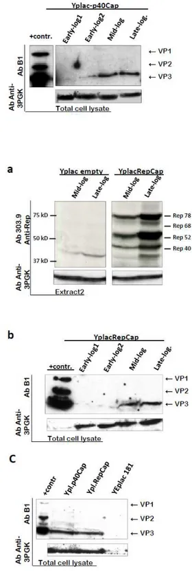
2.3.1. Expression of AAV structural and nonstructural proteins from natural viral promoters and influence of Rep proteins on capsid protein expression in yeast S.cerevisiae ... 27
2.3.2. Inducible Yeast Promoter for Regulation of AAV2 Structural Protein Expression ... 35
2.3.3. Simultaneous expression of AAV2 capsid proteins and modulation of their stoichiometry toward successful capsid assembly. ... 38
2.3.4. Assembly of yeast-cell derived recombinant capsid proteins ... 44
3. PROJECT II: Formation of AAV single stranded DNA genome from a circular plasmid
in Saccharomyces cerevisiae ... 56
3.1. The aim of the project ... 56
3.2. The background of the project ... 56
3.2.1. AAV replication in the recombinant background ... 56
3.3. Results ... 56
3.3.1. Rep68 increases the frequency of yeast clones containing the AAV genome. .. 57
3.3.2. Expression of Rep proteins ... 58
3.3.3. AAV ssDNA formation in yeast ... 60
3.3.4. Expression of Adenovirus helper factors E1b55K and E4orf6 in yeast cells and their influence upon rAAV ssDNA formation in the presence of Rep ... 66
3.4. Conclusion and future prospective ... 68
4. To sum up… ... 73
5. Materials & methods ... 74
5.1. Strains, media, yeast transformation and cultivation ... 74
5.2. Plasmid construction ... 75
5.3. RNA isolation and Retro-Transcription ... 77
5.4. Analysis of protein expression. ... 78
5.5. AAV VLP extraction and purification ... 79
5.6. Transmission electron microscopy... 80
5.7. DNA isolation and Southern blotting ... 80
References ... 82
List of figures
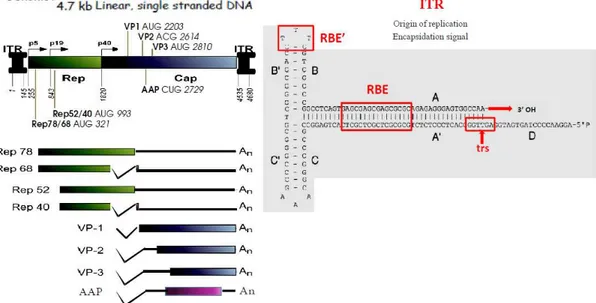
Figure 1.1: AAV genome organization (left) and Inverted Terminal Repeat (right). ... 2
Figure 1.2: AAV life cycle ... 5
Figure 1.3: Mechanism of AAV DNA replication. ... 6
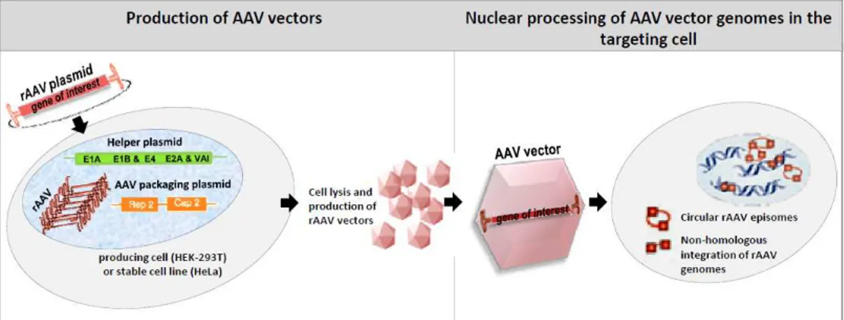
Figure 1.4: AAV vector production and rAAV genome processing in the targeting cell nuclei ... 10
Figure 1.5: Different AAV serotypes target different groups of tissues. ... 12
Figure 1.6: Transient-transfection based protocols for rAAV production from HEK-293 cells. ... 15
Figure 2.0: Cap gene organisation. ... 26
Figure 2.1: Expression of AAV2 Cap and Rep proteins from natural promoters in yeast. . 28
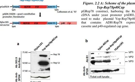
Figure. 2.2 A: Scheme of the plasmid Yep-Rep78p40Cap ... 30
Figure 2.2 B: Cap expression from p40 promoter in presence of Rep 78, expressed from the constitutive ADH1 promoter. ... 30
Figure 2.3: Distribution of AAV2 Rep proteins to soluble and insoluble protein fraction obtained by glass-bead extraction method. ... 31
Figure 2.4: Recovery of AAV2 proteins by ”Optimized post-alkaline” extraction. ... 33
Figure 2.5: Reverse Transcription (RT)-PCR-based analysis of p40mRNAs. ... 34
Figure 2.6: Expression of AAV2 structural proteins from the galactose-inducible promoter pGal1 ... 36
Figure 2.7 A, B: VP1 & VP3 expression in double-transformed yeast cells, listed in Table 2.1. ... 39
Figure 2.8: The “short induction” strategy for preventing decrease of VP3 protein. ... 41
Figure 2.9 A: “High glu-high gal” - strategy for VP1:VP3 ratio optimization: ... 42
Figure 2.9 B: “Low glu-high gal” - strategy for optimization of VP1:VP3 ratio: ... 43
Figure. 2.10: Concentrating VP assembly products by high-speed ultracentrifugation through 40% sucrose-cushion. ... 45
Figure.2.11. Purification of yeast cell-derived AAV2 capsid like-structures by CsCl-gradient ultracentrifugation. ... 49
Figure 3.2: The frequency of colonies carrying rAAV genome increase when Rep68 is
expressed. ... 58
Figure 3.3: Western blot analysis of the Rep protein expression in yeast. ... 59
Figure 3.4: AAV replication in yeast. ... 62
Figure 3.5: S1 and Mung Bean nuclease sensitivity. ... 63
Figure 3.6: Characterization of newly replicated DNA. ... 64
Figure 3.7: ssDNA formation is dependent on ITRs and Rep68. ... 66
Figure 3.8: Effect of the expression of E4orf -E1b55k on ss DNA formation. ... 67
Figure 3.9: Model of ssDNA formation from a plasmid containing rAAV genome in yeast. ... 70
List of tables
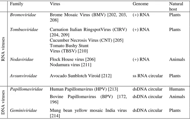
Table 1.1: S. cerevisiae cell factory for production of “virus-like” pharmaceuticals ... 20Table 1.2: Viruses that replicate in yeast ... 23
Table 2.1: Constructs used to cotransfrorm RSY12 cells for simultaneous expression of VP1 &VP3 ... 38 Table 2.4. Comparison of the G/C content in the genomes: Total GC composition and the GC content at the 1st, 2nd, and 3rd codon position in each genome are listed as indicated55
1. Introduction
This work communicates two separate research lines that merge to open new prospective for adopting yeast Saccharomyces cerevisiae as a novel model-host organism for studying still elusive aspects of Adeno-associated virus (AAV) biology. The first part relates to using yeast cell expression system for production of AAV structural proteins from the cognate and heterologous yeast promoters and analyses the ability of these proteins to assemble in authentic virus-like particles (VLPs) inside of yeast cell. The second part investigates the aptitude of the Saccharomyces cerevisiae replication machinery to carry out the replication of a recombinant AAV (rAAV) genome in the presence of yeast-cell expressed AAV Rep 68 protein. Together, the results obtained from these two research lines evaluate the properties of yeast S. cerevisiae as a novel, putative production host for rAAV-based vectors.
1.1. Adeno-Associated Virus Biology and Life Cycle
1.1.1. Virus classification and structure
Adeno-Associated Virus (AAV) belongs to Parvoviridae family, a family of the smallest and the simplest viruses, whose single stranded DNA (ss-DNA) is enclosed in a non-enveloped icosahedral capsid. These viruses infect numerous species, starting from insects (subfamily Densovirinae) to highly evolved vertebrates, including man (subfamily Parvovirinae). The vertebrate subfamily is divided in 3 genera, Parvo-, Erythro- and Dependo-virus, whose members share similar genomic structure, but are distantly related in sequence (~15% identity) [1]. The first two genera, Parvovirus (Canine parvovirus and murine one, the minute virus of mice-MVM), Erythrovirus (human prototype B19) are pathogens that can replicate on their own in the host cell and hence, are called “autonomous”. By contrast, non-pathogenic Dependoviruses, with AAV as a type-species, are dependent upon “helper” virus co-infection for completion of their replicative cycle. In particular, the name “AAV” derives from first identification of this virus, nearly fifty years ago, in a form of a super-infection (contaminant or a satellite virus) of the cells infected with Adenovirus (Ad) [2-4]. Although Ad helper functions have been most extensively studied (reviewed in [5]), several DNA viruses are later shown to provide helper functions, such as herpesvirus, human papillomavirus and vaccinia virus [6-9].
To date, over 100 AAV variants have been isolated from adenovirus preparations (stocks) of human and non-human primate tissues while new ones are continuously emerging; around 14 serotypes (variants of a known serology) have been well characterized [10, 11] and 11 members recognized by the International Committee for Taxonomy of viruses, including AAV-1, AAV-2, AAV-3, AAV-4, AAV-5, AAV-6, avian AAV, bovine AAV,
http://www.ictvdb.org/Ictv/index.htm). All these variants share similar virion structures and genomic organization. The virion shells of icosahedral symmetry (T=1) enclose single-stranded, approximately 4.7kb DNA genomes, comprised of two functional regions-terminal repeats and two viral genes (ORFs), rep and cap gene. Small, 60 subunit capsids, 20-25 nm in diameter, are made of three types of protein subunits, VP1, VP2 and VP3. For attachment and internalization, all AAV serotypes use ubiquitously expressed cell receptors , such as heparin sulfate proteoglycans-HSPGs (AAV2/3) [12-14] or sialic acid (AAV4/5) [15] (reviewed in [11, 16, 17]). However, important differences between serotypes consisted in the variations in amino acid compositions of their capsid proteins, result in the usage of different cell-surface receptors and coreceptors, causing different cell entry pathways and distinct tissue tropism among serotypes. For instance, both AAV1, AAV5 and AAV6, utilize N-linked sialic acid [15, 18] while O-linked 2,3-sialic acid serves as a binding receptor for AAV4 [15]. Ever since the first infectious clone of AAV serotype 2 (AAV2) was established in 1982 [19], its genome has been studied most thoroughly among all serotypes and AAV2-based vectors quickly gained great popularity in gene therapy applications. Its crystal structure has been recently published [20].
1.1.2. AAV2 genomic organization
For more than 25 years (since AAV2 clones were first established) all knowledge about AAV2 biology accumulated around the 4.7kb, single-stranded DNA AAV2 genome, comprising two large genes, rep and cap. All Parvoviruses share the same genome organization where the 5’ rep gene, located on the left half of the genome, codes for nonstructural proteins, essential for viral replication, while 3’ cap gene, the right half located, codes for structural proteins of the capsid (fig. 1.1 left). All AAV2 mRNAs terminates at a single poly-adenylation site at map unit 96 [21].
Top-left part: The AAV-2 genome divided into 3 segments: rep, cap and ITRs and the nucl. positions indicated are: viral promoters, p5, p19 and p40, and translational start sites of Cap and Rep proteins. Middle-left: Rep proteins (boxes) and corresponding mRNAs (with polyA tails). The bottom-left: p40 encoded capsid proteins VP1, VP2 and VP3 and regulatory protein of capsid assembly, AAP, and corresponding mRNAs. Right: Secondary structure of an AAV-2 ITR showing the RBEs (RBE, GAGCGAGCGAGCGCGC and RBE’, CTTTG) and the TRS (AGTTGG).
ITRs. AAV genes are flanked by 145 nucleotide-long inverted terminal repeats (ITRs).
The first 125 nucleotides of the ITR have multipalindromic structure whose complementary regions folds upon themselves to maximize base pairing and forms aT-shaped hairpin structure, while the other 20 bases, called the D sequence, remain unpaired (fig.1.1- right). ITRs contain all cis-acting sequences necessary for packaging, replication, integration of the viral genome in the host cell DNA and for the subsequent rescue from the integrated state (reviewed elsewhere [22, 23]). In addition, it’s been proposed that ITRs possess some transcriptional promoter activity, although relatively weak [24]. A key role of the ITR is that T-hairpin secondary structure provides 3’-hydroxyl group for the initiation of viral replication, thus being the origin of replication and serving as a primer for second-strand synthesis by a host DNA polymerase. This activity and the cis-acting ITRs’ function in integration necessitate, in trans, activity of the two large Rep proteins, Rep78 and 68, which specifically bind at the Rep binding elements, RBE and RBE’ of ITRs and nick in a strand- and site-specific manner (endonuclease activity) at the terminal resolution site, TRS located downstream of RBEs [22].
The rep gene = rep ORF, by the use of two promoters, p5and p19 (map units 5 and 19),
encodes four nonstructural proteins, longer Rep78/-68 and shorter Rep52/-40, respectively, designated by apparent molecular weights. Rep68 and 40 differ from Rep78 and 52 due to an alternative splicing which replaces 92 residue amino-acid element in the later ones with 9 element in the former ones, while amino-acid sequence in the central portion of the gene is identical for all Rep proteins, translated from the same ORF (fig.1.1-left). Rep proteins are involved in a number of processes of viral life cycles , starting from the control of viral gene expression, over regulation of AAV-2 DNA replication (reviewed in [22, 25]). Large Rep proteins, Rep 78 and 68, perform the activities required for AAV DNA metabolism and site-specific integration [26-28] by means of their specific site-specific DNA binding and endonuclease activities as well as nonspecific ATP-ase and helicase activities, common to all four Rep proteins [29-33]. Large Rep proteins coordinate the viral gene expression mainly by transactivation of p19 and p40 promoter and repression of p5 promoter, both through the interaction with the RBEs in the TR and within the p5 promoter [34]. Repression of p5 by Rep68 and 78 is well described during productive infection, when the presence of helper proteins enable its derepression and consequential activation of the other two AAV promoters [34-37]. Conversely, during latent infection, AAV transcripts are not detected [38], suggesting that p5 repression may be attributed to the negative effect of some cellular factors such as YY1 [39]. Besides, large Rep proteins have been shown to affect the expression of various cellular and viral genes although the exact
level of action is often unknown [40-43]. Moreover, inhibition of the cell proliferation or S-phase arrest by Rep 78 is the activity mainly associated with the protein’s ability to induce cellular DNA damage in the cell [44, 45]. Probably related to its ability to arrest the cell cycle, Rep has been shown to promote p53-independent apoptosis [46]. Finaly p19 promoter products, Rep 52 /40 play an essential role in the generation and accumulation of ss progeny genomes used for packaging in preformed capsids [47, 48].
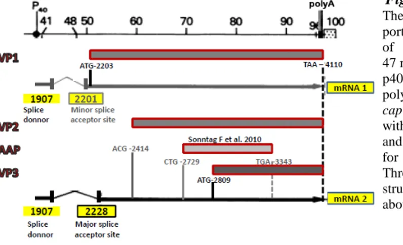
Cap gene. For more than 25 years it was believed that AAV2 cap gene = cap ORF, where
three capsid proteins VP1 (87 kDa), VP2 (73 kDa) and VP3 (62 kDa), with common C-terminus, are generated from the same p40 promoter and overlapping sequences in a single ORF (ORF1). Only recently, a new AAV2 gene product has been identified to derive from the alternative ORF2 of the cap gene, coding for a nonstructural viral protein involved in promotion of capsid assembly [49] (Fig.1.1-left, bottom part). The relative ratio of single protein subunits in mature virions is approximately 1:1:10 for VP1, 2 and 3 respectively, reflecting the intracellular stoichiometry of the three VPs inside the producing cell. This, in turn, is regulated on transcriptional and translational level. The two splicing events use the same splice donor site, but two different acceptor splice sites, among which the one closer to p40 promoter is poorly used, resulting in lower abundance of VP1 coding mRNA and thus, lower intracellular VP1 protein level. VP2 and VP3 are translated from the shorter mRNA whose greater abundance is due to the use of conventional splice acceptor site. Lower intracellular level of VP2 is the result of the poor translation initiation from uncommon ACG initiator, while high expression of VP3 protein owes to the usage of common AUG initiator in the favorable Kozak context [50, 51].
Although little is known about AAV capsid assembly in vivo, generally accepted concept of AAV virion assembly suggests the two step mechanism where empty capsids are firstly formed into which singles stranded DNA genomes are then introduced [52]. This concept is based on early studies which demonstrated that Cap protein localized in the punctuate nucleolar structures in the absence of Rep or AAV DNA [36, 53]. The nucleolus may provide factors/chaperones that help assembly process, the notion further supported by the discovery of two nucleolar proteins, nucleolin and nucleophosmin, found to associate with AAV capsids in vitro [54, 55]. The recent finding of Sontag et al. complemented this concept, showing that a newly identified AAV protein, AAP, not only stimulates transport of VP proteins to the nucleolus, but also, in some way, promotes the assembly process itself [49].
1.1.3. Productive (replicative) and latent AAV virus infection
Viral entry and trafficking to the nucleus. On their way to the nucleus where AAV2 has
to deliver its genome in order to start replication or latent integration, virions have to pass multiple barriers: receptor binding, cell entry, intracellular trafficking, endosomal release, viral uncoating or nuclear entry. AAV2 infection on the cellular level starts with multiple contact with glycoprotein receptor (HSPG), the primary receptors for AAV2 binding, and
is further assisted by various coreceptors that are thought to be essential for its internalization and first post-internalization events (reviewed in [56]). These include: fibroblast growth factor receptor-1 [57], hepatocyte growth factor receptor [58], ανβ5 and ανβ1 integrin [12, 59] and the 37/67 kd laminin [60], that act as co-receptors. It’s currently believed that virions enter the cell by endocytosis in a clathrin & dynamin-dependent process, facilitated by integrins and other coreceptors, and are rapidly transported toward perinuclear area via endosomal compartment: through early, late, recycling endosomes and lysosomes [56, 61]. When and how exactly, endosomal release occurs is still under investigation and is thought to be cell type specific [11]. However, recent studies have identified certain capsid components necessary for subcellular trafficking, thought to be prone to conformational changes and critical for passage from one cellular compartment to another. In particular, higher acidity of endosomes is thought to trigger a capsid conformational change which brings out to the capsid surface two functional regions located near the N termini of VP1 and VP2 (stretches of hydrophilic, basic regions-BRs), hidden in the capsid interior during assembly process. These elements, phospholipase A2 (PLA2) domain and putative nuclear localization signals (NLS) in BRs are thought to be crucial for vesicular disruption followed by endosomal escape and nuclear entry, respectively [62-65]. Although it’s known that mutations in these regions have severe negative impact on AAV2 infectivity, their direct role in trafficking has not yet been demonstrated for AAV2. After endosomal escape, capsids can be either degraded by proteasomes or imported into nucleus. Although several groups have separately demonstrated that nucleus may be the place of AAV2 uncoating, there is still lot of contradiction regarding the mechanism and efficacy of virion/genome import into the nucleus, being the focus of current investigations (reviewed in [11]). Once inside the nucleus, AAV will undergo lytic or latent life cycle depending on the physiological state of the host cell, permissive or non permissive cellular environment for AAV productive infection as reviewed in the Figure 1.2.
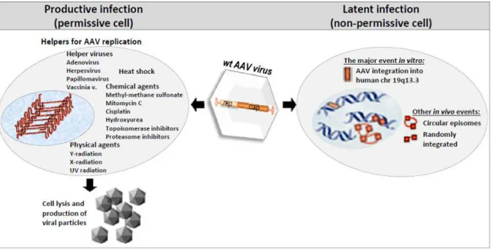
Figure 1.2: AAV life cycle
Replicative cycle. Infection with another virus (one of above listed AAV helpers) or
genotoxic stress caused by various physical and chemical agents are the factors that determine the permissive cell-state that support conversion of single-stranded AAV2 genome to a duplex DNA. This is done by the host DNA polymerase acting on the 3’ end of the viral ITR. The resulting incomplete double-stranded ds-monomer (the form with the priming ITR closed in a hairpin and the other one replicated) is transcriptionally competent thus yielding Rep78/68 proteins, which are necessary to bind RBE and RBE’ of priming ITR and make a nick at TRS site, allowing ITR to unwind and the synthesis of ds-monomer can be completed (schematically presented in the fig. 1.3). Ds-ds-monomer is essential molecular form both for viral protein expression and for second round of self-priming replication which will generate double-stranded DNA dimer molecules by strand-displacement mechanism.
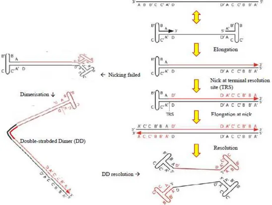
Figure 1.3: Mechanism of AAV DNA replication.
Letters indicate specific sequences in the inverted terminal repeats (AA’, B-B’, C-C’, D-D’ denote complementary sequences). Input viral DNA is in black and newly synthesized DNA in red. The arrow indicates the 3’OH available as a primer for DNA synthesis. TRS: terminal resolution site.
The two most abundant replicative intermediates, replicating-form monomer and RF-dimer serve as reservoir of ss viral DNA which, at some point, is displaced from these templates, probably along with ss DNA encapsidation in preformed capsids. AAV capsids package both positive (+ polarity) and negative DNA strand (- polarity) with equal efficiency (reviewed elsewhere: [17, 22, 48]).
All the steps involved in AAV DNA replication have been reproduced in cell/free systems using over-expressed recombinant Rep proteins [66, 67]. These studies further strengthened the concept that in the presence of Rep, AAV replication can rely mostly, if not uniquely, on direct help from cellular factors, since purified cellular proteins, such as replication protein A (RPA), replication factor C (RFC), proliferating cell nuclear antigen (PCNA), minichromosome maintenance (MCM) proteins, and DNA polymerase δ (Pol δ), were sufficient to replicate the AAV genome in vitro. Studies conducted on the contribution of helper viruses to AAV replication are important not only to identify helper activities that can be used to produce recombinant AAV vectors but also to understand how AAV adapts its replication strategy to the helper virus and to the nuclear environment in general.
Adenovirus (Ad) helper functions have been most extensively studied among AAV
helpers and four of its proteins: E1A, E1B, E2A, E4orf6, as well as one virus-associated RNA (VAI RNA), have been identified as indispensable for AAV replication. These Ad functions help AAV productive life cycle by stimulating viral gene expression and by enhancing AAV genome replication, mainly indirectly (reviewed in [5]). The E1A gene product relieves repression from AAV p5 promoter, probably through direct binding of the YY1 element [39]. Similarly, the Ad DNA-binding protein (DBP), the E2a gene product, was reported to be able to activate transcription from the p5 promoter [68]. Moreover, early studies showed that Adenovirus DBP protein is able to modestly enhance the processivity of AAV genome replication in vitro [69]. The E4orf6 gene has been shown to be essential for replication on several levels. First, it was demonstrated that its product can overcome the rate-limiting step of second-strand synthesis of the single-strand virus genome [70]. More recently, it was shown that the adenovirus proteins E1b55k and E4orf6 can stimulate AAV genome replication by degrading the cellular Mre11/Rad50/Nbs1 (MRN) complex that restricts AAV genome replication during adenovirus coinfection [71,72]. The function of the virus-associated RNA in AAV DNA replication remains somewhat elusive and has been suggested as a translational enhancer of AAV RNAs [73].
Herpes Simplex Virus type 1or 2 co-infection with AAV results in AAV titers similar to
those obtained in the presence of Ad and the very first studies in this field also indicated that this virus enabled more rapid AAV life cycle than the one with Ad [5, 6], which was later confirmed in experiments with recombinant AAV plasmids [74]. Early studies showed that the HSV-1 helicase-primase (HP) complex (UL5/8/52) and DBP (ICP8) were sufficient to replicate rAAV-2 plasmids [75] and it was later shown that the helicase activity, but not primase activity, of HP complex was required for this effect [76]. More recently, a comprehensive study of HSV-1 helper activities demonstrated that the HSV-1 immediate-early proteins ICP0, ICP4, and ICP22 could stimulate rep gene expression and that HSV-1 DNA polymerase encoded by UL30, along with its associated processivity factor (UL42), although not strictly required, significantly increased AAV replication
levels induced in the presence of the HP complex and ICP8 [9, 77]. Only recently, a novel HSV1 protein has been shown to be directly associated with Rep proteins within AAV replication centers (RC), further demonstrating that this viral exonuclease plays a critical role during AAV replication by enhancing the formation of discrete AAV replicative forms, suitable for packaging, thus increasing the titer of infective AAV particles [78]. Having identified five HSV-1 factors in direct association with Rep proteins opposing to a single adenoviral helper protein [79], this study also points out a more direct role of HSV-1 in AAV replication than that of adenovirus, indicating that AAV may be able to differentially adapt its replication strategy to a nuclear environment induced by a helper.
Latent cycle. Under non-permissive conditions (in the absence of helper functions or
stress) AAV genomes that reached the nucleus of the infected cell have the capacity to establish long-term latency by integrating into a specific locus, accordingly named AAVS1, on chromosome 19 (19q13.3-qter) (see fig. 1.2, right half ). Upon nuclear entry, initial conversion of ss AAV genomes to ds forms enables basal expression of Rep 78 and 68, the only viral trans acting factors essential for integration, who form the bridge between ITR and p5 promoter- located Rep Binding Sites (RBS) on one side and the homologous sequences located in the AAVS1 locus on the other [22, 48, 80]. Various RBS-like elements have been identified in human genome, but the vicinity of RBE-like and TRS-like region in 33 bp region of AAVS1 make it almost identical to the analogous regions in ITR which probably enable viral integration via semi-homologous recombination between the two regions (reviewed in [17]). Another proposed model for imprecise integrational mechanism is based on simultaneous replication of AAV genome and AAVS1 region, both mediated by AAV large Rep proteins with intrinsic tendency of a Rep-mediated replication complex to switch template strands. In any case, if latently infected cells are subsequently infected with a helper virus, the AAV genome is rescued and re-enters the replicative phase of the life cycle [81]. However, recent studies with rAAV plasmids have shown that in contrast to AAVS1, replication at the viral terminal resolution site does not appear necessary for the integration event to occur [82] and the viral sequences required for site-specific integration has been identified. This element, designated p5IEE for ‘p5 promoter sequence residing integration efficiency element’, is the sole cis requirement for high-efficiency integration of AAV genome into AAVS1 [83]. Although approximately 0.1% of infecting wt AAV genomes integrate at AAVS1, this mechanism could nonetheless play a role in the natural history of the virus, and AAV proviruses have been identified in AAVS1 after natural infection [84]. The wild-type AAV genome can also integrate at sites in the genome other than AAVS1, as demonstrated by the identification of a provirus on chromosome 1q31.1 in human tonsillar tissue and in multiple different chromosomes in vitro [85, 86]. These events were presumably Rep-independent and thus analogous to the integration of rep-deficient vectors described in the next section. Remarkably, latent AAV genomes have been detected in various tissues derived from humans and non-human primates, including bone marrow, brain, spleen,
colon, heart, liver, lymph node and kidney (reviewed in [87]). While the presence of these latent genomes is consistent with integrated proviruses, in some cases episomal genomes may also persist for prolonged periods [88]. A simplified scheme of the latent AAV infection is shown in the figure 1.2, right part.
1.2. AAV-based vectors in gene therapy
1.2.1. Brief introduction to gene therapy
Gene therapy uses variety of nucleic acids as therapeutic agents (“genes as drugs”). Depending on the desired function and the disease profile, the therapeutic can be in a form of protein coding DNA /non-coding DNA / RNA or analogous molecules, such as: mRNA targeting antisense oligos, siRNA, decoys, ribozymes etc. Their usage is wide, starting from prevention (e.g when used as adjuvants for vaccination), over gene augmentation in acute treatments, to compensation for “genetic loss-off function” in chronic diseases. Moreover, therapeutic genes can provide cytotoxicity in cancer cells or be used for correction of endogenous genes, which presents extremely important branch of gene therapy yet, still limited to experimental studies in cell-culture. In each gene therapy approach the choice of a therapeutic gene (transgene), a target cell/organ and a delivery route have to be matched in the best way to provide maximal efficiency, specificity and
persistence of gene transfer, while minimizing toxicity associated with: gene
administration method and/or the transgene itself. Gene therapy uses three major routes of delivery: 1. ex-vivo, gene transfer is performed in laboratory, in isolated hematopoietic or stem cells, followed by re-administration of “transgenic” cells back into the patient; 2. In-vivo systemic delivery (intra-venous/arterial/peritoneal); and 3. In-In-vivo topical delivery, directly in tissues hardly reachable by systemic delivery (e.g brain, eye, joints) or where systemic delivery could cause unwanted effects (e.g tumors). Naked nucleic acid therapeutics are generally difficult to deliver and are destined to rapid elimination from the circulation for couple of reasons: rapid clearance by serum nucleases, lack of organ-specific distribution and low efficiency of cellular uptake. The use of virus-based vectors is currently the best way for improving delivery efficiency and long term transgene expression; while most gene therapy strategies are coalescing around two types of viral vector: lentivirus vectors for ex vivo gene transfer and AAV vectors for in vivo transfer into postmitotic tissues [89]. Nevertheless, these viral vectors have various limitations: i) inherent packaging constraints limit the size of the therapeutic gene; ii) safety concerns related to: immunotoxicity (harmful immune response) of capsids, envelope proteins, residual viral genes and risks associated with high probability of mutagenic integration (retroviral & lentiviral vectors); iii) transcriptional silencing of integrated vectors leading to reduced transgene expression over time. In addition, even weak immune responses generated against vectors can dramatically decrease transgene expression upon
re-human population, such as Ad and AAV is one of the main limitations of these vectors. Thus, it is imperative to develop efficient producing and engineering technologies to render the vectors capable of escaping immune recognition and avoid inflammation. (Literature used for this section: [17, 90-93])
1.2.2. Properties of AAV-based vectors
Several marked features of AAV life cycle such as the ability of these viruses to infect non-replicating cells, capacity to integrate its proviral DNA into a specific site of the human genome and almost non-immunogenic nature of AAV infection, indicate how well these viruses adapted to long-term persistence and coexistence with the host cell without causing any deleterious effect. These aspects continue to inspire still growing use of AAV viruses in gene transfer technology. The first successful cloning of AAV2 in early 1980s led to more than two decades of constant effort in improving recombinant technologies for AAV-based vector production followed by extensive studies of unknown aspects of AAV biology (particularly serotype 2). So far, AAV vectors have shown therapeutic efficacy in a range of animal models (mice, pigs, dogs, ship etc.), owing to a number of properties which make them preferential with respect to other viral vectors. First of all, these vectors are “gutless”, constructed by complete deletion of all viral genes, rep and cap, and insertion of “therapeutic” gene (gene of interest) between ITRs, which are the only viral cis-acting element indispensible for replication and packaging or recombinant AAV genomes, as long as rep, cap and helper functions are provided in trans by the packaging cell [94] (see fig. 1. 4, left part). Since only 300 nucleotides of viral DNA are retained in the vectors, the risk of their recombination with wt virus is minimal. Further, such “gutless” AAV vectors, deprived of the previously mentioned cis integrational element and trans acting integrational factors (p5IEE element and rep68/78 proteins, successively) are not capable of integrating their genome site-specifically. Once they enter the nucleus of a targeting cell, these vectors may persist in couple of molecular states (Fig. 1.4 - right part).
One common outcome is the conversion of the AAV genome to a double-stranded circular episome either by second-strand synthesis or complementary strand pairing [95, 96]. These episomes can be further converted to high-molecular-weight multimeric concatamers [97], formed by the recombination of monomer genomes [98], and probably are the main source of long-term transgene expression, particularly in non-dividing cells [85, 97]. Such a transductional mechanism of AAV vectors almost eliminates the risk of insertional mutagenesis or silencing, seen for integrating retroviral vectors. Accordingly, in the absence of the immune response to a therapeutic gene, or the pre-existing immunity to common AAV serotypes, transgene expression from AAV vectors in vivo can last for months or years. Such a success was specifically documented in AAV-based gene transfer approaches in animal models [99], which lack memory immunity to most of the human AAV serotypes used in these approaches. However, a large body of data demonstrates that AAV vectors can also integrate at non-homologous sites in the host genome, both in vitro and in vivo, either as a single-copy proviruses or concatamers [84, 87, 100]. More recent studies focused on analysis of large number of vector : chromosome junctions (e.g. Inagaki et al. characterized approx.1000 integration sites from liver, heart and skeletal muscle [101]) and provided insights into AAV vector integration process. The integration of AAV vectors occurred preferentially at specific sites, ‘hot spots’, in the genome such as: ribosomal DNA repeats that encode ribosomal RNAs [102, 103], CpG islands and within 1kb of transcription start sites [101-103]; segmental duplications, satellite DNA and palindromes [101, 102]. Moreover, in mouse hepatocytes, around 60 % of AAV vector integrations occurred in active genes [103]. These integration hot spots may represent regions of genomic instability and together with the evidence that AAV vectors integrates at chromosomal double-strand breaks [104] (generated by the endonuclease I-Sce I, or by treatment with etoposide and γ –irradiation), suggest the model of AAV vector integration. According to Deyle and Russell, these data suggest that the nonhomologous integration of AAV vectors is not a random event and that the preferential integration at certain sites presumably reflects availability of free chromosomal ends that can ligate to AAV, such as regions prone to double-strand breaks or other forms of DNA damage [87]. Even though a rare event, the nonhomologous AAV vector integration can be associated with chromosomal deletions and rearrangements [105], where the most concerning, possible genotoxic effect is the malignant transformation of a transduced cell. The majority of experimental data in animal models have demonstrated that AAV vectors are safe in this regard. Still, some convincing evidence for AAV-induced tumorigenesis derives from a follow up study with mice with mucopolysacharidosis VII (resulting from mutations in β-glucuronidase) injected with the vector carrying human β-glucuronidase gene [106]. Both mutant and non-mutant mice injected with the same vector had an increased incidence of hepatocellular carcinoma (~50% compared with ~8% in controls) and showed the occurrence of four independent tumors containing vector proviruses in the same locus (but not in the surrounding normal liver tissue) suggesting that insertional mutagenesis by AAV
vectors was the cancer trigger, although these loci were not previously associated with hepatocellular carcinoma. Besides, AAV vectors also integrate into tissues other than the liver, heart, skeletal muscle [101], thus pointing to the need for further research to fully assess the risk associated with AAV vector genome integration into the host genome. (Other literature used for this section: [16, 17, 80, 107].
1.2.3. Strategies for improving properties of AAV2 vectors
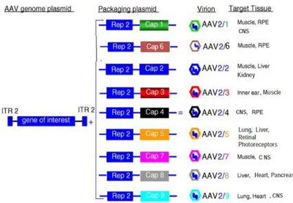
Albeit transduced with different efficiencies, skeletal muscle, hart, liver, brain, retina and lungs are the main targets for AAV2-based gene transfer approaches, while many cell types and relevant diseased tissues still remain non-permissive to transduction by AAV2 based-vectors (and vectors based on other serotypes). The reasons for these differences in tissue transduction efficiencies go beyond the receptor choice and internalization and reflect cell-type related differences (limitations) at various points essential for efficient transgene expression, such as : viral subcellular trafficking to the nucleus, nuclear entry, uncoating and second-strand synthesis (reviewed in [17, 80]). The last limitation was partially overcome with development of self-complementary (sc) AAV vectors, which once in the nucleus can fold upon themselves and elicit rapid expression of a therapeutic gene without the need for inefficient second-strand synthesis [108]. Moreover, packaging of AAV2 based-recombinant genomes into capsids of other serotypes, which show different tissue tropism from the one of AAV2 serotype, “pseudotyping technology” (fig.1.5), can meliorate efficiency of transduction for some tissues, but only slightly widen the range of AAV2 permissive tissues [17, 109, 110].
Figure 1.5: Different AAV serotypes target different groups of tissues. Information taken from www.aaveye.eu and Ref. [17]
Beside pseudotyping, other similar production technologies were developed in order to overcome transduction limitations of AAV2 vectors and further expanding tissue tropism.
These technologies tend to synergistically combine properties of naturally occurring serotypes and further improve them by introducing certain modifications (e.g. targeting peptides), leading to generation of mosaic, chimeric and targeting vectors (direct and
indirect). The mosaic vectors are composed of mixed capsid subunits of various AAV
serotypes [111], while chimeric vectors are produced by domain swapping approach among multiple parental serotypes, involving either entire capsid loops or parts thereof or individual residues [112, 113]. For example, domain swapping between serotypes 1 and 2 yielded a chimeric vector who could be purified by heparin affinity chromatography (purification method designed for AAV2 serotype), had increased transduction efficiency of muscle cells (AAV1 property) and was less efficiently recognized by anti-AAV2 neutralizing antiserum [113]. Furthermore, genetically engineered capsids can lead to more restrictive targeting to a diseased tissue. Numerous groups have developed novel strategies to engineer “designer” AAVs tailored for better transduction of clinically relevant organs (reviewed in detail in ref [11, 114, 115]) These strategies can be grouped in “direct”, physical capsid modifications approaches or “indirect” approaches that base on chemical binding of targeting molecules (ligands) to the viral surface, forming a conjugate ideally able to retarget the capsid to a refractory cell type. Examples of such ligands are: bispecific antibodies [116], avidin-coupled ligands [117] or cellular receptors. In the direct targeting approach, targeting ligands are genetically inserted into the viral capsid proteins [118, 119]. The insertion site has to be chosen in a way to avoid interference with assembling, packaging and infectivity properties of modified virions [120]. In this view, N-terminus of VP1 and VP2 was shown to be a good choice for such modifications [121]. In addition, conferring selective tissue targeting often demands elimination of broad tissue tropism, since a considerable safety issues are raised if non-target tissues are transduced. It’s been observed that systemically routed AAV vectors accumulate in the liver and spleen (mainly associated with HSPG receptor recognition of AAV vectors), thus reducing the transduction efficiency in non-liver target tissues [122, 123]. Some success in combining vector de-targeting from liver and spleen was achieved by insertion of targeting ligands in positions of or close to HSPG binding domains [124, 125].
The aforementioned example of domain swapping between AAV1 and AAV2 serotypes demonstrate that the use of different AAV serotypes present one way for avoiding vector clearance by pre-existing anti-AAV2 immunity to vector capsids. However, results in humans indicate that AAV capsid-primed CD8+T cells are likely to cross-react with alternate serotypes thus urging further development of new strategies for circumventing immunotoxicity [126]. One of possible strategies is introduction of mutations in capsid epitopes recognized by anti AAV2-neutralizing antibodies (A20). Mutations are either results of clever guesses [127] or the use of “evolutionary strategies”, where combinatorial libraries of mutated cap regions are subjected to high-throughput screenings for selection of desired characteristics [125, 126, 128].
(For detailed reviews of all aforementioned strategies see Ref: [10, 11, 80, 126]).
Two inherent limitations of AAV vectors are the small, aprox.5kb, packaging capacity of the capsids and infrequent integration in quiescent target cell-genomes. The first limitation can be overcome, in a part, by skilful engineering of transgenes and their regulatory regions (minimizing the size) and by use of trans-splicing approach for expression of transgenes greater than 9kb [129]. Although rare and mostly related to chromosomal rearrangements, integration events point to the need for fully assessing the associated risk in the future. Finally, long-term expression of the therapeutic level of transgene is the major challenge associated with clinical usage of AAV vectors. As indicated from large number of AAV -based pre-clinical and phase I (less phase II and III) clinical trials, these challenges are tissue-specific and embrace cell-type dependant variables related to administration rout and predominant immune responses directed to the vector or to the transgene product [130].
1.2.4. Historical overview of current methods for AAV-vector
production
Extensive studies on AAV-vectors in the last decade resulted in a variety of currently existing methods for their production. However, the rising number of AAV vector-based gene therapy trials that require high vector doses, over 1013 genome copies (g.c.)/kg of body weight [131], impose the need for creating new production technologies with better “yield versus cost” parameters.
When the first foreign gene was expressed from AAV2 vectors in mammalian cells in the early 1980’s, the stocks of virions carrying recombinant AAV2 genome were obtained by
transfection of Ad2 infected HEK293 cells (a human Adenovirus E1A and E1B genes
-transformed cell line) with two bacterial plasmids, one carrying rAAV genome and the other one providing missing cap gene in trans [132]. Soon later, concerns surrounding the use of replication-competent vectors lead to elimination of rep gene form recombinant genomes and providing it in trans together with cap gene, while infection with Adenovirus was still used for providing helper functions. Frequent contamination of AAV2 stocks with Ad urged cloning of Ad genes into bacterial plasmid, thus eliminating infection [94, 133]. Concerns regarding generation of replication competent vectors were reduced by placing rep and cap genes in opposite orientations. The triple transfection-based AAV production method was the subject of further improvements associated with extensive studies on minimal helper function requirements, leading to currently used protocols for HEK-293T production system which involves following components: (1) vector containing recombinant rAAV genome - the transgene expression cassette flanked by two ITRs (AAV2 derived), (2) plasmid providing Rep and Cap proteins in trans, and (3) plasmid containing adenovirus genes encoding E1, E2A, E4, and virus-associated RNA (VA-I RNA) comprising minimal requirements for inducing cell permissivity to productive AAV replication [5]. The next upgrading introduced to this system was placing AAV2 rep and
cap genes together with Ad-helper genes in a single “helper” plasmid, thus diminishing number of transfecting constructs from three to two
for production of “pseudotyped” AAV2 vector genomes; instead of AAV2 cap gene sequence, the helper construct comprises of serotype
expression cassettes [134].
Vector generation in a producing cell involves following steps: Rep and VP proteins are expressed from helper plasmid and the type2
from the vector genome and replicated in Rep78/68 dependent manner followed by encapsidation of ssDNA into preformed capsids of desired serotype (reviewed elsewhere [17, 135]). The vector yield from transfected HEK293 cells was further improved with fine tuning of Rep78-Rep52 expression level with better results obtained
inferior to Rep52. Although advanced, the productivity of transient transfection method (titers of viral stocks obtained) was still limited by transfection efficiency of the cell line utilized. So far, the best results are obtained with HEK293 cells when calcium phosphate precipitation is utilized for transient transfection (reviewed in
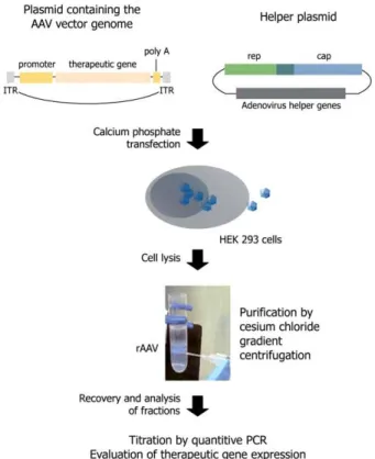
Figure 1.6: Transient-transfection based protocols for rAAV production from HEK The system presented is using two plasmid componen
cassette, flanked by (ITRs); plasmid encoding for: Rep, Cap and adenoviral proteins providing helper functions. Twenty-four hours after transfection, cells are lysed and the vectors are purified by standard cesium chloride ultracentrifugation. Figure reproduced from r
To overcome transfection limitations, infection
helper genes in a single “helper” plasmid, thus diminishing number of transfecting constructs from three to two (fig.1.6). The same procedure is used for production of “pseudotyped” AAV2 vector genomes; instead of AAV2 cap gene equence, the helper construct comprises of serotype-specific cap and AAV2 rep
producing cell involves following steps: Rep and VP proteins are expressed from helper plasmid and the type2-ITR flanked transgene cassette is rescued from the vector genome and replicated in Rep78/68 dependent manner followed by to preformed capsids of desired serotype (reviewed elsewhere ). The vector yield from transfected HEK293 cells was further improved with fine Rep52 expression level with better results obtained when Rep78 was inferior to Rep52. Although advanced, the productivity of transient transfection-based of viral stocks obtained) was still limited by transfection efficiency of the cell line utilized. So far, the best results are obtained with HEK293 cells when calcium phosphate precipitation is utilized for transient transfection (reviewed in [135, 136]).
transfection based protocols for rAAV production from HEK-293 cells. The system presented is using two plasmid components: 1. AAV vector itself, harboring transgene expression
plasmid encoding for: Rep, Cap and adenoviral proteins providing helper four hours after transfection, cells are lysed and the vectors are purified by standard cesium
ref.[17].
Adenovirus, Herpesvirus or Baculovirus-based vectors (reviewed in [135]), carryng both rAAV genome and helper functions. Although high rAAV titers were obtained with Herpesvirus - Baby-Hamster-Kidney cell line (BHK) double-infection system [137], this method is still not widely accepted. On the contrary, recombinant Baculovirus infection of evolutionary distant insect cells has gained lot of popularity since originally established [138]. The original system consisted of three different baculoviruses: the 1st one carrying rep genes, the 2nd one-cap genes and the 3rd one carrying ITR flanked transgene.
Finally, great effort has been made for creating “packaging” and “proviral”, stable cell
lines (mammalian), considered as the best systems for scaling up manufacturing procedure.
Packaging cell lines contain rep and cap genes integrated in the host genome, while the second one contain integrated recombinant AAV genome. The best currently existed stable-cell line systems are based on HeLa cell-lines, but BHK and 293T cells have been also successful so far. Opposing the HeLa packaging cell line, the major difficulty in generating a 293-based AAV producer cell line is the E1A mediated activation of AAV p5 promoter, which control AAV Rep78/68 proteins, which are known to be cytotoxic if constantly expressed [46]. Thus, this difficulty is overcome by creating cell lines with highly inducible Rep systems [139-141]. However, a main limitation which renders stable-cell line approach insufficiently efficient is the multi-step and time-consuming procedure. Due to multiple steps of transfection and/or infection and selections, several weeks to months are needed to produce a high-yield cell line [135, 136, 140, 141].
Since AAV particles are very resistant to harsh manipulation and solvents, conventional
methods for purification of AAV vectors are based on cesium chloride density gradient
ultracentrifugation, which provides efficient separation of empty capsids from genome containing virions [17] (Fig.1.5-bottom). Still, it’s been observed that longer exposure of particles to CsCl can cause a certain decrease in vector infectivity, leading to development of alternative purification strategies. Zolotukhin et al. described the use of nonionic iodixanol gradients followed by ion-exchange or heparin-affinity column chromatography for the purification of AAV2 and other serotypes able to bind heparan sulfate [142]. So far the most commonly used rAAV production method, based on transient transfection of mammalian cells, holds one crucial limitation: the use of adherent cell cultures which make this methods difficult to scale-up. Growing vector requirements for large animal-preclinical and human-clinical trials practically cannot be met with the use of adherent cell-solid supports, such as tissue culture plates (around 50-15 cm plates needed for 293-cell production of 1014 rAAV particles with current transfection methods). This issue is being addressed with new technologies using shake flasks and bioreactor-based
suspension cultures of HEK-293 cells, suitable for scaling-up, although expensive and
manpower demanding [143, 144]. Aside of great improvements achieved in traditional production in mammalian cells, Baculovirus-Sf9 suspension culture system has been the subject of active optimization in the last years and hence, developed into a reliable and
scalable process. Although producing as much rAAV particles per Sf9 cell as per mammalian cells, the original system was mainly limited to small-scale production for two reasons: (i) it required high MOI of each of the three Baculoviruses (Bacs) for efficient co-infection and (ii) proliferation of infected cells had to be limited due to high instability of rep Bac helper and the consequent loss of Rep protein expression following passage 2/3 [145]. These deficiencies were alleviated with optimal expression of Rep78 and Rep52 from a single polycistronic transcript and placing the Cap ORF into the same baculovirus expression vector (BEV), in the opposite orientation with respect to the Rep cassette [146]. Stabile expression of the VP and Rep proteins from consolidated “RepCap”- Bac was observed through at least six amplification passages. This enabled couple-fold increase in the cell-density of suspension cultures, while keeping the same particle per cell yield. Further advances involved optimization of feeding and growth conditions as well as improvements in processing protocols for efficient recovering of rAAV vectors from large volumes and biomass (40-100l bioreactor cultures). Currently, Bac-Sf9 system can meet requirements of commercializing rAAV production, offering lower production costs, high productivity and relatively easy scalability [147, 148].
Efficient Rep78-dependent rAAV replication and production of biologically active vectors in invertebrate cells inspired us to explore the permissivity of the most simple, eukaryotic, single-cell organism, the yeast Saccharomyces cerevisiae, for rAAV genome replication on one side and AAV virus-like particles (VLPs) assembly on the other. In view of the greater complexity of the cell biology and genetics of metazoans, yeast has already demonstrated its usefulness for virus research, which will be discussed in the following section together with the advantages of yeast-based heterologous expression system over the other currently applicable systems for foreign protein production (viral proteins in particular).
1.3. Budding yeast, Saccharomyces cerevisiae, as a cell factory for
viral proteins and model organism for studying virus biology
So far, structural and nonstructural viral proteins have been expressed from practically all recombinant cell-expression systems, from bacteria to human cells. The producer cell choice depends on the characteristics of foreign protein/protein complex (e.g. VLPs) on one hand and from downstream applicability of the produced protein/VLPs on the other, such as:
1) Basic research (e.g. Structure–function analysis [149]); 2) Biomedical applications: diagnostic research and seroepidemiological studies employing various pathogenic viruses; production of vaccines and vaccine candidates, either in a form of surface viral protein or peptide or multisubunit VLPs (in detail in the following sections, table 1.1); 3) production of VLP for nanotechnology applications (e.g. nanoparticles for DNA and drug delivery [150]).
While small scale production satisfies the needs of basic research, commercialized biomedical applications, designed for use in humans and animal models, demand usage of safe, easy to scale up and possibly, low-cost, recombinant expression systems.
The first part of this section addresses some of the major advantages of the high yield-yeast expression system over other heterologous systems, underlying the notable importance of this organism in the field of vaccine production, mostly based on efficient VLPs assembly in yeast cellular background. The second part provides a brief overview of the S. cerevisiae permissivity to replication of many lower and higher eukaryotic viruses and of a further use of this phenomena in elucidating still unknown aspects of viral life cycles. Together, they suggest the potential of extending current usage of yeast-cell factories to production of viral vectors for gene transfer approaches.
1.3.1. Cell factories for recombinant pharmaceuticals. Why yeast?
Since the beginnings of recombinant DNA (rDNA) technologies in the late 70's, large set of methodological platforms have been developed for production of foreign proteins of an important therapeutical, industrial and research values in both eukaryotic and prokaryotic systems. The first recombinant protein-based pharmaceutical to enter the market in early 80’s (Food & Drug Administration of USA (FDA) approved) was human insulin derived from recombinant Escherichia coli. E.coli was the first microorganism used for production of recombinant therapeutics saving uncountable lives, albeit, this expression system was often facing obstacles related to frequent aggregation of overexpressed proteins, thought to be associated with aberrant protein folding and/or stress response of the host to the foreign product [151]. The attempt to produce recombinant Hepatitis Virus B small (s) surface antigen-sHBAg in E.coli expression system failed due to instability of the protein and its deleterious effect upon the host cell. In addition, this first recombinant subunit vaccine was abortive since it failed to induce immune response in “immunized” test animals [152]. Soon after, the platform of heterologous systems for expression of recombinant pharmaceuticals incorporated: bakery yeast Saccharomyces cerevisiae, other less common yeast species (Pichia pastoris, Hansenula polymorpha), mammalian and insect cells and more recently, transgenic plants and animals adapted to the same purpose. The preference in using bacterial and yeast cell-factories over the other expression platforms is hold, on one hand, in a simple microbial cultivation and instrumentation procedures, and, on the other hand, in a genetic flexibility of these microorganisms, apt to engineering and manipulation [151]. Stable, autonomously replicating plasmid-based protein expression from high-density microbial cell cultures enabled inexpensive, controllable and highly scalable production of polypeptides of interest. However, the aforementioned drawbacks of E. coli expression platform mainly consisted in its inability or inefficiency to authentically process foreign proteins and introduce certain post-translational modifications (PTMs) present in most proteins processed by mammalian cells (e.g. glycosylation or phosphorylation) [153, 154]. Among PTMs, glycosylation is the most common andimportant one, playing a crucial role in protein folding, processing, stability, final biological activity, tissue targeting, serum half-life and immunogenicity of the protein. Therefore, failures of bacterial cell in e.g disulfide bond formation, correct proteolytic processing and/or introduction of correct PTMs, may result in a production of insoluble, unstable or inactive proteins. Advances in genetic engineering enabled some of these hurdles, such as protein misfolding and aggregation, to be overcome by stable introduction of various chaperons and foldases [154, 155]. Otherwise, yeast is generally considered advantageous microbial cell-based expression system since its eukaryotic intracellular environment is suitable for the most of authentic posttranslational processing events, typical of higher metazoans (and most importantly of mammalian cells); whereas the low-cost production from this organism is the advantage over higher eukaryote expression systems [156]. Furthermore, the long history of S. cerevisiae industrial applicability in brewing, winemaking and baking demonstrate the absolute absence of hazardous pyrogens, pathogens or viral inclusions in the final products obtained from these cells. Accordingly, this microorganism has been classified as a GRAS (generally regarded as safe) [157, 158]. The era of its successful utilization in pharmaceutical biotechnology started already in the early 80’s, when the first commercialized recombinant vaccine against Hepatitis B (sHBAg) was made in Saccharomyces cerevisiae, soon after the production in E.coli failed [159]. Its wide utilization has been further improved with genetic modifications of selected S. cerevisiae strains, facilitated by the availability of its complete genome sequence, published in 1996 [160]. Subsequent sophisticated approaches of genetic engineering enabled manipulation of entire yeast metabolic pathways performing gene deletions or introductions of single/multiple genes, creating “improved” strains with novel characteristic [158]. For example, addition of humanized N-glycans of the intermediate mannose type or even the complex types provides the option to produce biopharmaceuticals with human protein modifications [161]. In the last decade, heterologous expression systems for biopharmaceutical production based on S. cerevisiae and other yeast species are given the highest commercial value [162]. In a big part this is because S.cerevisiae is an extensive tool-kit of plasmids for high-level expression from large number of wild type (wt) and artificial, regulative and constitutive yeast promoters, of different strengths and substrate dependences [163, 164]. Besides, new regulation systems continue to be added to the existing expression platform and together with a range of engineerized “gracious strains” [165] continue to offer more refined transcriptional to -posttranslational control. Shortly, yeast cell allows refined control of “when” (e.g. triggering expression by means of inducible promoters), “where” (intracellular accumulation vs. secretion) and “how much” (easily scaled-up expression) of the recombinant product will be obtained.
1.3.2. Saccharomyces cerevisiae as a cell factory for VLPs
Since yeast derived HBV vaccine was certified for human use in early 80’s, high-yield production from S. cerevisiae system has been reported for large number of viral structural proteins derived from both prokaryotic and eukaryotic viruses. The viral biopharmaceuticals include: 1. surface proteins, useful candidates for recombinant subunit vaccines but also used for structural analysis [156]; 2. viral core and nucleocapsid proteins, mostly used for diagnostic tests and seroepidemiological studies; 3. authentic and chimeric VLPs, which are of the greatest interest to this work (1-3 reviewed in the table 1.1). Table 1.1: S. cerevisiae cell factory for production of “virus-like” pharmaceuticals
Fammily & Genus
Virus species
(+1parasite) Downstream application
The type of “virus-like” pharmaceuticals
Hepadnaviridae Hepatitis B -FDA approved vaccine;
[159, 166, 167] last generation vac. “RecombivaxHB” [168] Surface protein assembled in Autentic Virus-Like Particles (VLPs): -Drug delivivery nanoparticles [150] Polyomaviridae Human papilloma virus
(PAP) type: 6,11,16,18
FDA approved vaccine- “Gardasil [169-171] Bovine PAP virus[172]
Vaccine candidates Diagnostics Seroepidemiology Cottontail Rabit PAP
[173]
Polyomaviridae Human polyomavirus[174]
Parvoviridae Human Parvovirus B19
[175]
Leviviridae Bacteriophage Q3[176] “Nano”-drug delivery of:
Peptides (e.g -antigens) and mRNAs (vaccines) Bacteriophage MS2 [177] Retroviridae HIV-1[178] Vaccine candidates Authentic &chimeric VLPs
Reoviridae Rotavirus [179] Triple-layered
VLPs Paramixoviridae Human Parainfluenza
Virus (HPIV) type1,3 [180] Vaccine candidates Diagnostics Seroepidemiology Core and nucleocapsid proteins assembled in Nucleocapsid -like Particles (NLPs) Henipaviruses [181] Menangle Virus [182] Tioman Virus [183] Measles Virus [184] Mumps Virus [185] Sendai Virus [186] Bunyaviridae Hantaviruses [187]
Orthomyxoviridae Influenza A virus [188]
Vaccine candidates
Surface protein: Influenza Hemaglutinin
Rhabdoviridae Rabies virus [189, 190] Surface
glicoproteins Herpesviridae Herpessimplex virus [191]