UNIVERSITÀ DI PISA
Facoltà di Medicina e Chirurgia
Scuola di Specializzazione in Radiodiagnostica
Direttore: Prof. Carlo Bartolozzi
TESI DI SPECIALIZZAZIONE
Infrapopliteal revascularization in diabetic foot ischemia:
correlation between morphologic and clinical results
Relatore:
Chiar.mo Prof. Carlo Bartolozzi
Candidata:
INDEX
A
BSTRACT 2I
NTRODUCTION 4M
ATERIALS ANDM
ETHODS 6RESULTS
12DISCUSSION 15
T
ABLES ANDF
IGURES 20ABSTRACT
Purpose: To retrospectively evaluate angiographic and clinical outcomes of
percutaneous revascularization in diabetic patients with Fontaine stage IV critical limb ischemia (CLI).
Materials and Methods: Clinical and procedural data were retrospectively
collected of consecutive diabetic patients with Fontaine stage IV CLI who underwent percutaneous limb revascularization from January 2009 to June 2011. Pre- and post-procedural angiographic images were retrospectively reviewed to classify lower limb arterial involvement according to 3 systems: 1) TASC II; 2) Graziani’s morphological classification; 3) Joint Vascular Society Council calf and foot scores. Foot lesions were graded according to the Texas University (TU) classification. Clinical results (healing, stability or major amputation) were compared to baseline clinical data and angiographic results.
Results: In the study period, 202 percutaneous procedures were performed,
with an immediate technical success rate of 93.6%. The majority of ulcers (75.7%) were deep (TU grades 2 and 3). Mean preprocedural calf and foot scores were 7.8±1.6 and 7.3±2.3, respectively; 132 patients (65.3%) were in Graziani’s morphological classes from 4 to 7 (at least two arteries occluded); in 112 (55.4%) cases TASC II was inapplicable (only infrapopliteal
involvement). After the procedure, mean calf and foot scores were 4.8±2.3 and 5.9±2.6, respectively, and 86.6% of cases were into the Graziani’s classes 1 and 2, whereas TASC II was inapplicable in all cases. Healing rate was 66.8% and major amputation rate was 3.9%. Among all the clinical and angiographic variables included in the analysis, only pre- and post-procedural foot scores were significantly associated to the clinical outcome (p=0.04).
Conclusion: TASC II is inadequate to describe vascular involvement in the
vast majority of diabetic patients with CLI. In this population, percutaneous revascularization should represent the first-line treatment option, leading to high healing and limb salvage rates. Pre- and post-procedural foot scores significantly affect chances of healing, thus aggressive treatment of foot arteries should be attempted whenever possible.
INTRODUCTION
Lower limb peripheral arterial disease (PAD) is a common complication of diabetes mellitus and is associated with more severe clinical manifestations and a higher risk of critical limb ischemia (CLI) and limb loss in diabetic patients. Many recent studies have demonstrated the benefit derived from the percutaneous revascularization procedures on diabetic patients with CLI, in terms of high rate of technical success and limb salvage, facing low periprocedural complication rates [1-7].
However, it’s still very difficult to compare the clinical results of the different trials, even the ones regarding only a diabetic population, in the absence of uniform criteria which describe the involvement of the peripheral vascular system [8]. It is already known that the international guidelines, produced by the Intersociety Consensus for the Management of Peripheral Arterial Disease (TASC II) [9], don’t provide any description of PAD in patients with a infrapopliteal arteries damage, and, therefore, they are not useful in the diabetic population. An interesting review performed by Graziani L. et al. [10] on 417 consecutive diabetic patients has allowed the development of a more specific morphological classification describing the infrapopliteal arterial involvement in diabetic PAD; this classification seems to be correlated to the clinical severity of the disease. Furthermore, it is well-known from the
clinical experience that the angiographic presentation of peripheral diabetic macroangiopathy does not directly correspond to the clinical situation of every specific patient, and in the absence of a direct dialogue with the clinicians, sometimes there could be difficulties in understanding which one is the limb or the target lesion to treat. Furthermore, recently, the so-called “angiosome model” has been successfully proposed, underline the need to treat specifically the wound-related artery allowing direct blood flow to the lesion to improve the final clinical result [11,12].
The aim of this study was to retrospectively evaluate the correlation between the clinical scenario, in terms of lesion severity and clinical success of treatment, and the vascular damage evaluated by the angiographic study, before and after the treatment, in a homogeneous series of diabetic patients with ischemic foot, consecutively treated with percutaneous revascularization from January 2009 to June 2011.
MATERIALS AND METHODS
We retrospectively collected the data of all the diabetic patients with chronic critical limb ischemia, defined by the presence of trophic lesions and/or ischemic rest pain, according to the definition of the TransAtlantic InterSociety Consensus [9], submitted to infrainguinal percutaneous revascularization between January 2009 and June 2011, and admitted from the Diabetic Foot Clinic of our University Hospital.
All pre-procedural data were collected, with particular reference to: age, gender, comorbidities, diabetes type and duration in years, serum levels of HbA1c and creatinine, lesion location and severity, according to the classification of the Texas University (Table 1) [13]. All the patients with suspected or proven diagnosis of PAD underwent digital subtraction angiography (DSA; digital subtraction angiography; GE Innova 4100), by percutaneous transfemoral arterial access controlateral to the target limb, under local anaesthesia, with 4Fr Pigtail catheter (Cordis Corporation; Miami, FL, USA), in order to map the arterial tree.
Every single case was discussed in a multidisciplinary team composed by interventional radiologists, diabetologists, and vascular surgeons, in order to establish the most proper therapeutic option. This study included only patients indicated for endovascular revascularization.
Technique of endovascular revascularization
The endovascular revascularization was performed, after obtaining written informed consent, with an orthodromic ispilater puncture of the common femoral artery (4- to 5-Fr introducer sheath), under local anaesthesia; in selected cases, a contralateral access was chosen followed by the cross-over the iliac axis. The steno-obstructions were overcome with hydrophilic guidewires and multiple angioplasties were performed with 3- to 6-mm balloon catheters with variable length depending on the target vessel.
Treatment was considered successful in the presence of a direct flow in the treated vessels with residual stenosis <30% of the vessels’ diameter. In the presence of angioplasty-resistant stenosis, or dissection complicating the flow, a stent was deployed. In specific cases, the revascularization was performed by subintimal angioplasty, according to the technique described by Bolia et al [14,15]. To reduce the risks of thrombo-embolic phenomena, all patients were administered a bolus of heparin (routinely, 5000 IU) just before treatment.
Other Treatments
Following the procedure, an anticoagulation protocol was required in all patients, including fractioned heparin (1 mg per day in the first week, 0.5 mg per day from the second to the fourth week) and ticlopidine (250 mg per day)
or acetylsalicylic acid (300 mg per day) starting from the second week. Medical therapy of comorbidities was carefully performed when required. The lesions were treated, when necessary, with local surgical approach (such as debridment, minor amputations) in the days following the revascularization, with the exception of infected lesions for which a surgical treatment was performed in emergency the day before revascularization.
Major amputations were considered and carried out, with the patient consent, in cases where revascularization did not lead to a meaningful lesion improvement.
Post-Procedural Follow-Up
All patients underwent color-coded Duplex Ultrasound 24 hours after the procedure. After discharge, follow-up clinical examinations were carried out (with different time intervals, depending on each single case) at the Diabetic Foot Clinic, for the adaptation of the systemic antibiotic therapy based on the results obtained from the microbiological exams, for the local medications and for the drainage until complete wound healing.
Data Collection
The clinical and epidemiological data were collected from the clinical files. The angiographic data were collected via the analysis of the reports and images stored on the computer-system of R.I.S.-P.A.C.S (Radiology Information System-Picture Archiving and Communication System) of the Interventional Radiology Unit. The data concerning the surgical operations and the follow-up of patients were collected by the analysis of the computerized clinical folders, stored on the information system E-upodi@ of the Diabetic Foot Clinic.
Evaluation of the clinical results
The clinical result was defined, based on lesion evolution, as healing, stability and worsening, the latter leading to major amputation. Time for healing was calculated from the time of the revascularization procedure, where healing was defined as the complete and durable re-epithelization of the lesion, according to the International Consensus on Diabetic Foot [16]. Ipsilateral or contralateral recurrence was defined as recurrence of rest pain, onset of new lesions or relapse of the primary lesion after recovery.
Evaluation of the angiographic results
The angiographic acquisitions of the peripheral arteries, before and immediately after treatment, were retrospectively reviewed by two interventional radiologists in consensus, in order to classify the preprocedural and post-procedural arterial involvement, using three different scoring systems:
1) TASC II classification for the femoro-popliteal lesions [9] (Table 2);
2) Classification according to Graziani L. et al [10] which provides 7 different classes with progressively increasing arterial involvement (Table 3); 3) The Joint Vascular Societies Council classification [17,18] which provides a score from 0 to 3 on the basis of the most severe stenosis (score 0 fro stenosis less than 20%, score 1 for 20-49% stenosis, score 2 for 50-99% stenosis, score 2.5 for occlusion of less than half the total length of the vessel, score 3 for longer occlusion). The following arteries were included in the analysis: anterior tibial (AT), posterior tibial (PT), peroneal including the tibio-peroneal trunk (Per), dorsalis pedis (DP), lateral plantar (LP), and medial plantar (MP). We evaluated the DP on its entire length from the ankle joint level to the level at which it gives off its arcuate branch. We counted up a “foot score” (sum of the 3 foot vessels scores plus 1) and a “calf score” (sum of the 3 calf vessels scores plus 1).
Statistical analysis
The data were evaluated via descriptive statistics (mean, median and standard deviation, DS) and compared with the chi-square or Fisher test for the categorical data and the Student's t test for the continuous variables.
Lesion severity and clinical results were evaluated in relationship to the clinical and pre- and post-procedural angiographic variables.
The statistical analysis was performed with the SAS software (SAS Institute, Cary, NC). A P-value of less than 0.05 was considered statistically significant.
RESULTS
The study included 202 percutaneous revascularizations in 166 patients (males/females: 115/51, mean age: 72.8±9.8 years); the clinical and demographic data, collected at the time of each procedure, are reported in Table 4.
Lesion severity resulted to be significantly associated with patients' age (P=0.03), with higher age (mean 74.7 years) in patients with 1C lesions compared to patients with 2D lesions (mean 62.9 years) (Table 5). Moreover, type D lesions (i.e. with infectious component) were significantly more frequent in patients with diabetic retinopathy (P=0.0007) and with higher levels of HbA1c (P=0.01).
At the pre-procedural angiograms, the mean calf score was 7.8±1.6 (median 8), while the mean foot score was 7.3±2.3 (median 8); in 112 angiographies (55.4%) the TASC II classification resulted to be inapplicable; finally, 132 angiographies (65.3%) fell within the Graziani’s morphological classes ranging from 4 to 7, with 7 (3.5%) cases that were not classifiable for the absence of below-the-knee arterial occlusions (Table 6). We observed a significant correlation between Graziani’s morphological classes and calf (p<0.0001) and foot (p<0.001) scores. The pre-procedural calf and foot scores
were statistically lower in patients with 2D lesions (P=0.002 and P=0.03, respectively).
The revascularization procedure was technically successful in 189 cases (93.6%). At the final angiographic examination, the mean calf score was 4.8±2.3 (median 4) with an mean reduction of 3±1.9 (range 0-8), while the mean foot score was 5.9±2.6 (median 6.5), with an mean reduction of 1.4±1.6 (range,-2,5-8). At the end of the procedure, 175 angiographies (86.6%) were classified in Graziani’s morphological classes of 1 and 2, while the TASC II classification resulted to be inapplicable in all cases, following the successful revascularization of the femoro-popliteal axis in all the required patients.
Clinical follow-up was available in 181 cases. At latest follow-up, 121 (66.8%) procedures had determined complete ulcer healing, with a mean healing time of 28.4±23.7 weeks. In 53 cases (29.3%), the lesion was clinically stable, whereas in 7 cases (3.9%) major amputation was required a mean of 30.7±24.6 weeks after the revascularization procedure. Minor amputations were performed in 19 cases a few days before the procedure and in 59 cases, a mean of 6±5 days after the revascularization procedure.
Ulcer healing was significantly associated with lower pre-procedural (mean score 7 in healed patients and 7.8 in non-healed patients; P=0.049) and post-procedural (mean score 5.5 in healed patients and 6.3 in non-healed patients;
P=0.047) foot scores. All the remaining clinical and angiographic variables included in the analysis did not result to significantly affect the clinical outcome (Tables 7,8,9).
Ipsilateral recurrence rate was 37.8% (n=68) after a mean of 28±23 weeks from revascularization (range 8-981 days). For the contralateral limb, the incidence of lesion recurrence was 23.3% (n=42) after a mean of 24.6±25.7 weeks (range 5-903 days). Ipsilateral recurrence was significantly more frequent in patients with type D lesions (P=0.004).
DISCUSSION
PAD is a relatively common complication of diabetes mellitus that can be underestimated, although it is associated with a higher risk of CLI, major amputation and death compared to the non-diabetic population. Revascularization is therefore required either by percutaneous or surgical approach, providing evidence of clinical benefits.
In the Eurodiale study, which involved 1300 patients of 14 specialized centers in 10 European countries, it has emerged how the severity of PAD heavily influences the risk of major amputation as well as the incidence of co-morbidities [19].
Our retrospective study on diabetic patients with CLI focused on the possible correlation between severity of arterial involvement and clinical results.
Our baseline clinical data show how diabetic patients with CLI are aged patients, with multiple co-morbidities and microvascular as well as macrovascular multiple diseases. Coronary artery disease was present in about one third of the patients, whereas 10% of the population was affected by cerebrovascular disease, and hypertension was found in over 50% of cases.
Among micro-vascular complications, diabetic retinopathy was registered in 53% of patients, nephropathy in 32.2%, and neuropathy in 16.3% of patients.
Regarding the limb lesions, the vast majority (75.7%) of our patients had either type 2 or 3 lesions according to the Texas University, which means that the vast majority of the lesions involved the deeper levels.
The lesion severity was in accordance to the degree of arterial involvement. In fact the median calf and foot scores were 8, which means that there were at least two obstructed arteries for each district in the vast majority of our cases. These scores were significantly correlated to the Graziani’s morphological classification, which seems to nicely represent the arterial involvement in the diabetic population. in our series, the majority of patients were in Graziani’s classes >4, with percentages in the different classes which are comparable to the ones reported by Graziani et al. in their wider cohort of patients [10]. However, Graziani’s classification does not take into consideration the foot level, which, in accordance to our results, seems to be the only factor significantly influencing the final clinical outcome.
To date, TASC II guidelines are the only available international guidelines providing recommendations for the treatment of PAD. However, limitations of these guidelines have been pointed out, particularly regarding the lack of a proper description of below-the-knee arterial involvement, which is the prevalent involvement in the diabetic population [20]. Indeed, our data demonstrate that TASC II classification is often inadequate in diabetic
patients with CLI to describe the arterial involvement. In fact, more than half of the patients included in the analysis weren’t referable to any of the TASC II classifications, and such percentage reaches 100% after treatment, either due to lack of femoro-popliteal involvement or to the successful percutaneous treatment of femoro-popliteal stenoses or occlusions when present.
In agreement with previous published data [1-7], The immediate technical success rate of percutaneous treatment was 93.6%, while healing rate was almost 70%.
Healing time is however a major concern. Our mean healing time was about 6 months, suggesting that the healing process requires careful follow-up and a remarkable effort both from the care provider and from the patient. Efforts are required to try and reduce the time required for healing, by improving revascularization techniques and patency rates. By reviewing post-procedural angiographic results, we noticed how we are nowadays able to revascularize most of the infrapopliteal arterial districts, also due to the introduction of low-profile dedicated materials, with a remarkable reductions in the post-procedural calf scores. On the other hand, we did not observe the same evident reduction in the post-procedural foot scores. At this level the presence of heavy calcifications, long-lasting chronic occlusions and multiple tiny collateral arteries might interfere with our technical chances of obtaining the
revascularization of an entire native pedis artery. In these conditions, implementing the blood flow into the collateral arteries is often the only available possibility. However, the need has already been pointed out of moving from “below-the-knee” to “below-the-ankle” and feasibility has been reported in a few pioneer complex experiences, reserved to selected patients [21]. Indeed, our data demonstrate that the preprocedural and postprocedural foot scores represent important predictive factors for ulcer healing, thus suggesting the need for further technical improvements in order to provide direct blood flow to the target area [11,12].
The clinical result was not significantly associated with any of the remaining angiographic scores. This result might represent a further confirmation of the unnecessary achievement of an “optimal” angiographic result, by revascularizing each single infrapopliteal artery, while efforts should concentrate on improving the direct blood flow to the wounded area [12], as confirmed by good healing percentage and the low percentage of major amputations. The accomplishment of a “cosmetic result” could increase the incidence of possible complications, due to the increasing interventional manipulations and procedural time.
Our major amputation rate (about 4%) seems to be lower in comparison to previous published data [1-7]. This result could be biased by the retrospective
nature of our study, in which only patients candidates to percutaneous treatment were included. However, the low ampuation rate might also represent a confirmation of the effectiveness of percutaneous treatment and of a multidisciplinary organization dedicated to diabetic foot care.
Complete ulcer healing depends on many variables, such as patient age, presence of co-morbidities, glycometabolic control, lesions size and duration [7], as well as patients’ compliance to treatment and prevention. In other words, the “diabetic foot” has to be considered as a complex multifactorial disease. The therapeutic approach should, therefore, be multidisciplinary through the organization of a dedicated multidisciplinary team, for pre-procedural evaluation, treatment and follow-up [22,23].
In conclusion, the percutaneous revascularization represents a useful and valuable treatment in diabetic patients with CLI but it should be included inside a complex treatment algorithm to take care not only of the limb, but of the patient as a whole. The foot score represents the most significant angiographic parameter to evaluate chances for ulcer healing, and, consequently, to state the success of the revascularization procedure.
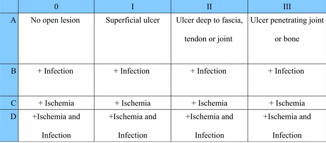
Table 1: Texas University score system
0 I II III
A No open lesion Superficial ulcer Ulcer deep to fascia, tendon or joint
Ulcer penetrating joint or bone
B + Infection + Infection + Infection + Infection
C + Ischemia + Ischemia + Ischemia + Ischemia
D +Ischemia and Infection +Ischemia and Infection +Ischemia and Infection +Ischemia and Infection
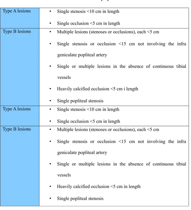
Table 2: TASC classification of femoro-popliteal lesions Type A lesions Single stenosis <10 cm in length
Single occlusion <5 cm in length
Type B lesions Multiple lesions (stenoses or occlusions), each <5 cm
Single stenosis or occlusion <15 cm not involving the infra geniculate popliteal artery
Single or multiple lesions in the absence of continuous tibial vessels
Heavily calcified occlusion <5 cm i length
Single popliteal stenosis
Type A lesions Single stenosis <10 cm in length
Single occlusion <5 cm in length
Type B lesions Multiple lesions (stenoses or occlusions), each <5 cm
Single stenosis or occlusion <15 cm not involving the infra geniculate popliteal artery
Single or multiple lesions in the absence of continuous tibial vessels
Heavily calcified occlusion <5 cm in length
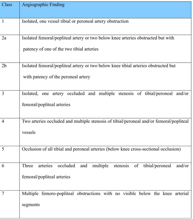
Table 3: Graziani’s classification of below-the-knee arterial lesions Class Angiographic Finding
1 Isolated, one vessel tibial or peroneal artery obstruction
2a Isolated femoral/popliteal artery or two below knee arteries obstructed but with patency of one of the two tibial arteries
2b Isolated femoral/popliteal artery or two below knee tibial arteries obstructed but with patency of the peroneal artery
3 Isolated, one artery occluded and multiple stenosis of tibial/peroneal and/or femoral/popliteal arteries
4 Two arteries occluded and multiple stenosis of tibial/peroneal and/or femoral/popliteal vessels
5 Occlusion of all tibial and peroneal arteries (below knee cross-sectional occlusion) 6 Three arteries occluded and multiple stenosis of tibial/peroneal and/or
femoral/popliteal arteries
7 Multiple femoro-popliteal obstructions with no visible below the knee arterial segments
Table 4: Patients’ demographic and clinical data
Variable N
Age (years, mean ± SD) 71.8 ± 9.8 (range 39-89)
Gender (M/F) 142 / 60
Comorbidity
Coronary artery disease 65 (32.2%)
Other cardiac disease 44 (21.8%)
Cerebrovascular disease 22 (10.9%) Hypertension 124 (61.4%) Retinopathy 107 (53%) Nephropathy 65 (32.2%) Neuropathy 33 (16.3%) Charcot 10 (5%) HbA1c (%, mean ± SD) 7.8 ± 1.8 (range 0.9-14.5)
Creatinine (mg/dl, mean ± SD) 1.68 ± 1.9 (range 0.5-13.4) Diabetes type
1 / 2
3 / 199
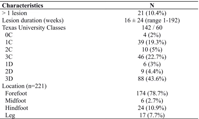
Table 5: Data regarding the foot lesion
Characteristics N
> 1 lesion 21 (10.4%)
Lesion duration (weeks) 16 ± 24 (range 1-192)
Texas University Classes 142 / 60
0C 4 (2%) 1C 39 (19.3%) 2C 10 (5%) 3C 46 (22.7%) 1D 6 (3%) 2D 9 (4.4%) 3D 88 (43.6%) Location (n=221) Forefoot 174 (78.7%) Midfoot 6 (2.7%) Hindfoot 24 (10.9%) Leg 17 (7.7%)
Table 6: Preprocedural angiographic classifications a) TASC B C D NA Total 63 (31.2%) 1 (0.5%) 26 (12.9%) 112 (55.4%) b) Graziani 1 2a 2b 3 4 5 6 7 NA Total (%) 5 (2.5) 4 (2) 4 (2) 50 (24.7) 66 (32.6) 15 (7.4) 42 (20.8) 9 (4.5) 7 (3.5)
c) The Joint Vascular Societies
Leg AT PT PER
Total 7.8±1.7 2.4±0.9 2.6±0.8 1.8±1.0
Foot DP LP MP
Table 7: Pre-and post-procedural morphologic evaluation according to TASC
II classification, compared to clinical outcome
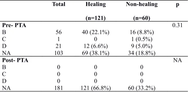
Total Healing (n=121) Non-healing (n=60) p Pre- PTA 0.31 B 56 40 (22.1%) 16 (8.8%) C 1 0 1 (0.5%) D 21 12 (6.6%) 9 (5.0%) NA 103 69 (38.1%) 34 (18.8%) Post- PTA NA B 0 0 0 C 0 0 0 D 0 0 0 NA 181 121 (66.8%) 60 (33.2%)
Table 8: Pre-and post-procedural morphologic evaluation according to
Graziani’s Classification, compared to clinical outcome
Total Healed (n=121) Not healed (n=60) p
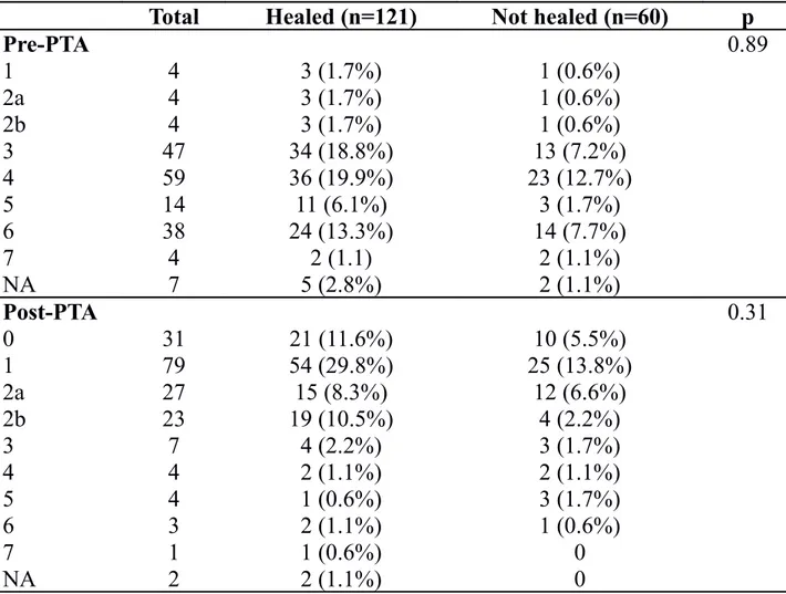
Pre-PTA 0.89 1 4 3 (1.7%) 1 (0.6%) 2a 4 3 (1.7%) 1 (0.6%) 2b 4 3 (1.7%) 1 (0.6%) 3 47 34 (18.8%) 13 (7.2%) 4 59 36 (19.9%) 23 (12.7%) 5 14 11 (6.1%) 3 (1.7%) 6 38 24 (13.3%) 14 (7.7%) 7 4 2 (1.1) 2 (1.1%) NA 7 5 (2.8%) 2 (1.1%) Post-PTA 0.31 0 31 21 (11.6%) 10 (5.5%) 1 79 54 (29.8%) 25 (13.8%) 2a 27 15 (8.3%) 12 (6.6%) 2b 23 19 (10.5%) 4 (2.2%) 3 7 4 (2.2%) 3 (1.7%) 4 4 2 (1.1%) 2 (1.1%) 5 4 1 (0.6%) 3 (1.7%) 6 3 2 (1.1%) 1 (0.6%) 7 1 1 (0.6%) 0 NA 2 2 (1.1%) 0
Table 9: Pre-and post-procedure morphologic evaluation according to the
guidelines of The Joint Vascular Societies, compared to clinical outcome
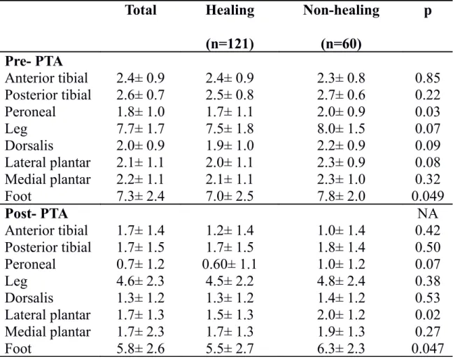
Total Healing (n=121) Non-healing (n=60) p Pre- PTA Anterior tibial 2.4± 0.9 2.4± 0.9 2.3± 0.8 0.85 Posterior tibial 2.6± 0.7 2.5± 0.8 2.7± 0.6 0.22 Peroneal 1.8± 1.0 1.7± 1.1 2.0± 0.9 0.03 Leg 7.7± 1.7 7.5± 1.8 8.0± 1.5 0.07 Dorsalis 2.0± 0.9 1.9± 1.0 2.2± 0.9 0.09 Lateral plantar 2.1± 1.1 2.0± 1.1 2.3± 0.9 0.08 Medial plantar 2.2± 1.1 2.1± 1.1 2.3± 1.0 0.32 Foot 7.3± 2.4 7.0± 2.5 7.8± 2.0 0.049 Post- PTA NA Anterior tibial 1.7± 1.4 1.2± 1.4 1.0± 1.4 0.42 Posterior tibial 1.7± 1.5 1.7± 1.5 1.8± 1.4 0.50 Peroneal 0.7± 1.2 0.60± 1.1 1.0± 1.2 0.07 Leg 4.6± 2.3 4.5± 2.2 4.8± 2.4 0.38 Dorsalis 1.3± 1.2 1.3± 1.2 1.4± 1.2 0.53 Lateral plantar 1.7± 1.3 1.5± 1.3 2.0± 1.2 0.02 Medial plantar 1.7± 2.3 1.7± 1.3 1.9± 1.3 0.27 Foot 5.8± 2.6 5.5± 2.7 6.3± 2.3 0.047
REFERENCES
[1] Faglia E, Mantero M, Caminiti M, et al. Extensive use of peripheral angioplasty, particularly infrapopliteal, in the treatment of ischaemic diabetic foot ulcers: clinical results of a multicentric study of 221 consecutive diabetic subjects. J Intern Med. 2002;252(3):225-32.
[2] Dorros G, Jaff MR, Dorros AM, Mathiak LM, He T. Tibioperoneal (outflow lesion) angioplasty can be used as primary treatment in 235 patients with critical limb ischemia: five-year follow-up. Circulation 2001; 104:2057-2062
[3] Faglia E, Clerici G, Clerissi J, et al. Early and five-year amputation and survival rate of diabetic patients with critical limb ischemia: data of a cohort study of 564 patients. Eur J Vasc Endovasc Surg 2006;32(5):484-490.
[4] Faglia E, Clerici G, Losa S, et al. Limb revascularization feasibility in diabetic patients
with critical limb ischemia: Results from a cohort of 344 consecutive unselected diabetic patients evaluated in 2009. Diabetes Res Clin Pract. 2012;95(3):364-71.
[5] Hinchliffe RJ, Andros G, Apelqvist J, et al. A systematic review of the effectiveness of
revascularization of the ulcerated foot in patients with diabetes and peripheral arterial disease. Diabetes Metab Res Rev. 2012;28 Suppl 1:179-217.
[6] Faglia E, Dalla Paola L, Clerici G, et al. Peripheral angioplasty as the first-choice
revascularization procedure in diabetic patients with critical limb ischemia: prospective study of 993 consecutive patients hospitalized and followed between 1999 and 2003. Eur J
Vasc Endovasc Surg. 2005;29(6):620-7.
[7] Uccioli L, Gandini R, Giurato L, et al. Long-term outcomes of diabetic patients with
[8] van Battum P, Schaper N, Prompers L, et al. Differences in minor amputation rate in
diabetic foot disease throughout Europe are in part explained by differences in disease severity at presentation. Diabet Med. 2011 Feb;28(2):199-205.
[9] Norgren L, Hiatt WR, Dormandy JA, et al. Inter-Society Consensus for the Management of peripheral arterial disease (TASC II). J Vasc Surg 2007; 45 Suppl S:S5-67. [10] Graziani L, Silvestro A, Bertone V, et al. Vascular Involvement in Diabetic Subjects
with Ischemic Foot Ulcer: A New Morphologic Categorization of Disease Severity. Eur J
Vasc Endovasc Surg 2007; 33:453-460.
[11] Clemens MW, Attinger CE. Angiosomes and wound care in the diabetic foot. Foot Ankle Clin. 2010;15(3):439-64.
[12] Manzi M, Cester G, Palena LM, Alek J, Candeo A, Ferraresi R. Vascular imaging of
the foot: the first step toward endovascular recanalization. Radiographics.
2011;31(6):1623-36.
[13] Le Frock JL, Joseph WS. Bone and soft tissue infections of the lower extremity in
diabetics. Clin Podiatr Med Surg 1997; 12: 87.
[14] Bolia A. Percutaneous intentional extraluminal (subintimal) recanalization of crural
arteries. Eur J Radiol 1998; 20(3):199-204.
[15] Ingle H, Nasim A, Bolia A. Subintimal angioplasty of isolated infragenicular vessels
in lower limb ischaemia: long term results. J Endovasc Ther 2002; 9:411-416.
[16] Apelqvist J, Bakker K, van Houtum WH, et al. International Consensus and practical guidelines on the management and prevention of the diabetic foot. International Working Group on the Diabetic Foot. Diabetes Metab Res Rev 2000; 16 Suppl 1; S84-92
[18] Toursarkissian B, D'Ayala M, Stefanidis D, et al. Angiographic scoring of vascular
occlusive disease in the diabetic foot: relevance to bypass graft patency and limb salvage.
J Vase Surg 2002; 35:494-500.
[19] Akhtar S, Schaper N, Apelqvist J, Jude E. A review of the Eurodial studies: what
lessons for diabetic foot care? Curr Diab Rep 2011; 11:302-9
[20] Graziani L, Piaggesi A. Indications and clinical outcomes for below knee endovascular therapy: review article. Catheter Cardiovasc Interv 2010; 73:433-443
[21] Manzi M, Palena LM, Brocco E. Is digital arteries recanalization useful to preserve
the foot functionality and avoid toes amputation, after pedal recanalization? Clinical results. J Cardiovasc Surg (Torino). 2012;53(1):61-8.
[22] Schaper NC, Andros G, Apelqvist J, et al. Diagnosis and treatment of peripheral arterial disease in diabetic patients with a foot ulcer. A progress report of the International Working Group on the Diabetic Foot. Diabetes Metab Res Rev 2012; 28 Suppl 1:218-24 [23] Valabhji J, Gibbs RG, Bloomfield L, et al. Matching the numerator with an appropriate denominator to demonstrate low amputation incidence associated with a London hospital multidisciplinary diabetic foot clinic. Diabet Med 2010; 27:1304-7