Full Terms & Conditions of access and use can be found at
http://www.tandfonline.com/action/journalInformation?journalCode=kccy20
Cell Cycle
ISSN: 1538-4101 (Print) 1551-4005 (Online) Journal homepage: http://www.tandfonline.com/loi/kccy20
Recognition mechanism of p63 by the E3 ligase
Itch
Alessia Bellomaria, Gaetano Barbato, Gerry Melino, Maurizio Paci & Sonia
Melino
To cite this article: Alessia Bellomaria, Gaetano Barbato, Gerry Melino, Maurizio Paci & Sonia
Melino (2012) Recognition mechanism of p63 by the E3 ligase Itch, Cell Cycle, 11:19, 3638-3648, DOI: 10.4161/cc.21918
To link to this article: http://dx.doi.org/10.4161/cc.21918
Published online: 30 Aug 2012.
Submit your article to this journal
Article views: 43
View related articles
©2012
Landes
Bioscience.
Do
not
distribute.
Cell Cycle 11:19, 3638-3648; October 1, 2012; © 2012 Landes Bioscience RepORt
*Correspondence to: Sonia Melino; Email: [email protected] Submitted: 08/03/12; Accepted: 08/21/12
http://dx.doi.org/10.4161/cc.21918
Introduction
Ubiquitin ligases play a pivotal role in modulating intracellular protein levels, by ubiquitylation of the protein substrates for their proteasomal or lysosomal degradation.1 The differential status
for ubiquitination (mono- or poly-) may further contribute to the mark for proteasomal or lysosomal degradation.2 E6-AP
car-boxyl terminus (HECT)-type family of E3 ubiquitin ligases3
(EC 6.3.2.19) transfer ubiquitin directly to substrate proteins by acting as catalytic intermediates through a conserved cys-teine residue.4,5 They are involved in a wide variety of cellular
processes, including differentiation, endocytosis, signaling and
transcription.6-12 The oncogenic potential of the HECT-type
the HeCt-containing e3 ubiquitin ligase Itch mediates the degradation of several proteins, including p63 and p73, involved in cell specification and fate. Itch contains four WW domains, which are essential for recognition on the target substrate, which contains a short proline-rich sequence. Several signaling complexes containing these domains have been associated with human diseases such as muscular dystrophy, Alzheimer’s or Huntington’s diseases. to gain further insight into the structural determinants of the Itch-WW2 domain, we investigated its interaction with p63. We assigned, by 3D heteronuclear NMR experiments, the backbone and side chains of the uniformly 13C-15N-labeled Itch-WW2. In vitro interaction of Itch-WW2 domain with p63 was studied using its interactive p63 peptide, pep63. pep63 is an 18-mer peptide corresponding to the region from 534–551 residue of p63, encompassing the ppxY motif that interacts with the Itch-WW domains, and we identified the residues involved in this molecular recognition. Moreover, here, a strategy of stabilization of the conformation of the ppxY peptide has been adopted, increasing the WW-ligand binding. We demonstrated that cyclization of pep63 leads to an increase of both the biological stability of the peptide and of the WW-ligand complex. Stable metal-binding complexes of the pep63 have been also obtained, and localized oxidative damage on Itch-WW2 domain has been induced, demonstrating the possibility of use of metal-pep63 complexes as models for the design of metal drugs to inhibit the Itch-WW-p63 recognition in vivo. thus, our data suggest a novel strategy to study and inhibit the recognition mechanism of Itch e3-ligase.
Recognition mechanism of p63
by the E3 ligase Itch
Novel strategy in the study and inhibition
of this interaction
Alessia Bellomaria,1 Gaetano Barbato,1 Gerry Melino,2,3 Maurizio paci1 and Sonia Melino1,*
1Dipartimento di Scienze e tecnologie Chimiche; University of Rome “tor Vergata”; Rome, Italy; 2Medical Research Council (MRC); toxicology Unit; Leicester University;
Leicester, UK; 3Laboratorio di Biochimica IDI-IRCC c/o Dipartimento di Biochimica e Chirugia; University of Rome “tor Vergata”; Rome, Italy
Keywords: p63, p53 family, E3 ubiquitin ligases, HECT, ubiquitilation, cyclization, metal-peptide
Abbreviations: CSI, 13C-chemical Shift Index; DTNB, 5,5'-DiThiobis-(2-NitroBenzoic acid); HECT, homologous E6-AP carboxyl
terminus; HSQC, heteronuclear single quantum coherence; IPTG, IsoPropyl-β-D-ThioGalactoside; LAPTM5, lysosomal-associated protein transmembrane 5; LATS 1, large tumor suppressor homolog 1; LC/ESI-MS, liquid chromatography electrospray
ionization mass spectrometry; NOESY, nuclear overhauser effect spectroscopy; Melan-A, melanoma antigen recognized by T-cells 1; RP-HPLC, reverse phase high performance liquid chromatography; PPII, possible polyproline II; pep63, p63(534-551)
peptide; TNB, 2-Nitro-5-Thiobenzoic acid; TCEP, Tris (2-CarboxyEthyl)-Phosphine; TOCSY, total correlated spectroscopy
E3s is emphasized by the recognition of tumor-suppressor mole-cules among their protein targets.13 The E3 ubiquitin ligase Itch
or atrophin-1-interacting protein 4 (AIP4, here, after referred to as Itch; UniProtKB accession number Q96J02) is a member of the NEDD4 family of E3 ligases. Itch plays a key role in different cellular contexts and contributes to many cellular pro-cesses by targeting regulators of multiple signaling pathways.14
Itch-null mice develop systemic and progressive immunological disorders.15 Itch catalyzes the poly- and monoubiquitination of
important globular and membrane proteins such as Melan-A, ErbB4, CXC-chemokine receptor 4 and LAPTM5, regulating transport of intracellular vesicles to lysosome,2,16-18 and
tran-scription factors as c-Jun, JunB, p73, p63 and LATS 1, which
©2012
Landes
Bioscience.
Do
not
distribute.
RepORt RepORtfolded structure of the entire domain.27 The WW2 domain of
Itch E3 has been shown to bind both p73 and p6328,29 by
inter-action with the PPxY motif present in the C-terminal region of these transcription factors.30 Indeed, the in vitro interaction
between the p63 region of residues 534–551 (named pep63), encompassing the PPxY motif and WW2 domain of Itch, sug-gested a potential modulation effect of the downstream region to the PPxY motif on the Itch-p63 interaction. Accordingly, an extended PPxY consensus motif [P-P-P-Y-X(4)-(ST)-(ILV)] has been suggested to modulate the affinity and specificity of the interaction of p63 with Itch.30
In order to further investigate the influence of the individual residues in the recognition mechanism between Itch and p63, we have performed a structural analysis by NMR spectroscopy of the complex and identified the residues of Itch-WW2 domain involved in the interaction with the proline-rich domain of p63. Here, the NMR assignment of the Itch-WW2 backbone and the binding with a cyclic form of pep63 are indicated, demonstrat-ing that more stable complexes can be obtained usdemonstrat-ing the cyclic form of the pep63. The metal-binding ability of both forms of pep63 has also been investigated, demonstrating the ability of are crucial to determine cellular fate.14,19-23 Itch is a monomeric
protein with a modular structural organization that consists
of an N-terminal Ca2+-dependent phospholipid-binding C2
domain and 2–4 WW domains, which confer substrate specific-ity.14,24 The protein-protein recognition is a crucial event in the
ubiquitination by the E3 ligases and it is determined by recogni-tion of the WW domains with a specific proline-rich region of the targets. The WW domains are small stable proteins whose topology consists of a slightly curved, three-stranded β-sheet, comprising about 35 amino acids, including conserved tryp-tophan residues and proline-containing sequences that
medi-ate the recognition mechanism.25,26 According to their ligand
binding preferences, the WW domains have been grouped into four classes, Itch has group I WW domains, specifically inter-acting with the PPxY motif, a proline-rich consensus sequence. Generally, the first tryptophan residue of this type of WW domain, in conjunction with an externally exposed tyrosine residue, forms a concave hydrophobic binding surface for the first two proline residues of the PPxY motif. The hydrophobic buckle formed by these two prolines stack against the W and Y residue, which likely contributes to maintain a more stable
Figure 1. NMR assignment of Itch-WW2 domain. Region of the 15N-1H-HSQC spectrum (A) and 13C-1H-HSQC spectrum (B) of the 0.25 mM Itch- WW2
do-main in 10 mM potassium phosphate buffer, pH 6.0, 50 mM KCl and 10% D2O at 285K. (C) Secondary 13C chemical shift (Δδ 13C) of Itch-WW2 domain for
Cα, Cβ and C’ from CSI consensus values plotted and secondary structure prediction based on chemical shift of the backbone resonances using CSI.
©2012
Landes
Bioscience.
Do
not
distribute.
HBHA(CO)NH experiments and confirmed with the HCACO experiment. Side-chains were then assigned starting from the
known 1Hα and 1Hβ frequencies, and the spin system was
com-pleted using the HCCH-TOCSY experiment and CC(CO) NNH and HCC(CO)NNH. Side-chain NH2 frequencies of asparagines and glutamine residues were assigned from the 3D
15N-NOESY-HSQC and the CBCA(CO)NH spectra. Statistics
for resonance assignments includes 42/43 HN, 42/43 N, 46/47 Cα, 45/46 Hα, 45/46 Cβ, 71/72 Hβ, 31/32 Cγ, 41/42 Hγ, 15/16 Cδ, 23/24 Hδ. Figure 1B shows the 1H-13C α region of the 1H-13C HSQC with the relative assignments; only the N-terminal
Gly, the Pro-12 and the Arg 39 are missing in this spectra. The secondary structure was predicted using the 13C-chemical shift
index (CSI).33 Figure 1C shows the 13C chemical shift deviations
from the consensus CSI values for Itch-WW2; the lower pan-els report the predicted secondary structure. The data confirm a canonical structure for the Itch-WW2 domain, which encom-passes an antiparallel three-stranded-β-sheet. The residue Arg39, is positioned right after the end of the third strand of the β-sheet, in a position thus that could result in some conformational aver-aging, potentially explaining the broad signals detected experi-mentally as several resonances broadened beyond detection. The sequence-specific 1H, 13C and 15N resonance assignments for
the metal-complexes to induce an oxidative damage to the WW2 domain. Since Itch regulates p63 functions, cyclic pep63 may represent a potential therapeutic pharmacological target. Taken together, our results open the way to obtain more stable com-plexes of Itch with its recognition target sites, in order to perform the structure of the complexes in solution, and to design potential selective competitive inhibitors of the E3 ligase activity of the recognition mechanism.
Results
Assignment of ITCH-WW2 domain by NMR spectroscopy.
From previous interaction studies,30 we expressed the recombinant
15N-13C-uniformely-labeled-Itch-WW2 domain and performed
the backbone assignment of the Itch-WW2 domain, correspond-ing to region from D417 to E460 residues of the human Itch
protein. Figure 1A shows the 1H-15N HSQC spectrum with all
the amide groups assigned; a total of 42 signals are labeled in the spectrum due to the presence of six prolines at the residue N-terminal of the protein, and 94% of the residues (46 out of 49) was assigned. Sequential backbone assignment was obtained using the “building blocks strategy,”31,32 the 1Hα and 1Hβ
fre-quencies were assigned from the corresponding HBHANH and
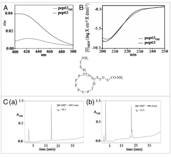
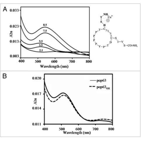
Figure 2. Oxidation reaction of pep63. (A) DtNB’s assay of the pep63 after oxidation, UV-vis spectra of the for pep63 (_____) and pep63
OX (- - -), the
maximum at 412 nm indicates the presence of tNB2+ and the thyols; (B) Rp-HpLC of the pep63 (a) and pep63
OX (b) the molecular masses were obtained
by eSI-MS of the corresponding peaks; (C) CD spectra of 50 μM pep63(____) and pep63
OX (___) in 10 mM sodium phospate pH 6.2, buffer 25 mM KCl.
©2012
Landes
Bioscience.
Do
not
distribute.
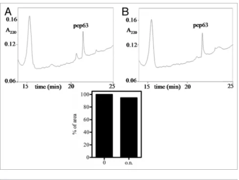
proteolysis, probably due to a reduced acces-sibility of the recognition sites to the protease, in addition to the presence of proline residues. Indeed, only 5.4% of digestion was detected, comparing the area of the undigested peak after 15 min and overnight (Fig. 3).
Interaction of Itch-WW2 with pep63OX by fluorescence and NMR spectroscopy. The
interaction between Itch-WW2 and both of pep63 forms was evaluated by the quench-ing of the intrinsic fluorescence of Itch-WW2 after addition of the peptides in the solution. The apparent Kd values, obtained by the curve fitting analyses, were 47.7 ± 0.2 μM and 13.5 ± 0.8 μM for pep63 and pep63OX, respectively (Figure 4). The cyclic peptide shows a higher affinity for the Itch-WW2 compared with the linear peptide.
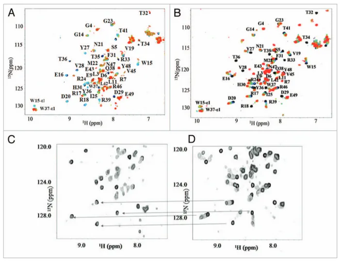
The interaction was also investigated by NMR spectroscopy, and Figure 5A shows the HSQC titration of Itch-WW2 domain
with pep63OX. The spectra show that the NH
resonances of Itch-WW2 domain affected by the binding to the cyclic-form are the same of those detected in the binding to the linear-form of the peptide. The assignment of the free
domain using the 15N-13C-labeled Itch-WW2
domain allowed the identification of the WW2 residues involved in the binding with the two forms of the peptide (Fig. 5A
and B). The identical amino acid residues of Itch-WW2 domain
are involved in the binding of both forms of the peptide, how-ever, with relevant differences in the exchange regime (Fig. 5C
and D). In fact, most of the residues affected by the binding for
the cyclic form are in slow-exchange regime (Fig. 5C). In both interactions the residues of the C- and N-terminal regions of the domain are not affected by the binding. Table 1 summarizes the residues involved, with their respective exchange regimes. In par-ticular, while R18 and the residues from T32 to W37 show a slow exchange regime in both complexes, the residues L11, E16, D20, R24, Y26, Y27, H30, Q38, T41 and V45 change their exchange
regime from fast to slow in case the WW2-pep63OX. The
slow-exchange regime indicates that the resident lifetime of the bound form is increased with respect of the open form, revealing a
higher affinity between Itch-WW2 and pep63OX and the
forma-tion of more stable complexes.
Metal binding of pep63 and oxidative damage to Itch-WW2 domain. Pep63 is characterized by the presence of a histidine residue
in third position forming a characteristic metal binding motif, the ATCUN motif, which binds metal ions such as Cu2+ and Ni2+.37,38
In order to assess the formation of the ATCUN-Cu2+complex, we
performed spectroscopic studies. Figure 6 shows the UV-Vis
spec-tra of pep63 and pep63OX in the presence of equimolar
concen-trations of Cu2+ at different pHs. Both pep63 forms exhibit the
characteristic maximum at 525 nm of the Cu binding in the pH range between 3.4 and 10.17 in presence of copper ions. The metal binding at low pHs indicated a characteristic of the formation of a Itch’s second WW domain were deposited in BioMagResBank
under the accession number BMRB 17715.
Cyclization and characterization of pep63. Peptide
cycli-zation is often used to synthesize conformationally stable mol-ecules, with limited flexibility when compared with their natural counterpart. Moreover, cyclic peptides are of considerable interest for several reasons, such as their resistant to proteolysis and deg-radation. In fact, cyclization affects metabolic stability, bioavail-ability and pharmacokinetics.34 Oxidation in solution needs to be
performed at high dilution, typically at 10–l00 μM, in order to avoid aggregation and intermolecular side reactions; accordingly, the oxidation of pep63 was performed at 100 μM in 25 mM Tris-HCl, pH 8.0, buffer for 48 h at 25°C. The oxidation reaction was monitored according to Ellman’s method (Fig. 2A).35,36
The major hydrophobicity in RP-HPLC of the pep63OX
(RT 21.5 min), as compared with pep63 (RT 19.7 min), is in agreement with the formation of disulfide bonds (Fig. 2C). LC/ ESI-MS analysis of pep63OX gives a m/z of 945.4 [M+2H]2+,
fur-ther confirming the intramolecular formation of disulfide bond. The pep63 CD spectrum shows a significant negative band at 202/204 nm characteristic ofa polyproline II (PPII) conforma-tion. Comparing the CD spectra of two linear and cyclic forms of pep63, a small shift of the dichroic band vs. β-strand con-formation of the oxidized form is detectable (Fig. 2B), probably due to the rearrangement of the poly-proline helix in the cyclic form. The biological stability of the peptide was also investigated by resistance to the proteolysis using chymotrypsin in E/S ratio 1:100 (w/w). The reaction products were analyzed by RP-HPLC (Fig. 3); the cyclic form pep63OX reveals a high resistance to the
Figure 3. Biological stability of pep63OX. the chimotrypsin proteolysis of the pep63OX was monitored by Rp-HpLC using the following solvent B gradient: 0–5 min, 0%; 5–25 min, 15%; 25–27 min, 15% and 27–30 min 90%. the eluted was monitored at 220 nm by UV detector; in (A) and (B) selected regions of the chromatograms of the digested products after 0 and overnight incubation with protease, respectively, are shown. Histogram of the percentages of the pep63OX peaks’ area are also shown.
©2012
Landes
Bioscience.
Do
not
distribute.
complex is different from that with the metal ions alone. The same experiment performed using Cu2+-pep63
OX complex showed only a
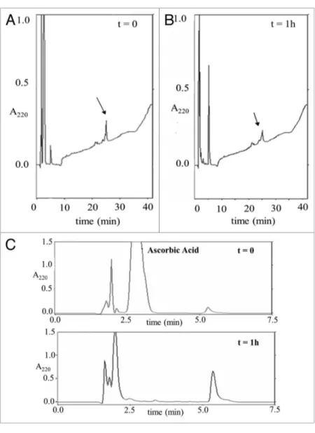
reduction of the intensity of the band corresponding to the WW2-Itch domain (Fig. 7B) suggesting a possible fragmentation of the WW domain. In agreement to this hypothesis, the RP-HPLC analysis of the products after the reaction showed an increase of hydrophilic peaks concurrently to the reduction of the peak cor-responding to the WW domain (Fig. 8).
Discussion
The studies presented here shed light on the molecular deter-minants of the key WW-ligand interactions. Itch-WW2 shows a canonical structure of the WW domain in solution, with its antiparallel three-stranded-β-sheet. The NMR assignment reported here represents a key step to assess the relevant resi-dues and the conformational changes due to the molecular rec-ognition mechanism of Itch with protein substrates. Titrating with increasing amounts of linear peptide, we detected distinct changes in the HSQC spectrum of WW domain. That is, we report resonance shifts, signal decrease or broadening and new signals, appearance, denoting a mixture of signal regimes respec-tively fast, intermediate and slow exchanging; all these charac-teristics well fit with a picture of a complex lifetime in the few ms timescale (< 4 ms).30 At atomic level description, the
resi-dues R18, T32, R33, T34, T35, T36, W37 are in slow exchange regime in the WW2-pep63 complexes, and the residues pres-ent in the third β strand seem to be predominantly involved in the binding of the peptide. Notable changes are induced by the interaction with the same peptide after cyclization, ten sig-nals of Itch-WW2 domain change their exchange regime from fast to slow. The third strand signals exchange regime is invari-ant, while both the second and the first strands are involved in the change of regime from fast or intermediate to slow. These
stable ATCUN-Cu2+-peptide complex in agreement with previous
studies.37,38 Previously, specific Ni/Cu-ATCUN complexes have
been shown to inhibit enzyme activity by inducing modification of the active site residues by reactive oxygen species cross-linking or cleavage.39,40 In fact, several amino acid residues are susceptible to
metal catalyzed oxidation.39,41 We have investigated the effects of
both metal-peptide complexes on the Itch-WW2 domain. Figure
7 shows the SDS-PAGE of Itch-WW2 domain after incubation
in the presence of either pep63 or pep63OX following the addition of ascorbic acid. A 25 kDa molecular weight band is detectable (Fig. 7A) after 30 min of incubation of Itch-WW2 in the presence
of Cu2+-pep63 at 37°C, presumably for the formation of
cross-links catalyzed by the formation of radical oxygen species during the reaction. Furthermore, the densitometry of the protein bands shows a 50% reduction of the WW2 band. By contrast,
follow-ing 30 min incubation of Itch-WW2 in presence of CuCl2 and
ascorbic acid, the higher molecular weight band is not visible; only after 1 h do we observe an initial formation of a new band corresponding to a dimeric form of Itch-WW2 (Fig. 7A), indicat-ing that the damage on the Itch-WW2 due to the metal-peptide
Figure 4. Interaction study between pep63OX and Itch-WW2. Intrinsic
fluorescence changes (λex 280 nm and λem 330 nm) of 5 μM Itch-WW2 at increased concentrations of pep63 (▲) and pep63OX (○) in 10 mM
sodium phosphate, 6.0 pH, buffer, 100 mM NaCl, 0.1 mM eDtA, 5 mM Dtt at 37°C.
Table 1. Amino acid residues of Itch-WW2 involved in the interaction
with pep63 and pep63OX
Residues Exchange Regime pep63 pep63OX
L11 FASt SLOW
W15 FASt FASt
e16 FASt SLOW
R17 - SLOW R18 SLOW SLOW V19 FASt FASt D20 FASt SLOW M22 - SLOW R24 FASt SLOW
I25 FASt FASt
Y26 FASt SLOW
Y27 FASt SLOW
V28 SLOW SLOW D29 SLOW SLOW H30 FASt SLOW F31 FASt FASt t32 SLOW SLOW R33 SLOW SLOW t34 SLOW SLOW t35 SLOW SLOW t36 SLOW SLOW W37 SLOW SLOW Q38 FASt SLOW R39 FASt FASt t41 FASt SLOW V45 FASt FASt
©2012
Landes
Bioscience.
Do
not
distribute.
Nδ1 atom of H30 in the Itch-WW2 domain might be improved by the cyclization of the pep63 stabilizing the complex. Thus, the cyclization may also be advantageous for allowing the PPxY peptides to attain a close molecular fit within the hydrophobic grooves of the WW domains. A less flexible conformation of the PPxY peptide, achievable by cyclization, could also result in a lower entropic penalty upon binding to the WW domain. Hence, the overall free energy of WW-pep63 interaction could be favored by a reduction in the entropic penalty.
The above data are in agreement with recent thermody-namic studies that suggested strongly that non-consensus resi-dues within and flanking the PPxY peptides are important for stabilizing the peptide conformation, and that peptides with distinct conformations are optimally suited for binding to WW
domains.46 The balance among rigidity and flexibility of both
WW domains and protein targets might also be an in vivo critical factor in this molecular recognition mechanism. Accordingly, the WW-ligand interactions are usually studied in vitro using short, proline-rich peptides, and the relatively low affinities observed (in 10 to 100 μM) are generally characteristic of several similar data strongly suggest that the interaction of the domain with
the cyclic form of the peptide results in a more stable complex with a longer lifetime than that resulting from the interaction with the linear form of pep63. This observation, from the sig-nals point of view, is strongly backed up further by the 4-fold decrease in Kd of the interaction as measured in the fluorescence studies. Thus, the data presented here show unequivocally that pep63 is able to form a more stable complex when in cyclic form. It is notable that the residues Y26, V28, H30 and W37 seem to form a hydrophobic groove within Itch-WW2 domain. These residues are highly conserved across the family of WW
domains that recognize PPxY ligands,42-46 such as in the four
WW domains of Itch, except for the substitution of the W with Y residue in the WW4 domain. In binding with cyclic form of pep63, the exchanging regime of two of these residues, Y26 and H30, change from fast to slow, indicating the formation of a less shaky complex induced by a more stable conformation of the
peptide. Similarly, as described for Yap2-WBP2 complex,46 in
the WW2-pep63OX complex, the formation of a hydrogen bond
between Hη phenolic hydrogen of Y within the PPxY motif and
Figure 5. NMR studies of the Itch-WW2-pep63 interaction. (A) titration of 0.6 mM 15N-labeled-Itch-WW2 domain in 10 mM potassium phosphate
buffer, 50 mM KCl, 5% D2O, pH 6.0, with unlabeled pep63OX at molar ratios pep63OX /WW ranging from 0 to 35, monitored by HSQC, at 285K; (B) 15N-1
H-HSQC spectra of 0.25 mM 15N-labeled-Itch-WW2; the titration was performed by sequential addition of pep63 (0–7.0 mM), seven different spectra were
collected using pep63/Itch-WW2 molar ratios ranging from 0 to 35. Selected regions of the (C) Itch-WW2/pep63OX (D) Itch-WW2/pep63 15N-1H-HSQC
spectra, the lines underline some of the major differences observable.
©2012
Landes
Bioscience.
Do
not
distribute.
suggest the possibility of using pep63, and in the particular the cyclic form, as a model for the design of metal-drug inhibitor of Itch-recognition mechanism. Our future efforts will focus on unraveling the effects of the metal-pep63OX complex on the ubiq-uitilation of p63 and p73 in cell.
In conclusion, the above data provide a suggestion for a novel strategy for studying the WW-p63 interaction and, more gener-ally, the key WW-ligand mechanism, with a potential use of the cyclic PPxY-containing peptide as a model for the inhibition of the recognition mechanism of HECT-E3-ligase.
Materials and Methods
Peptide synthesis. Synthetic peptides were purchased from
Spectra 2000. Analysis of the synthetic peptides by reverse phase high-performance chromatography (RP-HPLC) and mass spec-trometry revealed a purity > 98%. The two sequences of the
peptides had the following sequences: 18-mer pep63 NH2-TSH
CTP PPP YPT DCS IVS-CONH2 (1901.15 m/z).
Circular dichroism. CD measurements were performed
using a Jasco 710 spectropolarimeter (Jasco) equipped with a thermal controller calibrated with camphor-sulfonic acid. Far-UV CD experiments were performed to explore the con-formation of the peptides and of Itch-WW domain. CD spectra interactions.25,46-50 The low affinity in many
cases limited the possibility to obtain a high-resolution structure of the complex by NMR spectroscopy. Our results show the possibility that an increase of the stability of the complex in the solution may induce a major conforma-tion stability of the fragment, probably improv-ing the mimickimprov-ing of the local structure of the protein by using a cyclic form of the PPxY peptide. Such optimized cyclic forms of PPxY peptides could represent a good model to study the determinants of the molecular recogni-tion mechanism WW ligand. This cyclizarecogni-tion strategy could also be used to optimize pro-tein microarrays for analyzing the molecular determinants relevant in this key interaction. Furthermore, the S-S cyclization of pep63 leads also to the proteases’ resistance. This fea-ture can likely be attributed to a reduction in backbone flexibility of the peptide. Generally, in fact, potential cleavage sites within a pro-tein are more susceptible to attack by proteases if they are located in flexible regions of the backbone.51
The cysteine residues in the pep63 peptide are present in native p63 protein. Although cyclization of the pep63 backbone provides an excellent protection from proteolytic attacks while increasing the interaction with the WW domain, this strategy cannot be directly applied to other WW-ligand interactions. Indeed, for each individual case, it is necessary to select a
specific size of the linker in order to span the appropriate distance between the N and C termini of the ligand.
This optimization the WW-ligand interaction opens the way to the identification of novel potentially selective inhibitors of this molecular interaction. Protein-protein interactions play a key role in every biological process and represent a promising new class of biological drug targets.52,53 The modulation of the
E3 ligase-protein (namely Itch-p63) interactions could allow the development of innovative molecules able to modulate cel-lular functions, with potential therapeutic effects. In our study, the possibility to use the peptides to inhibit the p63-Itch rec-ognition mechanism has been investigated. To this purpose, we evaluated the metal-binding ability of pep63, which presents a histidine residue in the third position; thus, at the N terminus of this peptide is present an N-terminal Cu2+-and Ni2+-binding
motif.37,38 The ATCUN motif has a well-defined structure of
coordinated ligands around the metal center and is an optimal center for the design synthetic metal drugs. ATCUN metal peptides complexes have been studied for their ability to induce localized oxidative damage to nucleic acids or proteins, gener-ating not diffusible radical species,39,41,54 and, for this property,
their potential application in biotechnological and pharmaceuti-cal field has been proposed.55-57 The biological effects that we
describe here for both forms of the pep63 on the WW domain
Figure 6. Metal binding of the both forms of the pep63. UV-vis spectra of 0.5 mM pep63 (A)
in the presence of equimolar concentration of CuCl2 at different pHs; (B) UV-vis spectra of 0.5
mM pep63 (____) and pep63
OX (- - -) in the presence of 0.5 mM CuCl2 at pH 5.5, showing the
characteristic maximum of the Cu2+ bound at 525 nm.
©2012
Landes
Bioscience.
Do
not
distribute.
of peptide provides a titration curve. The ΔF was plotted vs. molar equivalent of peptide, and the following equation was used for gen-erating a Scatchard plot from which binding constants were determined Δf = nK[peptide] ΔFcomplex/ (1+K[peptide]) ΔF was the observed fluorescence changing after addition of peptide. Fluorescence was measured at the chemical equilibrium at the earliest 2 min after addition of the peptide. The results were plotted using GRAPHPAD PRISM v. 4.0 for Windows (GraphPad Software; www.graphpad.com).
Expression of the uniformly isotope labeled Itch-WW2 domain. The
GST-Itch-WW2 was overexpressed using E. coli BL21 strain in LB medium containing 100 μg/ml ampicillin. Cells were grown at 37°C, and the induction of the expression of the protein was performed by addition of 1 mM IPTG. Cells were grown at 37°C for a further 4 h, collected by centrifugation and after dis-rupted by sonication. The GST-Itch-WW2 was purified using a GST-Trap FF column (5 ml, GE-Amersham) equilibrated with 100 mM Tris-HCl, pH 7.5, 0.3 M NaCl buf-fer at 1.0 ml/min and was eluted using 50 mM Tris-HCl, pH 8.0, 10 mM GSH buf-fer. The Itch-WW2 domain was cleaved from the GST using the PreScission Protease
(GE-Amersham). The uniformly isotopically labeled 15N-13
C-GST-Itch-WW2 for NMR spectroscopy was generated in BL21 E. coli strain using the protocol for isotope labeling described by Marley et al.,58 the culture was grown at 37°C in the presence
of (15N)-NH
4Cl and (13C)-glucose as the sole nitrogen and
car-bon sources, respectively. The induction of the expression of the
protein was performed by addition of 1mM IPTG. The U-15
N-13C-GST-Itch-WW2 was purified using GST-Trap FF column
(5 ml, GE-Amersham) equilibrated with 100 mM Tris-HCl, pH 7.5, 0.3 M NaCl buffer at 1.0 ml/min and was eluted using 50 mM Tris-HCl, pH 8.0, 0.3 M NaCl and 10 mM GSH buf-fer. The Itch-WW2 domain was cleaved from the GST using the PreScission Protease (GE-Amersham). The expression-puri-fication procedure resulted in a 16 mg/L yield. The purified protein was concentrated and dialyzed with 10 mM phosphate
buffer, pH 6.0, 50 mM KCl. The incorporation rate for 15N and
13C were estimated by NMR to be ~90%.
NMR spectroscopy. 15N-13C-Itch-WW2 was prepared for
NMR experiments at 600 μM in 10 mM pH 6.0, 50 mM
KCl and 95/5% H2O/D2O using 250 μl of sample in Shigemi
microtubes (Shigemi Inc.). All the NMR experiments were performed using a three channel 700 MHz Bruker AVANCE instrument equipped with a triple resonance probe. For the
assignment of backbone 1H, 13C and 15N resonance, HNCA,
HN(CO)CA, CACBNH, CBCA(CO)NH, HBHANH,
HBHA(CO)NH, HNCO, HN(CA)CO,59 HCACO60 and
3D 15N-edited TOCSY-HSQC and NOESY-HSQC spectra
Figure 7. Oxidative effects on Itch-WW2. SDS-pAGe at 20% gel acrylamide of 30 μM WW2
in the presence of 0.3 mM CuCl2 or Cu-pep63 complex (1:1 Cu2+/peptide c/c) in the linear (A)
and cyclic form (B) after incubation of 0, 30 and 60 min at 37°C in 10 mM tris-HCl, pH 7.0. the reactions was started by addition of 3 mM ascorbic acid and the densitometries of the Itch-WW2 bands (c bands) are shown.
were obtained between 200 and 250 nm using a path-length of 0.1 cm, 1.0 sec time constant, 2 nm bandwidth, 2 nm/min scan rate and at 20 or 50 mdeg of sensitivity. Each spectrum was averaged over four scans and subjected to smoothing following subtraction of the buffer background. The measured ellipticity data were converted to mean molar ellipticity per residue [(θ), deg x cm2 × dmol-1].
Cyclization of the pep63. The cyclic form of pep63 was
obtained by disulfide bond formation incubating the peptide in 50 mM Tris-HCl, pH 8.0, buffer in presence of oxygen for 48 h at 25°C. The oxidation reaction was monitored by estimation of free thiol groups according to the method described by Ellman et al.35,36 Briefly, the sample was added to the working solution:
100 μM of Ellman’s reagent 5,5'-dithiobis-2-nitrobenzoic acid (DNTB), which was previously solubilized in 50 mM sodium acetate solution, in 1M Tris-HCl, pH 8.0, buffer. The reaction between DTNB and thiols groups gave the mixed disulfide and 2-nitro-5-thiobenzoic acid (TNB), which was quantified by monitoring of the absorbance of TNB anion at 412 nm. The oxidized form was then analyzed by LC/ESI-MS spectroscopy (C4T).
Fluorescence ivnteraction studies. The interaction of
Itch-WW2 with pep63 peptides was monitored by quenching of
the emission band of Itch-WW2 excited at 280 nm using an λex
and λem slit of 5 nm in 10 mM phosphate buffer, pH 6.0, buffer, 100 mM NaCl at 37°C. Binding constants were determined by fitting the titration curve of fluorescence changes vs. equivalents
©2012
Landes
Bioscience.
Do
not
distribute.
were recorded. For side chain resonance
assign-ment, 2D 1H-13C HSQC, 3D 13
C-edited-NOESY-HSQC, HCCH-TOCSY, CC(CO)NNH and HCC(CO)NNH spectra were recorded. All NMR spectra were recorded at 298 K, processed using
NMRPipe61 and analyzed using the program
NMRViewJ (OneMoon Scientific Inc.).62
Spectroscopic characterization of the itch-WW2-pep63 binding. The NMR experiments were
performed using 250 μl of sample in Shigemi micro-tubes. Itch-WW2 domain as well as peptide pep63 were dissolved in 10 mM phosphate buffer, 50 mM KCl, 1 mM tris(2-carboxyethyl)-phosphine (TCEP), pH 6.0. Titrations of the Itch-WW2 domain were conducted twice at 12°C and followed using two dif-ferent techniques. In the first, a sample of Itch-WW2 at 250 μM was titrated by addition of small aliquots of a 5 mM pep63 or pep63OX solution dissolved in the
same buffer. Itch-WW2 uniformly 15N-labeled was
used. The titrations were repeated following all the
WW2 domain amide region with 2D 1H-15N HSQC
experiments using a pulse sequence with water flip back selective pulses to avoid water saturation. For these experiments small aliquots of the peptide were added stepwise using a 50 mM stock solution of the pep63 or pep63OX dissolved in the same buffer. All the NMR experiments were performed using a 600 MHz Bruker AVANCE instrument equipped with a z-gradient triple resonance proton cry-probe. The NMR spectra were processed and analyzed with an Octane Silicon Graphics workstation operating
under Irix 6.5, using NMRPipe61 and NMRView J
6.2.2 (One Moon Scientific Inc., 2009).
Proteolysis resistance Chymotripsin diges-tion was performed using a 1:100 E:S (w/w) in 50 mM Tris-HCl, pH 8.04, the reaction mixture was incubated at 37°C for the indicated times and then stopped by addition of 0.1% TFA solu-tion, vortex and freezing. Before the RP-HPLC analysis, 10 mM DTT was added at the samples in
order to have a reduction of the disulfide bond of the pep63OX
and, potentially, the separation of the fragments. The samples were analyzed using RP-HPLC system chromatography (mod. LC-10AVP Shimadzu equipment) with an solvent B gradient (0–5 min, 0%; 5–25 min, 15%; 25–27 min, 15% and 27–30 min 90%), using 0.1% TFA as solvent A and 80% CH3CN, 0.1% TFA as solvent B and a Brouwnlee C-18 column (OD-300, 250x4.6mm, 7 μm). The eluted was monitored at 220 nm by UV detector (Shimadzu). The same conditions of analysis was also used to analyze the WW domain after oxidative
dam-age using the metal-pep63OX complex.
Spectrophotometric metal binding characterization. The
peptides were dissolved in water to a concentration of 0.5 mM CuCl2 was added in a 1:1 molar ratio with the peptides. The pH
Figure 8. Oxidative cleavage of Itch-WW2. Rp-HpLC of Itch-WW2 (30 μM) after
incu-bation in presence of Cu2+-pep63
OX complex (0.3 mM) and of 3 mM ascorbic acid (A)
after 0 and (B) 60 min of incubation at 37°C. (C) Selected region of the chromatograms (from 0 to 7.5 min.) showing the presence of fragments. the arrows indicate the peak corresponding to Itch-WW2.
of the solution was decreased or raised using 0.1 M HCl or 0.2 M NaOH respectively, and the solutions were equilibrated at each pH before the spectrophotometric measurements. The visible spectra of the peptide-metal complexes were acquired on Perkin-Elmer Lambda-Bio 20 double-beam spectrophotometer using a 1 cm path length quartz cells at 25°C.
Disclosure of Potential Conflicts of Interest No potential conflicts of interest were disclosed.
Acknowledgments
The authors are grateful to F. Bertocchi for technical support for the NMR experiments and to Dr. V. Fiordalice for her support in the preparation of some experiments.
©2012
Landes
Bioscience.
Do
not
distribute.
31. Bax A, Ikura M. An efficient 3D NMR technique for correlating the proton and 15N backbone amide
reso-nances with the alpha-carbon of the preceding residue in uniformly 15N/13C enriched proteins. J Biomol
NMR 1991; 1:99-104; PMID:1668719; http://dx.doi. org/10.1007/BF01874573.
32. Bax A, Ikura M, Kay LE, Barbato G, Spera S. Multidimensional triple resonance NMR spectroscopy of isotopically uniformly enriched proteins: a power-ful new strategy for structure determination. Ciba Found Symp 1991; 161:108-19, discussion 119-35; PMID:1814691.
33. Wishart DS, Sykes BD. The 13C chemical-shift index: a
simple method for the identification of protein second-ary structure using 13C chemical-shift data. J Biomol NMR 1994; 4:171-80; PMID:8019132; http://dx.doi. org/10.1007/BF00175245.
34. Tyndall JD, Reid RC, Tyssen DP, Jardine DK, Todd B, Passmore M, et al. Synthesis, stability, antiviral activity, and protease-bound structures of substrate-mimicking constrained macrocyclic inhibitors of HIV-1 protease. J Med Chem 2000; 43:3495-504; PMID:11000004; http://dx.doi.org/10.1021/jm000013n.
35. Ellman GL. Tissue sulfhydryl groups. Arch Biochem Biophys 1959; 82:70-7; PMID:13650640; http:// dx.doi.org/10.1016/0003-9861(59)90090-6. 36. Riddles PW, Blakeley RL, Zerner B. Reassessment of
Ellman’s reagent. Methods Enzymol 1983; 91:49-60; PMID:6855597; http://dx.doi.org/10.1016/S0076-6879(83)91010-8.
37. Sigel H, Martin RB. Coordinating properties of the amide bond: Stability and structure of metal ion com-plexes of peptides and related ligands. Chem Rev 1982; 82:385-426; http://dx.doi.org/10.1021/cr00050a003. 38. Laussac JP, Sarkar B. Characterization of the copper(II)-
and nickel(II)-transport site of human serum albumin. Studies of copper(II) and nickel(II) binding to pep-tide 1-24 of human serum albumin by 13C and 1H NMR spectroscopy. Biochemistry 1984; 23:2832-8; PMID:6547847; http://dx.doi.org/10.1021/ bi00307a046.
39. Stadtman ER. Metal ion-catalyzed oxidation of pro-teins: biochemical mechanism and biological con-sequences. Free Radic Biol Med 1990; 9:315-25; PMID:2283087; http://dx.doi.org/10.1016/0891-5849(90)90006-5.
40. Nepravishta R, Bellomaria A, Polizio F, Paci M, Melino S. Reticulon RTN1-C(CT) peptide: a potential nuclease and inhibitor of histone deacetylase enzymes. Biochemistry 2010; 49:252-8; PMID:20000484; http://dx.doi.org/10.1021/bi9012676.
41. Stadtman ER. Oxidation of free amino acids and amino acid residues in proteins by radiolysis and by metal-catalyzed reactions. Annu Rev Biochem 1993; 62:797-821; PMID:8352601; http://dx.doi. org/10.1146/annurev.bi.62.070193.004053. 42. Macias MJ, Hyvönen M, Baraldi E, Schultz J, Sudol
M, Saraste M, et al. Structure of the WW domain of a kinase-associated protein complexed with a proline-rich peptide. Nature 1996; 382:646-9; PMID:8757138; http://dx.doi.org/10.1038/382646a0.
43. Pires JR, Taha-Nejad F, Toepert F, Ast T, Hoffmüller U, Schneider-Mergener J, et al. Solution structures of the YAP65 WW domain and the variant L30 K in com-plex with the peptides GTPPPPYTVG, N-(n-octyl)-GPPPY and PLPPY and the application of peptide libraries reveal a minimal binding epitope. J Mol Biol 2001; 314:1147-56; PMID:11743730; http://dx.doi. org/10.1006/jmbi.2000.5199.
44. Huang X, Poy F, Zhang R, Joachimiak A, Sudol M, Eck MJ. Structure of a WW domain containing fragment of dystrophin in complex with beta-dystroglycan. Nat Struct Biol 2000; 7:634-8; PMID:10932245; http:// dx.doi.org/10.1038/77923.
45. Kanelis V, Rotin D, Forman-Kay JD. Solution struc-ture of a Nedd4 WW domain-ENaC peptide complex. Nat Struct Biol 2001; 8:407-12; PMID:11323714; http://dx.doi.org/10.1038/87562.
16. Lévy F, Muehlethaler K, Salvi S, Peitrequin AL, Lindholm CK, Cerottini JC, et al. Ubiquitylation of a melanosomal protein by HECT-E3 ligases serves as sorting signal for lysosomal degradation. Mol Biol Cell 2005; 16:1777-87; PMID:15703212; http://dx.doi. org/10.1091/mbc.E04-09-0803.
17. Sundvall M, Korhonen A, Paatero I, Gaudio E, Melino G, Croce CM, et al. Isoform-specific monoubiqui-tination, endocytosis, and degradation of alterna-tively spliced ErbB4 isoforms. Proc Natl Acad Sci USA 2008; 105:4162-7; PMID:18334649; http://dx.doi. org/10.1073/pnas.0708333105.
18. Marchese A, Raiborg C, Santini F, Keen JH, Stenmark H, Benovic JL. The E3 ubiquitin ligase AIP4 medi-ates ubiquitination and sorting of the G protein-coupled receptor CXCR4. Dev Cell 2003; 5:709-22; PMID:14602072; http://dx.doi.org/10.1016/S1534-5807(03)00321-6.
19. Zhang P, Wang C, Gao K, Wang D, Mao J, An J, et al. The ubiquitin ligase itch regulates apoptosis by targeting thioredoxin-interacting protein for ubiquitin-dependent degradation. J Biol Chem 2010; 285:8869-79; PMID:20068034; http://dx.doi.org/10.1074/jbc. M109.063321.
20. Ishihara T, Tsuda H, Hotta A, Kozaki K, Yoshida A, Noh JY, et al. ITCH is a putative target for a novel 20q11.22 amplification detected in anaplastic thyroid carcinoma cells by array-based comparative genomic hybridization. Cancer Sci 2008; 99:1940-9; PMID:19016753.
21. Ho KC, Zhou Z, She YM, Chun A, Cyr TD, Yang X. Itch E3 ubiquitin ligase regulates large tumor suppres-sor 1 stability [corrected]. Proc Natl Acad Sci U S A 2011; 108:4870-5; PMID:21383157; http://dx.doi. org/10.1073/pnas.1101273108.
22. Melino G. p63 is a suppressor of tumorigenesis and metastasis interacting with mutant p53. Cell Death Differ 2011; 18:1487-99; PMID:21760596; http:// dx.doi.org/10.1038/cdd.2011.81.
23. Levine AJ, Tomasini R, McKeon FD, Mak TW, Melino G. The p53 family: guardians of maternal reproduction. Nat Rev Mol Cell Biol 2011; 12:259-65; PMID:21427767; http://dx.doi.org/10.1038/ nrm3086.
24. Perry WL, Hustad CM, Swing DA, O’Sullivan TN, Jenkins NA, Copeland NG. The itchy locus encodes a novel ubiquitin protein ligase that is disrupted in a18H mice. Nat Genet 1998; 18:143-6; PMID:9462742; http://dx.doi.org/10.1038/ng0298-143.
25. Macias MJ, Wiesner S, Sudol M. WW and SH3 domains, two different scaffolds to recognize pro-line-rich ligands. FEBS Lett 2002; 513:30-7; PMID:11911877; http://dx.doi.org/10.1016/S0014-5793(01)03290-2.
26. Zarrinpar A, Lim WA. Converging on proline: the mechanism of WW domain peptide recognition. Nat Struct Biol 2000; 7:611-3; PMID:10932238; http:// dx.doi.org/10.1038/77891.
27. Sudol M, Hunter T. NeW wrinkles for an old domain. Cell 2000; 103:1001-4; PMID:11163176; http:// dx.doi.org/10.1016/S0092-8674(00)00203-8. 28. Rossi M, De Laurenzi V, Munarriz E, Green DR, Liu
YC, Vousden KH, et al. The ubiquitin-protein ligase Itch regulates p73 stability. EMBO J 2005; 24:836-48; PMID:15678106; http://dx.doi.org/10.1038/ sj.emboj.7600444.
29. Rossi M, De Simone M, Pollice A, Santoro R, La Mantia G, Guerrini L, et al. Itch/AIP4 associates with and promotes p63 protein degradation. Cell Cycle 2006; 5:1816-22; PMID:16861923; http://dx.doi. org/10.4161/cc.5.16.2861.
30. Bellomaria A, Barbato G, Melino G, Paci M, Melino S. Recognition of p63 by the E3 ligase ITCH: Effect of an ectodermal dysplasia mutant. Cell Cycle 2010; 9:3730-9; PMID:20855944; http://dx.doi.org/10.4161/ cc.9.18.12933.
References
1. Hershko A, Ciechanover A, Varshavsky A. Basic Medical Research Award. The ubiquitin system. Nat Med 2000; 6:1073-81; PMID:11017125; http:// dx.doi.org/10.1038/80384.
2. Ishihara T, Inoue J, Kozaki K, Imoto I, Inazawa J. HECT-type ubiquitin ligase ITCH targets lysosomal-associated protein multispanning transmembrane 5 (LAPTM5) and prevents LAPTM5-mediated cell death. J Biol Chem 2011; 286:44086-94; PMID:22009753; http://dx.doi.org/10.1074/jbc.M111.251694. 3. Schwarz SE, Rosa JL, Scheffner M. Characterization
of human hect domain family members and their interaction with UbcH5 and UbcH7. J Biol Chem 1998; 273:12148-54; PMID:9575161; http://dx.doi. org/10.1074/jbc.273.20.12148.
4. Ingham RJ, Colwill K, Howard C, Dettwiler S, Lim CS, Yu J, et al. WW domains provide a platform for the assembly of multiprotein networks. Mol Cell Biol 2005; 25:7092-106; PMID:16055720; http://dx.doi. org/10.1128/MCB.25.16.7092-7106.2005.
5. Kerscher O, Felberbaum R, Hochstrasser M. Modification of proteins by ubiquitin and ubiquitin-like proteins. Annu Rev Cell Dev Biol 2006; 22:159-80; PMID:16753028; http://dx.doi.org/10.1146/ annurev.cellbio.22.010605.093503.
6. Abriel H, Loffing J, Rebhun JF, Pratt JH, Schild L, Horisberger JD, et al. Defective regulation of the epithelial Na+ channel by Nedd4 in Liddle’s syndrome. J Clin Invest 1999; 103:667-73; PMID:10074483; http://dx.doi.org/10.1172/JCI5713.
7. Zhu H, Kavsak P, Abdollah S, Wrana JL, Thomsen GH. A SMAD ubiquitin ligase targets the BMP path-way and affects embryonic pattern formation. Nature 1999; 400:687-93; PMID:10458166; http://dx.doi. org/10.1038/23293.
8. Kavsak P, Rasmussen RK, Causing CG, Bonni S, Zhu H, Thomsen GH, et al. Smad7 binds to Smurf2 to form an E3 ubiquitin ligase that targets the TGF beta receptor for degradation. Mol Cell 2000; 6:1365-75; PMID:11163210; http://dx.doi.org/10.1016/S1097-2765(00)00134-9.
9. Flores SY, Debonneville C, Staub O. The role of Nedd4/Nedd4-like dependant ubiquitylation in epithe-lial transport processes. Pflugers Arch 2003; 446:334-8; PMID:12698368.
10. Bai Y, Yang C, Hu K, Elly C, Liu YC. Itch E3 ligase-mediated regulation of TGF-beta signaling by modulat-ing smad2 phosphorylation. Mol Cell 2004; 15:825-31; PMID:15350225; http://dx.doi.org/10.1016/j. molcel.2004.07.021.
11. Anindya R, Aygün O, Svejstrup JQ. Damage-induced ubiquitylation of human RNA polymerase II by the ubiquitin ligase Nedd4, but not Cockayne syndrome proteins or BRCA1. Mol Cell 2007; 28:386-97; PMID:17996703; http://dx.doi.org/10.1016/j.mol-cel.2007.10.008.
12. Liu YC. The E3 ubiquitin ligase Itch in T cell activa-tion, differentiaactiva-tion, and tolerance. Semin Immunol 2007; 19:197-205; PMID:17433711; http://dx.doi. org/10.1016/j.smim.2007.02.003.
13. Bernassola F, Karin M, Ciechanover A, Melino G. The HECT family of E3 ubiquitin ligases: multiple players in cancer development. Cancer Cell 2008; 14:10-21; PMID:18598940; http://dx.doi.org/10.1016/j. ccr.2008.06.001.
14. Melino G, Gallagher E, Aqeilan RI, Knight R, Peschiaroli A, Rossi M, et al. Itch: a HECT-type E3 ligase regulating immunity, skin and cancer. Cell Death Differ 2008; 15:1103-12; PMID:18552861; http:// dx.doi.org/10.1038/cdd.2008.60.
15. Matesic LE, Copeland NG, Jenkins NA. Itchy mice: the identification of a new pathway for the develop-ment of autoimmunity. Curr Top Microbiol Immunol 2008; 321:185-200; PMID:18727493; http://dx.doi. org/10.1007/978-3-540-75203-5_9.
©2012
Landes
Bioscience.
Do
not
distribute.
58. Marley J, Lu M, Bracken C. A method for efficient isotopic labeling of recombinant proteins. J Biomol NMR 2001; 20:71-5; PMID:11430757; http://dx.doi. org/10.1023/A:1011254402785.
59. Bazzo R, Cicero DO, Barbato G. A new 3D HCACO pulse sequence with optimized resolution and sen-sitivity. Application to the 21 kDa protein human interleukin-6. J Magn Reson B 1995; 107:189-91; PMID:7599953; http://dx.doi.org/10.1006/ jmrb.1995.1077.
60. Bazzo R, Cicero DO, Barbato G. A new 3D pulse sequence for correlating intraresidue NH, N and CO chemical shifts in C-13 N-15 labeled proteins. Application to the protein human interleukin 6. J Magn Reson B 1996; 110:65-8; http://dx.doi.org/10.1006/ jmrb.1996.0008.
61. Delaglio F, Grzesiek S, Vuister GW, Zhu G, Pfeifer J, Bax A. NMRPipe: a multidimensional spectral processing system based on UNIX pipes. J Biomol NMR 1995; 6:277-93; PMID:8520220; http://dx.doi. org/10.1007/BF00197809.
62. Johnson BA. Using NMRView to visualize and analyze the NMR spectra of macromolecules. Methods Mol Biol 2004; 278:313-52; PMID:15318002.
52. Wells JA, McClendon CL. Reaching for high-hanging fruit in drug discovery at protein-protein interfaces. Nature 2007; 450:1001-9; PMID:18075579; http:// dx.doi.org/10.1038/nature06526.
53. Dömling A. Small molecular weight protein-pro-tein interaction antagonists: an insurmountable challenge? Curr Opin Chem Biol 2008; 12:281-91; PMID:18501203; http://dx.doi.org/10.1016/j. cbpa.2008.04.603.
54. Jin Y, Cowan JA. DNA cleavage by copper-ATCUN complexes. Factors influencing cleavage mechanism and linearization of dsDNA. J Am Chem Soc 2005; 127:8408-15; PMID:15941274; http://dx.doi. org/10.1021/ja0503985.
55. Brittain IJ, Huang X, Long EC. Selective recog-nition and cleavage of RNA loop structures by Ni(II).Xaa-Gly-His metallopeptides. Biochemistry 1998; 37:12113-20; PMID:9724523; http://dx.doi. org/10.1021/bi9806605.
56. Melino S, Gallo M, Trotta E, Mondello F, Paci M, Petruzzelli R. Metal-binding and nuclease activity of an antimicrobial peptide analogue of the salivary histatin 5. Biochemistry 2006; 45:15373-83; PMID:17176059; http://dx.doi.org/10.1021/bi0615137.
57. Hocharoen L, Cowan JA. Metallotherapeutics: novel strategies in drug design. Chemistry 2009; 15:8670-6; PMID:19685535; http://dx.doi.org/10.1002/ chem.200900821.
46. McDonald CB, McIntosh SK, Mikles DC, Bhat V, Deegan BJ, Seldeen KL, et al. Biophysical analy-sis of binding of WW domains of the YAP2 tran-scriptional regulator to PPXY motifs within WBP1 and WBP2 adaptors. Biochemistry 2011; 50:9616-27; PMID:21981024; http://dx.doi.org/10.1021/ bi201286p.
47. Kay BK, Williamson MP, Sudol M. The importance of being proline: the interaction of proline-rich motifs in signaling proteins with their cognate domains. FASEB J 2000; 14:231-41; PMID:10657980.
48. Linn H, Ermekova KS, Rentschler S, Sparks AB, Kay BK, Sudol M. Using molecular repertoires to identify high-affinity peptide ligands of the WW domain of human and mouse YAP. Biol Chem 1997; 378:531-7; PMID:9224934; http://dx.doi.org/10.1515/ bchm.1997.378.6.531.
49. Dalby PA, Hoess RH, DeGrado WF. Evolution of binding affinity in a WW domain probed by phage dis-play. Protein Sci 2000; 9:2366-76; PMID:11206058. 50. Webb C, Upadhyay A, Giuntini F, Eggleston I,
Furutani-Seiki M, Ishima R, et al. Structural fea-tures and ligand binding properties of tandem WW domains from YAP and TAZ, nuclear effectors of the Hippo pathway. Biochemistry 2011; 50:3300-9; PMID:21417403; http://dx.doi.org/10.1021/ bi2001888.
51. Hubbard SJ. The structural aspects of limited pro-teolysis of native proteins. Biochim Biophys Acta 1998; 1382:191-206; PMID:9540791; http://dx.doi. org/10.1016/S0167-4838(97)00175-1.