1
UNIVERSITÀ DI PISA
Facoltà di Farmacia
Corso di Laurea in
Chimica e Tecnologia Farmaceutiche
“Synthesis and biological evaluation of novel
N,N-dialkyl-2-arylindol-3-ylglyoxylamide TSPO ligands„
Relatori:
Dott.ssa Sabrina Taliani
Prof.ssa Claudia Martini
Candidata:
Fanelli Serena
2
3
A
BSTRACT:
The 18kDa Translocator protein (TSPO) was discovered in 1977 and initially named “Peripheral-type benzodiazepine receptor” (PBR). It is located at the point of contact between inner and outer mitochondrial membrane. It is associated with other membrane proteins to form the mitochondrial permeability transition pore (MPTP). The expression of TSPO is ubiquitary in peripheral tissues (steroid producing tissues, liver, heart, kidney, lung, immune system), and in the central nervous system is mainly located in glial cells and in neurons. It is involved in numerous functions including steroidogenesis, the transport and the synthesis of heme, immunomodulation, the regulation of apoptosis and generation of radical oxigen species (ROS). In the 1995 Dalpiaz et al. proposed a pharmacophore model later refined by Da Settimo et al. in 2008. On the basis of these studies a series of N,N-dialkyl-2-phenylindol-3-ylglyoxylamide derivatives was synthesized and tested as TSPO ligands. The SARs of these compounds were rationalized in light of the pharmacophore/topological model of TSPO binding site made up of three lipophilic pockets (L1, L3 and L4) and a H-bond donor group (H1). The aim of this work is the design and the synthesis of a novel series of N,N-dialkyl-2-phenylindol-3-ylglyoxylamide featuring suitable substituentes on the indole nucleus for good pharmacokinetic parameters. The affinity towards TSPO of all the new compounds was tested by binding assays
performed on rat kidney membranes. For those compounds which showed a Ki value in the
sub-nanomolar range, the ability to induce functional effects trough the TSPO was also tested. In particular, since TSPO-related apoptosis and steroidogenesis are events which play key roles in the homeostasis of cellular processes, we carried out experiments on cell growth in metabolic stress conditions. The status of cellular stress was created by nutrient deprivation using low serum medium culturing conditions (1% fetal bovine serum FBS). The effects of some compounds (the most promising ones in term of affinity) were evaluated by proliferation MTS assay and crystal violet staining. The obtained results showed an increase in cell growth after 48 hours of treatment with compounds, in respect to the control. If such preliminary results are confirmed even in a cellular model of primary neurons or in a neurodegenerative cell model, these compounds may be used as versatile scaffold for the design of novel neuroprotective agents.
4
T
ABLE OF CONTENTS
ABSTRACT... 3
INTRODUCTION...6
ROLE OF TSPO IN CELLULAR FUNCTION...12
REGULATION OF STEROIDOGENESIS...
12
REGULATION OF APOPTOSIS...
14
IMMUNOMODULATION...
16
TSPO AND NEUROINFLAMMATION...
17
OTHER FUNCTIONS...
17
TSPO LIGANDS...19
TSPO ENDOGENOUS LIGANDS...
19
TSPO SYINTHETIC LIGANDS...20
INTRODUCTION OF EXPERIMENTAL SECTION...24
N,N-DIALKYL-2-PHENYLINDOL-3-YL LYOXYLAMIDES…….…...……
24
EXPERIMENTAL SECTION...
31
5
CHEMISTRY...
41
BIOCHEMICAL SECTION...46
MATERIALS AND METHODS...
50
DETERMINATION OF PROTEIN CONCENTRATION...
50
DETERMINATION OF THE BINDING OF RADIOLIGAND TO THE TSPO IN PRESENCE OF NEW SYNTHESIS COMPOUNDS...
52
PROLIFERATION STUDIES...53
THAWING PROCEDURE………..……….54
CONDITIONS OF CELL CULTURE………
.54
SPLITTING TECHNIQUE...
55
CELL SEEDING...
56
MTS ASSAY...
57
CRYSTAL VIOLET. ...
58
RESULTS AND DISCUSSION...60
REFERENCES...65
6
I
NTRODUCTION:
The first benzodiazepine, chlordiazepoxide (Librium), was discovered accidentally in 1955 by Leo Sternbach, and made available in 1960 by Hoffmann-La Roche, who also marketed as diazepam (Valium) since 1963.[1] Starting in the sixties and
seventies benzodiazepines have begun to be widely prescribed clinically. The extensive clinical use is based on their activities of powerful anxiolytics, anticonvulsants, sedative-hypnotics and muscle relaxants.[2] In humans exist two
different forms of benzodiazepine receptors: central benzodiazepine receptors (CBRs) and peripheral benzodiazepine receptors (PBRs).
The first type (CBRs) is found in the brain and forms an allosteric site on the GABAA receptor complex. In fact, ligands acting at this allosteric site, such as
diazepam and chlordiazepoxide, enhance the affinity of the γ-aminobutyric acid (GABA) toward the CBR and, in this way, influence chloride (Cl-) influx at the
GABAA receptor pore, causing downstream effects on GABA-mediated
inhibition.[3] Different studies have shown benzodiazepines (BZs) acting on CBRs to
be responsible for different GABAA-induced effects.
The second type (PBRs) is first described in 1977 by Braestrup and Squires[4] as an
alternative binding site in non-neuronal tissue for the diazepam, a centrally acting benzodiazepine. It was named peripheral according to this tissue distribution and benzodiazepine receptor because BZs is the class of ligands by which PBR was discovered. However, multiple other names have been used to refer to this protein, including mitochondrial benzodiazepine receptor (MBR), mitochondrial diazepam
7
binding inhibitor (DBI) receptor complex (mDRC), PK11195 binding sites (PKBs), isoquinoline binding protein (IBP), Omega3 receptor. [5]
Recent data have increasingly supported the renaming of this protein with the aim to represent more accurately its sub-cellular role and putative tissue-specific functions. In 2006, a team of scientists has proposed the new name Translocator Protein (18 kDa), TSPO. [6] In fact the name PBR was widely accepted mainly for
historical reasons in the scientific community, but it didn’t take into account the new findings regarding its structure, subcellular roles and tissue distribution. It has also been shown that many ligands structurally different from BZs bind to this protein, which is not a receptor in the traditional sense, but rather a translocator of molecules. TSPO is an evolutionarily well-conserved and tryptophan rich 169-amino acids protein with five trans-membrane domains which consist of extended α-helices composed of 21 residues, long enough to spam an entire membrane bilayer. These α-helices are linked by hydrophobic loops, with a carboxyterminal and a short amino-terminal tails located outside and inside the mitochondria, respectively. Furthermore, site-directed mutagenesis studies demonstrated that the portion of the receptor that recognized the ligands is located on the first cytoplasmatic loop TSPO forms complexes of four to six molecules whose organization is postulated to form a single pore, reflecting the function of TSPO as a transporter protein in the mitochondrial membrane. [5] The TSPO active shape
8
However, the exact three-dimensional structure of TSPO has not yet been determined, as its close association to the membrane makes the purification and crystallization processes very difficult to accomplish. At a subcellular level, TSPO is mainly located on the mitochondrial outer membrane and particularly concentrated at outer/inner mitochondrial membrane contact sites. In addition, TSPO is also expressed at low levels in other subcellular compartments, such as plasma membranes and nuclear fraction of cells. In 1992 McEnery et al. reported that TSPO is strictly associated in a trimeric complex with VDAC, a voltage-dependent anion channel of 32 kDa located at sites of contact between outer and inner mitochondrial membrane that acting as a channel allowing passage of small molecules and ions into the mitochondria, and ANT, adenine nucleotide translocase, a specific antiporter of 30 kDa, located in the inner mitochondrial membrane, for the exchange of ATP and ADP as part of oxidative
FIGURE (1): Three-dimensional structure of TSPO. (A) View perpendicular to the plane of the membrane of the TSPO
dimer. (B) View parallel to the plane and after rotation of 40 °. (C) View perpendicular to the plane after 90 ° rotation. (D) Monomer TSPO view parallel to the plane from the side of the interface with the dimer. (E) TSPO monomer seen from the lipid bilayer.
9
phosphorylation. Together with other proteins, these three sub-units constitute the mitochondrial permeability transition pore (MPTP).[8]
FIGURE (2): Molecular structure of 18kDa TSPO and localization. It is also shown some proteins as-sociated with TSPO
in the MPTP complex (VDAC, ANT and PRAX-1)[8]
Furthermore, it has been identified four cytosolic TSPO-associated proteins which most of time play an important role in the biological processes where TSPO is involved. p10 is the first cytosolic protein associated to TSPO identified, but whose bio-chemical role has not been understood yet. It is a protein of 10 kDa that coimmunoprecipited with 18 kDa TSPO using the isoquinoline carboxamide radioactive probe PK14105, a ligand selective for mitochondrial benzodiazepine receptors, to photolabel rat mitochondrial preparations.[9]
PBR associated protein-1 (PRAX-1) is isolated, cloning and characterized by Galiègue et al. in 1999.[10] It is a protein of 1857 amino acids with a molecular mass
of 240 kDa, discovered using the yeast two-hybrid screening strategy. Exhibiting various domains involved in protein-protein interaction, such as proline-rich domains, leucine-zipper motifs and Src homology region 3-like (SH3-like)
do-10
domains, it has been suggested that PRAX-1 acts as an adaptor protein to recruit additional targets to the vicinity of TSPO so as to modulate its function. In addition, it was assumed that a single PRAX-1 protein interacts with the C-terminal end (14 amino acids) of several molecules of TSPO (at least two of them).
FIGURE (3): Chromosomal localization of the human PRAX-1 gene. A, FISH mapping of the human PRAX gene. The
left panel shows the FISH signals on chromosome. The arrow indicates the specific site of hybridization to chromosome 17. The right panel shows the same mitotic figure stained with DAPI to identify chromosome 17. B, idiogram of the human chromosome 17 illustrating the distribution of labeled sites for the human PRAX-1 probe. Each dot represents the double FISH signals detected on human chromosome17.
Another important protein functional associated to TSPO is Steroidogenic Acute Regulatory Protein (StAR), that mediates the flow of cholesterol from the outer to the inner mitochondrial membrane, permitting steroid formation in steroidogenic cells. Further studies indicated that StAR acts at the outer mitochondrial membrane and it is not needed to allow the entry of cholesterol into mitochondria. Therefore, Hauet et al. suggest that TSPO and StAR work in concert to bring cholesterol into mitochondria and in particular TSPO serves as a gatekeeper in cholesterol import into mitochondria and StAR plays the role of the hormone-induced activator.[11, 12]
11
PKA-associated protein 7 (PAP7) is a cytosolic protein with a molecular mass of 52 kDa involved in the hormonal regulation of steroid formation, interacting with both the cytosolic RIα subunit of PKA and TSPO. Particularly, PAP7 targets the PKA
isoenzyme, linked to increased steroid synthesis, which phosphorylating specific protein substrates induces the reorganization of TSPO topography and function.[13]
FIGURE (4): Biological Assembly Image for 3P0L.Human steroidogenic acute regulatory protein.
FIGURE (5): mRNA Expression of PAP7 in Mouse Tissues Examined by in Situ Hybridization with an antisense
12
R
OLE OF TSPO IN CELLULAR
FUNCTION:
REGULATION OF STEROIDOGENESIS.
The location of TSPO on the outer mitochondrial membrane and the extremely high density in steroidogenic endocrine tissues, such as adrenocortical cells and Leydig cells, suggest that TSPO plays an important role in steroidogenesis. Moreover, different publications report that this protein ligands stimulate steroid biosynthesis in adrenal, placental, testicular, ovarian and glial systems.[14, 15]
The biosynthesis of steroids begins with the enzymatic transformation of cholesterol into pregnenolone, which occurs through cholesterol side-chain cleavage by the cytochrome P450scc (CYP11A1) and auxiliary electron transferring proteins, localized on the matrix side of the inner mitochondrial membrane. Pregnenolone then leaves mitochondria to move to the endoplasmic reticulum, where it is transformed in the final steroid products. [6, 7, 11]
The rate-limiting step is the translocation of cholesterol from the cellular stores across the aqueous intermembrane space to the inner mitochondrial membrane and P-450scc. The fundamental role of TSPO in this mitochondrial cholesterol transport and thus in steroids synthesis is supported by numerous data, and particularly knockout and antisense experiments in vitro have demonstrated that down-regulation of TSPO causes a decrease in steroid synthesis.[7, 14, 15]
13
FIGURE (6): Transport of cholestrerol, across the mitochondrial membrane, from the intracellular space within themitochondria, by the TSPO.
The topographic study of TSPO in the mitochondrial membranes have also shown that after treatment of Leydig cells with a steroidogenesis stimulator, such as choriogonadotrophin hormone (hCG), there are various morphological changes, such as a formation of large complexes of 15-25 molecules of TSPO and a rapid reorganization of their localization in the mitochondrial membrane, as well as a rapid increase in TSPO ligand binding.[7, 14]
Moreover, amino acids deletion, site-directed mutagenesis and structural studies have permitted to identified a cholesterol recognition amino acid consensus (CRAC) sequence in the cytosolic carboxy-terminal domain of the TSPO that could be part of the binding site for the uptake and translocation of cholesterol (channel-like interaction) through a channel delimited by five α-helixes of TSPO. However, for cholesterol delivery into mitochondria TSPO-mediated is also required the interaction of TSPO with StAR, a protein acting as hormone-induced activator.[6, 7,
14
14] It has been observed a relationship between steroid levels, TSPO levels and
anxiety, principally due to the fact that neurosteroids are endogenous modulators of the GABAA receptor. In general TSPO levels, determined by radioligand binding to
platelets, decrease in patients with anxiety disorders. Therefore, ligands binding to TSPO located in glial cells, such as N,N-dialchyl-2- pheniylindol-3-ylglyoxylamides synthesized by Da Settimo et al.,[16] provide the cholesterol necessary to restore
neurosteroid synthesis and increase pregnenolone formation, which is then metabolize to form allopregnanolone, a potent allosteric modulator of the GABAA
exerting anxiolytic effects. In this sense TSPO could be considering a promising target for the psychiatric disorders that involve dysfunction in steroid biosynthesis.[11, 17]
It also has been observed that the systemic levels of steroids increase as a result of a lesion, to a pain or a fever, in response to stimulation by certain cytokines secretion of corticotropin releasing factor. The involvement of TSPO in the synthesis of steroids can contribute to this defense mechanism.[20]
REGULATION OF APOPTOSIS
MPTP plays an important role in the modulation of signaling pathways mediating apoptotic and necrotic cell death. The exact composition of the MPTP is not yet established, but it has been recognized various proteins implicated in pore formation and its regulation: an hexokinase, in the cytosol; a trimeric complex constituted by VDAC, ANC and TSPO; a creatine kinase in the intermembrane space and cyclophilin D in the matrix. MPTP allows the transfer of solutes, including ATP/ADP exchange, from the mitochondrial matrix to cytosol, through
15
the VDAC/ANC conduit, and there-fore facilitates the crossing of the highly impermeable mitochondrial inner membrane. This periodic transient increase in permeability by the MPTP allows the pumping of protons from the inner membrane by the electron transport chain and creates the transmembrane electrochemical gradient that derives ATP synthesis.[7, 18]
Several factors cause the opening of the MPTP: high [Ca2+] is the fundamental
trigger but alone is not enough, also low adenine nucleotide concentrations, high phosphate concentrations, oxidative stress and pro-apoptotic proteins. Pore opening leads to the dissipation of transmembrane electrochemical gradient, uncoupling of mitochondria and swelling, resulting in the release of cytochrome c and apoptosis inducing factor (AIF) into the cytosol. Once in the cytosol, the first induces the caspase cascade ending in the destruction of cell nucleus, cytoskeleton and plasma membrane; the second principally leads to nuclear chromatin condensation, DNA fragmentation and then to cell death.[18]
16
The exact events which TSPO modulates apoptosis are still unknown. In 2008 Veenman et al.[34] suggested that interaction between VDAC and TSPO is
fundamental to initiate the mitochondrial apoptosis pathway. The intermediary agent between TSPO and VDAC was supposed to be provided by mitochondrial
ROS (reactive oxygen species) generation under the control of the same TSPO.
ROS lead first to dissociation of cytochrome c from oxidized cardiolipins located at the inner mitochondrial membrane and, subsequently to its release in the cytosol via formation of a pore due to assemblage of VDAC molecules. In 2007 Azarashvili et al.[19] supposed another possible mechanism that provided evidence for TSPO
involvement in MPTP opening, controlling the Ca2+-induced Ca2+ efflux and AIF
release from mitochondria, important stage of initiation of programmed cell death. It has also been observed a modulation by TSPO of interactions between VDAC or ANT and pro-apoptotic or anti-apoptotic proteins (Bcl-2 and Bax). Therefore, it has been designed TSPO ligands with pro-apoptotic effects acting as anticancer agents.[20]
IMMUNOMODULATION
The presence of the TSPO in a large number of cells immunomodulatory such as the microglia, the blood monocytes, lymphocytes, and leucocit, led to think of an involvement of TSPO in the immune response. It has been shown, in special studies in mice, that some TSPO ligands, specifically benzodiazepines, have an immunosuppressive action, inhibiting the capacity of macrophages to produce ROS and inflammatory cytokines such as IL-1 (interleukin-1), TNFα (tumor necrosis
17
elimination of foreign antigens. Moreover, the TSPO is involved in oxidative metabolism by phagocytes, a process necessary to permanently delete the foreign antigens. The immunosuppressive action of certain ligands for the TSPO shown the important role played by this protein in the inflammatory response. [7]
TSPO AND NEUROINFLAMMATION
The neuroinflammation are characteristic of many pathological conditions including neurodegenerative diseases such as Alzheimer, Parkinson's disease, metabolic and hepatic encephalopathy and repair processes following damage to cerebral and peripheral level.[21]
Specific ligands with nanomolar affinity for the TSPO, such as PK 11195 and Ro5-4864, were used to determine the parameters of the interaction ligand-receptor in brain tissue. The TSPO has been identified in the ependymal cells in the olfactory bulb and the choroid plexus later in glial cells, including microglia and astrocytes. Using the radioligand [3H]-PK 11195 in animal models has been found an increase
of the concentration of TSPO in numerous neurological diseases including multiple sclerosis, brain trauma, encephalitis and stroke. In most of these studies it was found an up-regulation of TSPO where the microglia is activated. Thanks to recent studies is possible obtain images of activated microglia through the use of PET( positron emission tomography) with ligands for the TSPO labeled with 11C or 18F.
OTHER FUNCTIONS
Numerous studies on the functions of TSPO shown the modulation of its expression in physiological and pathological states, including the cellular response
18
in viral infections. One of the strategies adopted by viruses to bypass the protective mechanisms of cells against infection is to block apoptosis. Several pathogenic viruses using just the TSPO as a target to implement this block. This discovery therefore offers numerous prospects for new antiviral strategies.[20]
Other functions included protein import, important for membrane biogenesis and confirmed by the observation that TSPO is necessary for the import of StAR protein into mitochondria; TSPO, binding of dicarboxylic porphyrin and transport into mitochondria, have been reported as well as a relationship between TSPO and heme biosynthesis pathway; ion transport and calcium homeostasis, since it has been shown that TSPO regulates the Ca2+ flow into the cell. Furthermore, TSPO
plays an important role in cellular respiration and mitochondrial oxidation, and affects cellular proliferation and differentiation in a number of cell types.[6]
The activity of TSPO is also implicated in stress. Its activation during acute exposure to stressors can be seen as a predisposition to metabolic and neuronal better adaptation to stress.[20]
High expression of this receptor has also been observed in neoplastic cells and tissues of the ovary, and adenocarcinom in the liver and colon cancer. This increase is correlated with the degree aggressiveness of tumor. The monitoring of the expression of this receptor, therefore, may be relevant in a clinical procedure for diagnosing and/or to follow the progression of the disease.
19
T
SPO LIGANDS:
TSPO ENDOGENOUS LIGANDS
A wide variety of endogenous molecules with affinity for the TSPO and different chemical structures have been identified. One of the first molecules identified, isolated both at central and pheripheral tissues (adrenal gland, kidney and testes), is a residue of neuropeptides (11 kDa) composed of 86 aa, which inhibits the binding of diazepam with the receptor site of the BZs called "inhibitor of the binding of diazepam (DBI)". In addition to this molecule and its metabolites have been isolated other endogenous ligands: Protoporphyrins (protoporphyrins IX,
mesoporphyrins IX, deuteroporphyrins IX, hemin) exhibit a very high affinity for
TSPO. As several steroidogenic tissues, such as the adrenal gland and testis, show high TSPO and porphyrin levels, it has been suggested a physiological role for the interaction of these two molecules. Furthermore, having a plane of symmetry, these molecules could bind dimerized form of TSPO, confirming in this way the postulated two-binding site model.[7, 15]
20
Another endogenous ligand is cholesterol, as previously reported discussing the fundamental role of TSPO in the regulation of cholesterol transport and thus in the steroidogenesis. Again it has been shown that a dimeric form of TSPO possesses an enhanced binding capacity to cholesterol. [15]
Finally, anthralin, isolated in the stomach of rat, a 16 kDa protein that has been demonstrated interact with both TSPO and dihydropyridine binding site, and phospholipase A2 have also been proposed as endogenous ligands for TSPO. [15]
TSPO SYNTHETIC LIGANDS
Synthetic TSPO selective ligands have been developed with the aim to deepen the knowledge of the exact pharmacological role of TSPO, to define its involvement in several patho-physiological conditions and to establish the structural requirements needed for an optimum of affinity. Initially, the most of these ligands have been designed starting from classical selective CBR ligands, such as benzodiazepines, making the necessary structural modifies in order to shift the affinity toward TSPO. Until to date, the prototypic ligands, used as reference compounds in the development of TSPO pharmacophore models and in SAR studies, are the benzodiazepine Ro 5-4864 (3) and the isoquinolinecarboxamide PK11195 (1), classified by LeFur and coworkers the first as a TSPO agonist or partial agonist and the second as an antagonist.[7]
The ligands for the TSPO not have a fixed chemical structure, but are very different among them. We can distinguish nine classes of compounds:
In the class of benzodiazepines the selectivity toward the TSPO or CBR results very sensitive even to slight structural modifications: in fact, in the
21
case of Ro 5-4864 (3) the insertion of a chlorine at the para position of the pendant phenyl rings of the diazepam, equipotent at the two receptors, shifts the selectivity toward TSPO. Ro 5-4864 has been used in a number of studies aimed at characterizing TSPO, and particularly by Gavioli and colleagues to study the putative role of TSPO as a target for the treatment of psychiatric disorders.[5]
Benzothiazepines, a class of TSPO ligands featuring a 6,7 bicyclic nucleus,
were initially developed by Campiani and coworkers as selective ligands for CBR and GABA receptor subtypes. Some pyrrolobenzothiazepines derivatives possess an unexpected significant inhibitory activity at L-type calcium channels, equal to or higher than those of reference calcium antagonists such as verapamil and (+)-cis-diltiazem. [5]
On the basis of pyrrolobenzothiazepine skeleton, the pyrrolobenzoxazepine scaffold has been developed, featuring the replacement of the endocyclic sulfur (S5) with an oxygen (O5), that increases affinity by 2-3 fold. Some of
these compounds showed an high affinity toward the TSPO (Ki values in the low nanomolar-subnanomolar range) and the capacity to stimulate steroidogenesis in mouse Y-1 adrenocortical cell line. [5]
Isoquinolinecarboxamide PK 11195 (1) is the first non-benzothiazepine ligand
binding the TSPO with nanomolar affinity and it is widely used for studies aimed to define and map the binding site. In 2008 Chelli et al.[22] showed
that treatment of a human astrocytoma cell line (ADF) with PK 11195 actives an autophagic pathway followed by apoptosis mediated by mitochondrial potential dissipation. Moreover, has been showed that PK
22
11195 has a multidrug resistance modulating activity increasing the efficacy of a daunorubicin treatment on human multidrug-resistant leukemia cell line in vitro by 5-7 fold and blocking p-glycoprotein efflux, a transporter whose activity contributes to limit antitumor drug efficacy.[20]
Alpidem (4) can be considered the progenitor of the class of imidazopyridines
known to bind both TSPO and CBR with the nanomolar affinity (Ki 0.5-7 nM and 1-28 nM, respectively). [5]
Phenoxyphenylacetamide derivatives, such as DAA1097 (9), were designed by
a process of molecular simplification involving the opening of the diazepine ring of Ro 5-4864. Some compounds of this class of TSPO ligands have also showed potent anxiolytic properties in laboratory animals. [5]
Pyrazolopyrimidineacetamides, i.e. DPA714 (10), were first described by
Selleri and coworkers as bioisosters of the imidazopyridines and thereby closely related to alpidem.
Indoleacetamide derivatives, collectively named FGIN-1 (9), was developed
by Kozikowski and colleagues as a new class of compounds binding with high affinity and selectivity for TSPO. [5]
In a recent study SSR180575 (11), an indoleacetamide compound, was shown to have neuroprotective properties in different models of progressive degeneration of the PNS and CNS: precisely it promotes neuronal survival and repair following axotomy through the regulation of apoptosis of glial cells and/or the production of mediators such as neurosteroids, cytokines or other neurotrophic factors that support nerve survival.[20]
23
Indol-3-ylglyoxylamides.
DAA 1097 (9) DPA 714 (10) SSR 180575 (11)
24
I
NTRODUCTION TO EXPERIMENTAL
SECTION:
N,N-DIALKYL-2-PHENYLINDOL-3-YLLYOXYLAMIDES
TSPO expression is up-regulated in several human pathologies, including gliomas and neurodegenerative disorders (Huntington’s and Alzheimer’s diseases) as well as in various forms of brain injury and inflammation. Under neuroinflammatory conditions, TSPO markedly increases in activated microglia.[26] Changes in TSPO
level have been found in patients affected by generalized anxiety, panic, post-traumatic stress, obsessive-compulsive disorders, and separation anxiety. Consequently, TSPO has been suggested as a promising target for a number of therapeutic applications[20] and also as a diagnostic marker for related disease
progression. Therefore, many groups have been searching for TSPO ligands with improved performance for quantifying TSPO expression.
Subsequently, a number of diversified structures have been developed obtaining in some cases good results in term of affinity and selectivity, and leading to drawing various TSPO pharmacophore models useful to design novel synthetic derivatives. Neverthless, the topology of the TSPO binding cleft has not yet been completely defined. In 2004 Primofiore et al.[24] prepared and tested a series of
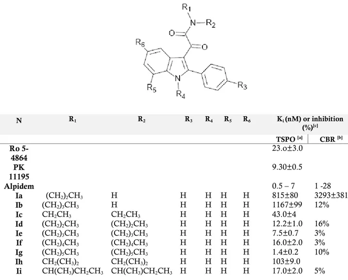
N,N-dialkyl-2-phenylindol-3-ylglyoxylamide derivatives I (TABLE 1) as TSPO ligands, designed as conformationally constrained analogues of the indoleacetamides of the FGIN-1 (5) series previously described by Kozikowski and coworkers. Most of these
25
compounds showed a high affinity for TSPO in the nanomolar / subnanomolar range and a high selectivity for TSPO over CBR. The TSPO/CBR selectivity was evaluated by binding studies using membranes from rat brain tissues and [3H]flumazenil as radioligand. For some of these TSPO ligands with high affinity,
the ability to stimulate pregnenolone formation from rat C6 gliomas cells was evaluated.
TABLE 1: TSPO binding affinity of N,N-dialkylindolylglyoxylamide derivatives Ia-Iaah. [a]The concentration of tested
compounds that inhibited [ 3H-PK 11195 ] binding to rat kidney mitochondrial membranes ( IC
50 ) by 50% was determinate
with six concentrations of the displacers, each performed in triplicate. Ki values are the mean ± SEM of the three
determinations. [b] the inhibition percent of [ 3H ]-flumazenil specific binding at 10µM of the compound are the mean ±
SEM of five determination Ki values are the mean ± SEM of three determinations. [ c ] Data taken from ref. 23.
N R1 R2 R3 R4 R5 R6 Ki (nM) or inhibition (%)[c] TSPO [a] CBR [b] Ro 5-4864 23.o±3.0 PK 11195 9.30±0.5 Alpidem 0.5 – 7 1 -28 Ia (CH2)2CH3 H H H H H 815±80 3293±381 Ib (CH2)3CH3 H H H H H 1167±99 12% Ic CH2CH3 CH2CH3 H H H H 43.0±4 Id (CH2)2CH3 (CH2)2CH3 H H H H 12.2±1.0 16% Ie (CH2)3CH3 (CH2)3CH3 H H H H 7.5±0.7 3% If (CH2)4CH3 (CH2)4CH3 H H H H 16.0±2.0 3% Ig (CH2)5CH3 (CH2)5CH3 H H H H 1.4±0.2 10% Ih CH2(CH3)2 CH2(CH3)2 H H H H 103±9.0 Ii CH(CH3)CH2CH3 CH(CH3)CH2CH3 H H H H 17.0±2.0 5%
26
Ij CH2CH3 CH2C6H5 H H H H 11.0±1.0 Ik -(CH2)4- H H H H 2400±125 4% Il -(CH2)5- H H H H 665±30 3% Im -(CH2)6- H H H H 33.0±3.0 3% In (CH2)2CH3 (CH2)2CH3 F H H H 4.28±0.32 4.3% Io (CH2)3CH3 (CH2)3CH3 F H H H 2.40±0.81 0% Ip (CH2)5CH3 (CH2)5CH3 F H H H 0.37±0.13 0% Iq CH2CH3 CH2C6H5 F H H H 1.68±0.12 10% Ir (CH2)2CH3 (CH2)2CH3 Cl H H H 4.65±0.52 14% Is (CH2)3CH3 (CH2)3CH3 Cl H H H 1.00±0.27 0% It (CH2)5CH3 (CH2)5CH3 Cl H H H 0.55±0.19 4.2% Iu CH2CH3 CH2C6H5 Cl H H H 1.30±0.15 7.3% Iv (CH2)2CH3 (CH2)2CH3 NO2 H H H 0.95±0.1 Iw (CH2)3CH3 (CH2)3CH3 NO2 H H H 0.23±0.07 Ix (CH2)5CH3 (CH2)5CH3 NO2 H H H 0.27±0.10 Iy CH2CH3 CH2C6H5 NO2 H H H 0.55±0.02 Iz (CH2)2CH3 (CH2)2CH3 CF3 H H H 1.69±0.2 Iaa (CH2)3CH3 (CH2)3CH3 CF3 H H H 1.16±0.1 Ibb (CH2)5CH3 (CH2)5CH3 CF3 H H H 1.0±0.1 Icc CH2CH3 CH2C6H5 CF3 H H H 1.0±0.1 Idd (CH2)2CH3 (CH2)2CH3 CH3 H H H 5.50±0.98 Iee (CH2)3CH3 (CH2)3CH3 CH3 H H H 3.80±0.91 Iff (CH2)5CH3 (CH2)5CH3 CH3 H H H 1.60±0.13 Igg CH2CH3 CH2C6H5 CH3 H H H 2.64±0.1 Ihh (CH2)2CH3 (CH2)2CH3 H H H F 2.67±0.48 Iii (CH2)3CH3 (CH2)3CH3 H H H F 4.00±0.15 Ijj (CH2)5CH3 (CH2)5CH3 H H H F 0.37±0.12 Ikk CH2CH3 CH2C6H5 H H H F 1.33±0.2 Ill (CH2)2CH3 (CH2)2CH3 H H H Cl 2.80±0.3 10% Imm (CH2)3CH3 (CH2)3CH3 H H H Cl 4.91±0.4 13% Inn (CH2)5CH3 (CH2)5CH3 H H H Cl 58.4±6 3% Ioo CH2CH3 CH2C6H5 H H H Cl 4.6±0.5 Ipp (CH2)2CH3 (CH2)2CH3 H H H NO2 20.2±2.02 0% Iqq (CH2)3CH3 (CH2)3CH3 H H H NO2 21.6±2.15 1% Irr (CH2)5CH3 (CH2)5CH3 H H H NO2 30.3±9.15 0% Iss CH2CH3 CH2C6H5 H H H NO2 18.3±0.15 0% Itt (CH2)2CH3 (CH2)2CH3 H H H OCH3 328±45 8.6% Iuu (CH2)3CH3 (CH2)3CH3 H H H OCH3 65.2±3.4 8.8% Ivv (CH2)5CH3 (CH2)5CH3 H H H OCH3 35.5±8.7 7.3% Iww CH2CH3 CH2C6H5 H H H OCH3 69.5±3.6 5.7% Ixx (CH2)2CH3 (CH2)2CH3 F H H Cl 2.83±0.08 14% Iyy (CH2)3CH3 (CH2)3CH3 F H H Cl 3.05±0.45 17% Izz (CH2)5CH3 (CH2)5CH3 F H H Cl 7.75±1.55 Iaaa CH2CH3 CH2C6H5 F H H Cl 4.01±0.26 9.7% Ibbb (CH2)2CH3 (CH2)2CH3 F H H F 6.73±1.39 Iccc (CH2)3CH3 (CH2)3CH3 F H H F 4.36±0.05 Iddd (CH2)5CH3 (CH2)5CH3 F H H F 0.95±0.1 Ieee CH2CH3 CH2C6H5 F H H F 1.67±0.37 Ifff (CH2)2CH3 (CH2)2CH3 Cl H H Cl 0.62±0.06 5% Iggg (CH2)3CH3 (CH2)3CH3 Cl H H Cl 1.9±0.2 0% Ihhh (CH2)5CH3 (CH2)5CH3 Cl H H Cl 5.8±0.6 3%27
Iiii CH2CH3 CH2C6H5 Cl H H Cl 3.33±0.3 Ijjj (CH2)2CH3 (CH2)2CH3 H H Cl H 14.0±1.5 0% Ikkk (CH2)3CH3 (CH2)3CH3 H H Cl H 3.40±0.3 Illl (CH2)5CH3 (CH2)5CH3 H H Cl H 2.4±0.3 0% Immm CH2CH3 CH2C6H5 H H Cl H 5.0±0.4 0% Innn (CH2)2CH3 (CH2)2CH3 H H CH3 H 25.0±3.0 Iooo (CH2)3CH3 (CH2)3CH3 H H CH3 H 6.0±0.6 Ippp (CH2)5CH3 (CH2)5CH3 H H CH3 H 1.90±0.1 Iqqq CH2CH3 CH2C6H5 H H CH3 H 2.30±0.2 Irrr CH3 CH2CH3 H H H H 940±120 15% Isss CH3 (CH2)3CH3 H H H H 53.3±4.0 Ittt CH3 (CH2)4CH3 H H H H 12.1±1.0 Iuuu CH2CH3 (CH2)3CH3 H H H H 12.6±1.0 Ivvv CH3 CH2CH3 Cl H H Cl 9.54±1.29 15% Iwww CH3 (CH2)3CH3 Cl H H Cl 0.15±0.02 Ixxx CH3 (CH2)4CH3 Cl H H Cl 0.18±0.02 Iyyy CH2CH3 (CH2)3CH3 Cl H H Cl 0.36±0.04 Izzz CH3 CH2C6H5 H H H H 12.0±1.0 Iaab CH3 CH2C6H5 F H H H 1.8±0.1 Iaac CH3 (CH2)3CH3 Cl H H H 11±1.0 Iaad CH3 (CH2)4CH3 Cl H H H 3.4±0.4 Iaae CH2CH3 (CH2)3CH3 Cl H H H 3.6±0.4 Iaaf CH3 (CH2)3CH3 H H H Cl 3.9±0.5 Iaag CH3 (CH2)4CH3 H H H Cl 3.6±0.5 Iaah CH2CH3 (CH2)3CH3 H H H Cl 1.8±0.2In 2008 the same research group[16] refined the TSPO pharmacophore/topological
model through the synthesis and the biological evaluation of novel indole derivatives with the general formula I, bearing different combinations of substituents R1-R6. (TABLE 1)
28
Within this class, SAR findings were rationalized in the light of a pharmacophore/receptor model made up of three lipophilic pockets (L1, L3, and L4) and an H-bond donor group. Specifically, the second carbonyl group of the oxalyl bridge engages an H-bond with the donor site H1; the two lipophilic substituents on the amide nitrogen, R1, and R2 (linear or ramified alkyl, arylalkyl
groups) interact hydrophobically with the L3 or L4 lipophilic pockets; the 2-phenyl
moiety establishes a putative π-stacking interaction within the L1 pocket. The high affinities of these 2-phenylindolglyoxylamide derivatives have recently permitted the development of new fluorescent probes useful for investigating the localization and the expression level of TSPO.[32, 26]
In TABLE 1 the binding affinity of compounds Ia-Iaah [16, 24 ] and of the standard
TSPO ligands Ro5-4864 (2), PK 11195 (5) and Alpidem (6), expressed as Ki values has been reported. It has been observed that, among the unsubstitued derivatives
Ia-Im (R3 = R4 = R5 = R6 = H), N-monosubstitued compounds Ia-Ib show the lowest
affinity probably because they cannot occupy both the L3 and L4 lipophilic pockets.
The N,N-disubstitued indolylglyoxylamides instead exhibit a high affinity and in particular compound Ig, bearing two n-hexyl groups, is the most potent, with a Ki of
1,4 nM. Therefore, increasing the length of the linear N-alkyl groups has been observed an enhancement of affinity, due to the better filling of L3 and L4 lipophilic
pockets.[24] Subsequently, the three derivatives Id, Ie, Ig and Ij has been selected as
leads for further affinity optimization efforts. It has been observed that the insertion of an electron-donating lipophilic group, such as a methyl group (Idd-Igg), in the 4’-position (R3) of the 2-phenyl ring does not produce any gain in affinity respect to
29
substituent such as Cl, F, NO2 and CF3 (In-Icc), it has been instead obtained
compounds with higher affinity, and among these ones, the N,N-di-n-hexyl derivatives Ip (Cl), It (F), Ix (NO2) and Ibb (CF3) have been revealed the most
potent, with Ki values of 0.37 nM, 0.55 nM, 0.27 nM and 1 nM, respectively. These
results suggest that the R3 substituent has to be electron-withdrawing to reinforce
the putative π-stacking interaction with an electronrich aromatic ring within the L1 pocket. The data of affinity of compounds Ihh-Iww bearing a substituent in the 5-position of the indole nucleus (F, Cl, NO2, OCH3) suggest that R6 has to be
electron-withdrawing and also very small for optimal binding, and only Ijj with a fluorine at the 5-position features these properties (Ki = 0.37 nM). The introduction
of two halogens in both 4’- and 5-positions (Ixx-Iiii; Ivvv-Iyyy) does not increase affinity in additive manner, but it has supposed a correlation with the nature of the
N,N-dialkyl chain. Substitutions at the 7-position of the indole nucleus (R5) with an
electron-withdrawing (Cl, Ijjj-Immm) or an electron-donating (CH3, Innn-Iqqq)
lipophilic group do not produce any gain in affinity.
The binding data of asymmetrical N,N-dialkyl derivatives (Irrr-Iaah), designed to probe the L3 and L4 lipophilic pockets at the TSPO binding site, indicate that the L3
and L4 pockets are probably different in their dimensions, and that R1 and R2 have
to be of different size to obtain the best-performing substitution on the amide nitrogen. Therefore, an aromatic moiety on R1/R2 substituents is equivalent to an
aliphatic moiety of similar size in interacting with the two lipophilic pockets.
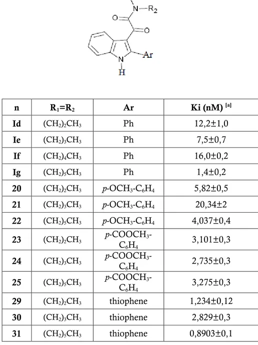
In the light of all these findings, research is currently focused towards the synthesis and biological evaluation of a series of N,N-dialkyl-2-phenylindol-3-ylglyoxylamides (II) bearing a polar group at the 4’-position of the 2-phenyl ring (R3 = NH2, OH,
30
COOH), and of a series of N,N-dialkyl-2-(3-thienyl)indol-3-ylglyoxylamides (III). These compounds have been synthesized as symmetrical amides featuring linear alkyl chains of different length on the amide nitrogen (n-propyl, n-butyl, n-hexyl) to evaluate any changes in the interaction with the lipophilic pockets L3, L4 of the pharmacophore. The TSPO affinity of intermediary compounds featuring a OCH3
and a COOCH3 substituent in 4’-position has also been estimated.
FIGURE(11): Chemical structures of N,N-dialkyl-2-phenylindol-3-ylglyoxylamides (II) and
N,N-dialkyl-2-(3-thienyl)indol-3-ylglyoxylamides (III).
To make these molecules are used as potential drugs or radiopharmaceuticals (as biomarkers of neuroinflammation), they must have a good pharmacokinetic profile. Therefore, the choice to insert a polar group in the scaffold of indolylglyoxylamide derivatives has the purpose to improve the lipophilic/hydrophilic balance, thus avoiding a high level of non-specific binding, a poor signal-to-noise ratios, a high plasma protein binding and thus a relatively poor penetration of the blood-brain barrier, resulting in accumulation of tracer in the brain.
31
E
XPERIMENTAL SECTION:
In this thesis work compounds 29, 30, 31 (2-(3-thienyl)), 31, 32 (R3 = NH2), 35, 36
(R3 = OH), 37, 38, 39 (R3 = COOH). It has also been estimated the Ki value of 20,
21, 22 (R3 = OMe), 23, 24 , 25, (R3 = COOMe) were prepared and biologically
evaluted.
The general synthetic procedures employed to prepare these compounds are shown in Scheme 1 and 2. They involve, as first step, the synthesis of the 2-phenylindoles
16a-c and 2-(3-thienyl) indole 27 through a one-step Fischer indole synthesis[16]
consisting in warming phenylhydrazine hydrochloride and the appropriate ketone acetophenone, methyl 4-acetylbenzoate, acetylthiophene directly with an excess of acid catalyst PPA (polyphosphoric acid). The reaction mixture was then poured into ice and the solid precipitated was collected by filtration and purified by recrystallization from toluene.
32
Scheme 1: n R1 = R2 R3 1v (CH2)2CH3 NO2 18 (CH2)3CH3 NO2 19 (CH2)5CH3 NO2 20 (CH2)2CH3 OMe 21 (CH2)3CH3 OMe 22 (CH2)5CH3 OMe 23 (CH2)2CH3 COOMe 24 (CH2)3CH3 COOMe 25 (CH2)5CH3 COOMeMMMecccCOOMe COOMe
COOMe
39 40
33
The indoles 16a-c and 27 were then acylated with oxalyl chloride in anhydrous ethyl ether at 0°C to give the corresponding indolylglyoxylyl chlorides 17a-c and
28, which were allowed to react in dry toluene solution and nitrogen atmosphere
with the appropriate amine, in presence of triethylamine in equimolar quantity to neutralize the hydrochloridric acid formed during the reaction of condensation.[24]
The crude compounds, after washing with a solution of 5% NaHCO3 dil. and then
with H2O to eliminate the amine not reacted, with HCl dil. 10% and again H2O to
remove the excess of Et3N, were tritured at 0°C with ethylether to yield the
indolylglyoxylamides 1v, 18-24, and 29, 30. Their chemical-physical properties were determined, and their structures were confirmed by 1H-NMR. (TABLE 2) Scheme 2:
34
The synthesis of the target compounds 32 and 33 (R3 = NH2), 35 and 36 (R3 = OH),
and 38-40 (R3 = COOH) was achieved by the procedures outlined in Schemes 3, 4
and 5. Briefly starting by indoles, previously prepared 1v, 18-25, we obtain, through appropriate reactions, the corresponding compounds featuring a polar group at the 4’-position of the 2-phenyl ring.
Scheme 3:
1v, 18, 19 32, 33, 34
The Scheme 3 describes the general procedure for the synthesis of the 2-phenylindolylglyolamide derivatives bearing a NH2 at para position at the 2-phenyl
ring:[26] N,N-dialkyl-[2-(4-nitrophenyl)indol-3-yl]glyoxylamide derivatives 1v, 18 and
19 were catalytically hydrogenated over palladium to yield the relative amines 32,
33 and 34. Their chemical-physical properties and 1H-NMR data are reported in
TABLE 2. Scheme 4: H2 / Pd-C abs. EtOH BBr3 anh. CH2Cl2 20-22 35, 36
35
The indolylglyoxylamide derivatives featuring a OH group at R3 (35, 36) were
obtained through a demethylation of the corresponding methoxy compounds 20 -
22[34] (Scheme 4) by treatment with BBr3, in anhydrous dichloromethane, in a
nitrogen atmosphere. At the end of the reaction (TLC analysis), methanol was added to the mixture to hydrolyze the excess of BBr3, and crude compounds 35, 36
were recovered as a solid precipitated after evaporation of the solvent under reduced pressure. Their chemical-physical properties and 1H-NMR data are
reported in TABLE 2.
Scheme 5 report the synthetic procedure to obtain compounds 37 - 39 by hydrolysis
of the methyl ester[35] of methyl 4-(3-dialkyl-aminoglyoxylylindol-2-yl)benzoate
derivatives 23–25 by treatment with lithium hydroxide monohydrate to a MeOH/H2O (3:1) solution. The chemical-physical properties and 1H-NMR data of
derivatives 23–25 are reported in TABLE 2.
Scheme 5: 23 –25 37 – 39 LiOH · H2O Me / H2O
36
F or m ul a C23 H26 N2 O3 C25 H30 N2 O3 C29 H38 N2 O3 1H -NMR (D MSO – d6 , pp m ) 0 .64 -0 .7 9 ( m , 6 H , CH 2 CH 2 CH 3 ); 1 .2 1 -1 .2 6 (m , 2 H ,CH 2 CH 2 CH 3 ); 1 .39 -1 .4 7 ( m , 2 H ,CH 2 CH 2 CH 3 ); 2 .93 -3 .06 ( 2 t, 4 H , J= 7 Hz , CH 2 CH 2 CH 3 ); 3 .83 ( s, 3 H , O CH 3 ); 7 .06 ( d , 2 H , J= 8 Hz , ArH ); 7 .0 8 -7 .29 (m , 2 H , ArH ); 7 .4 2 (d, 1 H , J= 8 Hz , ArH ); 7 .5 0 (d, 2 H , J=8 Hz , ArH ); 8 .0 1 (d, 1 H , J=8 Hz , ArH ); 1 2 .3 4 (S, 1 H , NH e xch. with D2 O). 0 .68 -0 .8 5 ( m , 6 H , CH 2 CH 2 CH 2 CH 3 ); 1 .02 -1 .1 0 ( m , 4 H , CH 2 CH 2 CH 2 CH 3 ); 1 .13 -1 .4 1 ( m , 4 H , CH 2 CH 2 CH 2 CH 3 ); 2 .99 -3 .0 3 ( 2 t, 4 H , J =7 Hz , C H2 CH 2 CH 2 CH 3 ); 3 .83 (1 s, 3 H , O CH 3 ); 7 .0 6 ( d , 2 H , J=8 H z, ArH ); 7 .2 0 -7 .2 8 ( m , 2 H , ArH ); 7 .4 6 (d, 1 H , J=8 Hz , ArH ); 7 .53 ( d , 2 H , J= 8 H z, ArH ); 8 .0 5 ( d , 1 H , J= 8 H z, ArH ); 1 2 .5 3 ( S, 1 H , NH ex ch. w ith D2 O). 0 .73 -0 .8 6 ( 2 t, 6 H , J =7 Hz , C H2 CH 2 CH 2 CH 2 CH 2 CH 3 ); 1 .04 -1 .2 1 ( m , 1 2 H , CH 2 CH 2 CH 2 CH 2 CH 2 CH 3 ); 2 .98 -3 .0 5 (m , 4 H , CH 2 CH 2 CH 2 CH 2 CH 2 CH 3 ); 3 .6 8 -3 .7 1 (m , 4 H , CH 2 CH 2 CH 2 CH 2 CH 2 CH 3 ); 3 .83 ( 1 s, 3 H , O CH 3 ); 7 .0 5 (d, 2 H , J=7 Hz , ArH ); 7 .2 0 -7 .2 5 (m , 2 H , ArH ); 7 .45 ( d , 1 H , J=6 Hz , ArH ); 7 .5 3 ( d , 2 H , J= 7 Hz , ArH ); 8 .0 2 (d, 1 H , J=6 Hz , ArH ); 1 2 .3 3 ( S, 1 H , N H e xch. wi th D 2 O). m p (C°) (cri stal lizat ion solv ent) 121 -1 26 (to luen e) 117 -1 21 (to luen e) oil Yeld (%) 52 55 45 Ar p-OCH 3 -C6 H4 p-OCH 3 -C6 H4 p-OCH 3 -C6 H4 R1 = R 2 (CH 2 )2 CH 3 (CH 2 )3 CH 3 (CH 2 )5 CH 3 n 20 21 2237
F or m ul a C24 H26 N2 O4 C26 H30 N2 O4 C30 H38 N2 O4 C20 H22 N2 O2 S 1H -NMR (D MSO – d6 , pp m ) 0 .66 -0 .7 7 ( m , 6 H , CH 2 CH 2 CH 3 ); 1 .2 0 -1 .2 7 (m , 2 H , CH 2 CH 2 CH 3 ); 1 .4 5 -1 .4 9 (m , 2 H , CH 2 CH 2 CH 3 ); 2 .9 3 -3 .10 ( 2 t, 4 H , J= 8 Hz , CH 2 CH 2 CH 3 ); 3 .92 (1 s, 3 H , COOCH 3 ); 7 .2 7 -7 .3 1 ( m , 2 H , ArH ); 7 .5 2 ( d , 1 H , J= 8 H z, ArH ); 7 .7 4 ( d , 2 H , J= 8 H z, ArH ); 8 .02 -8 .0 9 ( m , 3 H , Ar H ); 1 2 .6 4 ( S, 1 H , NH ex ch . with D2 O). 0 .69 -0 .8 0 ( m , 6 H , CH 2 CH 2 CH 2 CH 3 ); 1 .00 -1 .1 5 ( m , 4 H , CH 2 CH 2 CH 2 CH 3 ); 1 .25 -1 .4 1 ( m , 4 H , CH 2 CH 2 CH 2 CH 3 ); 2 .9 5 -3 .0 9 ( 2 t, 4 H , J =7 Hz , C H2 CH 2 CH 2 CH 3 ); 3 .89 (1 s, 3 H , CO OCH 3 ); 7 .2 5 -7 .3 0 ( m , 2 H , ArH ); 7 .4 9 ( d , 1 H , J=8 Hz , ArH ); 7 .7 2 ( d , 2 H , J= 8 Hz , ArH ); 8 .0 4 -8 .0 9 ( m , 3 H , ArH ); 1 2 .5 9 ( S, 1 H , NH ex ch. w ith D 2 O). 0.66 -0.77 (2t, 6H , J =7 H z, CH 2 CH 2 CH 2 CH 2 CH 2 CH 3 ); 0.98 -1.17 (m , 1 2H, CH 2 CH 2 CH 2 CH 2 CH 2 CH 3 ); 1.32 -1.4 6 (m , 4H , CH 2 CH 2 CH 2 CH 2 CH 2 CH 3 ); 2.85 -2.97 (m , 4H , CH 2 CH 2 CH 2 CH 2 CH 2 CH 3 ); 3.81 ( 1s , 3H , COO CH 3 ); 7.18 -7.21 (m , 2H , ArH ); 7.4 0 (d , 1H , J= 7 H z, ArH ); 7.64 (d , 2H , J= 8 H z, ArH ); 7.96 -8.00 ( m , 3H , Ar H ); 12.53 ( S, 1H, N H exch . w ith D2 O). 0 .65 ( t, 3 H , J =8 Hz , CH 2 CH 2 CH 3 ); 0 .8 1 ( t, 3 H , J=8 Hz , CH 2 CH 2 CH 3 ); 1 .3 0 -1 .5 0 (m , 4 H , CH 2 CH 2 CH 3 ); 3 .0 0 -3 .14 ( m , 4 H , CH 2 CH 2 CH 3 ); 7 .21 -7 .2 7 ( m , 2 H , ArH ); 7 .46 -7 .5 0 ( m , 2 H , ArH ); 7 .6 9 -7 .7 3 ( m , 1 H , ArH ); 7 .9 1 (d, 1 H , J= 7 Hz , ArH ); 8 .1 3 ( d , 1 H , J= 2 Hz , ArH ); 1 2 .4 5 (S, 1 H , NH ex ch. with D2 O). m p (C°) (cri stal lizat ion solv ent) 139 -1 41 (to luen e) 11 9-12 1 (to luen e) oil 68 -71 (to luen e) Yeld (%) 65 61 55 78 Ar p-COOC H3 -C6 H4 p-COOC H3 -C6 H4 p-COOC H3 -C6 H4 th io ph ene R1 = R 2 (CH 2 )2 CH 3 (CH 2 )3 CH 3 (CH 2 )5 CH 3 (CH 2 )2 CH 3 n 23 24 25 2938
F or m ul a C22 H26 N2 O2 S C26 H33 N2 O2 S C22 H25 N3 O2 C24 H29 N3 O2 1H -NMR (D MSO – d6 , pp m ) 0.66 (t, 3H , J= 7 H z, CH 2 CH 2 CH 2 CH 3 ); 0 .89 (t, 3H , J =7 H z, CH 2 CH 2 CH 2 CH 3 ); 0.98. -1. 09 (m , 4H , CH 2 CH 2 CH 2 CH 3 ); 1. 21 -1.36 (m , 4H , CH 2 CH 2 CH 2 CH 3 ); 3.00 -3.19 (2t, 4H, J =8 H z, CH 2 CH 2 CH 2 CH 3 ); 7.16 -7.3 0 (m , 2H , ArH ); 7.45 -7.49 ( m , 2H , ArH ); 7.68 -7.7 2 (m , 1H , ArH ); 7.90 (d , 1H , J= 8 H z, ArH ); 8.11 (S, 1H, ArH ); 1 2.43 ( S, 1H, N H exch . w ith D2 O). 0 ,6 6 -0 .7 2 (m , 3 H , CH3) ; 0 .8 7 -1 .1 2 ( m , 1 1 H , 5 CH 2 CH 3 ); 1 ,2 6 -1 .3 9 ( m , 8 H , 4 CH 2); 3 .0 0 -3 .2 0 (m , 4 H , 2 CH 2 ); 7 .1 8 -7 .2 9 ( m , 2 H , ArH ); 7 .4 5 -7 .5 1 ( m , 2 H , Ar H ); 7 .6 8 -7 .7 2 ( m , 1 H , ArH ); 7 .8 7 -7 .9 3 ( m , 1 H , Ar H ); 8 .1 4 ( S, 1 H , ArH ); 1 2 .4 4 (bs, e xch. wi th D 2 O, 1 H , NH ); 0 .63 -0 .8 4 ( m , 6 H , CH 2 CH 2 CH 3 ); 1 .2 9 -1 .3 7 (m , 2 H , CH 2 CH 2 CH 3 ); 1 .4 1 -1 .4 9 (m , 2 H , CH 2 CH 2 CH 3 ); 2 .9 7 -3 .08 ( 2 t, 4 H , J= 7 Hz , CH 2 CH 2 CH 3 ); 5 .57 ( s, 2 H , NH 2 ex ch. w ith D 2 O) ; 6 .62 ( d , 2 H , J=8 Hz , ArH ); 7 .1 7 -7 .4 3 (m , 5 H , ArH ); 7 .92 -7 .9 7 ( m , 1 H , ArH ); 1 2 .0 9 ( S, 1 H , N H e xch. wi th D 2 O). 0 .64 -0 .8 9 ( m , 6 H , CH 2 CH 2 CH 2 CH 3 ); 0 .98 -1 .0 0 ( m , 4 H , CH 2 CH 2 C H2 CH 3 ); 1 .20 -1 .3 6 ( m , 4 H , CH 2 CH 2 CH 2 CH 3 ); 3 .00 -3 .0 6 ( 2 t, 4 H , J =7 Hz , C H2 CH 2 CH 2 CH 3 ); 5 .52 (s, 2 H , NH 2 ex ch. w ith D2 O); 6 .6 1 (d, 2 H , J=8 Hz , ArH ); 7 .15 -7 .2 1 ( m , 2 H , ArH ); 7 .2 8 ( d , 2 H , J= 8 Hz , ArH ); 7 .39 ( d , 1 H , J= 7 Hz , ArH ); 7 .9 4 (d, 1 H , J=7 Hz , ArH ); 1 2 .0 8 (S, 1 H , NH e xch. with D2 O). m p (C°) (cri stal lizat ion solv ent) 118 -1 21 (to luen e) oil 19 7-198 (to luen e) 224 -2 27 (to luen e) Yeld (%) 79 82 52 61 Ar th io ph ene th io ph ene p-NH 2 - C 6 H4 p-NH 2 - C 6 H4 R1 = R 2 (CH 2 )3 CH 3 (CH 2 )5 CH 3 (CH 2 )2 CH 3 (CH 2 )3 CH 3 n 30 31 32 3339
F or m ul a C30 H37 N3 O2 C22 H24 N2 O3 C24 H28 N2 O3 C23 H24 N2 O4 1H -NMR (D MSO – d6 , pp m ) 0 .60 -0 .6 6 ( m , 3 H , CH 3 ); 0 .8 2 -0 .9 3 ( m , 1 1 H , 5 CH 2 CH 3 ); 1 .20 -1 .24 ( m , 8 H , 4 CH 2 ); 2 .9 7 -3 .1 2 ( m , 4 H , 2 CH 2 ); 5 .5 8 ( b s, ex ch. with D 2 O , 2 H , NH 2 ); 6 .6 0 ( d , 1 H , ArH , J= 8 .6 Hz ); 7 .16 -7 .4 1 ( m , 6 H , ArH ); 7 .9 1 ( d , 1 H , ArH , J= 7 H z ) ; 1 2 .0 8 ( b s, ex ch. w ith D 2 O , 1 H , NH ); 0 .64 -0 .8 2 ( m , 6 H , CH 2 CH 2 CH 3 ); 1 .2 7 -1 .4 6 (m , 4 H , CH 2 CH 2 CH 3 ); 2 .9 5 -3 .0 2 (2 t, 4 H , J=8 Hz , CH 2 CH 2 CH 3 ); 6 .86 ( d , 2 H , J= 8 Hz , ArH ); 7 .1 7 -7 .25 ( m , 2 H , ArH ); 7 .40 ( d , 3 H , J= 8 H z, ArH ); 8 .0 1 (d, 1 H , J=7 Hz , ArH ); 9 .89 ( s, 1 H , OH ex ch. w ith D2 O); 1 2 .2 7 ( s, 1 H , NH ex ch. w ith D 2 O). 0.68 -0.84 ( m , 6H , CH 2 CH 2 CH 2 CH 3 ); 1.0 2 -1. 14 ( m , 4H , CH 2 CH 2 CH 2 CH 3 ); 1.21 -1.3 6 (m , 4H , CH 2 CH 2 CH 2 CH 3 ); 2.94 -2.98 (2t, 4H , J =7 H z, CH 2 CH 2 CH 2 CH 3 ); 6.8 3 (d , 2H , J= 7 H z, ArH ); 7 .19 -7.21 (m , 2H , ArH ); 7.37 -7.40 (m , 3H , ArH ); 7.98 (d , 1H , J =7 H z, ArH ); 9.84 ( s, 1H , O H exch . w ith D2 O) ; 12.2 2 (s , 1H , N H ex ch . w ith D2 O). 0 .64 -0 .7 5 ( m , 6 H , CH 2 CH 2 CH 3 ); 1 .1 5 -1 .2 6 (m , 2 H , CH 2 CH 2 CH 3 ); 1 .4 3 -1 .4 7 (m , 2 H , CH 2 CH 2 CH 3 ); 2 .8 8 -3 .15 (2 t, 4 H , J= 8 Hz , CH 2 CH 2 CH 3 ); 7 .27 -7 .3 1 ( m , 2 H , Ar H ); 7 .5 1 ( d , 1 H , J=8 Hz , A rH ); 7 .7 1 ( d , 2 H , J= 8 Hz , Ar H ); 8 .0 4 ( d , 3 H , J=8 Hz , A rH ); 1 2 .6 3 ( s, 1 H , NH ex ch. w ith D 2 O). m p (C°) (cri stal lizat ion solv ent) 147 -1 49 (to luen e 210 -2 12 (to luen e) 249 -2 52 292 -2 94 Yeld (%) 58 59 68 85 Ar p-NH 2 - C 6 H4 p-OH - C 6 H4 p-OH - C 6 H4 p-COO H -C6 H4 R1 = R 2 (CH 2 )5 CH 3 (CH 2 )2 CH 3 (CH 2 )3 CH 3 (CH 2 )2 CH 3 n 34 35 36 3740
F or m ul a C25 H28 N2 O4 C29 H36 N2 O4 1H -NMR (D MSO – d6 , pp m ) 0 .68 -0 .8 0 ( m , 6 H , CH 2 CH 2 CH 2 CH 3 ); 1 .03 -1 .0 8 ( m , 4 H , CH 2 CH 2 CH 2 CH 3 ); 1 .10 -1 .4 0 ( m , 4 H , CH 2 CH 2 CH 2 CH 3 ); 2 .9 3 -3 .04 ( 2 t, 4 H , J= 7 Hz , CH 2 CH 2 CH 2 CH 3 ); 7 .2 5 -7 .2 8 (m , 2 H , ArH ); 7 .48 ( d , 1 H , J= 7 Hz , ArH ); 7 .6 8 (d, 2 H , J=7 Hz , ArH ); 8 .0 0 -8 .0 7 (m , 3 H , ArH ); 1 2 .5 6 ( s, 1 H , N H e xch. wi th D 2 O) . 0 .73 -0 .8 4 ( 2 t, 6 H , J =7 Hz , C H2 CH 2 CH 2 CH 2 CH 2 CH 3 ); 1 .00 -1 .1 2 ( m , 1 2 H , CH 2 CH 2 CH 2 CH 2 CH 2 CH 3 ); 1 .27 -1 .3 9 (m , 4 H , CH 2 CH 2 CH 2 CH 2 CH 2 CH 3 ); 2 .9 3 -3 .0 3 (m , 4 H , CH 2 CH 2 CH 2 CH 2 CH 2 CH 3 ); 7 .24 -7 .2 7 ( m , 2 H , ArH ); 7 .4 8 (d, 1 H , J= 7 Hz , ArH ); 7 .6 8 ( d , 2 H , J= 8 Hz , ArH ); 8 .0 0 -8 .05 ( m , 3 H , ArH ); 1 2 .5 6 ( s, 1 H , NH e xch. wi th D 2 O). m p (C°) (cri stal lizat ion solv ent) 147 -1 50 241 -2 44 Yeld (%) 87 79 Ar p-COO H -C6 H4 p-COO H -C6 H4 R1 = R 2 (CH 2 )3 CH 3 (CH 2 )5 CH 3 n 38 3941
M
ATERIALS AND METHODS:
Melting points were determined using a Reichert Köfler hot-stage apparatus and are uncorrected. Routine nuclear magnetic resonance spectra were recorded in
DMSO-d6 solution on a Varian Gemini 200 spectrometer operating at 200 MHz.
Evaporation was performed in vacuo (rotary evaporator). Analytical TLC was carried out on Merck 0.2 mm precoated silica gel aluminum sheets (60 F-254). Silica gel 60 Merck (230-400 mesh ASTM) was used for column chroma-tography. Anhydrous reactions were performed in flame-dried glassware under N2. All compounds showed ≥ 95% purity. All reagents used were obtained from commercial sources. All solvents were of an analytical grade.
C
HEMISTRY:
General procedure for the synthesis of:
2-phenyl-1H-indole derivatives (16a, 16b), methyl 4-(1H-indol-2-yl)benzoate derivative (16c), 2-(3-thienyl)-1H-indole derivative (27):
1 g of polyphosphoric acid (PPA) was added to a mixture of phenylhydrazine chlorhydrate (5.0 mmol) and methyl 4-acetylbenzoate (5.0 mmol), or the appropriate acetophenone (5.0 mmol), or acetylthiophene (5.0 mmol). The reaction was maintained at 75°C for 30 min for 16a (TLC analysis: petroleum ether/ethyl acetate, 7:3), at 120°C for 4 h for 16b (TLC analysis: toluene/acetonitrile, 9:1) and
27 (TLC analysis: chloroform), and at 60°C for 4 h for compound 16c (TLC
42
mixture was poured into ice and the precipitated solid was collected by filtration. All products were purified by recrystallization from toluene.
Yield, melting point, and spectral data of compound 16a were reported in literature.[16]
2-(4-methoxyphenyl)-1H-indole (16b). Yield: 84%; m. p.: 231-234 °C; 1H-NMR
(DMSO-d6, ppm): 3.80 (s, 3H, OCH3); 6.76 (d, 1H, J = 1.8 Hz, ArH); 6.96-7.09 (m,
4H, ArH); 7.36 (d, 1H, J = 8 Hz, ArH); 7.49 (d, 1H, J = 7.2 Hz, ArH); 7.77-7.81 (m, 2H, ArH); 11.40 (s, 1H, NH exch. with D2O).
Methyl 4-(1H-indol-2-yl)benzoate (16c). Yield: 78%; m. p.: 204-207 °C; 1H-NMR (DMSO-d6, ppm): 3.87 (s, 3H, COOCH3); 7.02-7.15 (m, 3H, ArH); 7.43 (d, 1H, J=8
Hz, ArH); 7.57 (d, 1H, J=8 Hz, ArH); 8.02 (s, 4H, ArH); 11.73 (s, 1H, NH exch. with D2O).
2-(3-thienyl)-1H-indole (27). Yield: 89%; m. p.: 243-245 °C; 1H-NMR (DMSO-d6,
ppm): 6.76 (d, 1H, J = 0.8 Hz, ArH); 6.94-7.12 (m, 2H, ArH); 7.37 (d, 1H, J = 8 Hz, ArH); 7.51 (d, 1H, J = 8 Hz, ArH); 7.59-7.67 (m, 2H, ArH); 7.87 (t, 1H, J = 1.5 Hz, ArH).
General procedure for the synthesis of:
(2-phenylindol-3-yl)glyoxylyl chloride derivatives (17a, 17b), methyl 4-(3-chloroglyoxylylindol-2-yl)benzoate derivative (17c), [2-(3-thienyl)indol-3-yl]glyoxylyl chloride derivative (28):
Oxalyl chloride (8.0 mmol) was added dropwise at 0°C to a well-stirred mixture of the appropriate indole 16a-c or 27 (4.0 mmol) in freshly distilled diethyl ether (10 mL). The mixture was maintained at room temperature for 2-24 h (TLC analysis: chloroform). A solid precipitate was obtained in the case of compounds 17b and
43
17c, which was collected by vacuum filtration, and immediately used in the
subsequent reaction. Instead, starting by compounds 16a and 27, it was not observed any formation of precipitate: thus, the solvent of reaction was evaporated under reduced pressure, and the generated solid was washed with portions of anhydrous diethyl ether to give the correspondent acyl chlorides 17a and 28.
[2-(4-nitrophenyl)indol-3-yl]glyoxylyl chloride (17a). Yield: 60% [2-(4-methoxyphenyl)indol-3-yl]glyoxylyl chloride (17b). Yield: 72% Methyl 4-(3-chloroglyoxylylindol-2-yl)benzoate (17c). Yield: 80% [2-(3-thienyl)indol-3-yl]glyoxylyl chloride (28). Yield: 65%
General procedure for the synthesis of:
N,N-dialkyl-(2-phenylindol-3-yl)glyoxylamide derivatives (1v, 18-22), methyl
4-(3-dialkylaminoglyoxylylindole-2-yl)benzoate derivatives (23-25), N,N-dialkyl-[2-(3-thienyl)indol-3-yl]glyoxylamide derivatives (29, 30, 31):
A solution of an appropriate ammine (2.0 mmol) in 5 mL of dry toluene was added dropwise to a stirred suspension, cooled at 0°C, of the (2-phenylindol-3-yl)glyoxylyl chloride derivatives 17a, 17b (2.0 mmol), or methyl 4-(3-chloroglyoxylylindol-2-yl)benzoate derivative 17c (2.0 mmol), or [2-(3-thienyl)indol-3-yl]glyoxylyl chloride derivative 28 (2.0 mmol) in 50 mL of the same solvent, followed by the addition of a solution of triethylamine (2.0 mmol). The reaction mixture was left under stirring for 2-24 h at room temperature (TLC analysis with appropriate eluent), and then filtered. The collected precipitate was triturated with a NaHCO3 5% aqueous
solution, washed with water, and collected again to give a first portion of crude product. The toluene solution was instead removed under reduced pressure, and the oily residue obtained was extracted with CHCl3 and purified by washing with 1) a
44
solution of NaHCO3 dil. 5%; 2) H2O; 3) HCl dil. 10%; and finally 4) H2O. After
drying with MgSO4, the chloroform solution was evaporated to dryness, and the
residue was tritured at 0 °C with ethylic ether to yield the crude product, that was collected by filtration. In the case of less soluble products, the crude compound precipitates together with the triethylamine hydrochloride and was collected, after washing with a solution of NaHCO3 dil. 5%, and dried over P2O5 in vacuo.
All products generally did not require additional purification steps, but in the case of dirty compounds they were purified by recrystallization from toluene; only the compound 1v, N,N-di-n-propyl-[2-(4-nitrophenyl)indol-3-yl]glyoxylamide, was purified by flash chromatography (CHCl3 as eluent), as reported in literature.[16]
Yields, melting points, and spectral data are listed in TABLE 2.
Yield, melting point, and spectral data of compound N,N-di-n-propyl-[2-(4-nitrophenyl)indol-3-yl]glyoxylamide were reported in literature.[16, 24]
N,N-di-n-butyl-[2-(4-nitrophenyl)indol-3-yl]glyoxylamide (18).
Yield: 57%; m. p.: 201-203 °C; 1H-NMR (DMSO-d6, ppm): 0.70-0.78 (m, 6H,
CH2CH2CH2CH3); 0.97-1.20 (m, 4H, CH2CH2CH2CH3); 1.32-1.53 (m, 4H,
CH2CH2CH2CH3); 3.07 (2t, 4H, J=8 Hz, CH2CH2CH2CH3); 7.22-7.37 (m, 2H,
ArH); 7.54 (d, 1H, J=7 Hz, ArH); 7.89 (d, 2H, J=8 Hz, ArH); 8.05 (d, 1H, J=7 Hz, ArH); 8.38 (d, 2H, J=8 Hz, ArH); 12.74 (s, 1H, NH exch. with D2O).
N,N-di-n-hexyl-[2-(4-nitrophenyl)indol-3-yl]glyoxylamide (19).
Yield: 60%; m. p.: 219-221 °C; 1H-NMR (DMSO-d6, ppm): 0.70-0.82 (m, 3H,
2CH3); 1.06-1.40 (m, 16H, 8CH2); 2.99-3.07 (m, 4H, 2CH2); 7.28-7.31 (m, 2H,
ArH); 7.50-7.53 (m, 1H, ArH); 7.84-7.89 (m, 2H, ArH); 8.01-8.03 (m, 1H, ArH); 8.31-8.37 (m, 2H, ArH); 12.72 ( bs, exch with D2O, 1H, NH).
45
General procedure for the synthesis of:
N,N-dialkyl-[2-(4’-aminophenyl)indol-3-yl]glyoxylamide derivatives (32-34):
Pd/C 10% (0.05 g) was added to a suspension of the appropriate N,N-dialkyl-[2-(4‘-aminophenyl)indol-3-yl]glyoxylamide derivatives 1v or 18 (0.65 mmol) in 150 mL of absolute ethanol. The mixture was hydrogenated under stirring for 5 h at room temperature and reduced pressure (TLC analysis with appropriate eluent). Once hydrogen absorption ceased, the catalyst was filtered off and the ethanolic solution was evaporated to dryness at reduced pressure. All products were purified by recrystallization from toluene. Yields, melting points, and spectral data of compounds 32-34 are listed in TABLE 2.
General procedure for the synthesis of:
N,N-dialkyl-[2-(4’-hydroxyphenyl)indol-3-yl]glyoxylamide derivatives (35-36)
To a stirred suspension of
N,N-dialkyl-[2-(4-methoxyphenyl)indol-3-yl]glyoxylamide derivatives 19 or 20 (0.5 mmol) in 10 mL of dry dichloromethane cooled at -10 °C were added dropwise 0.2 mL of BBr3. The mixture was left under
stir-ring for 30 min. at -10 °C, and subsequently at room temperature for 1h under nitrogen atmosphere (TLC analysis with appropriate eluent). Finally, the solution was cooled again, and was added 5 ml of methanol to hydrolyze the excess of BBr3.
The solvent was evaporated at reduced pressure, and the solid precipi-tate was washed several times with methanol. The residues obtained were purified by recrystallization from toluene (35) or by flash chromatography for compounds 36 (eluent system, petroleum ether/ethyl acetate in 5:5 ratios).
Yields, melting points, and spectral data of compounds 35-36 are listed in TABLE