Università di Pisa
Facoltà di Scienze Matematiche, Fisiche e Naturali
Corso di Laurea specialistica in
Scienze e Tecnologie Biomolecolari
Titolo della Tesi
“A candidate approach for the identification
of new genes in Myc induced tumor
suppressive mechanisms”
Relatori:
Candidato:
Dr. Bruno Amati
Rosato Umberto
Prof.ssa Luciana Dente
Table of contents
Elenco Abbreviazioni ... 3
Summary ... 5
Introduction ... 7
1. The Cell Cycle ... 8
1.1. The Restriction Point ... 10
2. Cancer ... 12
3. The Intrinsic Tumor Suppression ... 14
3.1. Apoptosis ... 15
3.2. Senescence ... 16
4. P53 ... 18
4.1. Direction of P53 transcriptional program ... 18
4.2. Postranslational modifications on P53 ... 20
4.2.1. Phosphorilation ... 20
4.2.2. Lysine Methylation and Acetylation ... 20
5. P53 and Apoptosis ... 22
5.1. P53 and the Canonical Apoptotic Pathway ... 22
5.2. P53 and survival signalings ... 23
5.3. Nontranscriptional Functions of P53 in Mediating Apoptosis ... 24
5.4. Coordination of P53 Apoptotic Program ... 24
6. P53 and Oncogenes ... 25
6.1. Oncogene induced Apoptosis: the P53 dependent part ... 25
6.2. Oncogene induced Apoptosis: the P53 independent part ... 25
6.3. Oncogene induced Apoptosis: crossing the final threshold ... 26
7. C-Myc ... 27
7.1. Myc in normal cells ... 27
7.2. Myc and Chromatin ... 29
7.3. Myc and Cancer ... 30
8. Myc induced Apoptosis ... 32
8.1. Myc induced Apoptosis: P53 dependent part ... 32
8.2 Myc induced Apoptosis: the P53 independent part ... 33
9. HIPK2 and SET7/9 ... 35
9.1. HIPK2 ... 35
9.2. SET7/9 ... 36
Aim of the Work ... 39
Results ... 40
SET7/9 ... 41
HIPK2 ... 47
HIPK2 in vitro part ... 47
HIPK2 in vivo part ... 53
Discussion ... 55
SET7/9 ... 57
HIPK2 ... 58
Matherials and Methods ... 59
1. Cell cultures ... 60
2. BrDU staining for FACS analysis ... 60
3. RNA extraction protocol ... 61
4. cDNA synthesis ... 62
5. Lin-/- cells isolation (MACS magnetic cell sorting) ... 65
6. Generation of protein extracts ... 63
7. MEFs preparation protocol ... 64
8. Bone marrow extraction ... 65
9. Lethally irradied mice reconstitution ... 65
10. Staining of peripheral blood for FACS analysis ... 67
11. Reagents and antibodies ... 67
Elenco Abbreviazioni
ADR = Adryamicin
AKT = v-akt murine thymoma viral oncogene homolog
APAF-1 = Assignment of apoptotic protease activating factor-1 protein ARF = Alternative reading frame
ATM = ataxia telangiectasia mutated BAK = BCL2-antagonist/killer 1 BAX = BCL2-associated X protein BCL2 = B-cell CLL/lymphoma 2 BCLX = BCL2-like 1
BID = BH3 interacting domain death agonist CBP = CREB binding protein
CHK1 = cell cycle checkpoint kinase 1 CHK2 = cell cycle checkpoint kinase 2 Cyclin dependent kinases = CDKs DDR = DNA Damage response DISC = Death inducing complex DNMT = DNA methyltransferase E2F = E2 Factor
FACS = Fluorescence activated cell sorting FAS/CD95 = fatty acid synthase
FLIP = FLICE-like inhibitory protein Gap phase 1 = G1
Gap phase 2 = G2
GFP = Green fluorescent protein HAT = Histone acetyltransferase Het = heterozygous
HIPK2 = Homeodomain interacting protein 2
hMOF = ortholog of Drosophila males absent on the first IAP = Inhibitor of apoptosis
INK4 = Inhibitor of kinase 4 KO = Knockout
LYS = Lysine
MDM2 = transformed mouse 3T3 cell double minute 2 MEFs = Mouse Embryonic Fibroblasts
mitosis = M phase
MLC1 = megalencephalic leukoencephalopathy with subcortical cysts 1 Myc = v-Myc myelocytomatosis viral oncogene homolog
PCAF = p300/CBP associated factor
PI3 kinase = phosphoinositide-3-phosphate kinase PIDD = “p53 induced protein with a dead domain” PIP2 = phosphoinositide-4-5-bisphosphate
PIP3 = phosphoinositide-3-4-5- trisphosphate PKB = thymoma viral proto-oncogene
pRB = Retinoblastoma R point = Restriction point
RAS = resistance to audiogenic seizures SER = Serine
THR = Threonine
TIP60 = Tat interacting protein of molecular weight 60kDa TNFR = tumor necrosis factor receptor
WNT = wingless-related MMTV integration site WT = wild type
Summary
Activated oncogenes direct cells toward uncontrolled proliferation, but concomitantly elicit tumor suppressive responses, such as apoptosis or senescence, that must be bypassed in order to allow tumor progression.
Oncogenes like RAS, for instance, generally induce senescence, while oncogenes like Myc, generally induce apoptosis. Even though apoptotic mechanisms have been widely uncovered during this decade; and their fundamental contribution to tumor suppression is widely accepted, how this phenomenon is linked to oncogene overexpression remains to be fully understood.
Myc induced apoptosis is a biological response requiring an active form of 53 protein for its correct establishment. P53 is accumulated in response to a wide variety of stress signals and, according to cell type, context and damaging stimulus, can induce three different transcriptional programs: senescence, cell cycle arrest and apoptosis. Remarkably, in conditions of Myc ectopic expression, p53 is committed to establish its apoptotic program.
My Thesis investigation consist on a screening, using “gene candidate approach” for genes involved in the establishment of the p53 apoptotic program upon Myc ectopic expression. The main criteria used for the selection of the genes was their ability to regulate p53 function in a way so as to influence its decision to induce apoptosis or cell cycle arrest. Accordingly, the genes chosen as a focus of the investigation are: the methyltransferase SET7/9, and the homeodomain interacting protein kinase 2 (HIPK2).
The idea of SET7/9 as a possible candidate for the regulation of Myc induced apoptosis came soon after the publication of the paper of J. Kurash (Kurash et al. 2008). In this paper, it was demonstrated how methylation of p53 LYS369, performed by SET7/9, is required in order to establish a cell cycle arrest or apoptosis in response to DNA damage. For instance, SET7/9 null MEFs were unable to arrest the cell cycle following treatment with adryamicin, a DNA damage inducing agent. This lack of cell cycle arrest correlate with p53 failure to
induce downstream targets required for its cell cycle arrest program such as p21 and PUMA.
HIPK2 was shown to phosphorylate p53 SER46 upon severe, nonreparable DNA damage, this kind of phosphorylation is considered a point of no return. p53 phosphorylated in this way shifts its transcriptional program from cell cycle arrest to apoptosis, thereby inducing the dead of cells carrying an excessive amount of DNA damage that is too high to be repaired. HIPK2 lacking cells for instance, show a significant decrease in apoptosis following adryamicin induced DNA damage.
These proteins thus, are crucial in regulating p53 function in response to genotoxic insults, but however, this role has been investigated only in response to genotoxic stress induced by UV irradiation or adryamicin, and little is known about whether they are able to regulate p53 in response to oncogenic stress, such as in conditions of Myc overexpression.
1. The Cell Cycle
The term “cell cycle” indicates an orderly series of events allowing a cell to duplicate its DNA, and then to divide in two. Following a cell division each daughter cell inherits a complete genome and a complete set of cellular organelles form its mother cell, allowing it to perform all cellular activities, such as growth, metabolism, responding to extracellular signals and, if necessary, to divide again.
All living organisms, ranging from bacteria and yeast to the complex multicellular metazoans, rely on this set of events to live and reproduce. In unicellular organisms, cell cycle is mainly dictated by nutrient availability: if nutrients in the surrounding environment can sustain the expenditure of free energy required for a cell division, the cell will divide. By contrast, in multicellular organisms, nutrients availability represent a necessary, but not sufficient, condition for undergoing cell division. Cells in these organisms indeed, behave like a community: controlling their behaviour by sending, receiving and integrating elaborate sets of extracellular signals that serve as a social control, dictating how a specific cell has to act in response to a specific condition.
Even if nutrients are sufficient, cells can divide only if extracellular signals allow this division. If cellular signals inhibit cellular proliferation, a cell will not divide despite the presence of sufficient nutrients in its surrounding. Though, multiple factors are required to drive cell division in multicellular organisms and their correct interpretation by the cell is fundamental in maintaining the community homeostasis.
Two main events characterize the cell cycle: DNA duplication, taking place during S phase, and the division of the mother cell in two daughter cells, occurring during mitosis (M phase).
The time interval between a cell division and DNA duplication of the daughter cells is called GAP phase 1 (G1), while the time interval between DNA
duplication and M phase is called GAP phase 2 (G2). In summary, cell cycle can be described as an orderly series of 4 phases: G1-S-G2-M.
The G1 phase covers a pivotal importance in eukaryotic cell cycle. During this phase, cells interpret extracellular signals and decide whether or not to divide accordingly. When a cell starts to duplicate its DNA, passing form G1 to S phase, it means that it is committed to divide and cannot stop. Consistently, when a cell starts S phase, it will continue through cell cycle even in the absence of growth factors and mitogens. If extracellular conditions cannot allow proliferation, a cell can exit from G1 and enter in a temporary, resting state, called G0. In this state, also called quiescence, cells stop proliferating and shut down components required for cell cycle progression. Nevertheless, if extracellular conditions become favorable and allow cell division, a cell can exit form quiescence, reentering in G1, and restart cell cycle.
Although some characteristics vary according to cell type and context, eukaryotic cells share many components of the complex system accurately monitoring cell cycle entering and progression. This system relies on highly conserved series of proteins that integrate extracellular signals, start DNA duplication as well as its segregation in two daughter cells. Notably, these mechanisms are highly accurate and allow the start of a given phase of cell cycle only if the preceding phase has been correctly completed. For example, they don’t allow segregation of chromosomes if DNA duplication is not completed and don’t allow DNA replication if extracellular signals are not permissive.
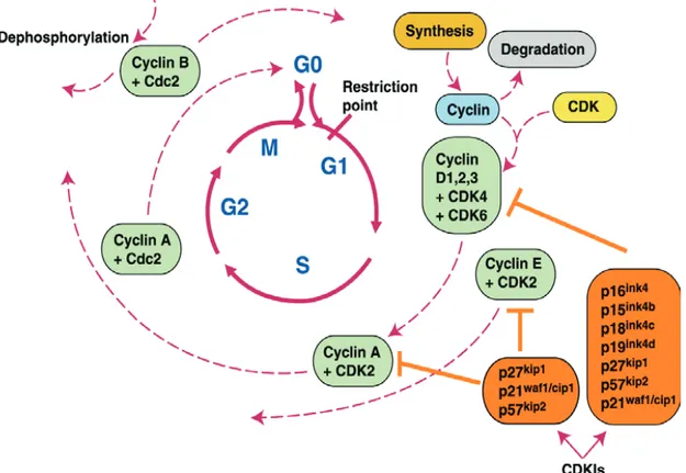
The “core system” of the cell cycle control machinery is constituted by a family of serine-threonine kinases called “cyclin dependent kinases” (CDKs) whose activity is tightly regulated by many cellular mechanisms. CDKs activity is mainly regulated by the interaction with their partners: the cyclins. Upon dimerization with these proteins, CDKs phosphorylate many cellular targets that can promote cellular proliferation and drive transition to the next phase of the cell cycle. Importantly, the same CDKs can phosphorylate different sets of proteins by binding with different cyclins partners. While CDKs are expressed homogeneously throughout cell cycle; specific cyclins are expressed only transiently during cell cycle progression and then are degraded. As a consequence, CDKs activity depend on the cyclins present in a given moment, at a given time, in the cell.
Every phase of cell cycle is dependent on different cyclin-CDKs complexes. Cyclin D (D1, D2 and D3) and E (E1 and E2) operate in G1 in complex with
CDK4 and CDK6 or CDK2 respectively. These cyclins integrate signals from the surrounding environment and lead to cell cycle entry, marked by the commitment to DNA replication, by driving the passage through the “Restriction Point” (R point) (described below). During S phase, DNA replication is mainly controlled by CyclinA/CDK2 complex. The same cyclin, in association with CDK1, mediates G2/M transition. Mitosis is mediated by CyclinB/CDK1 complex and its degradation is required from mitosis exit.
Two proteins families, INK4 and CIP/KIP, acting especially in G1 phase, counteracts CDKs activity. The INK4 family (p16, p15, p18 and p19) inhibits preferentially cyclin D in complex with CDK4 and CDK6, whereas the CIP/KIP family (p21, p27, p57) preferentially inhibits cyclin E or cyclin A in complex with the relative CDKs.
Moreover, CDKs are also regulated be regulated by phosphorylation or dephosphorylation events, that can activate or inhibit cyclins according to the site in which they occur.
1.1. The Restriction Point
In mammals, the G1 phase represents a crucial point during cell cycle. During this GAP phase, cells grow and decide to divide by integrating both external and internal stimuli. The passage through R point is driven by the cyclins operating in G1 in complex with their relative CDKs.
In quiescent cells, cyclins D are expressed following mitogenic stimulation and form complexes with CDK4 or CDK6. The neoformed complexes phosphorylate various proteins involved in cell cycle progression, including pRB family proteins (pRB, p107 p130). These proteins negatively regulate cell cycle by associating and repressing E2F. E2F in turn, regulates the expression of genes fundamental for G1-S transition. By phosphorylation, cyclinD/CDK4 and cyclinD/CDK6 complexes render pRB inactive and unable to efficiently bind E2F. As a consequence, E2F is released and stimulates its own transcription as well as transcription of its target genes such as Cyclin E. Cyclin E associates with CDK2 and phosphorylates pRB, increasing E2F activation. Thus, once this positive feedback loop has been established, it triggers the passage through R point by
hiperphosopohrylating pRB family proteins and allowing E2F release (Reviewed in Deshpande et al., 2005).
In conclusion, this G1 regulatory pathway is constituted mainly, but not only, by CyclinD/CDK4 cyclinD/CDK6, pRB, E2F, cyclinE/CDK2 and the G1 acting CDK inhibitors, triggers R point passage in response to external stimuli and is critical in preventing oncogenic transformation. This pathway appears to be mutated in many, if not all, human malignancies (figure I.1).
Fig. I.1 Summary of the genes involved in regulating cell cycle progression (Shah and Schwartz, 2001).
2. Cancer
In multicellular organisms each cell is a specialized member of a multicellular community. Cells have lost features needed for independent cell survival and acquired peculiarities that serve the needs of the body as a whole.
Cancer cells are mutant cells that escape all the rules required for a pacific coexistence with its neighbours, and are defined by two heritable properties: ability to reproduce continuously in defiance of the normal restraints on cell growth and division, and the ability to invade and colonize territories normally reserved for other cells. It is the combination of these properties that make cancer particularly dangerous. Thus, cancer cells have defects in the regulatory circuits governing normal cell proliferation and homeostasis.
Cells have evolved extremely efficient mechanisms for preventing the arise of cancer cells, that have to be bypassed in order to allow tumor progression. Normally, if one mechanism is inactivated by one mutation, the others can still compensate its loss. As a consequence, a single mutation is not enough to cause cancer. Rather, cancer development requires a gradual accumulation of mutations in a number of genes, different in different cancers, each one conferring a proliferative advantage and representing a step in tumor formation. Though, during tumor development, at each stage a cell can acquire mutations or epigenetic changes providing it a selective advantage over its neighbours, making it better able to proliferate.
There are more than 100 different types of cancer, and subtypes of tumors can be found within specific organs. Despite this, many, if not all, cancer cells acquire a variety of properties as they evolve, multiply and spread. These include alterations in cell signaling pathways, enabling the cells to ignore signals form the environment that normally keep cellular proliferation under tight control, or defects in the mechanisms that permanently stop cell division or induce apoptosis in response to cellular stresses. Moreover, many cancer cells escape the intrinsic cell division limit dictated by telomere shortening by reactivating telomerase, that prevents telomeres from getting shorter at each cell division, and provides cells
with a limitless replicative potential. Furthermore, many cancers are genetically unstable. This genetic instability is selected during cancer progression because it greatly accelerates the accumulation of further genetic and epigenetic changes that are required for tumor formation. All of these changes increase the ability of cancer cells to uncontrolledly survive, growth and divide in their original tissue, and to metastasize.
Cancer critical genes are genes that, if mutated, greatly contribute to tumor development and are divided in two groups: oncogenes and tumor suppressors. Oncogenes act in a dominant manner and contribute to tumor formation when overexpressed. By contrast, tumor suppressors normally restrain uncontrolled cellular proliferation and the in activation of both copies contribute to tumor progression. In many cases, oncogenes and tumor suppressors are genes that control the normal cellular proliferation and that, when mutated, contribute to damage this complex machinery. Many glioblastomas for example, have amplified the genes encoding oncogenes such as cyclinD and CDK4, thus favoring cellular proliferation (Buschges et al., 1999). Others instead, have mutations that inactivate the tumor suppressor pRB, that normally serves as a brake that restricts entry into S-phase (Ueky et al., 1996). Inactivation of p16 tumor suppressor, that blocks cell cycle under stress conditions, is also found in many cancers (Rocco and Sidransky, 2001).
Moreover many tumors bear mutations in p53 or in pathways that lead to its activation and therefore, continue through cell cycle even in presence of many cellular stresses, such as DNA damage or hypoxia. In addition, mutations in genes that stimulate proliferation in response to growth stimulation, such as Myc and RAS, lead to uncontrolled cellular proliferation by reducing their dependence from the extracellular milieu, and contribute to development of many tumors (Karreth and Tuveson, 2009; Gardner et al., 2002).
In summary, the genes responsible for the deregulated proliferation always found in cancer cells are mutated version of genes that, by contrast, are responsible for the tight regulation of cell cycle in normal cells.
3. The Intrinsic Tumor Suppression
Multicellular organisms maintain roughly the same number of cells in all tissues throughout their life. In renewable tissues, cell number is regulated by the balance between dying and proliferating cells. For example, in tissues such as epidermis or gut, cell divisions exactly balance the number of cells that are lost everyday, maintaining cell number equal. Therefore, in order to maintain the balance unaltered, cells need to have an extremely tight control of their cell cycle, allowing them to divide only in response to the organism needs as a whole. Yet, especially during cell divisions, cells can acquire mutations that disrupt or deregulate the complex “cell cycle control engine” contributing to the creation of cancer cells.
It is not surprising though, that metazoan cells have evolved, throughout millions of years, extremely efficient functions that prevent the arise of cancer cells: the tumor suppressive mechanisms. The most extensively studied processes that counteract tumor progression are senescence and apoptosis. Both of them, although with different ways, eliminate from the proliferative pool cells harboring mutations that can deregulate cell cycle. In the case of apoptosis, cells are eliminated by inducing cell death, whereas in senescence cells undergo a permanent cell cycle arrest, not allowing them reenter into cell cycle even in the presence of the appropriate stimuli. Importantly, these mechanisms have to be fired only when cells switch, or are prone to do so, from normal to deregulated cell cycle. In other words, a prerequisite for their activation is the ability of the cells to feel when proliferation starts to become aberrant.
Taken together, these mechanisms show a remarkable efficiency: in humans, despite trillions of cells, each one subject to a significant mutation rate, cancers arise less than once in life. Many, perhaps all malignancies, acquire mutations that disable these tumor suppressive mechanisms. In this way, cells are not able to couple the uncontrolled proliferation driven by mutations in the cell cycle control machine with the concomitant establishment of senescence or apoptosis.
3.1. Apoptosis
Apoptosis, known also a “programmed cell death”, is an essential process in development and in tissue homeostasis. In addition, it is an important tumor suppressive mechanism induced by many oncogenic stresses. For example, deregulated proliferation driven by c-Myc ectopic expression in a cell population is accompanied by an increased apoptotic level (Evan et al., 1992). In this way, apoptosis prevent cells harboring this deregulated Myc from accumulating further mutations that can lead to the formation of cancer cells. Consistent with this, many, if not all, Myc driven tumors harbor secondary mutations that disable the apoptotic pathway (Hoffman and Liebermann 2008).
Even though the link between oncogene expression and apoptosis is now well established, the molecular mechanisms of such link have not been completely uncovered. At least two different pathways can lead to apoptosis, each regulated at many levels.
The intrinsic pathway starts with the release of cytochrome C, from the mitochondria, its normal location, in the cytoplasm. When this event occur, the cytoplasmic protein APAF1 binds cytochrome C and activate caspase 9, a cysteine protease that initiates a downstream proteolytic cascade that also involve caspase 3 and caspase 6. Once activated, caspases cleave proteins important for cell and genome integrity, orchestrating the orderly death and engulfment of the cell.
The main way to regulate this pathway is by altering the mitochondrial membrane permeability in a way so as to promote, or not promote, cytochrome C release. Mitochondrial permeability is influenced by the balance between the pro-apoptotic BAX/BAK proteins and their antipro-apoptotic BCL2/BCLX cousins. The activity of these proteins is regulated either at transcriptional level or by members of the BH3-only family proteins, such as PUMA and NOXA. BH3 only proteins act as final effectors of distinct signaling pathways activated in response to a given cellular state, such as DNA damage or hypoxia. Once induced, they promote apoptosis by positively regulating pro-apoptotic members and inhibiting the antiapoptotic ones.
According to the current model, apoptosis occurs when the protective BCL2/BCLX buffer is breached by the sum of all the active BH3-only proteins, resulting in the net dominance of the pro-apoptotic BAX/BAK members, which then permeabilize the mitochondria and allow cytochrome C release. Regulation of the intrinsic pathway occurs at many levels. Above all, the transcriptional regulation of the BCL2/BH3 only family members, but also by expressing a class of caspase inhibitors known as inhibitors of apoptosis (IAP).
The other pathway that can lead to apoptosis is the extrinsic one. It is activated through the ligation of “cell surface death receptors” such as FAS/CD95, TNFR and DF-5 with their respective cognate ligands FAS TNF and TRAIL. Once ligated, these receptors form a death inducing signaling complex (DISC), which activates the apical caspase 8, leading to the proteolytic cascade mentioned above. In some cell types, this is sufficient to trigger apoptosis. By contrast, in other cells, death receptor induced apoptosis also requires the recruitment of the mitochondrial pathway through caspase 8 mediated activation of the BH3-only protein BID. The extrinsic pathway is subject to modulation by decoy receptors, which bind the ligand but are defective in signaling transmission, and by intracellular molecules such as FLIP that competes with caspase 8 for binding to DISC.
3.2. Senescence
Senescence is a tumor suppressive mechanism that, like apoptosis, acts by eliminating harmful cells from the proliferative pool (Mathon and Lloyd, 2001). In this case however, cells don’t dye but enter in a state of permanent growth arrest. Senescent cells are metabolically active and “live” but are unable to proliferate despite the presence of nutrients and growth factors. Nowadays, no known physiological stimuli, at least in vitro, can trigger escape from senescence (Campisi and D’Adda di Fagagna, 2007).
At molecular level, senescence seems to be triggered and maintained by p53 (through induction of ARF) and its induction of p21, and by p16, a cell cycle inhibitor that keeps pRB in an unphosphorylated state. Noteworthy, the relative contribution of p53 and pRB to senescence seems to be extremely variable on cell type. For example, MEFs seems to depend mainly on ARF-p53 pathway, whereas human fibroblasts also rely on p16-pRB functions (Lowe, Cepero and Evan, 2004).
The first example of senescence was described more than 40 years ago as the loosing of proliferative capacity of human fibroblast after a given number of passages in culture (Hayflick et al., 1961). Years later, this was shown to be the result of telomere shortening. Telomeres indeed, thanks to a peculiarity of the replication apparatus, gradually shortens at each replication cycle. When their length falls below a still undetermined threshold, they are recognized as a DNA double strand breaks and trigger a prolonged DDR response that causes this permanent cell cycle arrest (Herbig et al., 2004; D’Adda di Fagagna, 2008).
In recent years, overexpression of some oncogenes such as RAS was shown to trigger senescence in a manner independent from telomere shortening (Serrano et al., 1997). In an elegant study of 2006 Di Micco et al demonstrated that this kind of senescence is caused, and also maintained, by the DNA damage response (DDR) triggered by activated oncogenes (Di Micco et al., 2006). In the paper, the authors demonstrated that senescence following RAS expression is established after a brief hiperproliferative phase and is associated with DDR markers. Cells with impaired CHK2, an early mediator of DDR response, bypass senescence and continue to proliferate. Moreover, experimental inactivation of DDR leads to escape from senescence and resuming of cellular proliferation. Although much remains to be learned about how oncogenes induce DNA damage, it has proposed to arise during S phase as a consequence of replicative stress (D’Adda di Fagagna, 2008).
Senescent cells have been detected in early phases of neoplastic processes, leading to a model postulating the role senescence as a tumor suppressive mechanism during early stages of neoplasia (Bartkova et al., 2006). Many doubts concern the fate of senescent cells in vivo. Some evidences seems to indicate that the tissue of origins plays an important role in this regard. Whereas skin naevi, that are, clones of skin cells that have undergone senescence, seem to remain stable for years (Michaloglou, 2005), senescent cells occurring in sarcoma or carcinoma tumors following p53 reexpression seem to be cleaned by a mechanisms dependent on the immune system (Ventura et al., 2007).
4. P53
P53 is without any doubt the most extensively studied gene in the field of biology. It is a transcription factor induced by a wide variety of stress signals a cell can encounter during malignant progression, such as oncogene activation, excessive ROS accumulation, hypoxia, DNA damage, just to listen a few. Once induced, it shapes the global cellular transcriptome in a way so as to induce either cell cycle arrest, senescence or apoptosis (Vousden and Prives, 2009). Cell type, cell context, damaging stimulus and its duration, determine which of the aforementioned program will be fired.
In normal, unstressed cells, p53 is continuously synthesized, but its concentration remains very low because MDM2, a p53 specific ubiquitin ligase, continuously marks the protein for proteasome degradation. Different stress situations lead to p53 accumulation mainly by interfering with its binding with MDM2 (Haupt et al., 2002). CHK2 for example, is a protein kinase activated following DNA damage and phosphorylates p53 on SER20
. This phosphorylation makes MDM2 unable to bind and ubiquitinate efficiently p53, leading to p53 accumulation (Caspari, 2000). CHK2 however, is not a mean used by all stress signals to accumulate p53. Rather, different stress signals target the MDM2-p53 interaction with different ways. Overexpression of some oncogenes, Myc for instance, induces ARF. Importantly, ARF is induced only in response oncogenic stress and, once induced, it binds and and inactivate MDM2, leading, as in the case of DNA damage, to p53 accumulation (Zindy et al., 1998). In summary, different cellular stresses impair the activity of the common target MDM2 in order to lead to p53 accumulation, however, they adopt different ways to accomplish this.
4.1. Direction of P53 transcriptional program
Once p53 is accumulated, it can induce three transcriptional programs, leading to different outcomes: cell cycle arrest, senescence and apoptosis. In this
last decade, an outstanding research effort has been devoted in understanding how environmental factors, cell type and context, can influence p53 and direct its transcriptional program toward a different outcome.
Recent discoveries seem to indicate that posttranslational play an essential role in this regard.
For instance, UV or gamma radiation can induce different p53 target genes in the same cell types (Zhao et al., 2000). These different targets correlate with different posttranslational modifications decorating p53 (Webley et al., 2000; Lu et al., 1998). Posttranslational modifications are thought to help p53 to associate with a selective group of promoters, and to recruit different coactivators or corepressors. Supporting this, it has been shown that DNA damage and hypoxia induce different posttranslational modifications correlating with p53 ability to associate with different promoters and auxiliary proteins (Koumenis et al., 2001).
Thus, it seems that different stress stimuli might decorate p53 with different modifications, influencing its ability to associate with specific genes as well as to recruit additional proteins. Understanding which modifications decorate p53 in various cell types in response to different stimuli, the proteins that perform such modifications and how they are regulated, would provide milestone insights in the field of p53 biology.
Although posttranslational modifications seem to be fundamental determinants in influencing p53 activity, stress signals can also influence p53 transcriptional program by additional ways. When overexpressed, Myc induces p53 accumulation through ARF, and actively repress genes required for p53 mediated cell cycle arrest program while letting untouched those required for apoptosis (Hoffman and Liebermann, 2008). Thus, Myc indirectly shifts p53 transcriptional program toward apoptosis by rendering the genes required for its mediated cell cycle arrest uninducible.
In summary, specific stress signals can induce different arrays of p53 posttranslational modifications and proteins, that alter the transcription of its dependent genes. By targeting p53 activity in these two ways, stress signals direct p53 program toward a specific direction, that can vary according to cell type and context.
4.2. Postranslational modifications on P53
4.2.1. Phosphorilation
Phosphorylation of p53 at the N-terminal SER46
has clearly a discriminatory function in the firing of p53 apoptotic program. This modification is mainly performed by Homeodomain interacting protein 2 (HIPK2) and occurs only under excessive DNA damage conditions, that a cell is not able to repair. When HIPK2 marks p53 with this modification, p53 is committed to fire its cell death program. Thus, p53 activity could be regulated by the extent of DNA damage using HIPK2 as a sensor. SER46
modifications, as well as HIPK2 regulation, will be discussed in the apposite section (Section 9.1).
Beside SER46
, p53 activity can be influenced by phosphorylation in other residues. P53 SER315
is phosphorylated in response to growth promoting kinases and regulates p53 ability to repress nanog, a factor required for stem cell self renewal. Mice harboring mutant p53 where SER315
is mutated to alanine have an impaired nanog repression. Furthermore, SER315
facilitates nuclear retention of the transcription factor E2F (Fogal et. al., 2005).
Finally, SER366 and SER387, phosphorylated by CHK1 and CHK2 respectively, are also of pivotal importance in regulating p53. Downregulation of either CHK kinases or mutations of these two phosphorylation sites, selectively affect p53 activation, promoter binding, and modifications of N-terminal lysines (Ou et al., 2005).
4.2.2. Lysine Methylation and Acetylation
LYS120
is acetylated in vivo in response to DNA damage, by two histone acetyl transferases (HATs), tip60 (Tang et al., 2006) and hMOF (Sykes et al., 2006). Acetylated LYS120
increases the expression of p53 apoptotic targets such as PUMA (Nakano et al., 2001). Cells overexpressing mutant forms of p53 where LYS120
is mutated to alanine show an increased resistance to apoptosis (Zupnick and Prives, 2006).
P53 LYS320
is acetylated by PCAF, a substrate of the transcriptional coactivator p300/CBP. Cells expressing mutant p53 in which this lysine is mutated to glutammine display decreased apoptosis following DNA damage. Consistent with this, these cells can neither induce APAF1 nor repress IAPs such as survivin. Nevertheless, they are still capable of inducing cell cycle arrest targets such as p21 (Knights et al., 2006).
Another lysine modification important for p53 activity is the methylation of lysine residues. For example, LYS370
is methylated by SMID 2 and impairs p53 transcriptional activation (Huang et al., 2006). LYS372 is thought to be methylated by SET7/9. SET7/9 KO cells where shown to have impaired cell cycle arrest and apoptotic response following DNA damage (Kurash et al., 2008). However, this will be better discussed in the apposite section (Section 9.2).
5. P53 and Apoptosis
When p53 is committed to trigger its apoptotic program, it targets the cell death machinery at different levels and through different mechanisms. These mechanisms in turn, act in a synergistic way ensuring that the apoptotic pathway proceeds efficiently once established.
5.1. P53 and the Canonical Apoptotic Pathway
One way through which p53 promotes apoptosis is by altering mitochondrial membrane permeability so as to induce the release of cytochrome C. P53 is able to upregulate the transcription of proapoptotic members of the BCL2 family such as BAX (Miyashita et al., 1994), and members of the BH3 only family such as PUMA (Nakano and Vousden, 2001), NOXA (Oda et al., 2000) and BID (Sax et. al., 2002). Consequently, it enhances the proapoptotic buffer and enhances the probability to overwhelm the protective effect of BCL2 and BCLX, the antiapoptotic members of the BCL2 family.
In addition to control factors altering the mitochondrial permeability, p53 controls the expression of factors acting downstream of cytochrome C release. For instance, p53 can increase the expression of APAF1 (Kannan et al., 2001) and caspase 6 (MacLachlan et al., 2002), an effector caspase, and repress survivin, a member of the IAP family (Raj D., 2008). In this way, it contributes to a more prompt and effective apoptotic response once cytochrome C is released in the cytoplasm.
Furthermore, in addition to the intrinsic pathway, p53 appear to regulate also the extrinsic one. Although the contribution of p53 to this kind of cell death is poorly understood, the genes encoding FAS/CD95, and the DR5 (Wu et al., 1997) death receptors are all transcriptional targets of p53 (Owen-Schaub et al., 2005). One possibility though, is that p53 sensitizes cells to death receptor induced
apoptosis by increasing death receptors expression and the expression of their ligands.
Finally, p53 targets additional genes regulating apoptosis whose mechanism is not well defined and, as a consequence, cannot be classified as a part of the intrinsic or the extrinsic pathway. For example “p53 induced protein with a dead domain” (PIDD) was identified as a p53 responsive gene following shift of an erythroleukemia cell line containing temperature-sensitive p53 to a permissive temperature. Although its precise role in apoptosis remains to be determined, suppression of PIDD inhibits apoptosis whereas enforced expression induces cell death (Okamura et al., 2001).
5.2. P53 and survival signalings
Survival signals are required for a cell to survive. In different ways, they promote cell survival by upregulating antiapoptotic members and downregulating the proapoptotic ones. Furthermore, they trigger signaling cascades stimulating processes like cell growth, metabolism and nutrient uptake.
P53 can strength its apoptotic program by short-circuiting these antiapoptotic pathways.
Consistent with this, p53 was shown to negatively regulate the prosurvival pathway activated by phosphoinositide-3-phosphate (PI3) kinase. This pathway is activated by many survival factors, for example, by prosurvival cytochines. PI3 kinase phosphorylates phosphoinositide-4-5-bisphosphate (PIP2) and produce phosphoinositide-3-4-5- trisphosphate (PIP3). PIP3 acts as a platform recruiting protein complexes that can activate effectors proteins promoting cell survival, like AKT or PKB. Once activated, AKT phosphorylates and inhibits the proapoptotic member BAD, and activate mTOR a kinase that promotes cell growth by phosphorylating many effector proteins. The main way through which p53 negatively regulates this pathway is by activating PTEN (Stambolic et al., 2002), that inhibit PI3 kinase by dephosphorylating PIP3 so as to produce PIP2. Thus, p53 can counteract survival signals from the microenvironment, thereby reducing the threshold needed for proapoptotic factors to trigger cell death.
5.3. Nontranscriptional Functions of P53 in Mediating
Apoptosis
Although the main functions of p53 lie in its transcriptional activity, recent discoveries seems to indicate additional p53 transcription-independent functions. Some studies suggest that a p53 stress-induced accumulation can occur in the mitochondria (Mihara et al., 2003). Here, mitochondrial redistribution of p53 precedes cytochrome C release and caspase activation and occurs only during p53 dependent cell death. This mitochondrial p53 appears to be proapoptotic, since direct targeting of p53 to mitochondria can promote apoptosis in p53 deficient mice (Mihara et al., 2003). Nevertheless, loss of DNA binding appears to be the first function selected for promoting tumorigenesis and cells harboring a mutant p53 defective in transactivation capabilities don’t undergo apoptosis (Jimenez at al., 2000). Thus, it seems that these transcription independent functions have only an auxiliary role in potentiating p53 dependent cell death.
5.4. Coordination of P53 Apoptotic Program
By short-circuiting survival signals, increasing the mitochondrial proapoptotic buffer, sensitize cells to death ligand induced apoptosis, and by transcription independent functions, p53 targets the apoptotic machinery at multiple nodes. By doing so, p53 increases the probability that the apoptotic process goes forward and ensures a well coordinated program once the process is initiated. Moreover, the multiple death circuits targeted by p53 for inducing apoptosis can explain why no single effector molecule can account for all p53 proapoptotic activities.
Each circuit targeted by p53, is also affected in different ways by the cell type, microenvironment, apoptotic stimulus or genetic background. Different cells might commit p53 to trigger its apoptotic program by decorating it with different posttranslational modifications. These posttranslational modifications, by increasing or decreasing p53 ability to transcribe specific subsets of genes, might influence the main way through which p53 triggers apoptosis in a given cell. As a consequence, one or more circuits may stand out as the crucial elements in a particular cell, while others might cover only an auxiliary role (Lowe, Cepero and Evan, 2004).
6. P53 and Oncogenes
6.1. Oncogene induced Apoptosis: the P53 dependent part
P53 is induced by many oncogenes, including E1A, MYC and E2F (Volgenstein et al., 2000) and its inactivation severely compromises apoptosis in many instances. Consistent with is role in coupling proliferation to cell death, inactivation of p53 potently cooperates with diverse oncogenes, such as RAS and Myc, to promote transformation in vitro and tumorigenesis in vivo (Lowe, Cepero and Evan, 2004).
This in turn, raise the question of how deregulated oncogenes induce p53 while their normal counterpart does not. One important clue in this answer is ARF. ARF is induced only in response to oncogenic stress and leads to p53 accumulation by interfering with MDM2 (Lowe and Sherr, 2003). The ability of Myc and E1A to activate p53 is severely compromised in ARF null cells (Zindy et al., 1998; de Stanchina et al., 1998). Moreover, disruption of ARF in mice dramatically accelerates Myc induced lymphomas and carcinomas in a manner broadly comparable to p53 inactivation (Eischen et al., 1999). Thus, these experiments demonstrate the crucial role of ARF during oncogene dependent activation of p53. Some evidences suggest a role of DNA damage response in inducing p53 following oncogene expression. Indeed, experiments show that oncogene activation is associated DNA damage and DDR activation, which lead to p53 accumulation through CHK2 phosphorylation in SER20 (Khan et al., 2000).
6.2. Oncogene induced Apoptosis: the P53 independent
part
P53 mediated apoptosis is one fundamental way used by cells to establish the apoptotic program in response to many oncogenes. In addition, activated oncogenes can influence the apoptotic machinery with additional, p53
independent mechanisms. These mechanisms act synergistically with p53 and ultimately, drastically reduce the threshold needed for the firing of the “cell death program”. Furthermore, when p53 is deactivated, p53 independent mechanisms, although with low efficiency, can still trigger apoptosis. Thus, even in the absence of p53, a certain degree of apoptosis can be still present in oncogene expressing cell, making the bypass of this tumor suppressive response a not easy task.
Accordingly, Myc, E2F and E1A have pleiotropic effects on the expression of members of the BCL2 family. Myc can repress the expression of BCL2 and BCLX and induce the expression of BIM, whereas E1A and E2F suppress MCL1, another member of the BCL2 family (Croxton et al., 2002; Eischen et al., 2001). Finally, E2F can induce several downstream effectors of the apoptotic machinery, including various caspases (Nahle et al., 2002). In vivo experiments indicate that loss of BIM accelerate Myc induced lymphomas in a manner comparable to p53 loss (Egle, et al., 2004).
However, is worth notice that the relative contribution of p53 dependent vs independent apoptotic mechanisms can vary according to cell type and cell context, as well as external stimuli.
6.3. Oncogene induced Apoptosis: crossing the final
threshold
Oncogene activation appears to target the apoptotic machinery at multiple levels, and in a manner either dependent or independent of p53. All these mechanisms in turn, ensure a more robust and prompt apoptotic response, making it dependent on multiple regulation levels instead of a single one, and collaborate in influencing the ratio of proapoptotic vs antiapoptotic members. Such ratio in turn, determines whether or not the apoptotic response has to be fired.
Mutations in anyone of the components through which oncogenes promote apoptosis can suppress a significant amount of cell death and confer significant growth advantage. This makes the discovery of genes important for apoptosis promotion relatively easy, but in contrast, make extremely difficult investigating how, and at which level, they act.
7. C-Myc
The c-Myc (Myc) is a transcription factor belonging to the family of Myc proto-oncogenes (comprising also N-Myc and L-Myc). Deregulation of c-Myc is involved in development of many human cancers. Overexpression of Myc is found in 80% breast cancers, 70% colon cancers, 90% gynecological cancers, 50% of hepatocellular carcinomas and a variety of hematological tumors (Gardner et al., 2002). This reason, has made Myc one of the most extensively studied genes in biology, together with other genes such as P53 and RAS.
Myc gene is located on the chromosome 8q24: consists of three exons and contains three different promoters form which transcription can be initiated. The main isoform present in the cell is a 64kDa protein that contain 5 Myc homology boxes (conserved in all the Myc family) important for its cellular activities, a transactivation domain required for the activation of gene transcription, a nuclear localization domain, and an helix-loop-helix leucine-zipper motif. This last domain mediates dimerization with is obligate partner MAX and subsequent DNA binding to specific DNA responsive element called E-boxes (see below) and is essential for Myc activity (Blackwood, 1991).
7.1. Myc in normal cells
Myc is expressed at low and tightly regulated level in proliferating cells (and at a nearly absent level in quiescent cells) and these levels are essential to mediate a correct proliferation in response to external stimuli (Gardner et al., 2002). Mouse embryo fibroblasts lacking Myc are still able to proliferate, but the doubling time of the cell population as a whole is very much higher than their wild type counterpart (Mateyak et al., 1997). These data might indicate that, although not essential for proliferation, Myc dramatically enhances the probability of cell to enter into cell cycle in presence of appropriate factors. In normal cells, Myc is induced by major the growth stimulatory pathways (RAS, WNT, NOTCH
signalling) and integrates these signals by generating downstream cellular responses (Henriksson, and Luscher, 1996).
Many high throughput studies, such as CHIP-seq, CHIP-CHIP and CHIP-PET have shown that Myc can control the expression of 15% of the genes in the entire genome (Eilers and Eisenman, 2008). Myc dependent regulation of some genes is conserved in many cell types whereas others are regulated in a cell type specific manner, indicating a cell type specific effect of Myc. Furthermore, many studies indicate that Myc is necessary but not sufficient for the activation of its target genes, and that its activity depend on the specific array of proteins present in a given cell (Lawlor et al., 2006). Perhaps is for this reason that, although many profiling studies of Myc are available, Myc target genes that drive tumor progression have not been identified yet (figure I.2).
Fig. I.2 Summary of the functional categories of genes whose expression is modulated by augmented Myc expression (Eilers and Eisenman, 2008).
Nevertheless, all expression profiles are consistent with the role of Myc in regulating cellular proliferation. All these studies highlight Myc regulation of genes involved in cell division, cell growth, metabolism ribosome biogenesis and proteins synthesis (Patel et al., 2004). Many genes regulated by Myc are
transcribed by RNA polymerase2 but recent studies demonstrated that Myc also regulates RNA polymerase1 (rRNA) (Grandori et al., 2005) and RNA polymerase3 (tRNA) (Steiger et al., 2008) transcription, both important classes of genes for protein synthesis. Moreover, Myc can promote oxidative phosphorylation and glycolisis (Zhang et al., 2007; Morrish et al., 2008).
Concerning progression through cell cycle, Myc can regulate key genes involved in the passage of R-point, though, genes that commit a cell to replicate its DNA and divide in two. Myc stimulates the transcription of the R-passing feedback loop genes such as Cyclin D, cyclin E and E2F (Patel et al., 2004) and inhibits cell cycle inhibitors such as P21, P27, P15, P16 (Staller et al., 2001; Herold et al., 2002; Knoepfler et al., 2002). All these data underscore the capacity of Myc to both promote cell growth, metabolism and cell division, and to downregulate cell cycle inhibitors, a powerful combination that, when deregulated, may drive the limitless replicative potential characteristic of nearly all tumors.
7.2. Myc and Chromatin
Myc is a transcription factor able to activate or repress transcription. Therefore, most of its cellular activities can be explained in terms of its effects on gene transcription. As previously mentioned, Myc is estimated to bind 15% of the known genes. Therefore, although covering a significant part of the genome, Myc binding has somehow to be restricted.
The first determinant of Myc Binding occurs mainly at a DNA responsive element called E-BOX (CACGTG) located in many promoters of Myc responsive genes (Blackwood et al., 1991). However, two studies in Dr. Amati’s Lab. demonstrated that Myc binding is also influenced by the chromatin context (Fernandez et al., 2003; Guccione et al., 2006). In the study of Fernandez et al. it was shown that not all Myc binding is E-box dependent and that E-box sequences are bound by Myc with different affinity (the authors divided genes in high and low affinity for Myc binding). High affinity group genes are highly conserved among different cells, are bound independently of Myc expression level and are mainly located within CpG islands. The low affinity group genes instead, including non E-box DNA binding, are bound only at high or extreme level of Myc expression and are not conserved among various cell types. In the successive study of 2006, Guccione et al. (Guccione et al., 2006) revealed that high affinity E-Boxes are located in chromatin regions bearing euchromatic marks such as
H3K4 me3, whereas low affinity group genes are located in regions bearing eterochromatic marks. Thus, these studies indicate that, in general, the more an E-BOX is located in an euchromatic region, the more it is likely to be an high affinity site for Myc.
Myc can activate or repress transcription. Generally, Myc binding to E-boxes is correlated with transcription activation. Once bound to its target promoters, Myc interacts with many histone acetyl-transferases complexes (HATs) or subunit of HAT complexes that hyperacetylate histones in proximity of its binding site (Amati et al., 2001). Consistent with this, profile of histone marks after Myc binding showed enhanced acetylation of several H3 and H4 lysines as well as H3K79 me3, but levels of other methylation marks, such as H3K4me3 and H3K27me3 were not modulated (Martinato et al., 2008). Although a cause-effect mechanism remains to be established, all these changes correlate with activation of gene expression. Acetylation for instance, is tought to relax chromatin structure by lowering the strength of histones-DNA interaction and allow the transcription machinery to have access to responsive elements and assemble at the promoter (Fletcher and Hansen, 1995).
In some instances, Myc also mediates transcriptional repression. However, this case is less clear and it seems that Myc can repress gene transcription by interfering with coactivator proteins rather than by DNA binding. For instance, Myc represses p21 transcription by interfering and sequestering its activator protein Miz1 (Soane et al., 2002). Moreover, recent studies indicate that Myc can also direct DNA-methiltransferases (DNMT) on p21 promoter, further strengthening its repression (Brenner et al., 2005).
7.3. Myc and Cancer
Consistent with is pivotal role in stimulating cellular proliferation in response to growth stimulatory pathways, Myc activity can become oncogenic if deregulated. In many tumors, Myc per se is not mutated, rather, it appears to be deregulated by mechanisms such as translocation or genomic amplification (Meyer et al., 2008). In Burkitt lymphoma for example, a chromosomal translocation juxtaposes Myc with the promoter of the immunoglobulin heavy chain. As a consequence, Myc is deregulately expressed in B-lymphocytes, contributing to uncontrolled cell division (Janz, 2006). In colon cancer instead, deregulated expression of Myc is manly caused by genomic amplification: more than 30 copies of Myc can be found in this type of tumor (Yander et al., 1985).
Moreover, Myc can be deregulated by alteration of protein or RNA stability. For instance, mutations of the peptidase USP28, or the ubiquitin ligase FBW7, both regulating Myc turnover, can lead to an altered Myc stability and activity (Popov et al., 2007; Yada et al., 2004). Finally deregulated expression of Myc can be caused by deregulation of pathways which Myc is responsive to, such as RAS, NOTCH and WNT (Nesbit et al., 1999).
In summary, many mechanisms acting at DNA, RNA and protein level, can contribute to deregulate Myc activity letting its sequence immutated.
The proofs that overexpression of Myc is oncogenic are astounding. More than 20 years ago, it was shown that rat embryo fibroblasts can be transformed when contransfected with Myc and RAS (Lee et al., 1985). Nowadays, with the technologies to engineer mouse genome, overexpression of Myc has been shown to induce tumors in many tissues. For example, the Eμ-Myc transgenic mouse is a model of B-cell lymphoma bearing Myc gene under the control of immunoglobulin heavy chain promoter, like in the human Burkitt lymphoma (Harris et al., 1988). These mice develop lymphomas in the first year with mean latency of 25 weeks.
Although a link between c-Myc and cancer is well established, the cellular mechanisms of Myc mediated transformation are not fully understood. Nowadays for instance, although the great use of expression profiles, Myc target genes critical for tumor promotion, if any, have not been discovered.
Consistent with its induction in response to growth stimulatory pathways and its role in integrating all these signals by stimulating key processes such as cell growth, metabolism, and the passage or R point, a reasonable model can be proposed. When Myc is deregulated, it is no longer responsive to its inductive signals such as RAS or NOTCH, but it can still mediate their downstream effects. A constitutively expressed Myc, such as in Burkitt lymphoma, can continuously stimulate the transcription of target genes required for R point passage such as cyclin D, cyclin E and E2F independently from upstream proliferation signals. In this, way Myc can deregulate the cell cycle machinery and promote continuous cell divisions despite stimulatory signals, reducing the dependency of the G1 phase from external factors: an hallmark of neoplasia (Gardner et al., 2002).
8. Myc induced Apoptosis
Ectopic expression of c-Myc can enforces cellular proliferation. In a cell population expressing deregulated c-Myc however, this increase in cellular proliferation is counterbalanced by an increased apoptotic level (Evan et al., 1992). This increased apoptosis, or “Myc induced apoptosis” provides a built-in failsafe program to limit unchecked cell growth under inappropriate conditions. Other oncogenes such as E1A and E2F1 can trigger this mechanism when overexpressed (Frisch and Mymryc, 2002; Stanelle and Putzer, 2006). Many, if not all, Myc driven tumors, bear mutations disabling the network underlying the apoptotic response (Hoffman and Liebermann, 2008).
8.1. Myc induced Apoptosis: P53 dependent part
Many mechanisms control the apoptotic network at different nodes, ensuring a robust and regulated apoptotic program in response to Myc deregulation. One major player in such process is p53.
Its accumulation is induced by deregulated c-Myc (Zindy et al., 1998), and its transcriptional activity is directed to trigger the apoptotic program (Hoffman and Lyebermann, 2008). Loss of p53 for instance, severly enhances lymphomagenesis onset in Eμ-Myc transgenic mice (Hemann et al., 2005).
Like many oncogenes, ectopic expression of Myc leads to p53 accumulation by inducing ARF and blocking MDM2 (Zindy et al., 1998). Moreover, ARF negatively regulates the activity of Myc by relocating it from the nucleoplasm to the nucleolus, a zone where Myc is largely absent (Sarker and Fisher., 2006). Thus, together with is well established role in tumor suppression as a “p53 accumulator” ARF also has p53-independent tumor suppressive functions. Biallelic loss of ARF occurs in 25% of Eu Myc mice derived lymphomas whereas another 33% display p53 mutations (Schmitt et al., 1999, Eischen et al., 1999).
Consistent with the DDR induced by oncogenes and the following activation of p53, it has been shown that ATM, an early mediator of DDR, is critical for the induction of apoptosis in response to c-Myc ectopic expression (Pusapati et al., 2006). Thus, at least is some cases, ARF and DDR can collaborate in activating p53 under oncogenic stress.
8.2 Myc induced Apoptosis: the P53 independent part
Although in many contexts p53 is crucial for the correct establishment of Myc induced apoptosis, many studies indicate that Myc targets the apoptotic machinery with additional, p53-independent mechanisms.
An elegant study performed in the laboratory of Scott Lowe (Hemann et al., 2005) demonstrated how two common Myc mutant alleles found in Burkitt lymphoma are able to circumvent p53 inactivation during tumor development. These mutant alleles were transduced into murine hematopoietic stem cells used to reconstitute hematopoietic system of lethally irradiated mice. Lymphoma arised from these mice display ARF induction, p53 accumulation, and transcription of its apoptotic targets such as BAX, PUMA and NOXA, suggesting a normal p53 activity directed toward apoptotic program. Nevertheless, mutant alleles, but not wild type alleles, failed to repress the antiapoptotic BCL2 and to upregulate the proapoptotic BIM, both not controlled by p53. Thus these data suggest that Myc apoptotic program is not linear, but instead involves p53 dependent and independent signals acting synergistically to promote cell death and suppress Myc induced tumorigenesis.
BCL2 and BIM are two apoptotic effectors regulated by Myc but not p53 and are central in mediating Myc induced apoptosis (Eischen et al., 2001; Hemann et al., 2005). In over half of lymphomas from Eμ-Myc transgenic mice BCL2 levels are markedly elevated, independently of the loss of ARF of p53, indicating that these two apoptotic pathways are inactivated separately (Eischen et al., 1999). Finally Myc can sensitize cells to death ligand induced apoptosis, although the relative contribution of p53 in such effect is not well defined. Overexpression of Myc represses FLIP transcription, a negative regulator of the apoptotic pathway (Ricci et al., 2004), and promotes expression of FASL and the TRAIL receptor DR5 (Wang et al., 2004, Brunner et al., 2000).
Myc induced apoptosis thus, occurs by targeting the apoptotic machinery at different levels. In this way, different nodes act synergistically to promote cell
death contributing to the bypass pf the threshold needed for the firing of the “cell death program”.
8.3. Myc induced Apoptosis: putting all together
By both p53 dependent and independent mechanisms, Myc increases the ratio of pro vs anti apoptotic BCL2 proteins, promoting mitochondrial membrane permeabilization and release of cytochrome C. By activating p53, Myc induces APAF-1, caspases, and IAP inhibitors, and consequently increases the efficiency with which cytochrome C triggers caspase activation when released in the cytosol. Through p53 dependent increase in PTEN, Myc short circuit survival signalings, thereby reducing the cell’s ability to buffer apoptotic signals. Finally, by increasing cell death receptors and decreasing their antagonists, Myc sensitizes cells to the action of death inducing ligands in the microenvironment.
Importantly, these mechanisms act all synergistically with a common purpose: induce cell death. Because of their synergistic action, elimination of even one of them can cause a significant drop in the apoptotic level and contribute to tumor progression. This make, at a first glance, the identification of elements contributing to Myc induced apoptosis not extremely complicated. But by contrast, make extremely challenging to investigate how individual components work in this multisignal network.
9. HIPK2 and SET7/9
9.1. HIPK2
DNA damage, if remains unrepaired, poses a serious threat to the organism. The DDR occurring after DNA damage can provoke, according to cell type and extent of DNA damage, two different cell fates. Generally, if the DNA damage is not high and repairable (cytostatic) the cells transiently arrest the cell cycle, thereby preventing cells with damaged genome to continue cell divisions and allowing to the DNA damage repair machinery to have the right time to repair the lesion. Once the lesion has been repaired the cells can reenter into cell cycle. By contrast, if the DNA damage is too high and difficult to repair, cells can trigger the apoptotic program and undergo a programmed cell death. Thus, cells must discriminate between the various extents of DNA damage and decide how to behave accordingly. It would be deleterious if cells undergo apoptosis with low doses of DNA damage easily repairable by DNA repair systems.
One proteins mediating such decision is p53 (Vogelstein et al., 2000). More specifically, in the case of cytostatic DNA damage, p53 induces cell cycle arrest by inducing, for example, the cell cycle inhibitor p21 (Gartel, 2009). If the DNA damage is unrepairable, p53 induces its apoptotic program by transcribing genes such as PUMA (Yu et al., 2001). P53 activity is in turn influenced by many coregulators. These proteins act like a sort of “molecular switch” being activated in response to an appropriate stimulus or cellular condition, and influencing p53 activity by posttranslational modifications (Braithwiate et al., 2006). Thus, cellular conditions activate, or deactivate, many p53 posttranlational regulators, resulting in an dynamic array of p53 modifications that vary according to cell type and context, and that directly influence p53 transcriptional program.
Under lethal, but not cytostatic, DNA damage, p53 is phosphorylated in SER46 (SER46
in human, homologous SER58
in mouse) (D’Orazi et al., 2002; Cecchinelli et al., 2006). In particular, phosphorylation at this site promotes change in p53 affinity for different promoters, with a shift from cell cycle arrest related genes to apoptosis related ones (Mayo et al., 2005). Based on these findings, p53 SER46
phosphorylation is considered a point of no return because it would irreversibly drive cells toward apoptosis.
The kinase responsible of this phosphorylation is Homeodomain interacting protein 2 (HIPK2) (Rinaldo et al., 2007), a member of nuclear SER/THR kinases family originally identified as a corepressor for homeodomain transcription factors (Kim et al., 1998). Importantly, HIPK2 performs such modification on p53, only under severe, but not cytostatic, DNA damage (Rinaldo et al., 2007). Thus, HIPK2 acts as a bridge connecting the extent of DNA damage to p53 transcriptional program. In 2007, Rinaldo and colleagues (Rinaldo et al., 2007) demonstrated that HIPK2 stability, and though, its activity on p53 is regulated by p53-MDM2 feedback loop. Under normal conditions, HIPK2 is targeted to the proteasome by MDM2 dependent ubiquitination. In condition of cytostatic DNA damage, p53 induces further MDM2 accumulation and as a consequence, leads to a negative feedback loop responsible for its repression as well as the further repression of HIPK2. By contrast, in condition of severe DNA damage, p53 is not anymore able to induce MDM2 accumulation (Perry, 2004), lading to an increase in HIPK2 levels responsible for the phosphorylation of p53 SER46
. Consistent with this, p53 SER46
phosphorylation and HIPK2 levels show an opposite correlation with MDM2 levels, moreover, MDM2 overexpression significantly reduces HIPK2 induction, p53 SER46
phosphorylation, and cell death upon treatment with a lethal dose of adryamicin. In summary, HIPK2 is activated only in response to lethal level of DNA damage, that stops the expression of MDM2, its specific ubiquitin ligase that targets it to the proteasome, Once activated HIPK2 phosphorylates p53 SER46
and this phosphorylation drives p53 program toward apoptosis. thus HIPK2 acts as a “cellular alarm” being activated in response to high level of DNA damage and ordering to “p53 guard” to activate its apoptotic program. Probably however, this modification alone is not sufficient for influencing p53 activity, and has to be coupled to additional p53 posttranslational modifications, working all in synergy to shift its transcriptional program toward apoptosis.
9.2. SET7/9
SET7/9 is a protein originally shown to methylate histone H3 and TAF10 in vitro (Nishioka et al., 2002). In recent times, it was shown to modulate p53 activity by methylating its LYS372
(Kurash et al., 2008).
In this regard, a paper of Kurash et al. (Kurash et al., 2008) demonstrated the importance of this methylation during p53 activation. Since SET7/9 was the first part of my thesis work, and that I obtained different data from those published in the paper, this section will be focused on the description of the data obtained by Kurash and colleagues. In the discussion section, you will find a comparison between these and my data.
In the paper, the authors suggest that SET7/9 methylation of p53 LYS372
is necessary for p53 mediated cell cycle arrest or apoptosis following DNA damage. FACS analysis of cell cycle profiles after 48h 1 μM Adryamicin treatment (Kurash et al., 2008, figure I.3) showed a cell cycle arrest of wild type cells in G1. This arrest was not present in both SET7/9 HET and KO cells, arguing for an aploinsufficent effect of SET7/9. Moreover, also the sub-G1 peak, indicating apoptotic cells, could be seen only in WT cells but not in HET or KO cells, indicating that also the apoptotic response could have been compromised. Growth curve showed that at 48 h WT cell number declines in presence of adryamicin, whereas SET7/9 HET and KO continue to proliferate (figure I.4). Consistent with these data, western blot analysis of cells trated for 48h with 1um showed a failure of both SET7/9 HET and KO to induce p53 dependent targets required for cell cycle arrest, such as p21, and for apoptosis, such as PUMA. (data not shown).
Fig. I.3 FACS analysis of cell cycle profiles of Propidium.-iodide stained SET7/9 +/+, +/- and -/- MEFs either untreated or treated with 1 μM adryamicin for 48 hours (Kurash et al., 2008).
Fig. I.4 Growth curve of SET7/9 +/+, +/- and -/- MEFs either untreated or treated with 1 μM adryamicin for the indicated hours (Kurash et al., 2008).
In summary, based on these data, the authors suggest an aploinsufficient effect of SET7/9 in activating p53. SET7/9 HET or KO, recapitulate the behaviour of p53-/- cells (Attardi et al., 2004): they proceed through cell cycle and are unable to elicit p53 dependent responses such as cell cycle arrest or apoptosis in response to DNA damage. According to this model, the authors demonstrated that reduced SET7/9 levels cause cellular transformation susceptibility.
Aim of the Work
Myc induced apoptosis is a fundamental mechanism in preventing the formation of Myc driven tumors. One protein that plays a crucial role in its establishment is p53. P53 activation can induce different transcriptional programs (cell cycle arrest, senescence and apoptosis) according to cell type, context and damaging stimulus. Remarkably, overexpression of Myc directs p53 to trigger its apoptotic program. Thus, it means that, some, among the many proteins that modify p53, could be activated in response to Myc overexpression and perform posttranslational modifications directing its transcriptional program toward cell death. The aim of my thesis is to identify some of these regulators. By a gene candidate approach, I decided to study whether SET7/9 and HIPK2 play a role in this regard. These proteins indeed, have been shown to perform posttranslational modifications essential in mediating the apoptotic program of p53 following DNA damage. I examined in my thesis, whether these modifications are also required for the correct establishment of p53 apoptotic response during Myc ectopic expression.