The Notch pathway: a novel therapeutic target for cardiovascular diseases? 1 2 3 4 5 6 7 8 9 10 11 12 13 14 15 16 17 18 19 20 21 22 1
Abstract
Introduction: The Notch pathway is involved in determining cell fate during development and postnatally in continuously renewing tissues, such as the endothelium, the epithelium, and in the
stem cells pool. The dysregulation of the Notch pathway is one of the causes of limited response,
or resistance, to available cancer treatments and novel therapeutic strategies based on Notch
inhibition are being investigated in preclinical and clinical studies in oncology. A large body of
evidence now shows that the dysregulation of the Notch pathway is also involved in the
pathophysiology of cardiovascular diseases (CVDs).
Areas covered: This review discusses the molecular mechanisms involving Notch which underlie heart failure, aortic valve calcification, and aortic aneurysm.
Expert opinion: Despite the existence of preventive, pharmacological and surgical interventions approaches, CVDs are the first causes of mortality worldwide. The Notch pathway is becoming
increasingly recognized as being involved in heart failure, aortic aneurism and aortic valve
calcification, which are among the most common global causes of mortality due to CVDs. As
already shown in cancer, the dissection of the biological processes and molecular mechanisms
involving Notch should pave the way for new strategies to prevent and cure these diseases.
2 23 24 25 26 27 28 29 30 31 32 33 34 35 36 37 38 39 40 41 42 43 4 5
Article Highlights
The Notch pathway plays pivotal roles in the cardiovascular system, both during the development and postnatal life
The role of Notch in congenital cardiovascular diseases is well established
Dysregulated Notch pathway is increasingly linked to heart failure, aortic aneurism and aortic valve calcification
Cardiovascular diseases and cancer share risk factors and underlying molecular pathways, including the Notch signalling
The dysregulation of Notch in solid tumors and leukemias has been long investigated and clinical trials targeting Notch in cancer are ongoing
The accumulated experience of almost 30 years on the targeting of Notch for cancer therapy should expedite the development of novel Notch-based therapeutic approaches for cardiovascular diseases
44 45 46 47 48 49 50 51 52 53 54 55 56 57 58 59 60 61 62 63 64 65 7
1 Introduction
The Notch pathway, a mediator of the communication of molecular signals between adjacent
cells, plays a pivotal role in the cardiovascular system, both during development and postnatal
life. Whereas the role played by Notch during the development of the cardiovascular system has
been deeply investigated, we are just now beginning to dissect the role of this signalling pathway
in the molecular mechanisms involved in the postnatal homeostasis of the vasculature and of the
heart and, consequently, in the pathophysiology of cardiovascular diseases.
The aim of this review is to provide an overview of the current knowledge on the role of the
Notch pathway in the most common cardiovascular diseases, that is, aortic valve disease, aortic
aneurysm and heart failure and, from a translational perspective, to focus on those areas of
investigation close to the identification of novel therapeutic approaches targeting Notch. The
challenges in this field, represented by the multiple, often opposite roles played by the Notch
receptors in the cardiovascular system, and in general by the complexity of this signalling, will
be discussed.
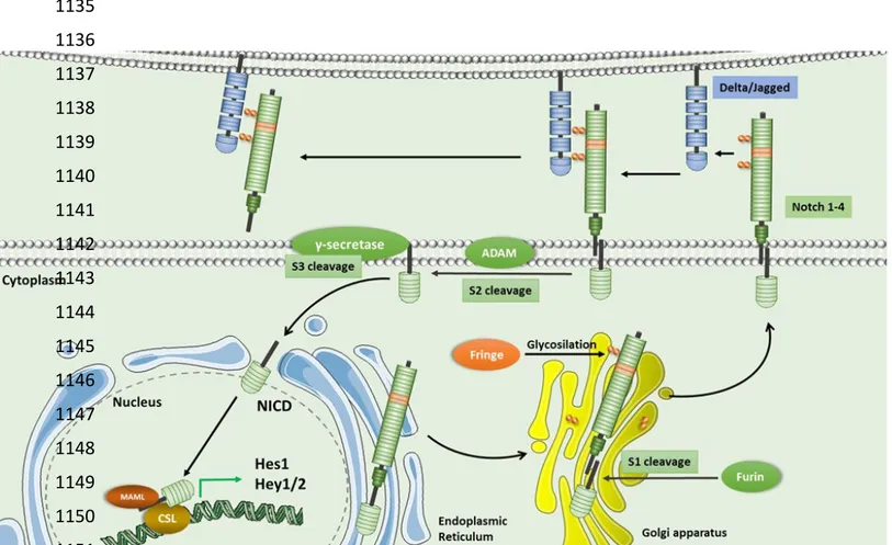
2 The Notch pathway
In mammals, the Notch family comprises four receptors (Notch 1- 4) and five ligands (Delta-like
ligand (Dll) 1, 3, 4 and Jagged (Serrate) 1, 2). The Notch receptors are type I single-pass
transmembrane proteins with a large extracellular ligand binding region (Notch extracellular
domain, NECD), a membrane-spanning and an intracellular domain (Notch intracellular domain,
NICD). The NICD consists of a recombination signal binding protein-1 for J kappa (RBP-Jκ)-4 66 67 68 69 70 71 72 73 74 75 76 77 78 79 80 81 82 83 84 85 86 87 10 11
associated molecule (RAM) domain, seven ankyrin repeats (ANK), edged by two nuclear
localization signals (NLS), a transcription factor scaffold domain or transactivation domain
(TAD), present in Notch 1, 2, 3, and a region rich in proline-glutamate-serine-threonine (PEST)
required for degradation of the protein (1). The Notch ligands are divided into two general
classes, depending on their homology to Drosophila prototypes Delta and Serrate, and are
collectively referred to as DSL family (Delta/Serrate/LAG-2). They are type I transmembrane
proteins with domain organization (Figure 1). Even though these Notch ligands are responsible
for the majority of processes regulated by the Notch signalling, other structurally unrelated,
non-canonical ligands, have also been identified (2). A detailed description of Notch receptors and
ligand and their function is available elsewhere (3).
The interaction between the Notch receptor on one cell and the ligand on the adjacent cell
(trans-interaction) results in conformational changes of the receptor extracellular domain exposing a
motif that is cleaved by ADAM (A Disintegrin And Metalloproteinase) (S2 cleavage site) (4).
The S2 cleavage creates membrane-tethered intermediate called Notch extracellular truncation
(NEXT) that is substrate for -secretase protease complex, containing presenilin1, presenilin2,
Pen-2, Aph-1 and nicastrin (5, 6). The γ-secretase cleaves the Notch receptors at the two distinct
sites, S3/S4, and releases NICD, which translocates to the nucleus to regulate gene transcription
(7). Nuclear Notch signals cause changes in gene expression mediated by the transcription factor
CSL (an acronym for CBF-1/RBP-Jκ in Homo sapiens/Mus musculus, respectively, Suppressor
of Hairless in Drosophila melanogaster, Lag-1 in Caenorhabditis elegans). In the absence of
NICD, CSL is bound by corepressor proteins, such as SMRT (NcoR) and SHRP (MINT/SPEN), 88 89 90 91 92 93 94 95 96 97 98 99 100 101 102 103 104 105 106 107 108 13
and inhibits the transcription of target genes by recruiting histone deacetylases (8). NICD/CSL
binding displaces corepressor complexes and allows recruitment of the transcriptional
coactivator Mastermind-Like-1 (MAML) and histone acetyltransferases such as p300 (9).
Formation of CSL/NICD/MAML complex results in direct transcriptional activation of target
genes (Figure 1). Recent data also show a direct implication of NICD in chromatin remodelling
(10, 11). In cancer and immune system, a Notch signalling active in the cytoplasm (referred to as
“non canonical” Notch signalling to distinguish it from the canonical, nuclear Notch signalling)
has been described (12). Non-canonical Notch signalling occurs without CSL involvement and
depends on interactions between the NICD and pathways such as mammalian target of the
rapamycin 2 complex (mTORC2)/ Akt, Wnt / β-catenin and Nuclear Factor kappa B (NF-κB) /
AKK-α / AKK-β (12). Non-canonical Notch signalling has also been observed in mitochondria
where interactions between Notch and PTEN-induced kinase 1 (PINK1) promote cell survival by
activating the mTORC2/Akt pathway. Notch signalling can be also triggered by ligands other
than Jagged/Dll. These alternative ligands include F3/contactin, Delta-like 1-2 (DLK1-2) and
epidermal growth factor domain 7 (EGFL7) and seem to influence Notch signalling by
competing with Jagged/Dll for Notch receptor binding (12, 13). Notch activation, by either
mechanisms described above, influences cell proliferation, apoptosis, and differentiation (13).
More recently, a role of Notch in the regulation of autophagy has been reported in cancer (14)
and immune cells (15).
6 109 110 111 112 113 114 115 116 117 118 119 120 121 122 123 124 125 126 127 128 16 17
Notch signalling is extremely dose-sensitive, due to the lack of a signal amplification step or
utilization of secondary messengers to transmit the signal from the cell surface to the nucleus
(16). For this reason Notch activity is fine tuned by post-translational regulation (17) and the
stability of the active form of Notch is tighly regulated, mainly by phosphorylation and
ubiquitination (18). Another peculiarity of this signalling pathway, is that, by altering the profile
of ligands and receptors expressed in the cells and/or the affinity of the receptors for a specific
ligand, through glycosylation mediated by a class of enzymes called Fringe (19) (Figure 1),
numerous scenarios of Notch activation patterns can be generated in a specific cell or tissue (20).
Of interest in this context, binding of receptor and ligand in the same cell (cis-interaction) can
lead to the inhibition of signalling (21). Additionally, interplays with other signalling pathways
are of crucial importance in Notch-mediated regulation of several physiological processes, both
during development and postnatally (13). Specifically, interactions between Notch and
Wnt/β-catenin control arterial specification of endothelial cells and regulate epithelial-mesenchymal
transition that initiates myogenesis during embryogenesis (13). Notch and Bone Morphogenetic
Protein (BMP) signalling crosstalk is implicated in cardiac valve formation, regulates blood
vessel branching, and controls chondrocytes proliferation during the formation of the cartilage
(13). Crosstalk between Notch and hypoxia-inducible factors (HIF) regulates hypoxic responses,
in which low oxygen levels result in the increase of NICD that in turn stabilizes HIF-1α and
HIF-2α (13). Interactions between Notch and NF-κB pathway are crucial for the regulation of
cellular immune responses and inflammation (22). Furthermore, in recent times, it has been 129 130 131 132 133 134 135 136 137 138 139 140 141 142 143 144 145 146 147 148 19
shown that the interactions between estrogen receptors and the Notch pathway regulate several
processes underlying cardiovascular homeostasis (23).
As a result of all these interactions, the outcome of Notch activation is cell type- and
context-dependent with multiple combinations of receptors and ligands transducing different biological
effects (10, 24).
2.1 Notch in cardiovascular development
The sequential expression of components of the Notch pathways and related genes is
indispensable during development of heart and vessels. The role of Notch in the cardiovascular
development has been deeply investigated and its discussion is beyond the scope of this review:
we give here a brief overview of the field, to set the stage for our discussion of the role of Notch
in cardiovascular disease, and refer the reader to several excellent reviews for details (25-27).
Notch ligands and receptors are sequentially expressed in the developing heart, thus ensuring
proper heart development. Notch ligand Jagged1 is expressed very early during heart
development, labeling the presumptive valve area of the atrioventricular channel (AVC) and the
trabecular myocardium, while ligand Dll4 and receptors Notch2 and Notch4 are expressed in the
endocardium. Then, Dll4 expression decreases whereas Jagged1 expression is maintained in the
endocardium and is activated in the compacted myocardium (25, 28). This sequential expression
of Notch genes Jagged1, Dll4, Notch2 and Notch4 supports myocardial patterning, maturation
and compaction and cardiac trabeculae formation (25, 28). The Notch pathway plays an
important role for the development of outflow tract (OFT) of the heart which starts with
8 149 150 151 152 153 154 155 156 157 158 159 160 161 162 163 164 165 166 167 168 169 22 23
endothelial-to-mesenchymal transition (EMT) in the endocardial cells leading to the formation of
cardiac valves. The involvement of Notch1 in this context is indicated by the expression of this
receptor in prospective valve endocardium at the beginning of EMT (29). Consistently with the
importance of Notch in the developing heart, mice lacking Notch target genes Hey1 and Hey2
die during embryogenesis due to severe cardiovascular malformations, including impaired
development of EMT (30).
The Notch pathway is crucial for vasculature development by determining arterial-venous
specification mainly through endothelial expression of Dll4 as demonstrated by severe vascular
defects and lack of arterial markers in Dll4-deficient embryos as well as in Rbpj mutants and
Hey1/Hey2 double mutant embryos (30, 31). Notch gain-of-function mouse embryos also develop arteriovenous malformations (32) and ectopic Notch4 and Notch1 expression in
endothelial cells results in the development of arteriovenous malformations and embryonic
vascular remodelling defects (33). Dll4 expression is required for vascular stabilization and
differentiation of the emerging vascular tree. Specifically, the decision to either form a new
sprout or widen the original vessel relies on differential expression patterns of Dll4, BMP and
vascular endothelial growth factor (VEGF) between endothelial cells (34).
Thus, Notch pathway is indispensable for embryonic development and postnatal maintenance of
cardiac and vascular tissues (Table1): its role in postnatal life and the consequent contribution of
its dysregulation to cardiovascular disease will be thoroughly discussed in the next paragraphs. 170 171 172 173 174 175 176 177 178 179 180 181 182 183 184 185 186 187 188 25
3 Congenital heart disease directly associated with Notch pathway mutations
Mutations in the genes of Notch family cause a wide range of congenital defects affecting the
heart and vessel development. These mutations are often characterized by incomplete penetrance
and variable expression. Mutations in human NOTCH1 gene were first described in association
with bicuspid aortic valve (BAV), a congenital heart defect (CHD) in which the aortic valve has
two leaflets instead of three, and this state is most commonly associated with pathologic
calcification of the aortic valve (35) and dilations and aneurysms of the aorta (35, 36). Recently,
we and others have described NOTCH1 gene variants and mutations in aortic stenosis patients
with normal tricuspid valve (37-39). Besides BAV, Preuss at al. studied families with a history
of left-ventricular OFT obstructions and revealed protein-altering mutations clustering
predominantly in genes NOTCH1, ARHGAP31, MAML1, SMARCA4, JARID2 and JAGGED1,
all belonging to the Notch signalling cascade (36).
NOTCH1 and JAG1 mutations have been associated with Tetralogy of Fallot (TOF), a severe developmental CHD (40), characterized by stenosis of right ventricular OFT, ventricular septal
defect, dextraposition of aorta and right ventricular hypertrophy. A recent study of whole exome
sequencing in 829 TOF patients has shown that the NOTCH1 locus is the most frequent site of
genetic variants predisposing to nonsyndromic TOF (41). Mutations in NOTCH1 are also
associated with hypoplastic left heart syndrome, a defect in which the left side of the heart is
underdeveloped (42) and with the Adams-Oliver syndrome (AOS), a rare congenital disease
characterized by cardiac, vascular and neurological symptoms, including valvular and ventricular
10 189 190 191 192 193 194 195 196 197 198 199 200 201 202 203 204 205 206 207 208 209 28 29
abnormalities, atrial septal defect, and TOF (43). In AOS have been also found mutations in
RBPJ and DLL4 (43) and in EOGT, encoding an EGF domain-specific O-linked N-acetylglucosamine transferase which presumably could regulate Notch receptors (44).
A recent study of a cohort of 428 patients with a spectrum of diseases affecting aortic
development such as aortic valve stenosis, a bicuspid aortic valve, aortic valve insufficiency
coarctation of the aorta, and hypoplastic left heart syndrome, subvalvular or supravalvular aortic
stenosis, hypoplastic aortic arch, interruption of the aorta, and mitral valve anomalies clearly
demonstrates that the phenotypic spectrum of NOTCH1 mutations includes a wide variety of
pathologies affecting the whole conotruncus of the heart (45). This is in agreement with the
described role of Notch pathway in determining the fate of neural crest–derived cells. Alagille
syndrome (ALGS), a congenital disease that mainly affects liver ducts and heart development, in
the vast majority (up to 96%) of patients, is caused by mutations in JAGGED1 and NOTCH2 (in
1- 2% of cases)(46).
Lastly, CADASIL (Cerebral Autosomal Dominant Arteriopathy with Subcortical Infarcts and
Leukoencephalopathy), a hereditary autosomal dominant disease, which affects the small
cerebral arteries, thus causing subcortical infarcts and damages to the white matter
(leukoencephalopathy), is associated with mutations in NOTCH3 (47).
4 Cardiovascular disease not always directly associated with defined mutations
As Notch is important for cardiovascular development, it is not surprising that mutations in
genes of the Notch family lead to various types of cardiac and vascular disorders. However, there 210 211 212 213 214 215 216 217 218 219 220 221 222 223 224 225 226 227 228 229 31
is accumulating evidence that a wider spectrum of cardiovascular diseases is associated with
dysregulation of the Notch signalling pathway, even without obvious mutations in Notch-related
genes.
4.1 Calcific aortic valve disease
Calcific aortic valve disease (CAVD) is a frequent heart valve disease characterized by
progressive mineralization of the valvular tissue. Both endothelial and interstitial cells, which
form the aortic valve, contribute to its calcification (48). To some extent, mineralization of the
aortic valve shares similarities with bone ossification, for which Notch is considered as one of
the most important pathways. The exact role of Notch in aortic valve calcification remains
unknown and the existing evidence is controversial. Acharya et al. demonstrated, through
chemical inhibition of Notch by γ-secretase inhibitor DAPT, that inhibition of Notch1 activity
resulted in accelerated calcification while stimulation of Notch signalling attenuated the calcific
process (49). Similarly, Nigam et al. showed that Notch1 in aortic valve cells represses Bmp2
and prevents the progression of osteogenic calcification (50). Furthermore, calcific aortic valve
disease has been associated with higher expression levels of lncRNA H19, which interferes with
the expression of NOTCH1(51). Contrary to these findings, Zeng et al. showed that Notch1
actually promotes osteogenic calcification in human valve interstitial cells (VIC)(52). Recent in
vitro work, using induced pluripotent stem cell (iPSC)-derived endothelial cells, showed that NOTCH1 haploinsufficiency disrupts endothelial cell response to shear stress and unlocks
pro-osteogenic and inflammatory network (53). Whether activation of Notch is pro- or
anti-12 230 231 232 233 234 235 236 237 238 239 240 241 242 243 244 245 246 247 248 249 34 35
osteogenic is unclear. Most probably, the described discrepancies arise from different in vitro
and in vivo experimental conditions used in different laboratories. Our studies show that in
CAVD patients dysregulated Notch signalling is associated with pathological mineralization of
the valve cells (54). Specifically, we report that the profile of Notch-related gene expression is
different in aortic valve interstitial cells of patients, compared to cells of healthy individuals.
This difference is associated with dysregulated Notch-dependent events in the cells of the
patients, such as NICD-dependent induction of EMT and calcification (54). Consistent with the
role of Notch in aortic valve calcification are genetic studies showing that the only proved
candidate gene for BAV, a risk factor of CAVD development, is NOTCH1 (55).
4.2 Aortic aneurysm
Thoracic aortic aneurysm (TAA) is a dangerous condition, which is manifested in patients by
progressive growth of the thoracic aorta diameter due to destructive changes in the aortic wall.
TAA could be a consequence of degenerative or hypertensive aortic enlargement or to less
common genetic disorders, such as Marfan syndrome, Ehlers-Danlos, or other connective tissue
diseases (56).
The aortic wall consists predominantly of endothelial cells and SMCs. Notch ligands expressed
by endothelial cells activate Notch signalling in the underlying SMCs, which in turn ensures
integrin adhesion to the endothelial basement membrane and induces maturation and
differentiation of these cells (57). There are several studies suggesting that lateral induction of
Notch signalling within multiple SMC layers, ensures differentiation induced by an endothelium 250 251 252 253 254 255 256 257 258 259 260 261 262 263 264 265 266 267 268 269 270 271 37
signal (58). Mice with an endothelial deletion of the Jagged1 gene show poor SMCs
differentiation and expression of SMCs markers (59). Furthermore, activation of Notch
signalling in SMCs by endothelial-expressed Jagged1 leads to increased expression of Jagged1
and Notch3 (60) which, together with Notch2, appears to be the most important Notch receptor
for SMCs (61). The critical role of Notch in blood vessels stabilization has been elegantly
demonstrated by the group of Duarte that showed that upregulation of Jagged1 in the
endothelium mediates the recruitement of pericytes needed for the maturation of the new vessels
(62). Lastly, we have recently shown that endothelial cells are capable of driving smooth muscle
osteogenic gene expression via cell-cell contact and activation of Notch signalling (63).
Mutations in NOTCH1 have been linked to BAV, which is associated with ascendic aorta
aneurysm, but a clear involvement of NOTCH1 mutations has not been described for aortic
aneurysm in patients with normal tricuspid aortic valve. In support for a role of Notch1
inactivating mutation in TAA, a recent study has shown that Notch1+/– mice develop aortic root
dilation (64). Consistent with the involvement of Notch in TAA, we have reported a
dysregulation of Notch in aortic endothelial and SMCs cells from TAA patients, regardless the
valve morphology and the presence of mutations in NOTCH1 gene (65-68). This was
concomitant with decreased expression of contractile markers in aortic SMCs of TAA patients
(69), in line with other observations suggesting that dysregulated Notch in endothelial and SMCs
could be involved in changes in differentiation state of SMCs and subsequent disruption of aortic
wall integrity. We have also shown that endothelial cells of TAA patients show dysregulated 14 272 273 274 275 276 277 278 279 280 281 282 283 284 285 286 287 288 289 290 291 292 40 41
Notch, BMP and Wnt/β-catenin related signalling and impairment of Dll4-mediated Notch
activation in response to flow (68). Aortic wall is subjected to a constant mechanical stress and
Notch, Wnt and BMP pathways are critical in maintaini endothelial integrity and proper
differentiation state of endothelial cells (70, 71). Furthermore, activation of Notch in response to
flow is an important differentiation and stress resistance mechanism (72). Our data show that
these Notch-regulated crucial protective mechanisms of the vascular wall are impaired in
aneurysmal patients (68).
In conclusion, the Notch signalling is involved in the development and maintenance of the
vessel wall (Figure 2). Concerted actions of several genes of the Notch family and Notch target
genes are needed for proper vessel function, thus the dysregulation of Notch pathway is
associated with pathological state of aorta and aortic valve.
4.3 Heart failure
Heart failure (HF), the last step of the so called “cardiovascular disease continuum”, is caused by
changes in size and shape of the ventricle (pathological remodelling) consequent to myocardial
infarction (MI), pressure or volume overload, inflammation, or cardiomyopathy. All these
conditions cause the dysregulation of common pathways, leading to altered gene expression,
cellular metabolism, protein turnover and, eventually, to the impairment of the heart contractile
function (73). 293 294 295 296 297 298 299 300 301 302 303 304 305 306 307 308 309 310 311 312 313 43
There is a limited number of studies in patients showing the involvement of Notch in HF
(reviewed in (74)). This is due to limited access to myocardium biopsies and to the scarce
availability of circulating markers to assess the status of activation of Notch in the failing heart.
Increased levels of the Notch ligand Dll1 were found in the serum of patients with HF (75) and
dilated cardiomyopathy (76): in both studies, the levels of Dll1 correlated with the number of
adverse cardiovascular events (75, 76). In patients with dilated cardiomyopathy, the levels of
periostin, a non-canonical Notch ligand, were higher and correlated with the degree of diastolic
dysfunction (76). The biological role and source of these soluble mediators in the serum of HF
patients need further investigation (77). A role for Notch in the pathophysiology of HF has also
emerged in the context of phase 1 clinical studies to assess safety of Dll4-blocking antibodies to
prevent tumor angiogenesis, that reported HF in some of the patients undergoing this treatment
(78, 79).
In contrast with the limited knowledge in patients, there is a large number of in vitro and in vivo
studies showing the critical role of Notch in the maintenance of the homeostasis of the heart and
arteries and the dysregulation of this pathway in HF development. In the next paragraphs, we
will focus on studies relative to the role of Notch in HF caused by atherosclerotic coronary artery
disease.
4.3.1 Role of Notch in plaque formation and progression
Coronary atherosclerosis, responsible in some patients for ischemic heart disease and its different
clinical manifestations, is characterized by a long, often silent phase preceding the plaque
16 314 315 316 317 318 319 320 321 322 323 324 325 326 327 328 329 330 331 332 333 334 335 46 47
formation. The process starts with endothelial dysfunction, defined as the disruption of
endothelium integrity (caused by increased apoptosis and permeability), impairment of its
function (reduced nitric oxide (NO) production) and increased expression of surface proteins
involved in the recruitment of inflammatory cells (80, 81). During the last decade, the role of
Notch in preventing endothelial dysfunctions caused by inflammation, turbulent shear stress,
dyslipidemia and low estrogen levels has been demonstrated by a plethora of in vitro and in vivo
studies (as discussed in (23, 80)) (Figure 3). The first detailed description of molecular
mechanisms underlying the protective role of Notch in the endothelium came from the work in
iPSC (inducible Pluripotent Stem Cell) by Theodoris et al. showing that Notch1 is required to
transduce the positive effects of shear stress and for the normal function of the endothelium (53).
More evidence was provided by the study of Briot et al showing that the downregulation of
endothelial Notch1 is involved in dyslipidemia-induced endothelial dysfunction in regions at risk
for plaque formation, such as the lower aortic arch exposed to pro-atherogenic, turbulent blood
flow (82). Conversely, ivabradine, a heart rate slowing drug, delays plaque formation in the
endothelium of the lower aortic arch of dyslipidemic mice by inducing the expression of
anti-atherogenic genes, including Notch1 (83). Consistent with these observations, Mack et al.
reported that loss of endothelial Notch1 results in increased inflammation and plaque burden in
the aorta of adult mice unequivocally showing that Notch1 is required to maintain cell-cell
junctions, elongated cell morphology, and endothelial alignment with blood flow (84). More
details were provided by Polacheck et al. that showed that shear stress maintains endothelial
barrier function by activating non-canonical Notch1 specifically required for intact adherens 336 337 338 339 340 341 342 343 344 345 346 347 348 349 350 351 352 353 354 355 356 49
junctions (85). Recent work by Jabs et al. has shown that lack of endothelial RBP-Jκ affects
also the heart, by inhibiting the fatty acid transport to cardiomyocytes and causing a switch to
glycolysis, increased expression of “fetal genes” and, consequentely, HF (86). The studies
discussed so far, suggestive of a protective role of Notch1 against inflammation- and/ or
dyslipidemia-induced endothelium damages, are in contrast with the results of a study showing
a role for Notch1/Jagged1 in the interleukin (Il)-1β mediated induction of vascular cell
adhesion protein 1 (VCAM-1) in endothelial cells (87). In line with this in vitro study, a high
cholesterol diet resulted in reduced atherosclerosis in the aortic arch in Apoliprotein E
(ApoE)-deficient mice carrying either endothelial-specific deletion of Rbpj or systemic deletion of
Notch1 (88). Furthermore, mice with endothelial Notch1 knockdown showed attenuated inflammation, due to reduced histone H3K27 acetylation at a subset of NF-κB-directed
inflammatory enhancers (89). The conflicting sets of data from the in vivo studies could be, at
least in part, explained considering that RBP-Jκ deficiency does not impair the Notch
non-canonical signal (90) and that systemic Notch1 deletion will affect the activity and
differentiation of several components of the immune system and different aspects of
inflammatory responses (91, 92). Overall these discrepancies indicate that the role of Notch in
the endothelium requires more investigations (90).
Vascular SMCs are important for plaque stability (93) and the Notch signalling is required for
their survival (94) and to maintain them in a non-proliferative, contractile phenotype (95, 96)
(Figure 3). Consistent with these studies, we found that cholesterol loading of rat aortic 18 357 358 359 360 361 362 363 364 365 366 367 368 369 370 371 372 373 374 375 376 377 52 53
vascular SMCs leads to reduction and induction of contractility and inflammatory markers,
respectively, in association with reduced levels of Jagged1 and Hey2 and increased levels of
Dll4 mRNAs (20). Jagged1/Notch3 axis seems to be the main player in determining the
quiescent phenotype of vascular SMCs (97) but recent work in endothelial Notch1 KO mice
has found that activation of Notch1 by Jagged1, expressed on the adjacent endothelial cells, is
required for Akt-mediated survival of SMCs in the artery wall (94). On the contrary, in the
context of vascular wall remodelling following artery damage, Chen and collaborators found
that miR-34a inhibited neointima formation, that is decreased vascular SMCs proliferation, by
reducing Notch1 expression (98). Similarly, neointimal formation and vascular SMCs
proliferation, following carotid artery ligation, were inhibited by perivascular injection of Notch1
siRNA (99) and -secretase inhibitor DAPT prevented migration and proliferation of SMCs of
ductus arteriosus induced by angiotensin I through the Notch3-Hes1/2/5 axis (100). These results
were confirmed by a study revealing that phospholipase C (PLC) 1, through Akt, specifically
activates Notch1, necessary for intima formation after vessel injury (101). These apparently
opposite actions of the Notch1 and 3 receptors on vascular SMCs functions highlights once again
the complexity of the Notch system. Based on the studies discussed so far they could be, at least
partially, explained by i) the different roles played by Notch1 and Notch3 or by ii) their different
expression levels in vascular SMCs and/or by iii) the different cellular context present in
atherosclerotic plaques and in the damaged artery wall. 378 379 380 381 382 383 384 385 386 387 388 389 390 391 392 393 394 395 396 397 55
Macrophages play a major role in plaque formation and evolution. The role of Notch receptors,
mainly Notch1, in promoting the proinflammatory, M1 phenotype, has been thoroughly
investigated (as reviewed in (92, 102)). Notch1, rather than Notch2 and Notch3, plays a crucial
role in determining the M1 phenotype through the activation of the NF-κB pathway (103). This
observation has been confirmed and extended by Singla and collaborators that have reported
reduction of M1 macrophages and pro-inflammatory cytokines production in monocytes
treated with Notch1 siRNA, followed by the enhancement of M2 macrophage differentiation,
characterized by the production of anti-inflammatory cytokines (104). Consistently,
miR-148a-3p, a Notch1-induced miRNA, promotes the differentiation of monocytes into macrophages,
inhibits M2 polarization and favors instead a M1 state (105). In macrophages isolated from
Notch1 KO mice and in the macrophage cell line Raw 264.7 treated with Notch1 siRNA, lipopolysaccharides (LPS)-induced NF-κB and hypoxia-inducible factors (HIF)-1α activation
were decreased, confirming that LPS requires Notch1 for the transcriptional upregulation of
M1 genes (NO synthase-2, Nos2, Tumor necrosis factor-alpha, Tnfα, and Interleukin-1β, Il-1β)
(106). Of interest, in this study Notch1 activation was linked to metabolic upregulation of
mitochondrial oxidative phosphorylation and reactive oxygen species (ROS) production (106).
In terms of ligands, both Dll4 and Jagged1 have been found involved in macrophages function.
Dll4, induced by Il-1β and LPS, exacerbates the inflammatory response of macrophages (107)
and interferes with Il-4-induced M2 phenotype, causing instead macrophages apoptosis (108).
Other groups have confirmed the role of Dll4, expressed on endothelial cells, in
Notch-mediated promotion of a M1 pro-inflammatory fate (109) and shown that blockade of Dll4-20 398 399 400 401 402 403 404 405 406 407 408 409 410 411 412 413 414 415 416 417 418 58 59
mediated pro-inflammatory activation of macrophages interferes with atherosclerosis
progression (110). Foldi et al. have reported instead that LPS strongly induces Jagged1/Notch1
signalling, leading to the amplification of inflammatory response (111) (Figure 3). In addition
to the studies described so far showing mainly a role for Notch1 in macrophages activation,
other authors have reported the involvement of other isoforms of Notch receptors in the
regulation of macrophages function. Specifically, Notch3-activated signalling has been shown
to be crucial for inflammatory phenotype of macrophages (107) and canonical Notch2
signalling, together with Notch1, seems to promote macrophages function in wound repair (112).
Furthermore in mice has been shown that Dll1 expressed in the endothelium activates Notch2
signalling in monocytes, promoting the conversion of Ly6Chi monocytes into Ly6Clo
monocytes, involved in repair of ischemic tissues (113).
Macrophages are not the only cells of the immune system involved in atherosclerosis: CD4 and
CD8 T-cells also contribute to plaques formation and the dysregulation of the functions of the
regulatory T-cells (Tregs) is involved in the progression of this disease (92). Trying to blunt this
complex response, without compromising the immune defenses, represents a major translational
goal of the field of immunology of atherosclerosis (114, 115). The role of Notch in the regulation
of immunity has been thoroughly investigated in physiological conditions (92, 116), and in
cancer (117). Little is known about Notch-mediated regulation of acquired immunity in the
context of atherosclerosis (92): this knowledge could provide extra tools to harness this host
defense program in our favor. 419 420 421 422 423 424 425 426 427 428 429 430 431 432 433 434 435 436 437 438 439 61
4.3.2 Notch in the ischemic heart and post-infarction remodelling
The role of Notch1 in stimulating the proliferation of immature cardiomyocytes and of their
precursors has been showed by a large number of studies (reviewed in (74)). Recent work
addressing heart regeneration in neonatal mice has shed more light on the mechanism by which
Notch 1 sustains cardiomyocyte proliferation by showing that acetylation extends the half-life
of the Notch1 intracellular domain and enhances its transcriptional activity (118). It is widely
accepted that Notch1 is turned off in mature cardiomyocytes (74) because of epigenetic
modifications at Notch-responsive promoters that appear to be irreversible (119). There are,
nevertheless, numerous studies showing that the reactivation of Notch1 in the border zone of
the infarct or in overloaded heart prevents cardiomyocytes apoptosis, reduces the extent of
fibrosis and limits the hypertrophic response (reviewed in (74)). Both in the developing and the
stressed heart, Notch1 signalling is mainly activated via expression of Jagged1 on the surface
of cardiomyocytes. After activating Notch1, Jagged1 is endocytosed by the signalling cells and
processed to a Jagged1 intracellular domain which binds to N1ICD and blunts its activity in the
nucleus, thus contributing to the regulation of Notch signalling (120).
22 440 441 442 443 444 445 446 447 448 449 450 451 452 453 454 455 456 457 458 459 64 65
The Notch1 signalling pathway has been widely investigated in animal models of
ischemia/reperfusion (I/R) injury. In the heart subjected to I/R injury, Notch1 has a protective
function by counteracting oxidative/nitrate stress, decreasing expression of inducible NOS
(iNOS), increasing endothelial NOS (eNOS) phosphorylation and increasing the
phosphorylation of Akt (121, 122). In addition to Notch1, Notch3 overexpression is involved
in cardioprotection during I/R injury, by activating Akt signalling and by maintaining
mitochondrial function (123). Consistently with the protective role of Notch during I/R injury,
diabetes seems to augment the severity of MI by downregulating Notch1 and Jagged1 (124).
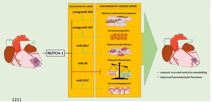
Furthermore, inhibition of miR-363, upregulated during in vitro ischemia, protects
cardiomyocytes through the activation of Notch1 and the induction of Hes1 and Hey1 (125).
Several compounds able to limit the I/R damage have been shown to activate Notch1
signalling, such as G1, an agonists of the G protein-coupled estrogen receptor, (GPER30)
(126), 2,3,5,4'-Tetrahydroxystilbene-2-O-β-D-glucoside, a compound extracted from
Polygonum multiflorum Thunb (127), and the alkaloid berberine (128). For both G1 and berberine, the cardioprotective action has been linked to Notch1-mediated activation of Akt.
(126) (128). Lastly, the Notch1 signalling is activated and contributes to the reduction of
cardiomyocyte apoptosis associated to ischemic pre- and post-conditioning, both approaches
being used to limit heart damages caused by a MI (129, 130).
An important question to be answered about the role played by Notch1 in heart repair is: in
which cell types of the damaged heart Notch1 is reactivated, and according to which molecular 460 461 462 463 464 465 466 467 468 469 470 471 472 473 474 475 476 477 478 479 480 67
mechanisms?. Felician et al. have shown that, due to the methylation of its promoter (119), it is
unlikely that Notch1 is reactivated in mature cardiomyocytes. On the contrary, work by Boni et
al. found that activation of Notch1 in c-kit-positive-cardiac precursor cells favors their myocytes
rather than fibroblast lineage, still maintaining them in a highly proliferative state (131). This
observation is consistent with the study by Nemir et al. in a mouse model of overloaded
myocardium in which Jagged1 overexpressed in cardiomyocytes stimulates the expansion of
Nkx2.5-positive cardiac precursor cells (132).
The extent of early ventricular remodelling following a MI is determined by a delicate balance
between timing and intensity of the inflammatory response, first required to remove the dead
cells debris, and the fibrosis, which will replenish the space left by the dead cardiomyocytes
(74, 133). In adult heart subjected to pressure overload, Notch1 reduces fibrotic response by a
Jagged1-mediated differentiation of cardiac precursor cells into cardiomyocytes rather than
fibroblasts (132). Additionally, Notch1 activation ameliorates cardiac fibrosis by inhibiting
transforming growth factor-β (TGF-β) signalling- induced fibroblast-myofibroblast transition
(134, 135). Noteworthy, relaxin, a natural hormone with antifibrotic capacity, reduces aberrant
TGF-β-mediated collagen deposition and fibrosis by activating Notch1 signalling (134). In
agreement with these data showing the involvement of Jagged1/Notch1- mediated signalling in
the damaged heart, intra-myocardial delivery of a peptide mimic of Jagged1 improves cardiac
function in rats subjected to MI by decreasing myocardial fibrosis (136).
24 481 482 483 484 485 486 487 488 489 490 491 492 493 494 495 496 497 498 499 500 501 70 71
MI elicited-inflammation leads to activation of interferon regulatory factor 3 (IRF3) and type I
interferons (IFNs) in cardiac macrophages which, in turn, induces further damages to the heart.
Interruption of IRF3-dependent signalling decreases cardiac expression of inflammatory
cytokines and attenuates ventricular dilation, thus improving cardiac function (137). Based on
this evidence, IRF3 and the type I IFN response could be a potential therapeutic target for
cardioprotection. Given the role played by Notch in the stimulation of inflammatory response in
macrophages and in the production of type I IFN by plasmacytoid dendritic cells (138), it would
be interesting to determine if Notch inhibition could reduce heart damages associated with
IRF3/IFN and, in general, with inflammatory response during I/R. In agreement with this
hypothesis, in an animal model of stroke, treatment with a Notch inhibitor reduced the size of
cerebral damage by interfering with the recruitment of neutrophils leukocytes (139).
A Notch involvement may be relevant also for arrhythmias. Inflammation-dominated
sympathetic sprouting adjacent to the necrotic area has been implicated in the etiology of
arrhythmias resulting in sudden cardiac death. At 3 days post-MI, high NICD levels were
observed in the macrophages infiltrating the infarct area. The administration of DAPT (30 min
before MI and then daily) decreased the number of macrophages and attenuated the expression
of nerve growth factor, thus preventing the process of sympathetic hyperinnervation and
arrhythmias (140).
4.3.3 Notch and post-infarction angiogenesis 502 503 504 505 506 507 508 509 510 511 512 513 514 515 516 517 518 519 520 521 522 73
Targeting angiogenesis could restore the microcirculation in the reperfused MI, reducing adverse
clinical outcome (141). The role of Dll4/Notch1 in regulating angiogenesis during development
and after birth, both under physiological conditions or tumor angiogenesis, has been thoroughly
investigated (80, 142). In the heart, Dll4 is expressed mainly in the endothelium, which also
expresses Notch1, nevertheless little is known about the role of Dll4/Notch1 in the ischemic
heart and in the progression to HF. Kratsios et al. were the first to show that overexpression of
Notch1 in the infarcted myocardium leads to increased angiogenesis and improved cardiac
function (143). Consistent with an active role of Dll4/Notch1 in regulating heart angiogenesis,
Jabs et al. reported reduced fractional shortening and ejection fraction following injection of an
anti-Dll4 antibodies in wild type C57BL/6 mice and increased blood vessels density, without
defects in blood perfusion, in the heart of mice lacking endothelial RBP-Jκ (86). It should be
noted that in the context of myocardial angiogenesis, RBP-Jκ may also act, independently of
Notch1, by binding and inhibiting HIF-1α and HIF-2α (144). In agreement with these
observations suggesting a role for Notch in angiogenesis in the context of HF, we showed that
treatment with serum of HF patients promotes sprouting angiogenesis and regulates Notch
signalling in human umbilical vein endothelial cells (145).
4.3.4 Notch in stem cells for heart repair
The role of Notch in the regulation of stemness is well defined, both in embryonic and adult
organs. In some tissues, as in the myocardium, Notch1 maintains cells in an undifferentiated,
26 523 524 525 526 527 528 529 530 531 532 533 534 535 536 537 538 539 540 541 542 543 544 545 76 77
high proliferative state and its downregulation is required for the acquisition of the differentiated
state (131). In other tissues, such as the skin, Notch1 is turned off in the stem cells and its
expression and activation is required for cells differentiation (146). Depending on its role as an
inhibitor or promoter of stemness, Notch1 will behave as an oncogene or tumor suppressor gene,
respectively, within a specific cell type (147).
In the last twenty years, a large body of data has been accumulated on the existence of cancer
stem cells (CSCs), derived from the transformation of tissue stem cells or from the acquisition of
progressive mutations of cancer cells (148). These cells, characterized by tissue-specific surface
markers (149), grow very slowly, compared to the bulk tumor cells, and therefore do not respond
to classical chemotherapy, thus causing tumor recurrence (150). Of interest, CSCs in tumors rely
on Notch signalling for their survival, thus inhibition of Notch in these cells seems to be a
promising attempt to prevent cancer growth and recurrence (151).
After a MI there is increased recruitment from the bone marrow of endothelial progenitor and
mesenchymal stem cells (EPCs and MSCs, respectively) that may contribute to the repair of the
damaged myocardium (74). This observation provided the basis for the use of intracoronary
injection of patients stem cells for heart regeneration following a MI. However and
unfortunately, so far there has been no clinical benefit from this treatments (152). It should be
noted, however, that this approach is relatively new, thus, more knowledge of the molecular
mechanisms regulating the function of these stem cells, together with more studies to identify the 546 547 548 549 550 551 552 553 554 555 556 557 558 559 560 561 562 563 564 565 566 79
best cell types to be used and/or new modalities to enhance engraftment and cells function,
should help to achieve heart regeneration by using patients stem cells (152). Of relevance in this
context, Notch1 modulates the recruitment and functions of EPCs and MSCs (as reviewed in (74,
80)). Noteworthy, hypoxic preconditioning enhances proliferation, migration of EPCs and the
secretion of NO and VEGF by activating the Notch-Jagged1 axis (153). Targeting Notch could
therefore help to obtain more “performing” stem cells to be used for myocardium repair.
One of the mechanism by which stem cell injection may favor heart repair is the formation of
new cardiomyocytes from resident cardiac progenitor cells (CPCs) (154). According to several
studies the myocardium has the potential to heal itself: cardiomyocytes renewal in human has
been documented, with a gradual decrease from 1% turning over annually at the age of 20 to
0.3% at the age of 75 (155) and CPCs have been identified in human explanted hearts (156).
Targeting Notch1 could enhance this intrinsic ability of the heart to repair itself since, as
already discussed, Notch1 maintains CPCs in a high proliferative state (131). Consistent with
this observation, Adeno associated virus- based gene transfer of activated Notch1 intracellular‐ ‐
or soluble Jagged1 increases number and size of cardiospheres originated from a‐
heterogeneous population of poorly differentiated cells outgrowing from in vitro cultured
cardiac explants (157). Interestingly, CPCs isolated from human heart differentiate
predominantly in smooth muscle cells but can be redirected to the cardiomyocyte fate by
transient activation, followed by inhibition, of Notch1 signalling (158). However, future studies
aimed to promote myocardium repair by targeting CPCs should consider a recent work using 28 567 568 569 570 571 572 573 574 575 576 577 578 579 580 581 582 583 584 585 586 587 82 83
single cell mRNA sequencing that found no evidence for the existence of a quiescent cardiac
stem cell population, further quenching the enthusiasm toward the promotion of heart repair by
activation of resident cardiac stem cells (159).
5 Conclusions
In summary, the Notch pathway plays a crucial role during the development and postnatally, for
the mantainance of the homeostasis of many tissues and organs, including the heart, arteries and
immune cells. Today, a large body of evidence shows that the dysregulation of Notch, as already
shown in cancer, is involved in congenital and not congenital cardiovascular diseases (Table 1).
6 Expert opinion: Notch a novel therapeutic target for cardiovascular disease?
Accumulating evidence shows that cancer and cardiovascular disease share risk factors and
numerous underlying molecular pathways. One of the most recent example of the overlapping
between these deadly diseases is the discovery that mutations in hematopoietic blood cells,
known to increase the risk of hematological malignancies, are also associated with nearly a
doubling in the risk of coronary heart disease in humans and with accelerated atherosclerosis in
mice (160). Additionally, the work by Meijers et al. has shown that a heart damaged by MI
releases soluble factors that promote cancer progression (161) shedding light on the possible
mechanisms underlying the increased risk of cancer observed in HF patients (162). This
connection between HF and cancer has also emerged from a study showing a 14% increase of
risk of lung cancer in patients under angiotensin converting enzyme (ACE) inhibitors (163). 588 589 590 591 592 593 594 595 596 597 598 599 600 601 602 603 604 605 606 607 608 85
Statins, which are widely used as 3-hydroxy-3-methyl-glutaryl-coenzyme A (HMG-CoA)
reductase inhibitors for LDL lowering therapies, have been recently under investigation for
possible effects on cancer incidence and progression (as reviewed in (164)).
Based on all these links, it is not surprising that drugs that are successfully used for cancer
therapy are now being used to treat cardiovascular disease, and vice versa. One of these drugs is
teniposide, a topo II (DNA topoisomerase II) inhibitor used for cancer treatment which recently
has been shown to reduce vascular calcification (135). Additionally, inhibitors of the EGFR
(Epidermal Growth Factor Receptor), such as erlotinib, used for targeted therapy of lung cancer,
also reduces atherosclerosis by inducing anergy in CD4 T cells (114, 165). Exploratory analyses
have found that anti-inflammatory therapy with canakinumab, a monoclonal antibody that
neutralizes Il-1β, not only reduces cardiovascular events in patients with cardiovascular disease
(166) and the development of chronic viral myocarditis (167) but also the incidence of mortality
attributable to lung cancer (115).
The Notch pathway represents another link between cancer and cardiovascular diseases. Since
Notch activation promotes survival of cancer cells in solid tumors and leukemias, large effort are
being put into the inhibition of Notch to render cancer more responsive to existing
chemotherapeutic agents (151, 168). Clinical trials are currently investigating small molecules
inhibitors of -secretase, but, in parallel, antibodies against the different isoforms of Notch
receptors and/or ligands together with decoy ligands, are also being tested in preclinical (151,
168) and pilot clinical (169) studies. The clinical trials conducted so far, targeting Notch alone or 30 609 610 611 612 613 614 615 616 617 618 619 620 621 622 623 624 625 626 627 628 629 88 89
in combination with other agents, have shown little, not extraordinary response (170). A recent
case study has reported total remission in a patient with early T-cell progenitor acute
lymphoblastic leukemia treated with -secretase inhibitor BMS-906024. Next Generation
Sequencing analyses have shown the presence of Notch mutations in leukemicblasts possibly
driving this tumor (171) suggesting the need for genomic analyses to identify super-responders
to anti-Notch treatment. When taking under consideration the challenges of targeting Notch in
cancer, it should be also considered that Notch is a suppressor gene in some tissues, such as the
skin (172) and not surprising skin tumors have been observed following treatment with Notch
inhibitors (168). Additionally, since Notch is a major player in the modulation of the immune
system (92, 173) long term studies should evaluate the effects on immunity of Notch inhibitors.
Also, based on the discussed crucial role of Notch in the cardiovascular system, the potential
cardiotoxicity of these Notch-targeting cancer drugs should be evaluated in long term studies
(174).
Conversely, Notch1 could be targeted to reduce the cardiotoxicity caused by some anti-cancer
agents. It is widely recognized that lack of estrogens causes endothelial dysfunction (23) and our
data show that estrogens protects the endothelium by activating Notch1 (175). Hence, the
cardiotoxicity observed in association with anti-estrogen treatment for breast cancer could be due
to endothelial Notch1 inhibition , and thus, it could be reduced by restablishing Notch1
signalling in this tissue (23). Similarly, given the pivotal role of Notch1 in the stressed heart,
studies aimed to investigate the involvement of this receptor in cardiomyocytes death, caused by 630 631 632 633 634 635 636 637 638 639 640 641 642 643 644 645 646 647 648 649 650 91
doxorubicin, trastuzumab and lapatinib treatments, could lead to novel, Notch-based, therapeutic
approaches to prevent cardiotoxicity linked to the treatment with these drugs (74, 174).
Due to the complexity of the Notch signalling, thoroughly discussed in the previous
paragraphs, the dissection of the mechanism of actions of the different Notch receptors and
their ligands, similarly to cancer, should be a crucial step for the targeting of this pathway for
cardiovascular diseases. Specifically, studies aimed to identify upstream or downstream
regulators and effectors of Notch signalling could facilitate the targeting of Notch in this
context. For example, it is known that Notch1 inhibition could prevent intima thickening, but
given the divergent effects of Notch receptors on vascular remodelling and in the endothelium,
the use of a -secretase inhibitor (GSI) to inhibit Notch1 may give undesired effects to other
Notch receptors. To overcome this problem Jiang et al, based on data showing that PLC
1-Akt–mediated Notch1 signalling is crucial for intima formation, found that specific inhibition
of the PLC1 and Akt interaction could be a promising therapeutic strategy for preventing
vascular remodelling (101). Another issue, given the multiple and opposite roles played by
Notch in multiple tissues, is the modality of administration of novel pharmacological agents.
Activation of Notch1 to reduce ischemia-caused damages in cardiomyocytes and to prevent
endothelial dysfunction, should be accomplished locally to avoid general activation of
inflammatory pathways. Nanoparticles, successfully utilized to reduce the MI damage by
delivering drugs targeting infiltrating cells (176), could be used to deliver miRNAs that
32 651 652 653 654 655 656 657 658 659 660 661 662 663 664 665 666 667 668 669 670 94 95
regulate Notch1 (125, 177, 178). These miRNAs could be also administrated systemically
(179) or locally, by intracardiac injection (180) or by hydrogels-functionalized with a peptide
mimic of Jagged1 (136) (Figure 3, 4). At this purpose hydrogels-functionalized with miRNAs,
similarly to those with a peptide mimic of Jagged1 (136) could be used. Similar approaches
could be exploited to inhibit Notch1 in leucocytes, to diminish inflammatory response, or to
reduce Dll4-mediated Notch signalling in plaques. In the context of atherosclerosis,
GSI-eluting stent could also be developed to prevent vascular SMCs proliferation and intima
thickening.
To the best of our knowledge, probably due to the many challenges to be overcome discussed
in this review, no clinical trials targeting Notch for cardiovascular disease have been conducted
so far. Based on our discussion, it emerges that effective strategies for the targeting of Notch in
cardiovascular diseases should benefit from the interaction between basic and clinical
researchers involved in cancer or cardiovascular diseases. This collaboration, aimed to further
understand the pathological role of Notch pathway and to identify novel therapeutic
interventions targeting both cancer and heart and vessels disease, will expand the established
field of the cardio-oncology, which today aims mainly to reduce the negative effect of cancer
drugs on the cardiovascular system.
Funding 671 672 673 674 675 676 677 678 679 680 681 682 683 684 685 686 687 688 689 97
Supported by the Grant of Russian Science Foundation 18-14-00152 to AM, Fondo Ateneo per
la Ricerca 2017, University of Ferrara to PR and Fondazione Annamaria Sechi per il Cuore
(FASC).
Declaration of interest
The authors have no relevant affiliations or financial involvement with any organization or entity
with a financial interest in or financial conflict with the subject matter or materials discussed in
the manuscript.
References
Papers of special note have been highlighted as either of interest (•) or of considerable interest (••) to readers
X1. Miele L (2006) Notch signaling. Clin. Cancer Res 12(4):1074-1079.
2. D'Souza B, Meloty-Kapella L, & Weinmaster G (2010) Canonical and non-canonical Notch ligands. Curr
Top Dev Biol 92:73-129.
3. Kovall RA, Gebelein B, Sprinzak D, et al. (2017) The Canonical Notch Signaling Pathway: Structural and Biochemical Insights into Shape, Sugar, and Force. Dev. Cell 41(3):228-241.
34 690 691 692 693 694 695 696 697 698 699 700 701 702 703 704 705 706 707 708 709 710 711 712 100 101
4. Brou C, Logeat F, Gupta N, et al. (2000) A novel proteolytic cleavage involved in Notch signaling: the role of the disintegrin-metalloprotease TACE. Mol Cell 5(2):207-216.
5. Struhl G & Greenwald I (2001) Presenilin-mediated transmembrane cleavage is required for Notch signal transduction in Drosophila. Proc Natl Acad Sci U S A 98(1):229-234.
6. Selkoe DJ & Wolfe MS (2007) Presenilin: running with scissors in the membrane. Cell 131(2):215-221.
7. Kopan R & Ilagan MX (2009) The canonical Notch signaling pathway: unfolding the activation mechanism. Cell 137(2):216-233.
8. Morel V, Lecourtois M, Massiani O, et al. (2001) Transcriptional repression by suppressor of hairless involves the binding of a hairless-dCtBP complex in Drosophila. Curr Biol 11(10):789-792.
9. Wallberg AE, Pedersen K, Lendahl U, et al. (2002) p300 and PCAF act cooperatively to mediate transcriptional activation from chromatin templates by notch intracellular domains in vitro. Mol
Cell Biol 22(22):7812-7819.
10. Bray SJ (2016) Notch signalling in context. Nat. Rev. Mol. Cell Biol 17(11):722-735.
11. Skalska L, Stojnic R, Li J, et al. (2015) Chromatin signatures at Notch-regulated enhancers reveal large-scale changes in H3K56ac upon activation. The EMBO journal 34(14):1889-1904.
12. Ayaz F & Osborne BA (2014) Non-canonical notch signaling in cancer and immunity. Front Oncol 4:345.
13. Siebel C & Lendahl U (2017) Notch Signaling in Development, Tissue Homeostasis, and Disease.
Physiol Rev 97(4):1235-1294.
• This review provides an overview of the role of Notch signaling in regulating tissue homeostasis, with examples from skin, liver, lung, intestine, and the vasculature.
14. Palomero T & Ferrando A (2009) Therapeutic targeting of NOTCH1 signaling in T-cell acute lymphoblastic leukemia. Clin. Lymphoma Myeloma 9 Suppl 3:S205-S210.
15. Marcel N & Sarin A (2016) Notch1 regulated autophagy controls survival and suppressor activity of activated murine T-regulatory cells. Elife 5.
16. Yamamoto S, Schulze KL, & Bellen HJ (2014) Introduction to Notch signaling. Methods in molecular
biology 1187:1-14.
17. Borggrefe T, Lauth M, Zwijsen A, et al. (2016) The Notch intracellular domain integrates signals from Wnt, Hedgehog, TGFbeta/BMP and hypoxia pathways. Biochim Biophys Acta 1863(2):303-313.
18. Andersson ER, Sandberg R, & Lendahl U (2011) Notch signaling: simplicity in design, versatility in function. Development 138(17):3593-3612. 713 714 715 716 717 718 719 720 721 722 723 724 725 726 727 728 729 730 731 732 733 734 735 736 737 738 739 740 741 742 743 103
19. Kakuda S & Haltiwanger RS (2017) Deciphering the Fringe-Mediated Notch Code: Identification of Activating and Inhibiting Sites Allowing Discrimination between Ligands. Dev. Cell 40(2):193-201.
20. Aquila G, Fortini C, Pannuti A, et al. (2017) Distinct gene expression profiles associated with Notch ligands Delta-like 4 and Jagged1 in plaque material from peripheral artery disease patients: a pilot study. J Transl. Med 15(1):98.
• This study reveals a possible role of Notch signaling in the pathophysiology of peripheral artery disease and suggests that ligand-specific activation of this pathway could determine the plaque characteristics and therefore progression of the disease.
21. Sprinzak D, Lakhanpal A, Lebon L, et al. (2010) Cis-interactions between Notch and Delta generate mutually exclusive signalling states. Nature 465(7294):86-90.
22. Osipo C, Golde TE, Osborne BA, et al. (2008) Off the beaten pathway: the complex cross talk between Notch and NF-kappaB. Lab Invest 88(1):11-17.
23. Fortini F, Vieceli Dalla Sega F, Caliceti C, et al. (2019) Estrogen-mediated protection against coronary heart disease: The role of the Notch pathway. J Steroid Biochem Mol Biol 189:87-100.
24. Aster JC, Pear WS, & Blacklow SC (2017) The Varied Roles of Notch in Cancer. Annu Rev Pathol 12:245-275.
25. Luxán G, D’Amato G, MacGrogan D, et al. (2016) Endocardial Notch signaling in cardiac development and disease. Circulation research 118(1):e1-e18.
26. Papoutsi T, Luna-Zurita L, Prados B, et al. (2018) Bmp2 and Notch cooperate to pattern the embryonic endocardium. Development.
27. de la Pompa JL & Epstein JA (2012) Coordinating tissue interactions: Notch signaling in cardiac development and disease. Developmental cell 22(2):244-254.
28. D'Amato G, Luxan G, & de la Pompa JL (2016) Notch signalling in ventricular chamber development and cardiomyopathy. FEBS J 283(23):4223-4237.
29. Del Monte G, Grego-Bessa J, Gonzalez-Rajal A, et al. (2007) Monitoring Notch1 activity in development: evidence for a feedback regulatory loop. Dev Dyn 236(9):2594-2614.
30. Fischer A, Steidl C, Wagner TU, et al. (2007) Combined loss of Hey1 and HeyL causes congenital heart defects because of impaired epithelial to mesenchymal transition. Circ Res 100(6):856-863.
31. Krebs LT, Shutter JR, Tanigaki K, et al. (2004) Haploinsufficient lethality and formation of arteriovenous malformations in Notch pathway mutants. Genes & development 18(20):2469-2473. 36 744 745 746 747 748 749 750 751 752 753 754 755 756 757 758 759 760 761 762 763 764 765 766 767 768 769 770 771 772 773 774 106 107
32. Duarte A, Hirashima M, Benedito R, et al. (2004) Dosage-sensitive requirement for mouse Dll4 in artery development. Genes & development 18(20):2474-2478.
33. Krebs LT, Starling C, Chervonsky AV, et al. (2010) Notch1 activation in mice causes arteriovenous malformations phenocopied by ephrinB2 and EphB4 mutants. Genesis 48(3):146-150.
34. Mack JJ & Iruela-Arispe ML (2018) NOTCH regulation of the endothelial cell phenotype. Current
Opinion in Hematology 25(3):212-218.
•• This study reveals that NOTCH1 is required to maintain junctional integrity and to prevent atherosclerosis. NOTCH1 signaling is also necessary in adult arteries to interpret hemodynamic forces and initiate appropriate biological responses required for vascular homeostasis and atheroprotection.
35. Garg V, Muth AN, Ransom JF, et al. (2005) Mutations in NOTCH1 cause aortic valve disease. Nature 437(7056):270-274.
36. Preuss C, Capredon M, Wünnemann F, et al. (2016) Family Based Whole Exome Sequencing Reveals the Multifaceted Role of Notch Signaling in Congenital Heart Disease. PLoS Genet 12(10):e1006335.
37. Ducharme V, Guauque-Olarte S, Gaudreault N, et al. (2013) NOTCH1 genetic variants in patients with tricuspid calcific aortic valve stenosis. J Heart Valve Dis 22(2):142-149.
38. Freylikhman O, Tatarinova T, Smolina N, et al. (2014) Variants in the NOTCH1 gene in patients with aortic coarctation. Congenit Heart Dis 9(5):391-396.
39. Irtyuga O, Malashicheva A, Zhiduleva E, et al. (2017) NOTCH1 Mutations in Aortic Stenosis: Association with Osteoprotegerin/RANK/RANKL. Biomed Res Int 2017:6917907.
40. Guida V, Chiappe F, Ferese R, et al. (2011) Novel and recurrent JAG1 mutations in patients with tetralogy of Fallot. Clinical genetics 80(6):591-594.
41. Page DJ, Miossec MJ, Williams SG, et al. (2019) Whole Exome Sequencing Reveals the Major Genetic Contributors to Nonsyndromic Tetralogy of Fallot. Circ Res 124(4):553-563.
42. Iascone M, Ciccone R, Galletti L, et al. (2012) Identification of de novo mutations and rare variants in hypoplastic left heart syndrome. Clinical genetics 81(6):542-554.
43. Southgate L, Sukalo M, Karountzos AS, et al. (2015) Haploinsufficiency of the NOTCH1 receptor as a cause of Adams–Oliver syndrome with variable cardiac anomalies. Circulation: Genomic and
Precision Medicine 8(4):572-581. 775 776 777 778 779 780 781 782 783 784 785 786 787 788 789 790 791 792 793 794 795 796 797 798 799 800 801 802 803 804 109