UNIVERSITA’ DEGLI STUDI DI MESSINA
Department of Chemical, Biological, Pharmaceutical and Environmental Sciences
PhD in: APPLIED BIOLOGY AND EXPERIMENTAL MEDICINE
XXXI Cycle
Coordinator: Prof.ssa Maria Assunta Lo Gullo
____________________________________________________________________________
NEW OMICS APPROACHES IMPROVING
CLASSIFICATION AND PERSONALIZED
RETINITIS PIGMENTOSA DIAGNOSIS
PhD Student: Tutor:
Dott. LUIGI DONATO Illustrious Professor
ANTONINA SIDOTI
____________________________________________________________________________
SUMMARY Page
1. INTRODUCTION AND OBJECTIVES OF THE WORK 1
2. CLINICAL, BIOCHEMICAL, MOLECULAR AND GENETIC
ASPECTS OF RP 4
2.1. Clinical signs in RP 4
2.2. Family history 5
2.3. Ophthalmic trial 6
2.3.1. The classic RP instrumental examination 6
2.3.2. Ocular findings linked to RP 7
2.4. Retinal functionality 8
2.5. Retinal imaging 10
2.6. Differential diagnosis for non – syndromic retinitis pigmentosa 14
2.7. Clinical aspects of RP genetic subtypes 32
2.8. Associated genes and proteins 39
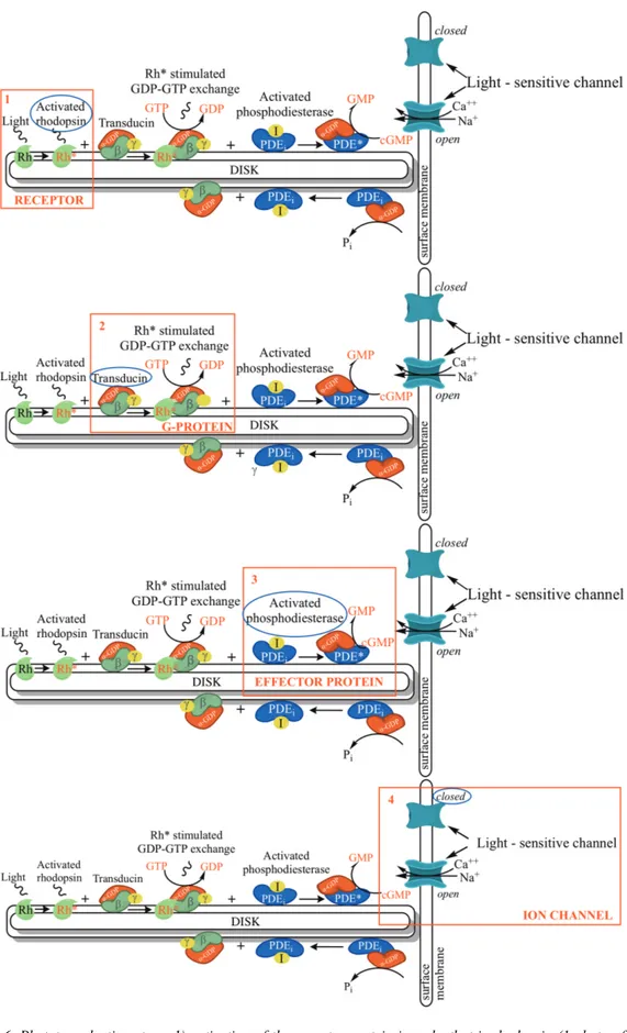
2.8.1. The phototransduction pathway 40
2.8.2. Visual cycle cascade 43
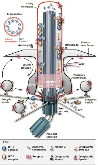
2.8.3. Connecting cilium trafficking 45
2.8.4. Photoreceptor outer segment assembly 47
2.8.5. The interphotoreceptor matrix 49
2.9. Handling of RP 51
2.9.1. Ophthalmic and genetic counseling 51
2.9.2. Visual rehabilitation 53
2.9.3. RP treatment possibilities 53
3. MATERIALS AND METHODS 57
3.1. Genomics 57
3.1.1. Patients’clinical features 57
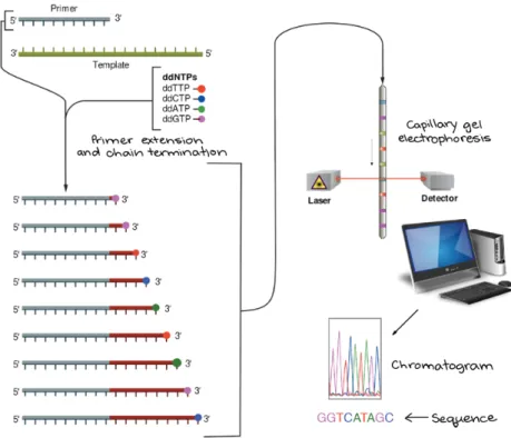
3.1.2. Genotyping (Sanger sequencing) 63
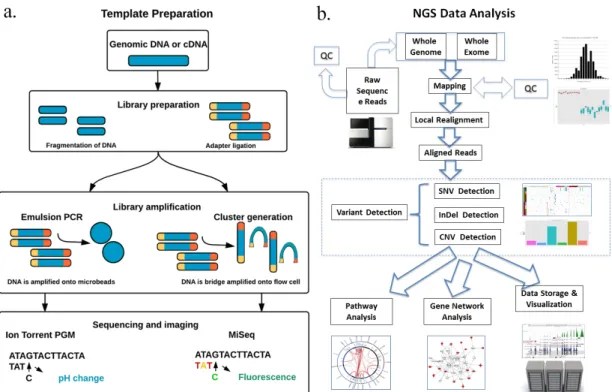
3.1.3. Whole Genome Sequencing (WGS) 64
3.1.4. In silico functional prediction analyses of variant effects 69
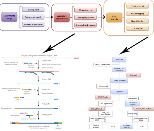
3.2. Transcriptomics 71
3.2.1. Cell cultures 71
3.2.2. Total RNA sequencing 72
3.2.3. Quality validation and read mapping 72
3.2.4. Differential gene and non – coding RNA expression and
statistical analysis 73
3.2.5. Functional gene annotations 74
3.2.6. Filtering and annotation of non – coding RNAs 75
3.2.7. Coding and non – coding genes pathway analyses 79
3.3. Functional and validation assays 82
3.3.1. Sanger validation and family member genotyping 82
3.3.2. Population screening 82
3.3.3. Data validation by qRT – PCR 82
SUMMARY Page
4. RESULTS 86
4.1. Genomics 86
4.1.1. Combined effects of known variants in genes still not
associated to RP 86
4.1.2. Combined effects of known variants and haplotypes in
Stargardt disease causative genes 94
4.1.3. Novel mutations in already known RP causative genes 98
4.2. Transcriptomics 100
4.2.1. Expression changes in RP candidate genes caused by
oxidative stress 100
4.2.2. Discovery of new regulative “hot – spot” withing new
found biochemical pathways 111
5. DISCUSSION 128
5.1. Classification of new RP forms by new genotype – phenotype
associations 128
5.2. Impaired bidirectional neutrotransmission in the inner retina
layers as RP pathogenic mechanism (and not as effect) 139
5.3. Evaluation of new consequences determined by already known
variants in RP causative genes 145
5.4. Classification of new non – syndromic RP forms by discovery
of new biochemical pathways 153
5.4.1. Candidate “macro – pathways” analysis 154
5.4.2. Pathway analysis of altered genes yet associated to retinal
diseases 160
5.4.3. New candidate genes and their possible impact on RP
etiopathogenesis 164
5.5. Classification of new non – syndromic RP forms caused by
alterations in regulation of new specific pathways 176
6. CONCLUSIONS 184
1. INTRODUCTION AND OBJECTIVES OF THE WORK
Retinitis Pigmentosa (RP) is a heterogeneous inherited ocular disease characterized by progressive retinal degeneration. It is considered an uncommon condition, but worldwide prevalence refutes this data, varying approximately from 1:9000 to 1:750 (Na et al., 2017), depending on the geographic location. RP affects 1-5 in 10,000 in Italy, while no data is available on RP frequency in Sicily. The term “pigmentosa” refers to the characteristic appearance of abnormal areas of pigment in the retina in the advanced stages of the disease. Such adjective was first coined by the eminent Dutch ophthalmologist F.C. Donders in 1857, although his colleague A.C. van Trigt described RP four years earlier, by the use of an ophthalmoscope (Naz et al., 2010). Interestingly, already during 19th century several forms of retinal degenerations had already been reported. Among them, we report that in 1744 R.F. Ovelgün described a form of familial night blindness closely resembling RP (Heckenlively, 1987), and the description by F.A. von Ammon of patients with poor vision and pigmented retinal lesions (Marre & Walther, 1990).
RP comprises a group of progressive inherited retinal distrophies (IRDs), generally characterized by the early degeneration of rod photoreceptors, followed by the loss of cone photoreceptors (O'Neal & Luther, 2018). The first symptom is usually reduced night vision, followed by a progressive loss of the peripheral visual field (tunel vision). Macula function is usually relatively well conserved until later stages of the disease. As already mentioned, fundus abnormalities include particular bone spicule pigmentation predominantly in the periphery and/or mid-periphery, along with attenuation of retinal vessels and a waxy pallor of the optic nerve head. Electroretinography can help to reveal the characteristic loss of photoreceptor function, primarily among rods rather than cones in early stages of the disease.
RP is clinically different from other IRDs, including IRDs occurring at birth or within the first few months of life (e.g. Leber congenital amaurosis, or LCA), dystrophies in which cone degeneration precedes rod degeneration (e.g. cone-rod dystrophy), macular dystrophies, and not – progressive disorders like achromatopsia and congenital stationary night blindness (CSNB) (Broadgate, Yu, Downes, & Halford, 2017). Additionally, about 25% of RP patients shows a syndromic form associated
2
with extra-ocular abnormalities. Collectively, these disorders constitute a big group of retinal dystrophies with significantly overlapping clinical and/or genetic findings (Fig. 1). This overlap represents the biggest challenge in IRDs specific classification. Moreover, only few therapeutic options are currently available in daily clinical practice. For that reason, it is fundamental to provide the patient with the best possible information about the expected clinical course and inheritance pattern. In order to realize such purpose, developing a classification system which merges the clinical diagnosis with the underlying genetic factors can provide precious prognostic information about the rate of progression and long-term outcome.
Fig. 1. Venn diagram summarizing the genetic overlap between RP and other inherited retinal dystrophies. Each circle represents a specific clinical diagnosis. The gene names listed in the overlapping areas indicate that mutations in these genes can lead to different phenotypes. Genes marked with an asterisk are candidate genes for non-syndromic RP.; Abbreviations: CRD: cone-rod dystrophy, CSNB: congenital stationary night blindness, ESCS: enhanced S-cone syndrome, LCA: Leber congenital amaurosis, MD: macular dystrophy, RP: retinitis pigmentosa.
3
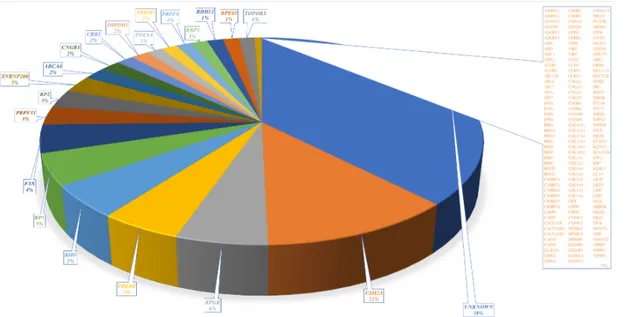
The high heterogeneity showed by RP patients is excellently illustrated by the wide number of genetic defects associated with RP. During the early 90s, the rhodopsin (RHO) gene was identified as the first gene involved in autosomal dominant RP (Shokravi & Dryja, 1993). Since then, mutations in more than 80 genes have been involved in non-syndromic RP (Ali, Rahman, Cao, & Yuan, 2017), and each year new genes are added to this list. Each of these genes belongs to a gene-specific subtype of RP with a specific spectrum of phenotypes. Furthermore, lots of factors can change widely within each of these particular subtypes, even between affected family members, suggesting the presence of unknown genetic and/or environmental factors that could influence the RP phenotype.
The main purpose of our work is to improve the classification of RP orphan forms, along with the clarification of etiopathogenesis mechanisms of already known ones. Such goals were realized by an omics approach, which exploits the modern technique of Next Generations Sequencing and derived “Big Data” to bring out the innermost and heterogeneous genetic sides of retinitis pigmentosa. We start providing a comprehensive overview of the clinical features associated with the different genetic subtypes of non-syndromic and syndromic RP. Additionally, in order to define the state of art about genotype – phenotype associations, we also discuss the role of proteins encoded by causative RP genes in the structure and function of the retina. Finally, we discuss the current therapeutic options and future perspectives for all analyzed RP forms.
4
2. CLINICAL, BIOCHEMICAL, MOLECULAR AND GENETIC ASPECTS OF RP
2.1. CLINICAL SIGNS IN RP
RP leads to a progressive photoreceptor and retinal pigment epithelium (RPE) degeneration, determining tunnel vision, night blindness and a gradual reduction of central vision. Nevertheless, the clinical findings in RP vary widely due to the huge number of genes involved, each of which can have several alleles. Now we examine the common clinical features considered representative of RP.
• Age of onset and progression rate: age of onset varies widely among RP patients. Several patients develop symptomatic visual loss during early childhood, while others can remain relatively asymptomatic up to mid-adulthood. Generally, difficulties with dark adaptation begin in adolescence, whereas typical visual loss during the mid-peripheral field becomes evident during young adulthood. Nevertheless, children’s ability to compensate for peripheral visual loss, along with dark adaptation difficulties detecting caused by our artificially illuminated nighttime environment, complicate the exact age of onset knowledge. Almost always, RP subtypes showing symptoms early in life tend to progress more rapidly. Additionally, pathology severity is correlated with the specific Mendelian pattern of inheritance. Generally, patients affected by X-linked forms (about 10% of RP patients) evidence a more severe disease course compared to patients with autosomal recessive ones (about 55% of RP patients), while autosomal dominant affected individuals (about 35% of RP patients) have the best long-term prognosis with great chances to maintain central vision (Farrar et al., 2017).
• Symptomatology: the beginning symptoms of RP include nyctalopia (night blindness) and dark adaptation difficulties. Interestingly, RP can also show loss
5
of the mid-peripheral visual field, although this is rarely reported as an initial symptom. Generally, the central retina maintains its integrity until the final stages of the pathology, even if anatomical abnormalities can appear early during the development of the disease. Such possibility is characteristic of middle age patients, in which central cone degeneration leads to visual acuity decrease. A large number of RP patients preserve the light detection ability, due to residual macular function or thanks to the retained integrity of peripheral temporal retinal island (Aleman et al., 2011). Photopsia is a widespread but often underestimated symptom (Ohguro et al., 2002) which can be very troubling to patients. This pathological manifestation may be caused by an impairment of afferent nerve synapses following photoreceptor degeneration or spontaneous self-signaling activity as consequence of inner retina remodeling (Jones, Marc, & Pfeiffer, 1995). Although photopsia can occur during early stages of RP, the most striking and disturbing effects are typical of the later stages of the disease (Bittner, Haythornthwaite, Diener-West, & Dagnelie, 2012). In advanced RP, as in Charles Bonnet syndrome (Franke, Rauschenberger, & Fluri, 2018), visual hallucinations can take animate forms. Finally, RP affected patients can also exhibit dyschromatopsia and photophobia (Riveiro-Alvarez et al., 2015).
2.2. FAMILY HISTORY
A detailed analysis of family history is very important in any patient suspected for RP. A pedigree for each proband is useful to assess the pattern of inheritance and may also help during diagnoses. For example, if an X-linked inheritance is suspected, RP2 and
RPGR genes should be sequenced before using next generation sequencing (NGS)
approaches. A pedigree may also exemplify which family members could develop RP and indicate subjects where non-penetrance should be suspected, as for mutations in
6
2.3. OPHTHALMIC TRIAL
2.3.1. The classic RP instrumental examination
Three clinical hallmarks represent the typical signs of RP: attenuation of retinal vessels, a waxy pallor of the optic nerve and, above all, bone spicule pigmentation. In the initial stages of RP, ocular fundus may appear normal, as bone spicule‒shaped pigment accumulation is either absent or sparse, vascular attenuation is not yet pronunciated, and the optic disc appears typical. Before such representative RP alterations, some patients may manifes aspecific abnormalities such as discontinuous whithish lesions localized around RPE, broadening of the foveal reflex and irregular reflexes from the internal limiting membrane.
Retinal vessel shrinkage in RP is not yet defined. Firstly, this clinical aspect was attributed to decreased metabolic demand following ganglion cell death after photoreceptor loss. Another possibility links the loss of oxygen-consuming photoreceptors to a hyperoxic state of the residual inner retina, leading to vasoconstriction and slowing down blood flow in retinal vessels (Sorrentino, Bonifazzi, & Perri, 2015). Moreover, Li et al. found that thinning of the extracellular matrix between the retinal vessels and the migrated RPE cells implies narrowing of the vessels (Z. Y. Li, Possin, & Milam, 1995). At last, Stone et al. hyphotesized that an impairment of synaptic input determined by photoreceptor cell death, along with the subsequent depleting of trophic factors, decreases the metabolism of the inner retinal cells, promoting vascular remodeling followed by vessel thickness (Grunwald, Maguire, & Dupont, 1996). Alternatively, Cellini et al. suggested that ocular blood flow was decreased more than expected by retinal atrophy, turning the debate on whether vascular changes in RP patients play a central role in the onset of RP or are simply secondary to neuroretinal remodeling (D'Orazi, Suzuki, & Wong, 2014). Regarding this, a crucial role for the vasoconstrictor endothelin-1 has been proposed, even if both boosted and reduced plasma levels of this protein have been described among RP patients, thus suggesting the need for further study (Olivares-Gonzalez, Martinez-Fernandez de la Camara, Hervas, Millan, & Rodrigo, 2018). Finally, because most of RP associated genes are involved in either the photoreceptor-RPE complex or
7
the inter-photoreceptor matrix, the most probable hyphotesis give a secondary role to these vascular alterations.
About the waxy pallor of the optic disc, typical of disease progression, it is probably determined by glial cell accumulation both on the surface and inside the optic disc, increasing light reflectance (Al Rashaed, Khan, Nowilaty, Edward, & Kozak, 2016). Curiously, not all RP patients develop characteristics bone spicules, substituted by dust-like pigmentation or nummular hyperpigmentation. The hyperpigmentation level can change among patients and it is not consequently related to the severity of the disease. Bone spicule pigmentation is the histological representation of RPE cell deteaching from Bruch membrane after photoreceptor degeneration, cell that migrate to intraretinal perivascular sites, where they constitute melanin pigment deposits (Jaissle et al., 2010). These bone spicules frequently arise in the mid-periphery, where the density of rods is the highest (Berson, 1993). Due to the high level of interdependence with the choriocapillaris and photoreceptors, the RPE migration might be triggered by the attenuation between the inner retinal vessels and the RPE itself, following photoreceptor degeneration in RHO knock-out mice (G. L. Wang et al., 2005).
2.3.2. Ocular findings linked to RP
Many other different eye – affecting conditions, such as disease-associated refractive error, nystagmus and macular complications, are frequenly associated with RP, and several are responsive to treatment. Cystoid macular edema (CME), together with macular hole and epiretinal membrane formation, represent the most displeasing conditions. CME, the most frequent of them, could be determined by several mechanisms: 1) Müller cell edema and dysfunction, 2) blood-retina barrier disruption, 3) vitreomacular traction, 4) RPE pumping activity alterations, and 5) anti-retinal antibodies (Strong, Liew, & Michaelides, 2017). About 40% of RP patients evidences epiretinal membrane constitution (Fujiwara et al., 2016), probably as consequence of idiopathic preretinal glial cell proliferation or of inflammatory processes (G. Liu, Du, Keyal, & Wang, 2017). The involvement of inflammatory pathways in RP is
8
abundantly proved but, if it was primarily considered as an event following rod and cone death, today inflammatory cells are believed to induce retinal degeneration by their cytotoxic effect on photoreceptors (Massengill, Ahmed, Lewin, & Ildefonso, 2018). Inflammation could be also associated to posterior subcapsular cataract, affecting about 45% of RP patients, which seriously impairs vision, but which can be resolved even during macular compromission (Bruno, Nebbioso, Rigoni, Gagliardi, & Vingolo, 2015). Furthermore, additional but uncommon abnormalities that can arise during RP are represented by vitreous cysts (N. Yoshida et al., 2015) and proteolipidic accumulation in optic nerve head and fiber layer (Garcia-Cazorla, Mochel, Lamari, & Saudubray, 2015). Finally, RP seems to be one of the most frequent hidden pathology in patients affected by secondary retinal vasoproliferative tumors (Honavar, 2018).
2.4. RETINAL FUNCTIONALITY
• Perimetry: one of distinguishing RP feature is progressive loss of the visual field. It is characterized by significant bilateral symmetry (Massof et al., 1979) and arises with mid-peripheral isolated scotomas which gradually merge in a partial or complete ring scotoma. During advanced stages of disease, this annular scotoma expands both inward and outward. Even in the absence of ring scotoma, other known patterns of visual field impairment could occur, as concentric or arcuate (from superior to inferior retina) visual field loss (Bellingrath et al., 2017). Kinetic perimetry is best suitable evaluation method of peripheral visual field loss; the annual rate of decline for target V4e of the Goldmann perimeter varies between 2 and 12%, in relation to gene-specific subtypes (A. Oishi et al., 2013). The degree of central visual field impairment is usually verified by static perimetry, but it was superseded by recent fundus-driven perimetry. Microperimetry, for example, exploits accurate eye tracking throughout the analysis, and provides direct structure-function correlations generating an annotated image of the posterior pole.
9
• Color vision: generally, dyschromatopsia is representative of later stages of RP, with the so-called type III (blue) acquired color vision impairments more diffused then type I (red-green) (Abdelhakim & Rasool, 2018). Blue cone defect has been attributed to the insufficient and irregular distribution of these short-wavelength cones at the fovea (leading to pericentral retinal function loss and tritanopia (blue-yellow color blindness) (Durlu, Koroglu, & Tolun, 2014). Reduction of visual acuity and associated central photoreceptor death increases the probability of developing a type I color dysfunction (Pokorny, Lutze, Cao, & Zele, 2008). Alternatively, vision impairment caused by CME should not have serious effect on color vision (Pinckers, van Aarem, & Keunen, 1993). • Dark adaptometry: an altered dark-adapted threshold is a typical sign of RP.
Reduction of rod sensitivity and related delayed recovery generally increase rod threshold (Birch, Wen, Locke, & Hood, 2011). Recent analyses highlighted increases in both photoreceptor thresholds, a delay in reaching the asymptotic rod threshold, or the entire loss of rod function (Bennett, Klein, Locke, Kiser, & Birch, 2017).
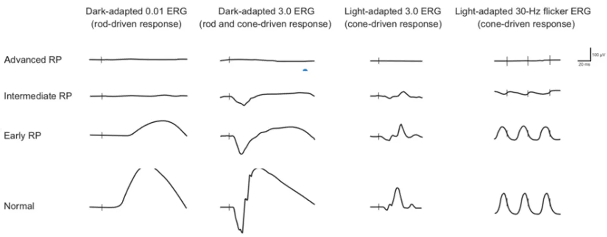
• Electroretinography: the quantitative evaluation of RP severity and progression is primarily realized by full-field electrophysiological testing, according to the ISCEV guidelines (Robson et al., 2018). Electroretinogram (ERG) alterations occur early and precede nyctalopia symptoms and fundus abnormalities (Fig. 2.). On the dark-adapted (scotopic), bright flash (mixed rod-cone) ERG, the a-wave is below the physiological standard. Moreover, isolated rod responses to a scotopic dim flash are delayed, decreased or absent in a full-field ERG recording. Cone responses may also be impaired in the early stages of RP, but this generally occurs after the onset of rod dysfunction. When present, cone compromission appears in the light-adapted (photopic) ERG as a delayed and reduced response to a bright flash and 30-Hz flicker stimuli. Oscillatory potentials may also be weakened in RP patients (Goo, Ahn, Song, Ryu, & Kim, 2011). The decay in central cone function is slower (Galli-Resta et al., 2013), as evidenced by a heterogeneous patient cohort including all three
10
inheritance patterns (autosomal recessive, autosomal dominant, and X-linked) and syndromic subtypes (Falsini et al., 2012). As the disease progresses, the full-field ERG may become unrecordable, even if a residual visual field is present. Such conditions require the use of full-field stimulus threshold (FST) or a multifocal ERG (mfERG) to obtain responses to follow RP progression, and delayed responses in the mfERG might predict visual field loss in a healthy-appearing retina (Lopez Torres, Turksever, Schotzau, Orgul, & Todorova, 2015).
Fig. 2. Schematic representation of ERG recordings in different stages of RP (i.e. early, intermediate and advanced RP). Vertical lines indicate the moment of stimulus flash. As the RP progresses, the amplitude of responses decreases, and the implicit time may increase. Cone dysfunction typically lags behind the onset of rod dysfunction. Eventually, the ERG—under both scotopic and photopic conditions—is extinguished.
2.5. RETINAL IMAGING
• Fundus imaging: standard fundus photography covers a visual field of of 30 – 50 degrees of the retina, with a decreased coverage for the peripheral area. Typical color fundus photography is qualitative superior, but limited by media opacities and insufficient pupillary dilation, thus requiring a high patient compliance. A better method could be represented by ultra-wide field imaging, which exploits confocal scanning laser ophthalmoscopy (cSLO) with red and green laser light. Although such technique captures up to 200 degrees of retina in a single acquisition (Dysli, Schurch, Pascal, Wolf, & Zinkernagel, 2018), it
11
presents several disadvantages: the peripheral image appears distorted due to the two dimensional reducted image of the three-dimensional globe, the colors are artificial, and many artefacts could have created by structures placed anterior to the retina (e.g. eyelashes and vitreous opacities). A recent method is multicolor imaging, which uses the reflectance of three lasers with a specific wavelength, to analyze the different layers of the retina. The final image results from merging of reflectance images from the individual lasers, and the coloring is also artificial. In RP patients, multicolor imaging improves border definition of the intact macular area compared to standard fundus photography (Andersen, Sauer, Gensure, Hammer, & Bernstein, 2018).
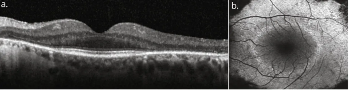
• Optical coherence tomography: thinning of photoreceptor outer segments represent the first histopathological RP (Milam et al., 1998). Such change is higligted in a spectral-domain optical coherence tomography (SD-OCT) image as disorganization of the outer retinal layers, starting from the interdigitation zone, following with the ellipsoid zone, and ending at the external limiting membrane (Battaglia Parodi et al., 2016) (Fig. 3a). RP progression determines dimensional reduction of the outer segments and of the outer nuclear layer, which includes photoreceptor cell nuclei, then completely loss during the advanced phases of RP (Hood, Lazow, Locke, Greenstein, & Birch, 2011). On the other hand, the inner retinal layers, as the deep nuclear layer and the ganglion cell layer, remain quite untouched, even if their thickening could occur, as consequence of edema creation in the retinal nerve fiber layer and/or neuronal-glial retinal remodeling following the thinning of the outer retina (Aleman et al., 2007). In advanced stages, several patients could show other retinal tubulations (N. R. Goldberg, Greenberg, Laud, Tsang, & Freund, 2013). Hyperreflective foci, reflex of RPE cells migration from ellipsoid area, are a frequently found in both nuclear layers and/or the in the subretinal space, and often fundoscopically visible as hyperpigmentation. Interestingly, many studies evidenced a correlation between visual acuity and the width of ellipsoid zone, also linearly correlated to an impairment of visual field and attenuation of the outer segments (G. Liu, Liu, Li, Du, & Wang, 2016). Finally, OCT exams may also help to detect other macular alterations present in more than
12
50% of all RP patients (Makiyama et al., 2014). Among them, CME is the most frequent, followed by vitreomacular traction syndrome, epiretinal membrane formation and macular hole. During CME development, cystoid spaces are mainly distributed in the inner nuclear layer, but they can also occur in the outer layers and/or in the ganglion cell layer (Hariri et al., 2016).
Fig. 3. Horizontal spectral-domain optical coherence tomography (SD-OCT; a) and fundus autofluorescence (FAF; b) images of the left eye of a patient with RP. The OCT image shows the perifoveal loss of the outer retinal layers. The central preservation of the ellipsoid zone corresponds to the internal edges of the hyperautofluorescent ring visible on FAF.
• Fundus autofluorescence imaging: fundus autofluorescence (FAF) can RPE metabolism abnormalities undetectable with other methods. With short-wavelength (SW) - FAF, using green or blue light, the signal derives predominantly from RPE lipofuscin aggregates (Delori et al., 1995), while near-infrared (NIR)-FAF shows also the autofluorescence signal from choroidal melanin or related fluorophores (Keilhauer & Delori, 2006). FAF represents an optimal tecnique to assess and monitoring the progression of RP, even if no detailed informations about the increased susceptibility to light toxicity dystrophic retinas - rich in photosensitizers as lipofuscin - can be obtained (Teussink et al., 2017). An abnormal foveal ring or curvilinear arc of amplified auto-fluorescence (Fig. 3b), detectable only by SW-FAF and NIR-FAF, occurs in more than 55% of RP patients (Mendis & Lois, 2014). Such hyperautofluorescent ring, generally characterized by a high interocular symmetry (Sujirakul et al., 2015), outlines a transition zone between altered (center of the ring) and undamaged (outside of the ring) retinal function. Autofluorescence degree immediately outside of the ring is generally conserved, even if retinal function is impaired. Additionally, photoreceptor degeneration in areas external to the ring corresponds to the loss of the ellipsoid
13
zone and the outer limiting membrane, along with thinning or depletion of the outer nuclear layer in an SD-OCT image (Lima et al., 2009). The autofluorescent ring itself reflects an area of outer segment dysgenesis and lipofuscin production, with progressive retinal attenuation, generally associated to dysgregation of the ellipsoid zone around the internal edge of the ring (Kominami et al., 2017). Most of patients do not show quantitative differences of the auto - fluorescence measured inside the ring compared to healthy eye (Schuerch et al., 2017). During disease progression, the ring diameter shrinks, with a high reduction rate for relatively large rings. Commonly, the increasing of cone dysfunction is reflected by the inner edge of the constricting ring, while rod sensitivity impairment is more diffused and involves the parafoveal area within the ring. Ultimately, the ring may disappear, as consequence of a widespread loss of sensitivity and visual acuity (Robson et al., 2012). Microperimetry in RP patients highlights that visual sensitivity is quite preserved within the ring, decreased in the ring zone itself, and frequently unrecordable outside the ring (Duncker et al., 2013). Besides the hyperfluorescent ring, other autofluorescence patterns can be detected. In the most of adult patients with RP, wide-field FAF reveals patchy and/or decreased autofluorescence in the mid-periphery, probably following the loss of peripheral vision (M. Oishi et al., 2014). Furthermore, central vision impairment could be reflected by an altered pattern of increased autofluorescence at the central macula (Wakabayashi, Sawa, Gomi, & Tsujikawa, 2010).
• Fluorescein angiography: fluorescein angiography represents a technique little used today for RP diagnosis. The angiogram shows chorioretinal atrophy, starting from periphery and expanding to the posterior pole. Generally, delay in the filling of retinal vessels is absent, but some leakage of dye could be present, due to vessels own attenuation. Moreover, CME presence and the uncommon choroidal neovascularization can be depicted by fluorescein angiography, even if the optical coherence tomography angiography (OCTA) is less invasive and more used today (Inooka et al., 2018).
14
• Adaptive optics scanning laser ophthalmoscopy: adaptive optics scanning laser ophthalmoscopy (AOSLO) is the most recent, non-invasive imaging tecnique that allows the visualization of photoreceptors at a microscopic level by correcting for ocular aberrations. In RP patients, the high resolution of AOSLO could early detect photoreceptor damage, as well as possible cone density reduction before the visual acuity is impaired, since a significant cone decrease is possible before the visual acuity becomes affected (Salmon et al., 2017). Summarizing, AOSLO represents a highly sensitive imaging method that may improve disease progression monitoring and evaluating treatment safety and efficacy in clinical trials.
2.6. DIFFERENTIAL DIAGNOSIS FOR NON-SYNDROMIC RETINITIS PIGMENTOSA
IRDs constitute a very heterogeneous and widespread family, made of disorders that especially affect the macula (e.g., Sorsby's fundus dystrophy and Stargardt disease) and of stationary disorders such as CSNB and achromatopsia. The inclusion criteria of RP depend on ophtalmological aspects (symptoms, fundus abnormalities and ERG results), as well as less objective evaluations, such as the patient's age at onset and/or historical factors. Finally, in order to minimize overlapping phenotypes, more evident at the late stages of disease, it has to be considered the whole pathology course when classifying an IRD. A comprehensive overview of the parameters take in account for differential diagnoses in RP is highlighted in Table 1.
• Other inherited retinal dystrophies: two retinal dystrophies, clinically and genetically indistinguishable, unclearly classified by the age of onset, are early-onset RP and Leber Congenital Amaurosis (LCA). Frequently, patients who manifest symptoms at birth or within the first few months of life are considered to be affected by LCA (Sharif & Sharif, 2017). In LCA, the excessively early loss of visual ability determines a group of symptoms that include photophobia,
15
poor pupillary response, nystagmus, and particular oculo-digital signs such as, pressing, poking and rubbing the eyes. Visual acuity is usually lower than 20/400, and the fundus can vary from a normal appearance to a wide atrophic RP-like pigmentary retinopathy. Scotopic and photopic ERG are frequently unrecordable or severely reduced. Early-onset RP can exhibit many of previously described symptoms, and the number of shared genes reflects the overlap with LCA (Fig. 1). Another IRD sharing clinical and genetic aspects with RP is cone-rod dystrophy. ERG exam is not always sufficient to assess which photoreceptors are primarily affected, especially during the later phases of the disease. Neverthless, the early symptoms of cone-rod dystrophy, such as early loss of visual acuity, variable achromatopsia, intense photophobia, and the initial absence of night blindness, can help to distinguish between cone-rod dystrophy and RP (Inui et al., 2014). Other specific retinal dystrophies show rod-cone degeneration, but their own particular phenotypes distinguish them from RP. Among them, most diffuse examples are gyrate atrophy (well-defined circular chorioretinal atrophy accompanied by high ornithine levels), choroideremia (patchy chorioretinal atrophy with physiological appearing retinal vessels) and late-onset retinal degeneration (perimacular drusen-like lesions together with long anterior lens zonules) (Stanton et al., 2017). Retinitis punctata albescens, even if presents a highly particular phenotype has been considered an RP subtype throughout most of the literature. Congenital stationary night blindness (CSNB) is an example of a stationary disorder principally identified by by rod abnormalities. Affected patients, generally present a normal fundus, except two particular subtypes of CSNB, called Oguchi disease and fundus albipunctatus. Neverthless, CSNB evidences a significant gene overlap with RP, especially for RDH5, RLBP1, PDE6B, SAG and RHO (Koenekoop, 2018).
• Pseudoretinitis pigmentosa: lots of disturbs can mimic the clinical features of RP (phenocopy) and are categorized as pseudoretinitis pigmentosa. Unlike real RP forms, the family of pseudoretinitis pigmentosa could be treatable and do not present an essential genetic base. Therefore, to distinguish them from
16
typical RP, a deep history, including lack of interocular symmetry, current and past medications, and lack of disease progression, is fundamental. For example, several patients who were diagnosed with “unilateral RP” fall in this category, even if a germline mutation in the RP1 gene was documented in a patient affected by strictly unilateral RP (Mukhopadhyay, Holder, Moore, & Webster, 2011).
Disease Clinical Features
Syndromic forms of retinitis pigmentosa
Ciliopathies
Usher syndrome RP with partial or complete neurosensory hearing
loss, sometimes vestibular dysfunction.
Bardet-Biedl syndrome
RP and obesity, postaxial polydactyly,
hypogonadism, renal dysfunction, cognitive
impairment.
Cohen syndrome RP and myopia, mental retardation, hypotonia,
fascial dysmorphism, short stature, neutropenia.
Joubert syndrome
RP/LCA with dysmorphic facial features, congenital hypotonia evolving in ataxia, developmental delay and unusual fast or slow breathing. Oculomotor apraxia and nystagmus may be present. The hallmark feature is the ‘molar tooth sign’ on MRI.
Senior-Løken syndrome RP/LCA and nephronophthisis (NPHP).
Sensenbrenner syndrome (cranioectodermal dysplasia) RP and craniosynostosis, ectodermal abnormalities.
Short-rib thoracic dysplasia with or without polydactyly (includes Jeune, Mainzer- Saldino, Ellis-van Creveld and short rib- polydactyly syndrome)
RP and thoracic hypoplasia, short stature, brachydactyly, polydactyly, chronic renal failure, (sometimes lethal) respiratory insufficiency.
Metabolic disorders
Alfa-tocopherol transfer protein deficiency (familial isolated vitamin E deficiency)
RP with (Friedrich-like) ataxia, dysarthria, reduced proprioception and hyporeflexia.
Bassen-Kornzweig syndrome (abetalipoproteinemia)
Atypical RP with onset 1st-2nd decade. Wide
spectrum of abnormalities including progressive
cerebellar ataxia, gastrointestinal disorders,
acanthocytosis and absence of apo-B containing lipoproteins.
Mucopolysaccharidoses
Group of disorders with RP, cloudy cornea and glaucoma and numerous symptoms in varying degree: cognitive impairment, developmental delay, hearing loss, hydrocephalus, facial abnormalities, dwarfism and hepato-splenomegaly.
Neuronal ceroid-lipofuscinoses, childhood onset (Batten disease)
RP with early vision loss, FAG: diffuse RPE atrophy with stippled hyperfluorescence, progressive neurodegeneration, seizures, may cause early death.
Refsum disease (phytanic acid oxidase deficiency) RP and anosmia, miosis, attenuated effect of
17
hearing loss, ataxia, polyneuropathy, ichthyosis, cardiopathy.
Mevalonate kinase deficiency (mevalonic aciduria (MEVA) and hyper-immunoglobulin D and periodic fever syndrome (HIDS)
Spectrum of clinical phenotypes, sometimes with
RP. HIDS: recurrent febrile attacks
lymphadenopathy, arthralgia, gastrointestinal
disturbances, skin rash and increased levels of serum immunoglobulin D. MEVA is the most severe form
with psychomotore retardation, progressive
cerebellar ataxia, dysmorphic features, recurrent febrile crises, and failure to thrive.
HARP syndrome (hypoprebetalipoproteinemia, acanthocytosis, RP and pallidal degeneration)
Part of the pantothenate kinase-associated neurodegeneration (PKAN) spectrum. RP with hypoprebetalipoproteinemia, acanthocytosis and pallidal degeneration (eye of the tiger sign on MRI). PHARC syndrome (polyneuropathy, hearing loss, ataxia, RP, and
cataract
RP with polyneuropathy, hearing loss, cerebellar ataxia and early-onset cataract.
Mitochondrial disorders
Kearns-Sayre Syndrome
RP with progressive external ophthalmoplegia, heart conduction defect, cerebellar ataxia or elevated protein concentration in cerebrospinal fluid. Onset <20 years.
NARP syndrome (Neuropathy, Ataxia, RP) RP and peripheral neuropathy, neurogenic muscle weakness, ataxia.
Inherited retinal dystrophies
Progressive retinal disease
Cone-rod dystrophy
Patients typically present with VA loss,
dyschromatopsia and photoaversion. May
experience nyctalopia. Primary loss of cone function on the ERG, followed by rod impairment. Syndromal associations.
Cone dystrophy
Progressive loss of VA and dyschromatopsia often accompanied by photoaversion and photophobia. Macula: ranging from normal to a bull’s eye maculopathy or RPE atrophy. Reduced or nonrecordable photopic ERG.
Leber congenital amaurosis
Early-onset retinal dystrophy at birth or in first months of life, nystagmus, hyperopia, amaurotic pupils, oculo- digital sign, extinguished photopic and scotopic ERG. Syndromal associations.
Macular dystrophies (Stargardt disease, Sorsby fundus dystrophy)
Progressive loss of VA, advanced disease sometimes associated with night blindness and loss of peripheral vision.
Bietti crystalline corneoretinal dystrophy
Yellow-white crystalline retinal deposits throughout posterior pole and sometimes in corneal limbus. Sclerosis of the choroidal vessels. Often marked asymmetry in retinal findings.
Late-onset retinal degeneration
Perimacular yellow-white drusen-like lesions, long anterior zonules, and hyperpigmentation in the midperiphery. Gradual loss of dark adaptation in fifth-sixth decade. Reduced visual acuity in advanced stages caused by scalloped areas of RPE atrophy or neovascularization, accompanied by ERG changes (rod-cone pattern). Normal caliber of retinal vessels.
18 Stationary retinal disease
Congenital stationary night blindness
Largely non-progressive. Nightblindness.
Nystagmus and myopia with decreased VA if onset early in life. Most common ERG is ’negative’ dark-adapted ERG. Oguchi disease and fundus albipunctatus are forms of CSNB.
Chorioretinal dystrophies
Choroideremia
X-linked, pigment clumping at RPE level, followed by patchy loss of RPE and choriocapillaris with visible underlying large choroidal vessels and sclera. Normal appearing retinal vessels.
Gyrate atrophy
Well demarcated, circular areas of chorioretinal atrophy often starting in far periphery, early onset cataract formation, myopia, CME, elevated plasma ornithine, type II muscle fiber atrophy, hair thinning. Helicoid peripapillary chorioretinal degeneration (Sveinsson
chorioretinal atrophy)
Autosomal dominant, peripapillary chorioretinal atrophy with radially extending wing-shaped atrophy, no attenuation of retinal vessels.
Progressive bifocal chorioretinal atrophy Slowly progressive, large atrophic lesions in macula and nasal to the optic disc. Nystagmus and myopia.
Vitreoretinal dystrophies
X-linked juvenile retinoschisis
VA loss from the 1st/2nd decade of life. Cystoid
macular lesions, typically in an spoke-wheel pattern, peripheral schisis in 50% of patients. ERG: selective reduction in b-wave amplitude.
Enhanced S-cone syndrome/Goldmann- Favre Syndrome
ERG: enhanced S-cone sensitivity (pathognomic). Variable phenotype, hallmarks are nummular pigmentations at RPE level and cystoid or schisis-like maculopathy. Night blindness from birth and decreased VA.
Wagner syndrome/erosive vitreoretinopathy
Optically empty vitreous with avascular vitreous strands and veils, presenile cataract, moderate
myopia, progressive chorioretinal atrophy
sometimes with diffuse pigmentary changes, reduced VA, night blindness and visual field constriction. Retinal detachment in advanced stages of disease.
Snowflake vitreoretinopathy
Autosomal dominant, corneal guttae, cataract, fibrillar degeneration of the vitreous, retinal detachment, and peripheral retinal degeneration, including crystalline deposits referred to as snowflakes, vascular attenuation and chorioretinal pigmentation.
Female carriers of inherited retinal dystrophies
Retinitis pigmentosa
Female carriers of XL-RP: highly variable presentation; from no abnormalities to RP phenotype. Tapetal-like reflex possible.
Choroideremia
Female carriers are generally asymptomatic, although chorioretinal atrophy and ERG changes similar to those in affected males can be observed.
19
Pseudoretinitis pigmentosa
Drug-induced
Thioridazine and chlorpromazine
Nummular areas with loss of RPE and choriocapillaris perfusion. Chlorpromazine often leads to posterior subcapsular cataract.
Quinolines (e.g. (Hydroxy)chloroquine)
Bull’s eye maculopathy, Asian patients: pericentral retinopathy. In case of poisoning: initially fixed dilated pupils, later miosis. Late fundus appearance.
Chorioretinal infections
Syphilis, Lyme disease, acute retinal necrosis and other viral infections (rubella, chicken pox, measles, cytomegalovirus)
Often unilateral or sectorial retinal disease. History of infectious retinal disease.
List of inflammatory disease
Sarcoidosis
Ocular clinical criteria: mutton-fat or small granulomatous KPs and/or iris nodules; nodules in trabecular meshwork or tent-shaped PAS; vitreous
snowballs; peripheral chorioretinal lesions;
nodular/segmental periphlebitis; optic disc nodule(s) and/or solitary choroidal nodule; bilaterality. Extraocular granulomas in: lymph nodes, lungs, skin, liver, spleen, salivary glands, heart, bones and nervous system.
Acute posterior multifocal placoid pigment epitheliopathy
Sudden loss of VA, blurred vision and central scotomas. Often self-limiting, good prognosis with visual recovery.
Birdshot chorioretinopathy
Gradual decline in VA due to CME and retinal atrophy, nyctalopia, floaters, glare, dyschromatopsia and photopsia. Cream-colored, irregular or elongated choroidal lesions radiating from optic disc. Supportive: HLA-A29+ and retinal vasculitis.
Serpiginous choroidopathy
Symptoms: VA loss, metamorphopsia or central scotoma. Signs: recurrent gray-yellowish subretinal infiltrates, centrifugally spreading from peripapillar region in a serpiginous manner. They resolve in atrophy. Bilateral, but often asymmetric.
Diffuse unilateral subacute neuroretinitis (DUSN)
Early stage: vitritis, papillitis, clustered
yellow-gray-white lesions.
Later stage: optic atrophy, arteriolar narrowing, increased ILM reflex (Oréfice’s sign), subretinal tunnels (Garcia’s sign), diffuse RPE degeneration, and afferent pupillary defect. Nematode sometimes visible.
Systemic lupus erythematosus (SLE) Extraocular SLE characteristics (fever, joint pain, rash, etc), cotton wool spots.
Miscellaneous
Vitamin A deficiency
Xerophthalmia and nightblindness. Yellow and white retinal spots may be present in the periphery. Symptoms may be reversible with vitamin A treatment.
Paraneoplastic Photopsias, history of primary tumor; most often
breast - or lung carcinoma or melanoma.
20
Siderosis bulbi Patient history, unilateral, inner retinal layers more severely affected than outer layers.
Old retinal detachment Unilateral, history of retinal detachment.
Pigmented paravenous retinochoroidal atrophy (PPRCA)
Pigment accumulation solely along retinal veins, no or very slow progression, often asymptomatic. Etiology unclear.
Acute zonal occult outer retinopathy
Acute onset, often initially unilateral; however, majority develops bilateral disease, scotoma, photopsias, fundus examination often apparently normal, later RPE disturbances. ERG: delayed implicit time of 30-Hz cone flicker response. EOG: reduction in the light rise.
Tab. 1. Differential diagnoses for non-syndromic retinitis pigmentosa. Abbreviations: CME: cystoid macular edema, ERG: electroretinography, ILM: internal limiting membrane, FAG: fluorescein angiography, KPs: keratic precipitates, PAS: peripheral anterior synechiae, RP: retinitis pigmentosa, RPE: retinal pigment epithelium, VA: visual acuity.
• Syndromic RP: the highest number of RP syndromic forms is related to mutations in genes involved in ciliary activities. The most common ciliopathy is Usher syndrome, whose symptomatology is specifically characterized by variable degree of neurosensory hearing loss (Mathur & Yang, 2015). Another well-known syndromic form of RP is Bardet-Biedl syndrome which presents, in addition to retinopathy, renal dysfunction, postaxial polydactyly, obesity, hypogonadism, and/or cognitive impairment (Forsythe, Kenny, Bacchelli, & Beales, 2018). Type and degree of such extra-ocular features are mainly related to the specific gene involved and the specific mutation within that gene. Syndromic RP is also associated with mitochondrial and systemic metabolic disorders. The extra-ocular features in syndromic RP can be extremely hidden (e.g. an impaired sense of smell) or easily detected by the examining clinician (e.g. evaluating cardiovascular and/or renal disease). An important element regards the possibility to surgically correct features, such as polydactyly, at an early stage. When, initially, clinical exams could not detect any extra-ocular anomalies, the unique distinguishing criteria that may help to diagnose syndromic forms of RP is the genetic testing. However, even if extra – ocular impairments are evident, it is necessary to match such abnormalities to involved genes, in order to assess a syndromic disease. Moreover, several genes associated with non - syndromic RP (e.g. USH2A and BBS1) may also determine syndromic RP (Table 2). Concluding, correct diagnosis of
21
syndromic RP could improve patients’ management until save his life, as for patients affected by metabolic disorders like Kearns – Sayre syndrome, a mitochondrial disease frequently associated to cardiac dysfunction (Shemesh & Margolin, 2018). Gene/locus RP type Inher. Pattern Decade
of onset Visual function Ophthalmic features ERG
Syndromic associations Other IRD phenot ypes ABCA4 19 AR 1 VA is severely affected: FC to NLP at higher age.
Bone spicule-like pigmentation may reach into the macular region. Severe chorioretinal
atrophy. Rod-cone pattern, later both responses NR - STGD, CRD (may occur simulta neousl y in RP familie s) AGBL5 75 AR 1-2 VA loss is highly variable. At 40-50 years, VA may range from
20/40 to NLP.
Macular involvement: macular atrophy, CME. PSC. Rod-cone pattern Mental retardation (correlation with AGBL5 unknown) - AHI1 NA AR 3-4 VA in 3rd decade
can range from 20/32 to HM or
even LP.
Macular involvement. PSC. Rod-cone pattern JS type 3 -
ARHGEF18 78 AR 3-4 VA: 20/30-20/60 in 4th decade, but may decrease to CF in the 6th decade. Photopsias.
Nummular pigment clumping, CME. Vitreous opacities (in 1
patient) Rod-cone pattern - - ARL2BP 66§ AR 3 Relative early loss of VA: HM (or even LP) in the 4th decade of
life. Yet, other patients may retain a VA of 20/40 up to the 6th decade of life.
Marked macular atrophy, PSC,
ERM. NR Situs inversus, primary ciliary dyskinesia (respiratory failure), otitis media - ARL6 55 AR NA No information on the visual function available.
No information on the retinal phenotype available. No further information available BBS type 3 - BBS1 NA AR 1-2, (earliest: 1y) Severe visual loss, may reach
LP by 5th - 6th decade of life. Severe constriction of VFs up to 5˚-10˚ in the 4th decade.
Nystagmus, possible macular atrophy, cataract (PSC and
cortical). Generally NR, severely disturbed in rod-cone pattern in 2nd decade BBS type 1 - BBS2 74 AR 1-2 Severe, relative early visual loss: HM or LP before the age 60. VFs
are severely constricted.
Macular atrophy, bull’s eye
22
BBS9 NA AR NA
No further information
available.
No information on the retinal phenotype available. No further information available BBS type 9 - BEST1 50 AD AR (1 family) 1-2, (5) Nightblindness may be absent. Loss of VA is a prominent symptom.
Yellow fundus flecks in the mid-periphery, pigmentation in
far periphery, CME, ERM. Macula relatively spared unless
serous macular detachments. NR scotopic responses, residual photopic responses - BVMD , AVM D, ARB, ADVI RC C2orf71 54 AR 1-2 (2 cases <5y) Night blindness may be absent. Ring scotomas in 4th -5th decade of life. Photophobia may occur.
Foveal atrophy. Early onset associated with severe chorioretinal atrophy. Rod-cone pattern, often NR Hearing loss, ataxia and cerebellar atrophy (digenic with RP1L1) - C8orf37 64 AR 1-2 Severe visual loss to HM/LP in the 4th decade. The VF is constricted to 5˚. Sometimes photophobia.
High myopia, cataract. Marked geographic macular atrophy.
Generally NR BBS type 21 CRD CA4 17 AD 2-3 VA levels of 20/200 (age 11) and LP (58 years).
Pigment clumping at the level of the RPE has been described (in
1 patient). Reduced or NR photopic and scotopic responses - - CDHR1 65 AR 2 VA loss to HM by the 4th or 5th decade of life. Severe color vision defects and VF constriction to 5-10˚, sometimes with mid- peripheral residue. Photophobia (3rd decade).
In early stage: sparse bone spicule pigment migration. Later stages: dense pigment migration,
macular atrophy. Generally NR, although ERG may show recordable rod- and cone-driven responses - CRD CERKL 26 AR 2-3 (mean: 23y) VA generally severely affected and may decrease to LP around the 5th decade. Photophobia.
Early macular involvement, sometimes with hyperpigmentation. Pericentral
localization of bone spicules. Normal appearance of optic
disc. Responses are SR in a rod-cone pattern, may be NR in the 3rd decade - - CLN3 NA AR 1-5 VA loss to HM by the 5th decade reported. Severe constriction of VFs up to 5˚-10˚ in the 6th decade.
Sparce bone spicule pigmentation, macular atrophy,
CME
NR or
rod-cone pattern JNCL CRD
CLRN1 61 AR NA Classic RP
phenotype. Typical RP features.
Rod-cone
pattern USH type 3 -
CNGA1 49 AR 1
A gradual decrease in VA may occur from the 4th decade
onwards. Concentric constriction of the VF during the 3rd decade of life.
Sometimes macular atrophy.
23 CNGB1 45 AR 1-2 Macular involvement with VA loss to LP. VF loss from a mean age of 33 years (13- 40). Sometimes photophobia.
Macular atrophy, pericentral RP (described once). Rod-cone pattern - - CRB1 12 AR 1-5 (median: 4y) 50% of patients have a VA <20/200 at age 35 years. Nystagmus (±40%), hyperopia, CME (50%), PPRPE, Coats-like vasculopathy, optic disc drusen,
retinal vascular sheathing, asteroid hyalosis, thickened retina with loss of the retinal laminations, bull’s eye maculopathy and yellow round
deposits in the posterior pole. Occasional dense pigmentation.
NR from 2nd – 3rd decade Nanophthalmo s LCA, PPRC A CWC27 NA AR 1 VF is severely constricted early in the disease course.
No information on the retinal phenotype available. No further information available Brachydactyly, craniofacial abnormalities, short stature, neurologic defects LCA DHDDS 59 AR 2-3 VA is generally mildly affected, although in some eyes VA decreases to LP levels.
Occasional CME. Parafoveal atrophy of the RPE. Pericentral
localization of pigmentation (reported once). SR or NR rod- and cone-driven responses from the 2nd decade. - - EYS 25 AR 2-3 (range 8-62y)
VA loss from the 4th decade to
levels of 20/200 to NLP in the 7th
decade.
Variable levels of bone spicule pigmentation and macular
atrophy. PSC. Rod-cone pattern, but often NR - - FAM161A 28 AR 2-3 Legally blind in 6th – 7th decade. Constriction of to 10˚.
Limited number of bone
spicules. Macular atrophy. PSC. NR
Hearing problems, hyposmia - FSCN2 30 AD 1 VA and VF relatively spared until the 4th decade, then VA loss to levels of HM.
Early vessel attenuation. Incidental macular atrophy.
SR at early ages, generally NR from the 4th decade - MD GNAT1 NA AR 2 Variable VA: 20/20 (80 years) to 20/80 (32 years).
Round pigment clumps and typical bone spicules. ERM.
Rod-cone pattern or NR - CSNB GUCA1B 48 AD NA Variable visual function: 20/20 (62 years) – 20/100 (47 years).
Highly variable retinal expression in Japanese patients:
normal fundi, sector RP with macular involvement, only macular atrophy or diffuse RP.
NR in patients with diffuse RP. Sector RP with macular atrophy leads to reduced scotopic and photopic responses - MD HGSNAT 73 AR 1-2 5-6 Severe VA loss to CF at age 60 years. VA is more preserved in case
CME, ERM, pericentral RP (described once). Reduced or NR rod- and cone-driven responses MPS type IIIC -
24 of late disease onset. HK1 79 AD, NP ≤15% 1-4 (range 4y-mid- 30s) Highly variable VA loss. VA loss to CF in 3rd decade reported. Photophobia.
Bull’s eye maculopathy, pericentral RP. Rod-cone pattern HMSN, nonspherocyti c hemolytic anemia - IDH3A NA AR 1-2 (range 1-11y) VA loss dependent on the presence of macular pseudocoloboma. Macular pseudocoloboma, CME. SR or NR rod-drive responses, cone- responses SR - - IDH3B 46 AR NA Classic RP
phenotype. Typical RP features, PSC.
SR amplitudes of both scotopic and photopic responses - - IFT140 NA AR 1-4 (range 2y-early- 30s)
Vision loss from 3rd - 5th decade,
VA may eventually reduce
to LP.
Macular atrophy, CME, ERM, early cataract or white dots.
Dense pigmentation in 7th
decade reported.
SR in a
rod-cone pattern SRTD type 9 LCA
IFT172 71 AR 1-2 Night blindness is the initial symptom. Further symptoms have not been specified.
Variable macular involvement: macular atrophy, CME, ERM.
No further information available SRTD type 10, BBS type 20 - IMPDH1 10 AD AR: (Asp226 Asn) 1-3 AD disease: variable degrees of VA loss. Legal blindness before the age of
40 has been described.
AD disease: CME, significant
vitreous disturbances, PSC. AD disease: SR or NR rod and cone responses - LCA AR disease: no information on the visual function available. AR disease: macular involvement. AR disease: NR IMPG2 56 AR 1-2 Central vision generally affected.
Macular atrophy and bull’s eye maculopathy. Sheathing of peripheral vessels. SR in a rod-cone pattern or NR - VMD
KIZ 69 AR 2 phenotype. Classic RP Macular thinning (in 1 patient).
NR at the age of 35 years Obesity, hearing problems (correlation with KIZ unknown) - KLHL7 42 AD 3 VA remains (near) normal up to the 5th or 6th decade. VF loss usually is the initial symptom.
Fundus appearance can be normal up to the 4th decade.
Later: CME and parafoveal atrophy. Rod-cone pattern, eventually NR. The mean (SD) decline in light-adapted 31-Hz flicker response is 3.0% (3.0) per year. CISS -
25 LRAT NA AR 1 (earliest: 2y) Severe, early VA loss to 20/100-20/200 and VF constriction to 30˚- 60˚ before the age of 10 years. Photophobia.
High hyperopia, nystagmus, poorly reactive pupils. Sparse or
absent bone spicules (age 9 years). RPA. Reduced
FAF signal. SR in a rod-cone pattern or NR - LCA MAK 62 AR 2-5
May show initial preservation of
nasal VF.
Macula generally not involved, but sometimes CME (in 1 patient) or macular atrophy.
Reduced in a rod-cone pattern, but often NR - - MERTK 38 AR 1-2 (earliest: 3y) VA loss to ≤20/200 in the 2nd decade. Variable VF loss: normal VF (3rd decade) – 5˚ (2nd decade). Legal blindness: ±40 years. Impaired color discrimination.
Nystagmus, bull’s eye maculopathy, macular atrophy.
Pallor of the optic disc may be absent. Photopic responses become NR during the 1st decade. Scotopic responses are NR - LCA MVK NA AR 3 VF loss may be the initial symptom.
Arterial tortuosity, PSC, ERM, thickening of the nerve fiber layer on OCT, CME (described
once). SR in a rod-cone pattern MKD (MEVA or HIDS) - NEK2 67 AR NA No information on the visual function available.
No information on the retinal phenotype available. No further information available - - NR2E3 37 AD, AR 1-3 (earliest: 3y) AD disease: VF
loss from the 2nd– 3rd decade.
AD disease: nummular and
spicular pigmentation. Early-onset cataract. FAF: 2 or 3 hyperfluorescent rings may be
visible. Pericentral RP. AD disease: rod-cone pattern. Rod responses are SR, and become NR in advanced disease. Cone-driven responses are affected late. - ESCS AR disease: early
VA loss. AR disease: clumped pigment
AR disease: rod-cone pattern NRL 27 AD, AR 1 (earliest: 1y) AD disease: VA
loss from the 4th
decade: 20/20 – 20/00. VF diameters: 50-60˚ in the 3rd decade and decrease up to 10˚ in the 8th decade.
AD disease: nystagmus, minimal
or absent hyperpigmentation in 2nd decade. Round pigment
clumps. Chorioretinal atrophy, macular atrophy, bull’s eye maculopathy, PSC. Peripheral
retinal telangiectasis (which may cause serous retinal
detachment). NR - - AR disease: visual function is more severely affected compared to AD disease.
AR disease: peripheral pigment
clumps. Retinal features are similar to NR2E3- associated
enhanced S-cone syndrome.
OFD1 23 XL (<2y) 1 Early loss of central vision. Only temporal and inferior VF residues.
Grayish spots at the level of the RPE. Granularity of macular
RPE. NR JS, OFDS type 1, SGBS type 2 -
26
PANK2 NA AR 5
VA reduction to HM in the 6th
decade.
No information on the retinal phenotype available. No further information available HARP syndrome, NBIA1 (also termed HSS) - PDE6A 43 AR 1 Marked peripheral VF loss.
CME, PSC and dense pigmentation. Rod-cone pattern - - PDE6B 40 AR 1 Loss of peripheral VF is a prominent symptom that occurs during the
2nd-3rd decade.
CME, PSC, dense pigmentation at high age (80 years),
pericentral RP. Rod-cone pattern Carriers: rod-driven responses may be reduced - CSNB PDE6G 57 AR 1 Marked constriction of the VF up to 5-10˚.
Normal vessels and optic discs in young patients. CME in all
patients (1 family). Both rod- and cone- driven responses are NR within the 1st decade of life. - - POMGNT1 76 AR 1-2 (3-4) Variable VA loss, may decrease to LP. VF constriction to 5˚ in the 6th-7th decade.
Macular involvement, CME. NR MEB -
PRCD 36 AR 1-3 Relative early visual loss.
Various macular involvement: bull’s eye maculopathy, macular
atrophy, CME. ERM, PSC. Fairly normal-colored optic disc.
Scotopically and photopically NR in an early stage of disease (earliest described: age 6 years) - - PROM1 41 AR 1 Visual loss during the 1st decade.
Large inter- and intrafamilial variability: from isolated (bull’s eye) maculopathy to pericentral
RP and severe RCD. Nystagmus. NR Polydactyly CRD, AD MD PRPF3 18 AD, full penetranc e 1 (4, once) Classic RP
phenotype. Classic RP phenotype.
Rod-cone pattern Rod- driven responses are abolished from the 2nd decade, cone-driven responses are SR by then - - PRPF4 70 AD 2-3 Variable visual loss, may reach
HM. VF constriction to
5-10˚ in the 6th
decade.
Variable degree of macular atrophy.
Generally
27
PRPF6 60 AD 2-4
VA initially spared, but may decrease to LP. Constriction of VFs to 30-40˚ (4th decade) and
±10˚ (6th decade).
Macular atrophy in later stages. Optic nerve heads may initially
be normal. PSC. SR responses in the earlier phases of the disease. Scotopic responses become NR over time, photopic responses tend to diminish more slowly. - - PRPF8 13 AD 1-2 VA may remain normal up to the 3rd – 4th decade, with progression to 20/200 in the 7th decade. VF constriction to ±10° in 4th decade.
Dense intraretinal pigment
migration (in 1 patient). NR - -
PRPF31 11 AD NP ≤10% 1-2 Variable presentation. Incomplete penetrance suggested in asymptomatic patients. Mean annual VF loss: 6.9%. Legal blindness: 4th decade.
Macular atrophy, CME, PSC. May present with para-arteriolar
absence of pigmentation or pericentral RP (described once).
No abnormalities observed in patients that lack penetrance.
The mean (SD) decline in light-adapted 30-Hz flicker response is 9.2% per year. Responses may be normal in patients that lack penetrance - - PRPH2 (formerly known as RDS) 7 AD digenic with ROM1 2-6 VA usually spared, but dependent on the degree of macular involvement.
Variable macular involvement, CME, RPA, pericentral RP
(described once). Rod-cone pattern, will become NR during 6th decade - MD, PD, CRD, LCA RBP3 66§ AR Early onset: 1 Early-onset disease: early visual loss. Strabismus.
Early-onset disease: (high)
myopia, PSC. Early-onset disease: SR responses, most often in a rod-cone pattern, although cone-rod patterns also occur. - - Late- onset: 4-6 Late-onset disease: blurred vision is an early symptom; night blindness may be absent.
Late-onset disease: PSC, (high)
myopia. Late-onset disease: SR rod- and cone-driven responses, often NR RDH12 53 AD, AR AD disease: 2-5 AD disease: classic RP phenotype.
AD disease: typical RP features.
AD disease:
no further information
available
28 AR disease: 1-3 AR disease: VA at presentation: 20/40- 20/200, may reach HM-LP. VF constriction to <5˚ in the 2nd – 3rd decade. Central scotoma may occur. Photophobia.
AR disease: nystagmus, macular
atrophy, dense intraretinal pigment migration with
para-arteriolar sparing. Hyperpigmentation may reach
into the macular region. Preservation of peripapillary RPE. CME, PSC. AR disease: NR or SR in both scotopic and photopic conditions. REEP6 77 AR 1-2 Gradual VA loss, although a decline to 20/400 at the age of 32 has been described.
CME, PSC, vascular sheathing.
SR in a rod-cone pattern or NR Anosmia - RGR 44 AR NA VA loss to ≤20/200. Severe VF constriction.
Macular atrophy in patients with severely affected VA.
Responses are reduced in a rod-cone pattern - - RHO 4 AD, AR 1-2, (4) Highly variable clinical course (also intrafamilial). Annual VA decline: 1.6%. Annual VF loss: 2.6%. Legal blindness: 6th – 8th decade.
Sector RP, and to a lesser extent pericentral RP. CME. Late-onset chorioretinal atrophy in patients with p.Met207Lys mutation.
Rod-cone pattern. Mean annual decline: 7.7 - 8.7%. - CSNB RLBP1 NA AR 2 Variable VF loss from the 3rd decade, to <5° residues.
Minimal or absent bone spicules, RPA. Rod-cone pattern - BRD, NFRC D, FA ROM1 NA Digenic (ROM1: Leu185Pr o + PRPH2) NA No information on the visual function available.
No information on the retinal phenotype available. SR scotopic and photopic responses - - RP1 1 AD, AR AD disease: 2-3 AD disease: moderate decrease in VA in 4-5th decade. AD disease: PSC. RP sine
pigmento (described once).
Rod-cone
pattern - -
AR disease: 1
AR disease:
relative early loss of VA to CF or HM in the 5th decade. VF constriction to 10˚ in the 3rd decade.
AR disease: macular atrophy,
CME, myopia. RP1L1 NA AR 4-5 Moderate decrease in VA to ±20/80. VF constriction to 5˚ in the 8th decade. Typical RP features. NA Hearing loss, ataxia, cerebellar atrophy (digenic with C2orf71) OMD RP2 2 XL 1 Early loss of central vision. Central scotoma in 50% of patients. Severe VF constriction in 2nd decade. Large
Myopia. Bull's eye maculopathy, macular atrophy,
sometimes choroideremia-like degeneration. A tapetal-like
reflex (reported once).
Rod-cone