Università degli Studi di Ferrara
DOTTORATO DI RICERCA IN
"Farmacologia e oncologia molecolare”
CICLO XXII
COORDINATORE Prof. Pier Andrea Borea
The Ca
2+
signal as common target
of three different proteins involved
in the apoptotic process
Settore Scientifico Disciplinare MED/04
Dottorando Tutore
Dott. Marchi Saverio Prof. Rimessi Alessandro
Index
Abstract ... 4
Riassunto ... 5
1. Introduction ... 6
1.1 The concept of Ca2+ as a cellular signal... 6
1.2 Generation (on reaction) and extinction (off reaction) of Ca2+ signalling... 7
1.3 Mitochondrial Ca2+ signalling: a turning point controlling cell fate... 11
2. Tools ... 14
2.1 Aequorin... 14
2.1.1 Recombinant Aequorin... 16
2.1.2 Chimeric Aequorin cDNAs... 17
2.1.3 Luminescence detection ... 18
2.1.4 Advantages and disadvantages of aequorin... 19
2.2 Green Fluorescent Protein (GFP) ... 21
2.3 Experimental set-up: collecting and analysing the GFP images... 24
2.4 Luciferase... 26
3. Protein Kinase C β and Prolyl Isomerase Pin1 Regulate Mitochondrial Effects of the Life-Span Determinant p66Shc... 29
3.1 Introduction... 29
3.1.1 p66Shc, an adaptor protein ... 30
3.2 Results ... 33
3.2.2 Effects of PKC β-dependent phosphorylation of p66Shc ... 37
3.2.3 Pin1 induces p66Shc mitochondrial translocation after Ser36 phosphorylation ... 41
3.3 Discussion ... 43
4. Intramitochondrial calcium regulation by the FHIT gene product sensitizes to apoptosis 45 4.1 Introduction... 45
4.2 Results ... 46
4.2.1 Subcellular localization of Fhit and effect on Ca2+ homeostasis... 46
4.2.2 Assessment of mitochondrial Ca2+-uptake capacity in permeabilized and intact cells.... 48
4.2.3 The [Ca2+]mt is affected by a mitochondrial Fhit chimera ... 52
4.2.4 The mitochondrial fraction of Fhit potentiates apoptotic effect of menadione... 53
4.3 Discussion ... 56
5. Akt kinase reducing endoplasmic reticulum Ca2+ release protects cells from Ca2+ -dependent apoptotic stimuli ... 59
5.2.1 Akt drastically reduces ER Ca2+ release, while leaving ER Ca2+ levels unaffected ... 62
5.2.2 Cytosolic and mitochondrial Ca2+ response to agonist stimulation is strongly impaired in m/p-AKT1 expressing cells ... 65
5.2.3 Akt overexpression protects against Ca2+-mediated cell death... 66
5.2.4 Akt inhibits [Ca2+]cyt increases induced by H2O2 and arachidonic acid ... 67
5.3 Discussion ... 69
6. Materials and methods ... 71
6.1 Cell culture, infection and transfection ... 71
6.2 Construction of mtFhit chimera ... 72
6.3 Aequorin measurements... 72 6.4 Luciferase measurements ... 73 6.5 Fura-2/AM measurements ... 74 6.6 Caspase 3 assay... 74 6.7 Apoptotic counts... 74 6.8 Annexin V assay... 75
6.9 Cell cycle analysis ... 75
6.10 Microscopic analysis of mitochondrial structure and PKC β translocation ... 75
6.11 Mitochondrial membrane potential measurements... 76
6.12 mtRP measurements... 76 6.13 Immunofluorescence ... 76 6.14 Isolation of mitochondria... 77 6.15 In vitro binding ... 77 6.16 Immunoprecipitation ... 77 6.17 Immunoblot... 78 7. Reference list ... 79
Abstract
Ca2+ signal plays a fundamental role in modulating diverse cellular responses as muscle contraction, exocytosis, motility, fertilization, proliferation and apoptosis. Therefore, several proteins involved in this wide range of biological processes “manipulate” intracellular Ca2+ to (dis)regulate these events. To analyse Ca2+ homeostasis at the subcellular level, we employed the Ca2+-sensitive photoprotein aequorin, targeted to defined intracellular locations (organelles, such as mitochondria, endoplasmic reticulum, and cytoplasmic regions).
Using this experimental approach, we investigated multiple effects on Ca2+ dynamics of three different proteins: i) the 66 KDa isoform of Shc protein (p66Shc) and its role on the alterations of mitochondrial Ca2+ homeostasis during oxidative stress. ii) The tumor suppressor protein Fhit and its role as sensitizer of the low-affinity Ca2+ transporters of mitochondria, and in turn potentiating the effect of apoptotic agents. iii) The proto-oncogene Akt, a potent inhibitor of apoptosis, through the reduction of Ca2+ release from the ER during apoptotic stimulation.
Taken together, these data remark the practical useful of Ca2+-dynamics investigation as a “reporter system” to understand different molecular mechanisms of different proteins and may provide ways to act on apoptotic cell death and its derangement in cancer.
Riassunto
Il segnale Ca2+ gioca un ruolo fondamentale nel modulare diverse risposte cellulari come contrazione muscolare, esocitosi, mobilità cellulare, fertilità, proliferazione ed apoptosi. Perciò, diverse proteine implicate in questa ampia gamma di processi biologici “manipolano” la concentrazione di Ca2+ intracellulare per (dis)regolare questi processi.
Per analizzare l’omeostasi del Ca2+ a livello sub-cellulare, il nostro laboratorio si avvale della tecnica dell’equorina, una foto-proteina sensibile al Ca2+ che può essere direzionata ai vari sub-compartimenti cellulari (organelli come mitocondri, reticolo endoplasmico e citoplasma).
Usando questo approccio sperimentale, abbiamo studiato i diversi effetti riguardanti le dinamiche del Ca2+ di 3 proteine: i) la proteina p66Shc e il suo ruolo nell’alterazione dell’omeostasi del Ca2+ in condizioni di stress ossidativo. ii) l’oncosoppressore Fhit e la sua capacità nel sensibilizzare i trasportatori del Ca2+a bassa affinità e nel potenziare gli effetti di agenti apoptotici. iii) il proto-oncogene Akt, un potente inibitore del processo apoptotico, capace di inibire il rilascio di Ca2+ dal reticolo endoplasmico dopo stimolo apoptotico.
Nel complesso questi dati sottolineano l’importanza dello studiare le dinamiche del segnale Ca2+, essendo questo un formidabile “sistema reporter” per meglio comprendere i meccanismi molecolari di proteine diverse. Inoltre, nello specifico, esso puo essere considerato un processo su cui poter agire per modificare il processo apoptotico e la sua disregolazione nel tumore.
1. Introduction
1.1 The concept of Ca2+ as a cellular signal
In the past two decades, our understanding of how extracellular signals are conveyed to eukaryotic cells via an increase in intracellular Ca2+ concentration has widely expanded. It is today common knowledge that a variety of extracellular stimuli (ranging from the binding of hormones, neurotransmitters, growth factors to phenomena such as cell-cell interactions), through diverse mechanisms (e.g. receptors that are themselves ion channels, or have an intrinsic enzymatic activity or are coupled to enzymatic effectors via G proteins) induce a rise in cytoplasmic Ca2+ concentration ([Ca2+]cyt) with defined
amplitude and kinetics [1, 2].
In most eukaryotic cells, a large electrochemical gradient for Ca2+ exists across the plasma membrane. The transmembrane potential across the membrane is 70 to 90 mV. The interior of the cell is the more negative, yet the cytoplasmic concentration of Ca2+ ([Ca2+]cyt) is less than one-ten thousandth of that in the extracellular milieu. There are also
intracellular organelles, such as the endoplasmic reticulum (ER) and secretory granules, which contain one- to ten thousand fold greater concentrations of Ca2+ than the cytoplasm.
Moreover, the technological advancements in probe design and imaging systems, by allowing the accurate measurement [Ca2+] at the single cell level, has revealed a marked asynchronicity in cell response and a high spatio-temporal complexity of the intracellular Ca2+ signal. We now know that the Ca2+ signal can be conveyed as repetitive [Ca2+]cyt
spikes (commonly referred to as Ca2+ oscillations) [3], as well as localised [Ca2+]cyt
increases that may either be confined or gradually propagate to the rest of the cell (“Ca2+ waves”) [4, 5].
An extensive Ca2+-signalling toolkit is used to assemble signalling systems with very different spatial and temporal dynamics. Rapid highly localized Ca2+ spikes regulate fast responses, whereas slower responses are controlled by repetitive global Ca2+ transients or intracellular Ca2+ waves. The Ca2+ has a direct role in controlling the expression patterns of its signalling systems that are constantly being remodelled in both health and disease. During the on reaction, stimuli induce both the entry of external Ca2+ and the formation of second messengers that release internal Ca2+ that is stored within the endoplasmic reticulum or Golgi apparatus. Most of this calcium is bound to buffers, whereas a small
exchangers and pumps. The Na+/Ca2+ exchanger (NCX) and the plasma membrane Ca2+ -ATPase (PMCA) extrude Ca2+ to the outside, whereas the sarco/endoplasmic reticulum Ca2+-ATPase (SERCA) pumps Ca2+ back into the ER.
Mitochondria also have an active function during the recovery process in that they sequester Ca2+ rapidly trough a uniporter, and release more slowly back into the cytosol to be dealt with by the SERCA and the PMCA. Cell survival is dependent on Ca2+ homeostasis, whereby the Ca2+ fluxes during the off reactions exactly match those during the on reaction.
1.2 Generation (on reaction) and extinction (off reaction) of Ca2+ signalling
Cells maintain a rigid control over the intracellular level of Ca2+, thus ensuring that the level is kept low during periods of inactivity. In order to use Ca2+ as a messenger, cells overcome this tight homeostatic control by using sophisticated mechanisms. An increase in the intracellular level of Ca2+ depends upon entry channels in the plasma membrane and release channels in the membranes of the internal stores, the endoplasmic reticulum (ER) or the Golgi apparatus. In first case, there are many different plasma membrane channels that control Ca2+ entry from the external medium in response to stimuli that include membrane depolarization, stretch, noxious stimuli, extracellular agonists.
Entry of Ca2+ is driven by the presence of a large electrochemical gradient across the plasma membrane. The cells use this external source of signal Ca2+ by activating various entry channels with widely different properties. We know the most about the voltage-operated channels (VOCs), which are found in excitable cells and generate the rapid Ca2+ fluxes that control fast cellular process such as muscle contraction or exocytosis at synaptic endings.
There are many other Ca2+-entry channels that open in response to different external signals, such as the receptor-operated channels (ROCs), for example the NMDA receptor that respond to glutamate. There also are second-messenger-opereted channels (SMOCs) that are controlled by internal messengers, such us the cyclic-nucleotide-gated channels that are found in sensory systems and the arachidonic-acid-sensitive channel. In addition to these more clearly defined channel-opening mechanisms, there are many other channel types that are sensitive to a diverse array of stimuli, such as the store-operated channels (SOCs) or G protein-operated-calcium channels (GOCCs).
The G protein-coupled receptors contains upwards of 1000 distinct members that mediate the vast majority of all responses to hormones, neurotransmitters, autocrine and paracrine factors. These integral membrane proteins have a conserved primary structure that contains seven stretches of 20-25 hydrophobic amino acid residues that span the plasma membrane. Hence, these important convertors of extracellular signals into intracellular signals are called seven transmembrane spanning domain receptors. Upon binding of agonist, GOCCs undergo a conformational change which then allows for productive coupling to heterotrimeric guanine regulatory proteins (G proteins). The active conformation of the agonist-occupied G protein-coupled receptor acts as a guanine nucleotide exchange factor for the heterotrimeric G protein and activates the G protein by promoting the dissociation of GDP and loading of GTP onto the G protein subunit. In order to limit the magnitude and duration of the extracellular stimulus, the signalling cascade has to be dynamically regulated. In fact, most stimuli are short lived (milliseconds to minutes) as their response wanes over time even in the presence of saturating concentrations of agonist. This phenomenon is termed desensitization. Several different processes with varying time frames are responsible for desensitization.
The release of Ca2+ from internal store is controlled by Ca2+ itself, or by an expanding group of messengers, such as inositol–1,4,5–triphosphate (IP3), cyclic ADP ribose
(cADPr), nicotinic acid adenine dinucleotide phosphate (NAADP) and sphingosine-1-phosphate, that either stimulate or modulate the release channels on the internal stores. The most ubiquitous of the intracellular Ca2+ release mechanisms involves the phosphoinositide specific phospholipase C-derived second messenger (IP3), which acts by
binding to a specific receptor (IP3R) on the endoplasmic reticulum. Distinct subtypes of the
receptor exist, representing products of at least three distinct genes, and additional forms arise as a result of alternative splicing of mRNA. The interaction of IP3 with its receptor
involves complex and poorly understood regulatory interactions among the receptor, IP3
and Ca2+, the latter exerting influence from both the cytoplasmic and luminal aspects of the receptor. Ca2+ in the lumen of the ER appears to sensitize the receptor to IP3. On the
cytoplasmic surface, low concentrations of Ca2+ sensitize the receptor, while higher concentrations are inhibitory. These actions may contribute to the "all-or-none" oscillatory behaviour of [Ca2+]cyt signals observed in some cell types.
The other major type of intracellular Ca2+-mobilizing receptor is the ryanodine receptor (RYR). The ryanodine receptor family comprising three members: RYR1 found in
brain and some other cells, and RYR3 found in smooth muscle, brain and other cells [6], [7]. In its most specialized setting, in skeletal muscle, the RYR is gated by a direct conformational interaction with a dihydropyridine receptor in the t-tubule membrane. This coupling allows for rapid release of stored Ca2+ when an action potential invades the t-tubule system. However, the physiological ligand for the RYR is usually Ca2+ itself; because of this, it is considered to be a Calcium-Induced Calcium Release receptor-Ca2+ channel. Because Ca2+ can sensitize the IP3 receptor to IP3, the IP3 receptor also can
exhibit a Calcium-Induced Calcium Release behaviour. However, some IP3 is always
required for its action, while the ryanodine receptor can function as a "pure"
Calcium-Induced Calcium Release receptor. Although the RYR is thought to be regulated primarily
by Calcium-Induced Calcium Release, there is also a small water-soluble molecule that can function as a regulatory ligand for at least some forms of the ryanodine receptor. Cyclic adenosine diphosphate ribose (cADPr) functions in a manner somewhat similar to IP3: it increases the probability of the ryanodine receptor channel opening by increasing its
sensitivity to Ca2+.
During the course of a typical Ca2+ transient, the generation of Ca2+ signalling is counteracted by the systems of switching off of Ca2+ signalling, during which time various pumps and exchangers remove Ca2+ from the cytoplasm.
The pumping mechanisms have important homeostatic functions in that they maintain the resting level of Ca2+ and ensure that the internal stores are kept loaded. Four different pumping mechanisms are responsible for the off reaction: the plasma-membrane Ca2+ -ATPase (PMCA), the Na+/Ca2+ exchanger (NCX), SERCA (sarco/endoplasmic reticulum Ca2+-ATPase) and the mitochondrial calcium uniporter (MCU). PMCA and SERCA pumps have lower transport rates but high affinities, therefore they can respond to modest elevations in Ca2+ levels and set basal Ca2+ levels. The NCX and mitochondrial uniporter have much greater transport rates, and can limit Ca2+ transient over a wider dynamic range.
Ca2+-ATPases constitute a large family of proteins that fall into two distinct groups, the sarco(endo)plasmic reticulum Ca2+-ATPase (SERCA), and the plasma membrane Ca2+ -ATPase (PMCA). Most eukaryotic cells co-express, in a tissue-specific and differentiation stage-specific manner, one or more types of SERCA and PMCA pumps.
The PMCA are calcium pumps of the plasma membrane, largely responsible for the regulated transport of Ca2+ between the intracellular and the extracellular milieu. Mammals have four genes encoding the proteins PMCA1 through PMCA4. Each gene transcript is
alternatively spliced to generate several variants. Their distribution is tissue- and cell-specific and undergoes regulation during cell development and differentiation. Traditionally, these pumps have been considered to play a housekeeping role in controlling basal Ca2+ levels, but more recently, it became clear that the presence (and the co-expression) of different isoforms must be related to a more specialized function [8].
The only known mechanism for accumulation of Ca2+ by the endoplasmic reticulum is through the actions of SERCA pumps. SERCA are calcium pumps involved in the transport of calcium from the cytosol to various intracellular stores such as the sarcoplasmic reticulum (SR), the endoplasmic reticulum (ER) and calciosomes. It catalyses Ca2+ transport to the lumen of the SR or ER by an active process that requires adenosine triphosphate (ATP). Enzyme phosphorylation and ATP hydrolysis result in translocation of the two Ca2+ ions bound to the enzyme from a high affinity site to a low affinity site. The two calcium ions are then released into the lumen of the intracellular stores [9].
Three separate genes encode the SERCA family. The SERCA 1 gene is exclusively expressed in fast twitch skeletal muscle. Developmentally regulated alternative splicing of SERCA 1 results in an adult isoform (SERCA 1a) and a neonatal isoform (SERCA 1b) [10-12]. The SERCA 2 gene is expressed in slow-twitch skeletal muscle, cardiac muscle, smooth muscle and non-muscle tissues. Tissue-dependant processing of the SERCA 2 gene transcript yields four SERCA 2 messenger ribonucleic acids (mRNAs) (classes 1–4). Class 1 mRNA encodes the SERCA 2a isoform, found in cardiac, smooth and slow twitch skeletal muscles. Class 2, 3 and 4 mRNAs encode the SERCA 2b isoform, ubiquitously expressed at low levels in all cell types [13, 14]. A third isoform, SERCA 3, is less well documented, but like the SERCA 2 gene, shows widespread tissue distribution [15]. Phospholamban (cardiac muscle) and sarcolipin (skeletal muscle) regulate SERCA activity, current models of Ca2+-ATPase regulation by phospholamban depict unphosphorylated phospholamban as an inhibitor of the Ca2+-ATPase, the inhibition is exerted by association of the two proteins [16, 17].
Na+/Ca2+ exchangers cooperate with the PMCA pumps to export Ca2+ from the cytosol using the energy of the Na+ (K+) gradient across the plasma membrane [18, 19]. Two basic types of Na+/Ca2+ exchangers have been characterized. They differ in the nature and stoichiometry of the transported ions: the protein found in the plasma membranes of cardiac and a number of other cells (the prototype of the Na+/Ca2+ exchangers denominated NCX) exchanges three Na+ for one Ca2+ [20], whereas the
cells as well (denominated NCKX) imports four Na+ ions in the cytosol in exchange for one Ca2+ and one K+ [21]. Thus, the exchanger functions electrogenically, using the energy potential stored in the transmembrane Na+ gradient to extrude the Ca2+ against its concentration gradient. The exchangers are expressed at high level in brain and a few other tissues, in particular skeletal muscles, where the presence of multiple isoforms underlines the critical role of the exchanger in the control of cytosolic Ca2+ concentration in neurons. A particular case is that of synaptic transmission, a process that demands that the cytosolic Ca2+ concentration is constantly re-adjusted and where the exchanger has a key role.
Mitochondrial Ca2+ uptake plays a key role in the regulation of many cell functions, ranging from ATP production to cell death. However, the molecular mechanism underlying this phenomenon has not yet been completely explained, indeed, while the contribution of OMM Ca2+ channels (VDAC) has been well characterized, little is known about the so-called mitochondrial Ca2+ uniporter (MCU). MCU is a highly selective ion channel located in the mitochondrial inner membrane, with a dissociation constant ≤ 2 nM over monovalent cations, reaching saturation only at supraphysiological [Ca2+]cyt. Ca2+ crosses the inner
mitochondrial membrane through the MCU thanks to the considerable driving force represented by the negative transmembrane potential [22]. Studies performed on isolated mitochondria allowed the identification of some regulatory molecules acting on MCU, in particular the most effective inhibitors are the hexavalent cation Rutenium Red (RuR) and its related compound RuR360 [23], clonazepam and CGP37157 [24]. Another important regulator of MCU is Ca2+ itself. As demonstrated by Moreau and its group [25], in fact, MCU has a biphasic dependence on cytosolic Ca2+ concentration: [Ca2+]cyt increase can
both activate or inactivate mitochondrial Ca2+ uptake.
1.3 Mitochondrial Ca2+ signalling: a turning point controlling cell fate
In the past years, mitochondria have been shown to be a critical checkpoint, capable of releasing structural components into the cytoplasm of cells doomed to die via apoptosis [26]. These proteins normally retained in the organelle (that include an important component of the respiratory chain, cytochrome c, as well as newly discovered proteins, such as AIF and Smac/Diablo) are released into the cytoplasm, where they activate effector caspases and drive cells to apoptotic cell death [27]. Interestingly, Ca2+ has been shown to play an important function in this process, thus representing a complex signal
within the organelle, which can be decoded into radically different biological consequences [28-31].
Indeed, work from various labs has revealed that the alteration of the Ca2+ signal reaching the mitochondria and/or the combined action of apoptotic agents or pathophysiological conditions (e.g., oxidative stress, Cadmium injury) can induce a profound alteration of organelle structure and function [32, 33]. Different effects have been described. Szalai et al. showed in hepatocytes that, upon treatment with sub-optimal doses of the lipid mediator of apoptosis ceramide, the repetitive spiking of cytosolic Ca2+ concentration ([Ca2+]cyt)
rather than triggering the activation of matrix dehydrogenases (and thus the stimulation of aerobic metabolism), causes opening of mitochondrial permeability transition pore (MPTP, the channel that allows the release in the cytosol of intermembrane-residing apoptotic factors) mitochondrial swelling and the release of cytochrome c [34]. Thus, a mechanism of coincidence detection of physiological agonists and apoptotic stimuli allows the specific induction of the apoptotic program (with mitochondria acting as the site where this differential decoding is operated). Subsequently, we have shown that in HeLa cells, ceramide directly causes the release of Ca2+ from the ER, and Ca2+ loading in mitochondria possibly in combination with a direct activity of the lipid mediator causes the morphological alteration of the organelle (fragmentation, swelling) and the ensuing release of cytochrome c and other caspase cofactors [33].
In all cases, the amount of Ca2+ released from the ER and the ability of mitochondria to accumulate it appear to play a critical role. In fact, a decrease of ER Ca2+ content protects cells from apoptotic cell death [35]; conversely, amplification of Ca2+ signals has a proapoptotic effect. In this respect, both in neurons and in hepatic cells it was observed that apoptotic signals (staurosporin in the former case, the proapoptotic X protein of Hepatitis B virus in the latter) cause the activation of caspase 3, with ensuing cleavage of the plasma membrane Ca2+ ATPase (that contain a consensus for caspase cleavage in its sequence) [36, 37]. Thus, the mechanism that allows the termination of the agonist induced Ca2+ signals is impaired, and [Ca2+]cyt rises are enhanced and prolonged. This
causes mitochondrial Ca2+ overload and the apoptotic morphological and functional alterations of the organelles [31].
Therefore, is a general agreement in the literature that the increased energy demand required for carrying out different processes, such as muscle contraction, exocytosis, motility, fertilization, proliferation and gene expression, is met, in part, from the
dependent activation of key intramitochondrial enzymes linked to ATP production [38, 39]. The efficiency of this process is greatly enhanced by placement of ER and mitochondrial membranes in close proximity [40]. Thus the physiological release of Ca2+ through IP3Rs
serves important signaling and “housekeeping” roles in maintaining normal function during agonist-mediated cell activation. However, in the presence of an apoptotic stimulus, the IP3R-mediated Ca2+ release can activate apoptotic pathways by inducing the release from
mitochondria of a number of pro-apoptotic factors including cytochrome c.
The present thesis is focused on studying the signal transduction pathways in living cells with particular emphasis on calcium signalling. In these studies I used a well established technique for measuring Ca2+ in subcellular compartments, that is based on the recombinant expression of the Ca2+-sensitive photoprotein aequorin, modified in order to include specific targeting sequences. This approach provides a formidable “read-out system” to understand how different proteins “manipulate” intracellular Ca2+ homeostasis in order to exert their biological functions.
2. Tools
The wide expansion of molecular biology techniques, with the possibility of modifying and expressing exogenous cDNAs in virtually all cell types, has been responsible for the growing success in the use of protein probes in cell biology. Two groups of reporter proteins are currently employed, derived from the wide variety of bioluminescent organisms: the chemiluminescent proteins (e.g. the different types of aequorin), and the fluorescent proteins (e.g. the green fluorescent protein, GFP).
The Ca2+-sensitive photoprotein aequorin was targeted to defined intracellular locations (organelles, such as mitochondria, endo- and sarcoplasmic reticulum, Golgi apparatus and nucleus, and cytoplasmic regions, such as the bulk cytosol and the subplasmalemmal rim) and used to analyse Ca2+ homeostasis at the subcellular level.
2.1 Aequorin
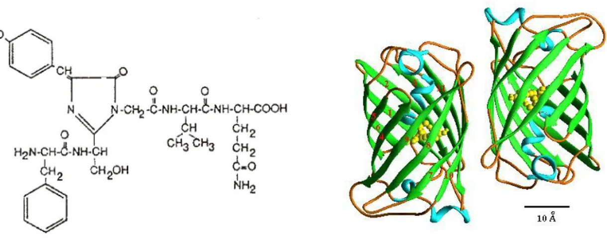
Aequorin is a Ca2+ sensitive photoprotein of a coelenterate, isolated from the jellyfish Aequorea Victoria. The protein is composed of a 21 kDa apoprotein and a hydrophobic prosthetic group, coelenterazine (MW~400 Da). The 2 components must be associated for the Ca2+-triggered light emission to occur. The holoprotein possesses 3 high affinity Ca2+ binding sites (homologous to the sites present in other Ca2+ binding proteins, such as calmodulin). Upon binding of Ca2+ ions, the coelenterazine is oxidized to coelenteramide, with a concomitant release of carbon dioxide and emission of light; this is an irreversible reaction where one photon is emitted.
Fig. 1: The aequorin reaction: Aequorin is composed of an apoprotein and a prosthetic group, coelenterazine, and its polypeptide sequence includes three high affinity Ca2+-binding sites. Ca2+ binding causes the rupture of the covalent link between the apoprotein and the prosthetic group, a reaction associated with the emission of one photon.
Although this reaction is irreversible, in vitro an active aequorin can be obtained by incubating the apoprotein with coelenterazine in the presence of oxygen and 2-mercaptoethanol. Reconstitution of an active aequorin (expressed recombinantly) can be obtained also in living cells by simple addition of coelenterazine to the medium. Coelenterazine is highly hydrophobic and has been shown to permeate cell membranes of various cell types, ranging from the slime mold Dictyostelium discoideum to mammalian cells and plants [41].
The possibility of using aequorin as a calcium indicator is based on the existence of a well-characterized relationship between the rate of photon emission and the free Ca2+ concentration. The rate of this reaction depends on the Ca2+ concentration ([Ca2+]) to which the photoprotein is exposed. In particular, at [Ca2+] between 10-7 and 10-5 M (the concentration normally occurring in the cytoplasm of living cells), there is a direct relationship between [Ca2+] and the fractional rate of consumption of the photoprotein.
Fig. 2: Relationship between the free Ca2+ concentration and the rate of aequorin photon emission. The fractional rate of aequorin consumption is expressed as the ratio between the emission of light at defined [Ca2+] (L) and the maximal rate of the light emission at saturating [Ca2+] (L max).
Figure 2 shows the Ca2+ response curve of aequorin, at physiological conditions of pH, temperature, and ionic strength. It is apparent that the fractional rate of aequorin consumption expressed as the ratio between the emission of light at a defined Ca2+
-8
-7
-7
-6
-6
-5
-5
-4
-4
-3
-2
-1
0
concentration (L) and the maximal rate of the light emission at saturating [Ca2+] (Lmax) is proportional to the 2nd-3rd power of [Ca2+]. Indeed, if all the light emitted by the photoprotein throughout an experiment, as well as that discharged at the end are collected it is possible to estimate Lmax and then calculate back the [Ca2+] to which the photoprotein is exposed in every moment.
Although aequorin luminescence is not influenced either by K+ or Mg2+ (which are the most abundant cations in the intracellular environment and thus the most likely source of interference in physiological experiments) both ions are competitive inhibitors of Ca2+ activated luminescence. Aequorin photon emission can be also triggered by Sr2+ but its affinity is about 100 fold lower than that of Ca2+, while lanthanides have high affinity for the photoprotein (e.g. are a potential source of artifacts in experiments where they are used to block Ca2+ channels). pH was also shown to affect aequorin luminescence but at values below 7.
In fact all experiments of this thesis with aequorin need to be done in well-controlled conditions of pH and ionic concentrations, notably of Mg2+.
2.1.1 Recombinant Aequorin
In the past, was widely employed to measure Ca2+ concentration in living cells, in fact the purified protein was widely used to monitor cytoplasmic [Ca2+] changes in invertebrate muscle cells after microinjection. However, due to the time-consuming and traumatic procedure of microinjection, the role of aequorin in the study of Ca2+ homeostasis remained confined to a limited number of cells (giant cells) susceptible to microinjection.
The cloning of aequorin cDNA [42] and the explosive development of molecular biology offered new possibilities in the use of aequorin, as microinjection has been replaced by the simpler technique of cDNA transfection, opening the way to recombinant expression and thus has largely expanded the applications of this tool for investigating Ca2+ handling in living cells. As a polypeptide, aequorin allows the endogenous production of the photoprotein in cell systems as diverse as bacteria, yeast, plants and mammalian cells.
In particular, recombinant aequorin can be expressed not only in the cytoplasm, but also in specific cellular locations by including specific targeting sequencing in the
Extensive manipulations of the N-terminal of aequorin have been shown not to alter the chemiluminescence properties of the photoprotein and its Ca2+ affinity. On the other hand, even marginal alterations of the C-terminal either abolish luminescence altogether or drastically increase Ca2+ independent photon emission [43]. As demonstrated by Watkins and Campbell [44], the C-terminal proline residue of aequorin is essential for the long-term stability of the bound coelenterazine. For these reasons, all targeted aequorins synthesized in our laboratory include modifications of the photoprotein N-terminal. Three targeting strategies have been adopted:
1. Inclusion of a minimal targeting signal sequence to the photoprotein cDNA.
2. Fusion of the cDNA encoding aequorin to that of a resident protein of the compartments of interest.
3. Addition to the aequorin cDNA of sequences that code for polypeptides that bind to endogenous proteins.
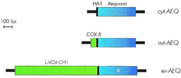
2.1.2 Chimeric Aequorin cDNAs (Fig. 3)
Below I briefly describe the constructs that were present in our laboratory and that I used for the thesis.
Aequorin targeted to cytoplasm (cytAEQ)
An unmodified aequorin cDNA encodes a protein that, in mammalian cells is located in the cytoplasm and, given its small size, also diffuses into the nucleus. An alternative construct was also available that is located on the outer surface of the ER and of the Golgi apparatus. This construct was intended to drive the localization of aequorin to the inner surface of the plasma membrane given that it derives from the fusion of the aequorin cDNA with that encoding a truncated metabotropic glutamate receptor (mgluR1). The encoded chimeric protein, however, remains trapped on the surface of the ER and Golgi apparatus, with the aequorin polypeptide facing the cytoplasmic surface of these organelles. The cytoplasmic signal revealed by this chimeric aequorin is indistinguishable from that of a cytoplasmic aequorin, but it has the advantage of being membrane bound and excluded from the nucleus.
Aequorin targeted to mitochondria (mtAEQ)
The mtAEQ probe has been successfully employed to measure the [Ca2+] of the mitochondrial matrix of various cell types. This construct includes the targeting presequence of subunit VIII of human cytochrome c oxidase fused to the aequorin cDNA [45].
Aequorin targeted to Endoplasmic reticulum (erAEQ)
The erAEQ includes the leader (L), the VDJ and Ch1 domains of an Ig2b heavy chain fused at the N-terminus of aequorin. Retention in the ER depends on the presence of the Ch1 domain that is known to interact with high affinity with the luminal ER protein BiP [46].
Fig. 3: Schematic representation of aequorin chimeras.
2.1.3 Luminescence detection
The aequorin detection system is derived from that described by Cobbold and Lee [47] and is based on the use of a low noise photomultiplier placed in close proximity (2-3 mm) of aequorin expressing cells. The cell chamber, which is on the top of a hollow cylinder, is adapted to fit 13-mm diameter coverslip. The volume of the perfusing chamber is kept to a minimum (about 200 µl). The chamber is sealed on the top with a coverslip, held in place with a thin layer of silicon. Cells are continuously perfused via a peristaltic pump with medium thermostated via a water jacket at 37°C. The photomultiplier (EMI 9789 with amplifier-discriminator) is kept in a dark box and cooled at 4°C. During manipulations on the cell chamber, the photomultiplier is protected from light by a shutter. During aequorin experiments, the shutter is opened and the chamber with cells is placed in close proximity of the photomultiplier. The output of the amplifier-discriminator is captured by an EMIC600 photon-counting board in an IBM compatible microcomputer and stored for further analysis. In order to calibrate the crude luminescent signal in terms of [Ca2+] an algorithm has been developed that takes into account the instant rate of photon emission
order to obtain the latter parameter, at the end of each experiment the cells are lysed by perfusing them with a hyposmotic medium containing 10 mM CaCl2 and a detergent (100
µM digitonin or 0,1% Triton X-100) in order to discharge all the aequorin that was not consumed during the experiment.
A schematic representation of custom-made luminometer used in my laboratory is shown in figure 4.
Fig. 4: Schematic representation of a custom-built luminometer. Cells loaded with functional aequorin probe are incubated in a perfusion chamber, at 37°C, in close proximity to a photon-counting tube. The complete assemblage is kept at 4°C, in the dark, to minimize extraneous signals. Acquisition of the data and subsequent calculations to transform light emission into [Ca2+] are performed by a dedicated computer algorithm.
2.1.4 Advantages and disadvantages of aequorin
In this section I will focus on the main characteristics that make aequorin such a flexible and powerful intracellular calcium sensor.
Advantages:
1) Selective intracellular distribution. Whereas recombinantly expressed wild-type aequorin is exclusively cytosolic, the intracellular fate of the photoprotein can be modified by adding specific targeting sequences.
2) High signal-to-noise ratio. Due to the low luminescence background of cells and the steepness of the Ca2+ response curve of aequorin, minor variations in the amplitude of the agonist-induced [Ca2+] changes can be easily appreciated with aequorin.
3) Low Ca2+ buffering effect. Although the binding of Ca2+ by aequorin may, in principle, affect intracellular Ca2+ homeostasis, this undesired effect is less relevant than with
4°C 37°C water circuit Photomultiplier Peristaltic pump for perfusion
fluorescent indicators. In fact, thank to the excellent signal to noise ratio, aequorin is loaded at a concentration which is 2-3 orders of magnitude lower than dyes, i.e. usually from <0.1 µM (for the recombinantly expressed photoprotein) to ~1 µM (in the case of microinjection of the purified photoprotein for single cell studies).
4) Wide dynamic range. It is clearly evident from Fig. 2 that aequorin can accurately measure [Ca2+] ranging from 0.5 µM to 10 µM, i.e. reaching concentrations at which most fluorescent indicators are saturated. Indeed, thank to these properties and to the low buffering effect, it is possible to estimate the large [Ca2+]c rises which occur, for example, in
neurons [48]. Moreover, by introducing point-mutations in the Ca2+-binding sites [49], using surrogate cations, such as Sr2+, and/or modified prosthetic groups, the sensitivity of the recombinant photoprotein can be further reduced, and thus the [Ca2+] can be monitored also in intracellular compartments endowed with high [Ca2+] (e.g. the lumen of the ER). 5) Possibility of co-expression with proteins of interest. A powerful approach for investigating the role, and the properties, of the various molecular components of the Ca2+ signalling apparatus is either the overexpression of the heterologous protein, followed by the study of the molecularly modified cell. This can be accomplished in two ways, either by generating stably transfected cell clones, or by transiently expressing the protein of interest in a cell type.
Disadvantages:
1) Overestimation of the average rise in cells (or compartments) with dishomogeneous behaviour. A disadvantages of the steepness of the Ca2+ response curve of aequorin is that, if the increase of the [Ca2+] is not homogeneous, the average estimate will be based towards the highest values. Indeed, by using targeted aequorin to monitor subplasmalemmal Ca2+ concentration, we measured a mean resting [Ca2+] of 1-2 µM, which most likely does not reflect the real average [Ca2+]pm value, but, rather, the
contribution of microdomains in the proximity of flickering Ca2+ channels [50].
2) Low light emission. In distinction to the fluorescent dyes (where up to 104 photons can be emitted by a single molecule, before photobleaching occurs), only 1 photon can be emitted by an aequorin molecule. Moreover, the principle of the use of aequorin for Ca2+ measurements is that only a small fraction of the total pool (varying, in a typical physiological experiment, from 10-7 to 10-2) emits its photon every second. This is not a major limitation in population studies, as averaging over 103-104 cells. Conversely, single cell imaging requires very high expression and special apparatuses [51] and it is endowed
2.2 Green Fluorescent Protein (GFP)
In recent years, two decisive methodological advances have greatly improved optical microscopy and allowed to follow biological processes in living cells: the improvement of the instrumentation (with the development of confocal microscopes and of mathematical methods for image analysis) and the discovery of the "green fluorescent protein" (GFP), a protein derived from a jellyfish that can be utilized as a highly efficient intracellular probe in fluorescence microscopy. It is thus now possible to follow directly in intact, living cells cellular events of major biomedical relevance, such as the movement of proteins (including their mutated variants in genetic diseases) or intracellular organelle distribution. The possibility of directly identifying intracellular organelles in living cells has a major relevance in these cell biology studies. Indeed, key events, such as organelle distribution and dynamics, can be monitored in a variety of physiological phenomena (e.g. localized Ca2+ rises), and also the intracellular location (and the relative relations between the different organelle) can be unambiguously identified.
Fig. 5: The chemical structure of the chromophore in Aequorea GFP. Structural formula of GFP. GFP has a cylindrical fold. Two protomers associate together to form a dimer in the crystal and also in solution at low ionic strengths, <100 mM. Dimerization has an effect on the excitation spectra and energy transfer of GFP. The dimer is involved in physiological interactions with aequorin for efficient energy transfer. The structure is comprised of two regular beta-barrels.
Green fluorescent protein, GFP, is in nature the legitimate partner of aequorin. Indeed, it is produced by the same jellyfish (Aequorea victoria) and packed in close association to the photoprotein, acting as a natural fluorophore that absorbs the blue light emitted by the photoprotein and re-emits photons of a longer wavelength (thus accounting
for its own name and for the greenish hue of the jellyfish luminescence) [41, 52, 53]. In research applications, GFP retains its fluorescence properties, and thus can be added to the long list of probes of the cell biologist’s toolbox [54]. Some of its unique properties account for its explosive success, and has made it, in relatively few years (the first report of GFP expression in heterologous systems dates back to 1994) [55], a powerful and versatile tool for investigating virtually all fields of cell biology (ranging from the study of gene expression to protein sorting, organelle structure, measurement of physiological parameters in living cells) [56, 57].
The main reason for the success of GFP is its own nature: the fluorescent moiety is a gene product (with no need for cofactors) that is open to molecular engineering and transient or stable expression in virtually every cell type. Moreover, its mutagenesis has allowed the adaptation of its fluorescent properties to different experimental needs (for a detailed description see reviews [54, 58]).
GFP mutants can be grouped in two major classes: the first are the “optimising” mutations, i.e. those that increase light emission by either altering the intrinsic properties of the fluorescent protein [59, 60] or increasing its production in mammalian cells [61]. The latter are the so called “humanised” versions of the cDNA, in which silent mutations are introduced in order to convert some of the codons into the most common and efficient for translation in mammalian cells.
As to the modification of GFP properties, mutations have been described that alter the stability of the protein and/or the quantum efficiency upon illumination with visible light (native GFP has a bimodal excitation peak, larger with UV than with blue light, while all currently employed green variants of GFP are best excited with blue light, thus minimising cytotoxicity and photobleaching). Among these mutations, the most useful appears to be the substitution of Ser65 into Thr (S65T), which causes the chromophore to be entirely in the anionic form: thus, when compared to wild-type GFP the quantum efficiency upon excitation with blue light is 6-fold increased, the rate of fluorophore formation is 4-fold faster and photobleaching is markedly reduced. The second class is represented by GFPs emitting light of a wavelength that can be clearly distinguished from the green colour of native GFP. This class includes some popular mutants commonly referred to as blue (Y66H, Y145F), cyan (Y66W) and yellow (T203Y) GFPs.
Fig. 6: Excitation (A) and emission (B) spectra of different GFP mutants.
Another green fluorescent protein, found in the sea pansy Renilla, has been biochemically characterised [62], while the cDNA has not been isolated yet. Although the chromophore of Renilla is similar to that of Aequorea, some of their biochemical properties differ. Renilla GFP has a much higher extinction coefficient, is an obligate dimer and is more resistant to pH-induced conformational changes, which could make it useful for some cell physiology studies.
Nowadays the most frequent application of the GFPs is to use them as a tag. In fact, fusing in frame the GFP cDNA to a cDNA coding for a protein of interest, it is possible to examine the function and fate of the resulting chimera in living cells. Moreover, chimeras with different spectral properties can be employed for visualising simultaneously two proteins of interest (e.g. two isoforms of a signalling molecule) or the morphology and spatial relationship between two intracellular compartments. The GFPs can also be used as a tool for analysing transfection efficiency or as sensors for physiological parameters. This approach stems from an interesting phenomenon, called fluorescence resonance energy transfer (FRET), occurring between two GFPs with different colours. FRET may occur only if the fluorescence emission spectrum of the “donor” GFP overlaps with the excitation spectrum of the “acceptor” GFP and if the two fluorophores are located within few nanometers in a favourable orientation.
As for aequorin I briefly describe the GFP constructs that were present in our laboratory and that I used for the thesis:
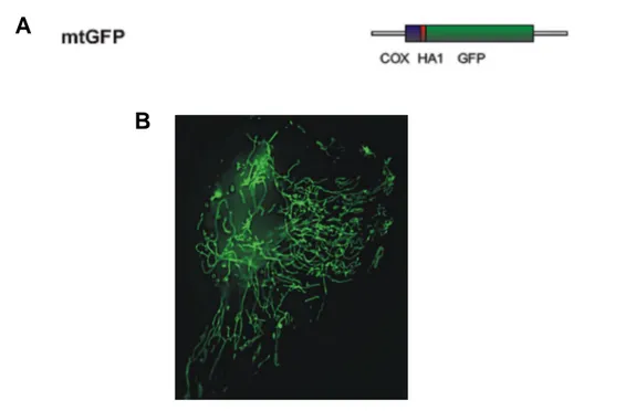
mitochondrial GFP probe: the previously employed mitochondrial presequence derived from subunit VIII of cytochrome c oxidase (encoding the 25 aa-long presequence and 6 aa of the mature polypeptide) to construct a mitochondrially targeted aequorin
chimera [45] was fused in frame with the GFP, in order to construct mitochondrially-targeted GFP (mtGFP) [63]. When expressed in mammalian cells, the chimeras show a typical mitochondrial distribution.
A
B
Fig. 7: The mitochondria-targeted GFP construct. The mitochondrial presequence of COX VIII protein was fused in frame with GFP at N-terminus (A). As aspected, this chimera shows a typical mitochondrial staining (B).
2.3 Experimental set-up: collecting and analysing the GFP images
The fluorophore of GFP is formed by the cyclization of three amino-acid residues of the primary sequence. This process directly follows, with few constraints (a relatively brief time lag), the synthesis of the protein, and thus GFP proved brightly fluorescent when expressed in a wide variety of cell types (mammalian cells, plants, fungi, bacteria, etc.) and intracellular locations (cytoplasm and virtually every organelle). Thus, also in our experiments the various GFP chimeras are transfected with the appropriate procedure (calcium phosphate, liposomes or particle gun, depending on the cell type), and directly visualised in living cells after allowing sufficient time for expression and chromophore formation (usually 24-36 hours). For this purpose, the coverslip with the transfected cells is fitted at the base of a thermostatted chamber, which is placed on the microscope stage.
Fig. 8: Microscope setting. A digital imaging system, built on an epifluorescence microscope, is equipped with filter-wheels placed on the excitation and emission light paths, a piezoelectric motor and a CCD camera. The system is operated by a software that also permits to analyse and computationally deblur the images.
Our microscope set up is presented in figure 8. In brief, a traditional wide-field, epifluorescence microscope is equipped with filter-wheels located both in the incoming and in the out-coming light paths (thus allowing to rapidly alternate excitation and/or emission wavelength) and a piezoelectric transducer (or motor drive) for rapid focussing in the z plane. The fluorescence image is collected by a back-illuminated and cooled (-40°C), charge coupled device (CCD) camera having high quantum efficiency (>70% at 500nm) and low noise (<10e- RMS at 1MHz) characteristics, and the image is stored as a digital file using the Metamorph/Metafluor program (Universal Imaging). This allows the direct monitoring of fluorescence intensity, important for some applications (e.g. the monitoring of FRET, or the pH measurements using the pH-sensitive GFP mutants, a topic that for reasons of brevity will not be discussed in this thesis).
A high-resolution, 3-D reconstruction of the distribution of a GFP chimera can be obtained with the technique of digital image restoration, also called deconvolution or deblurring. [64].
In situations where there may be significant intracellular motion of the GFP chimera, two approaches can be used to decrease the time for image acquisition. The area of the cell (i.e. the number of pixels in each optical section) imaged can be reduced, thus decreasing the time to transfer the image data to computer storage. A high-resolution image restoration of a limited volume of a cell has been obtained using a few as 5 to 7 optical sections [65], minimising the total acquisition time.
A high-speed version of this microscopy has been developed [66] that can acquire an entire through-focus image series of a GFP labelled cell in less than 1 second. This microscope system can be used to follow spatial and temporal intracellular dynamics (e.g. motor-protein based transport, signal transduction) too rapid for conventional fluorescence microscopy [66]. Conversely, if only the time course rather than the 3-D distribution of a fast process (e.g. Ca2+ signalling) needs to be assessed, single fluorescent images of the microscope field of interest can be acquired every 10-20 ms with no further image processing [67].
Finally, the filter-wheels allow the alternate imaging of two different fluorophores at different excitation and emission wavelengths, and thus the simultaneous visualisation of two different proteins of interest in the same cell, or the measurement of donor and acceptor fluorescence in FRET applications. When compared to laser scanning confocal microscopy, digital imaging is characterised by higher flexibility in the selection of excitation wavelengths, lower illumination intensity (thus reducing photobleaching and photodamage) and lower cost. Conversely, its disadvantages are the need for time-consuming off-line image processing and the unsuitability for the analysis of thick specimen (e. g. tissue slices).
2.4 Luciferase
Firefly luciferase was cloned in the late 1980s [68], and versions of the gene, optimised for thermostability and expression in mammalian cells, were generated. The enzyme uses an oxidisable substrate, termed luciferin, which is converted to an AMP adduct before final oxidation with molecular oxidation and the release of a photon of light (Fig. 9).
also be readily measured from living cells, after the addition of the (reasonably cell-permeant) cofactor, luciferin. Under most conditions, O2, and cofactors otherthan ATP, are
not limiting. Further, it can be calculated that the contribution of ATP consumption by luciferase represents only a tiny fraction of total cellular ATP turnover (<0.1 %, even at relatively high levels of luciferase expression, e.g. 1 x 10⁶ molecules/cell) and thus non-perturbing for normal cellular ATP homeostasis.
Fig. 9: The ATP-dependent luminescence reaction of Luciferase
The technology to the detection of changes in free intracellular ATP concentration require constantly high levels of luciferase expressed from strong viral promoters (e.g. the cytomegalovirus immediate early gene promoter, CMV-IE), so that small fluctuations in free ATP concentration can be monitored in cell populations, by detection of luciferase luminescence is possible with the photon-counting tube apparatus, as described above for the detection of aequorin luminescence.
Luciferase displays a Km for ATP close to 1 mM when assayed in cell homogenates
under approximate in vivo conditions of pH and physiological ionic strength [69] (compared to the low micromolar range under optimal in vitro conditions).
Confirming these values in living cells is complicated due to the distinct kinetics of the enzyme in the living cell (“glow” versus “flash” kinetics”). Although the basis for this difference is not fully understood, it may reflect a lack of the accumulation of inhibitory end product (oxyluciferin) in the cell, or a decreased sensitivity to this (or another inhibitor) mediated by other cellular cofactors (notably CoA). Whatever the mechanism, this makes monitoring [ATP] constantly in the living cell relatively straightforward, given sensitive photon detection equipment.
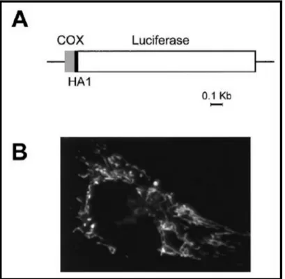
In order to measure [ATP] exclusively into the mitochondrial compartment ([ATP]mt),
our groups generated a mitochondrial-targeted luciferase, using the same strategies employed for creation of mtAEQ and mtGFP chimera (Fig. 10) [70]. Moreover, this paper
shown how stimulation with agonists evoking cytosolic and mitochondrial Ca2+ signals caused increases in [ATP]mt and [ATP]cyt [70].
Fig. 10: Map and correct localization of mtLUC construct. A) Schematic map of the chimeric mitochondrial luciferase. Lines and bars indicate the noncoding and coding regions [gray, cytochrome c oxidase subunit VIII (COX8); black, HA1; white, luciferase], respectively. (B) Immunofluorescence image of HeLa cells transiently transfected with mtLuc and stained with the anti-HA1 mAb (figure from [70]).
3. Protein Kinase C β and Prolyl Isomerase Pin1 Regulate Mitochondrial
Effects of the Life-Span Determinant p66Shc
3.1 Introduction
Many agents which induce apoptosis are either oxidants or stimulators of cellular oxidative metabolism. Conversely, many inhibitors of apoptosis have antioxidant activities or enhance cellular antioxidant defences. Mammalian cells exist in a state of oxidative siege in which survival requires an appropriate balance of oxidants and antioxidants, suggesting that eukaryotic cells may benefit from this perilous existence by invoking oxidative stress as a common mediator of apoptosis.
Apoptosis, the process that allows multicellular organisms to eliminate unnecessary, dangerous or damaged cells without evoking inflammation or tissue damage, occurs in both physiological and pathological contexts. Specific biochemical steps in the apoptotic pathway are inhibited by viruses and in tumors. On the other hand, excess apoptosis in non-proliferating cells has been proposed to be the basis of a number of degenerative disorders, as well as in progressive loss of organ function during ageing. The understanding of the control mechanisms of apoptosis is thus a major goal for the understanding of these pathophysiological events, and the development of novel therapeutic approaches.
Recently the mitochondrion has emerged as a key decoding station of the apoptotic process. Indeed, a large body of experimental evidence has unambiguously revealed that mitochondria trigger cell death, in addition to the well-established function of producing most of cellular ATP. Various apoptotic stimuli cause the release of specific mitochondrial proteins into the cytoplasm (including an essential component of the electron transfer chain, cytochrome c). Their assembly with cytosolic proteins forms a complex (the “apoptosome”), that recruits and activates effector caspases, which in turn trigger apoptotic death. The molecular mechanism of this release is still controversial, but may require the activity of a large-conductance channel, known as the permeability transition pore, PTP. Its opening induces the swelling of mitochondria, and this large-scale alteration of organelle morphology (with perturbation or rupture of the outer membrane) may allow the release of pro-apoptotic components into the cytosol.
There is no doubt that cell death belongs to the numerous cell functions on which Ca2+ exerts a complex regulatory role. As to the site of action of the “apoptotic” Ca2+ signal,
mitochondria again emerge as a critical site. Indeed, treatment with apoptotic stimuli, such as ceramide, causes a release of Ca2+ from the ER and induces dramatic changes of mitochondrial morphology [33]. If Ca2+ changes are prevented (e.g. by loading an intracellular Ca2+ buffer), mitochondrial morphology is preserved and the cells are protected from apoptosis. Much remains to be understood on the additional signals that converge on mitochondria and switch their function into apoptotic inducers. As much as Ca2+ appears to be involved, there is no doubt that coincident detection of other “pro-apoptotic” conditions needs to occur. Indeed, mitochondria can handle in physiological conditions large Ca2+ loads (e.g. in cardiac myocytes significant amounts of Ca2+ are accumulated at every heartbeat), with no deleterious effects. As to the additional “apoptotic signal”, the most important is considered to be oxidative stress. However, on this topic the information is very scant, as its real significance in apoptosis and the mechanism of action are still largely unknown.
In 1999, the group of Pelicci and co-workers published a paper that became a crucial point in studies on the oxidative stress [71]. They proposed that mammalian life span can be controlled by p66Shc protein due to regulation of cellular response to oxidative stress.
Ablation of the p66Shc gene causes life-span prolongation with no pathological consequence. In fact, mice lacking p66Shc gene had enhanced resistance to oxidative stress induced by paraquat (a potent inducer of oxidative stress in vivo).
3.1.1 p66Shc, an adaptor protein
The p66Shc protein is an alternatively spliced isoform of the growth factor adapter. Recently, three proteins from ShcA family have been identified (p66Shc, p52Shc and p46Shc). Each of them contains three functionally identical domains: the carboxy terminal Src homology 2 (SH2) domain, the central proline-rich domain (CH1), and the N-terminal phosphotyrosine-binding domain (PTB). PTB domain can bind to phospholipids and phosphotyrosine of the other proteins (Fig. 11).
P66Shc protein differs from p46Shc and p52Shc by the presence of an additional N-terminal proline-rich domain (CH2) with the serine phosphorylation site, Ser36. Phosphorylation of this residue plays an important role in the cellular response to the oxidative stress. Recently, it has been demonstrated that, in response to UV or H2O2
treatment, p66Shc is phosphorylated mainly at serine 36 in the N-terminal CH2 domain [71]. Also a p66Shc transfection with mutated Ser36 failed to restore apoptosis sensitivity in p66Shc lacking cells, what supports the importance of Ser36 in regulation of apoptosis.
The SH2 domain of Shc is important for certain receptor interactions, such as epidermal growth factor (EGF) receptor. Interaction between the SH2 domain and EGF receptor plays an important role in phosphorylation of tyrosines in the CH1 domain of p52Shc and p46Shc [72]. Tyrosine phosphorylation enables them to bind to the adaptor Grb2 protein–activator of Ras protein. Also p66Shc can be tyrosine phosphorylated, but there are no evidences about its ability to activate the Ras signalling pathway.
Further studies of the mechanism of lifespan-promoting activity of p66Shc deficiency, have suggested not only a role in ROS metabolism, but also in apoptosis. Steady-state ROS and p53-induced ROS are both decreased in p66Shc -/- Mouse Embryonic Fibroblasts (MEFs). Possibly as a consequence of this, hydrogen-peroxide-mediated release of cytochrome c is inhibited in p66Shc -/- MEFs, and transfection of p66Shc into these cells rescues the sensitivity. Furthermore, there is a reduction in the amount of oxidation-damaged DNA of p66Shc -/- mice, as well as for mitochondrial DNA [73].
P66Shc appears to sensitively detect oxidative stress, i.e. the pathophysiological condition that has been often associated to cellular ageing, and is known to sensitize to a number of apoptotic challenges. P66Shcthus appears as a very promising link between phenomenological observations of the ageing process and the identification of the molecular mechanism responsible for the cellular changes. This is a fundamental step as it highlights potential targets for pharmacological intervention.
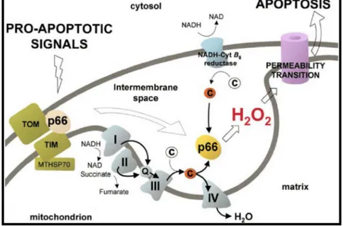
In this scenario, the portion of p66Shc located into mitochondria seems to be fundamental for its role in oxidative stress. In fact, a fraction of p66Shc localizes to mitochondria [74, 75], where it binds to cytochrome c and acts as oxidoreductase, generating reactive oxygen species (ROS) and leading to organelle dysfunction and cell death [76] (Fig. 12). The route leading to p66Shc activation is still unclear.
Fig. 12: Model of p66Shc redox activity during mitochondrial apoptosis. Proapoptotic signals induce release of p66Shc from a putative inhibitory complex. Active p66Shc then oxidizes reduced cyt c (red) and catalyzes the reduction of O2 to H2O2. PTP opening by H2O2 then leads to swelling and apoptosis (figure from
[76]).
In this work, we address this task, focusing on the Ca2+-mediated signals in mitochondria. The rationale for this strategy stems from a number of experimental observations. Mitochondria receive, under stimulation by physiological agonists or toxic agents, Ca2+-mediated inputs [34, 77, 78]. These Ca2+ signals are decoded within mitochondria into effects as diverse as stimulation of aerobic metabolism and alterations of organelle structure leading to release of caspase cofactors into the cytoplasm [79]. Recent
works showed that the responsiveness of mitochondria to Ca2+ signals can be tuned by the cross-talk with other signalling pathways and the activation of regulatory proteins, such
as kinases. Specifically, we could show that some PKC isoforms (i.e. the components of a wide molecular repertoire of kinases differing for biochemical properties and activation mechanisms) specifically affect mitochondrial Ca2+ responses to agonists (by reducing them, such as PKC, or enhancing them, such as PKCζ) [80]. Interestingly, PKCs were proposed to be activated directly in conditions of oxidative stress [81].
3.2 Results
3.2.1 Effect of oxidative stress and the aging protein p66Shc on the mitochondrial Ca2+
homeostasis
We used aequorin to monitor cellular concentrations of Ca2+, a green fluorescent protein with mitochondrial presequence (mtGFP) to monitor organelle structure (well explained in section 2), and other molecular tools to clarify the signalling route linking the oxidative challenge to the activation of p66Shc proapoptotic effect within mitochondria in mouse embryonic fibroblasts (MEFs).
Using mtAEQ, we investigated organelle Ca2+ responses to adenosine triphosphate (ATP), an extracellular agonist that causes the release of Ca2+ from the endoplasmic reticulum. P66Shc −/− and wild-type MEFs showed similar responses of mitochondrial calcium ([Ca2+]mt) to ATP, both in amplitude and in kinetics (Fig. 13, A and B). This reflects
a close similarity in the global Ca2+ signaling patterns. Indeed, the monitoring of concentration of free cytosolic Ca2+ ([Ca2+]cyt) showed that the [Ca2+]cyt rises evoked by
ATP in p66Shc −/− and wild-type MEFs were virtually superimposable (Fig. 13, A and B, insets).
To investigate the effect of an oxidative challenge, we treated cells for 30 minutes before the application of ATP with various concentrations of H2O2. Reduction of
mitochondrial Ca2+ responses and fragmentation of the threedimensional mitochondrial network [33] was observed in wild-type MEFs (Fig. 13, A and a) several hours before signs of apoptosis (cell shrinkage and nuclear condensation, for example) were detected, whereas minor changes in the Ca2+ response and morphology were detected in p66Shc −/− MEFs (Fig. 13, B and b). This alteration in Ca2+ response was characteristically mitochondrial, because no difference in the ATP-dependent [Ca2+]cyt rise was detected
between p66Shc −/− and wild-type H2O2-treated MEFs (Fig. 13, A and B, insets). The
reintroduction of p66Shc reestablished sensitivity to H2O2 in p66Shc −/− MEFs (Fig. 13, C
Fig. 13: Mitochondrial morphology and Ca2+ responses in p66Shc MEFs during oxidative stress.
Mitochondrial and cytosolic (inset) Ca2+ homeostasis in wild-type (wt) (A) and p66Shc −/− (B) MEFs. wt:
[Ca2+]mt) peak, 8.64 ± 0.32 mM; [Ca
2+
]cyt) peak, 2.90 ± 0.11 mM. p66Shc −/−: [Ca
2+
]mt peak, 8.71 ± 0.37 mM;
[Ca2+]cyt peak, 2.91 ± 0.15 mM. The dotted traces show the effect of treatment with H2O2 (1 mM, 30 min.) on
the ATP-dependent responses. wt: [Ca2+]mt peak, 5.84 ± 0.28 mM; [Ca2+]cyt peak, 2.60 ± 0.07 mM. p66Shc
−/−: [Ca2+]mt peak, 7.87 ± 0.33 mM; [Ca2+]cyt peak, 2.7 ± 0.09 mM. (a and b) Analysis of mitochondrial
structure in cells treated with or without H2O2 (1 mM, 30 min). (C and c) Reintroduced p66Shc reestablishes
Production of ROS by p66Shc [76] influences the opening of the mitochondrial permeability transition pore (PTP) [82]. We thus investigated whether the Ca2+ and morphology changes triggered by H2O2 could be prevented by the PTP blocker cyclosporine A (CsA).
In CsA-treated wild-type MEFs, the rise in [Ca2+]mt evoked by ATP stimulation in the
presence of H2O2 was largely restored (Fig. 14 A) and the integrity of the mitochondrial
network was preserved (Fig. 14 a). On the contrary, no effect of CsA on p66Shc −/− (Fig. 14 A, inset). Mitochondrial Ca2+ responses and morphology were not modified by H2O2
application to p66Shc −/− MEFs in which either the p66ShcE132Q-E133Q mutant (p66Shcqq), incapable of binding cytochrome c [76] (Fig. 15, A and a), or the p66ShcS36A mutant [71] (Fig. 15, B and b) had been reintroduced, indicating that both the oxidoreductase activity of p66Shc and the phosphorylation of Ser36 are essential for the H2O2-induced proapoptotic changes.
Fig. 14: Involvement of PTP in mitochondrial p66Shc action. Effect of treatment with CsA (4 mM, 10
min.) on H2O2-dependent reduction of [Ca2+]mt responses in wt MEFs. [Ca2+]mt peak, 5.84 ± 0.28 mM in cells
treated only with H2O2; 7.58 ± 0.33 mM, P < 0.01, in cells pretreated with CsA, then treated with H2O2 (A). No
effect of CsA on p66Shc −/− cells was detected. [Ca2+]mt peak, 7.87 ± 0.33 mM in cells treated only with
H2O2; 8.43 ± 0.54 mM in cells pretreated with CsA, then treated with H2O2 (inset). Morphology of H2O2-