Preclinical Findings
Monica Puligheddu1., Giuliano Pillolla1., Miriam Melis2,4, Salvatore Lecca2, Francesco Marrosu1, Maria Graziella De Montis3, Simona Scheggi3, Gianfranca Carta2, Elisabetta Murru2, Sonia Aroni2, Anna Lisa Muntoni4, Marco Pistis2,4*
1 Department of Public Health, Clinical and Molecular Medicine, University of Cagliari, Cagliari, Italy, 2 Department of Biomedical Sciences, University of Cagliari, Cagliari, Italy,3 Department of Molecular and Developmental Medicine, University of Siena, Siena, Italy, 4 C.N.R. Neuroscience Institute, Cagliari, Italy
Abstract
Nicotinic acetylcholine receptors (nAChRs) are involved in seizure mechanisms. Hence, nocturnal frontal lobe epilepsy was the first idiopathic epilepsy linked with specific mutations in a4 or b2 nAChR subunit genes. These mutations confer gain of function to nAChRs by increasing sensitivity toward acetylcholine. Consistently, nicotine elicits seizures through nAChRs and mimics the excessive nAChR activation observed in animal models of the disease. Treatments aimed at reducing nicotinic inputs are sought as therapies for epilepsies where these receptors contribute to neuronal excitation and synchronization. Previous studies demonstrated that peroxisome proliferator-activated receptors-a (PPARa), nuclear receptor transcription factors, suppress nicotine-induced behavioral and electrophysiological effects by modulating nAChRs containing b2 subunits. On these bases, we tested whether PPARa agonists were protective against nicotine-induced seizures. To this aim we utilized behavioral and electroencephalographic (EEG) experiments in C57BL/J6 mice and in vitro patch clamp recordings from mice and rats. Convulsive doses of nicotine evoked severe seizures and bursts of spike-waves discharges in ,100% of mice. A single dose of the synthetic PPARa agonist WY14643 (WY, 80 mg/kg, i.p.) or chronic administration of fenofibrate, clinically available for lipid metabolism disorders, in the diet (0.2%) for 14 days significantly reduced or abolished behavioral and EEG expressions of nicotine-induced seizures. Acute WY effects were reverted by the PPARa antagonist MK886 (3 mg/kg, i.p.). Since neocortical networks are crucial in the generation of ictal activity and synchrony, we performed patch clamp recordings of spontaneous inhibitory postsynaptic currents (sIPSCs) from frontal cortex layer II/III pyramidal neurons. We found that both acute and chronic treatment with PPARa agonists abolished nicotine-induced sIPSC increases. PPARa within the CNS are key regulators of neuronal activity through modulation of nAChRs. These effects might be therapeutically exploited for idiopathic or genetically determined forms of epilepsy where nAChRs play a major role.
Citation: Puligheddu M, Pillolla G, Melis M, Lecca S, Marrosu F, et al. (2013) PPAR-Alpha Agonists as Novel Antiepileptic Drugs: Preclinical Findings. PLoS ONE 8(5): e64541. doi:10.1371/journal.pone.0064541
Editor: Ste´phane Charpier, University Paris 6, France
Received January 9, 2013; Accepted April 15, 2013; Published May 27, 2013
Copyright: ß 2013 Puligheddu et al. This is an open-access article distributed under the terms of the Creative Commons Attribution License, which permits unrestricted use, distribution, and reproduction in any medium, provided the original author and source are credited.
Funding: This research was supported by the Italian Ministry of University (MIUR) to Marco Pistis (grant PRIN 2009: 200928EEX4) and to Francesco Marrosu and Anna Lisa Muntoni (grant PRIN 2009: 2009HST9YF) by the FIRE-AICE 2011 grant and Fondazione Banco di Sardegna grant 2012 to Marco Pistis. The funders had no role in study design, data collection and analysis, decision to publish, or preparation of the manuscript.
Competing Interests: The authors have declared that no competing interests exist. * E-mail: [email protected]
.These authors contributed equally to this work.
Introduction
Binding of nicotine to nicotinic acetylcholine receptors (nAChRs) elicits in laboratory animals dose-dependent effects that begin with hypermotility and culminate with clonic-tonic seizures and death at high doses [1]. Investigation on the mechanisms underlying nicotine-induced seizures might help to understand how nAChRs participate in the mechanisms of epilepsy. Interest for nAChR in several epileptic syndromes previously considered ‘‘idiopathic’’ was rekindled by the finding that altered functional properties of nAChR are implicated in the pathogenesis of nocturnal frontal lobe epilepsy (NFLE), and that seizures induced by nicotine in rodents model nAChR-related epilepsy. NFLE is linked with mutations of the a4 or b2 subunits [2,3,4,5,6,7], the most abundantly expressed subunits in the CNS [8]. Though in human NFLE, or in genetically engineered mice that model the disease, functional properties of nAChRs are intimately altered,
nicotine evokes seizures by over-activating a healthy system. However, several neurophysiological events ultimately leading to seizures may share common steps between these conditions. Indeed, the common trait in enhancing the epileptogenesis is the over-activation of cholinergic systems, either pharmacologically induced or mediated by the gain of function that mutated nAChRs exhibit toward their ligands [9]. These effects might be based on the extensive expression of nAChRs, particularly those containing the b2 subunit (b2*nAChRs) in thalamo-cortical, hippocampal and frontal regions [10]. Indeed, altered cholinergic activation of neocortical and/or thalamocortical networks plays a central role in the generation of both nicotine-induced and NFLE seizures, which originate in the frontal cortex (FCx) [11] and specifically affect GABAA-mediated inhibitory inputs to pyramidal neurons [12].
Based on this evidence, negative regulation of nAChRs might represent a potential therapeutic approach in nAChR-related forms of epilepsies.
We previously discovered that nicotine-evoked excitation of dopamine neurons both in vivo and in vitro, as well as nicotine addictive properties in rats and monkeys, are suppressed by ligands to the peroxisome-proliferator-activated receptor-a (PPARa) [13,14,15,16]. PPARa is one of three subtypes of the nuclear receptor PPAR family [17,18,19], expressed by neurons in many brain regions [20,21], and activated by endogenous ligands, the N-acylethanolamines oleoylethanolamide (OEA) and palmitoyletha-nolamide (PEA) [22], and by synthetic ligands such as hypolip-idemic fibrates [16].
Evidence points to a non-transcriptional interaction between PPARa and nAChR, via phosphorylation [13,14,23]. This mechanism might account for the blockade of neuronal and behavioral responses to nicotine [13,15,16].
Building upon these findings, we postulated that PPARa agonists might display anticonvulsant properties. Thus, we first investigated whether acutely or chronically administered PPARa ligands reduce the severity of nicotine-induced seizures. Next, we assessed whether these compounds regulate the phosphorylation status of the b subunit of the nAChR and abolish nicotine-induced enhancement of inhibitory currents on pyramidal neurons in layers II/III of the frontal cortex (FCx) in mice and rats.
Materials and Methods Animals
For behavioral experiments, electroencephalographic (EEG) and patch-clamp recordings male C57BL/6J mice (Harlan, San Pietro al Natisone, Italy) (n = 80; weight: 25–30 g each) were used. For immuno-blotting experiments and patch-clamp recordings, we utilized male Sprague-Dawley rats (250–300 g and 14–21 d, respectively, n = 30, Harlan, San Pietro al Natisone, Italy) Mice and rats were housed six per cage under a 12 h light/dark cycle (light on at 7:00 AM), in conditions of constant temperature (2162uC) and humidity (60%), with food and water ad libitum.
Ethics Statement
Experiments were performed in strict accordance with the EEC Council Directive of 24 November 1986 (86/609). All efforts were made to minimize pain and suffering and to reduce the number of animals used. The experimental protocols were also approved by the Animal Ethics Committee of the University of Cagliari.
Treatments
Animals were randomly assigned to the experimental groups undergoing behavioral seizure scoring or EEG recordings. In the first set of experiments a dose-response curve for nicotine (5, 7, 10 mg/kg) was carried out. Each mouse received a single dose of nicotine.
Subsequent acute experiments, aimed to assess the effect of the PPARa agonist WY14643 or the antagonist MK886, were carried out with the 10 mg/kg dose of nicotine. In these cases, mice received two injections, spaced by 10 min intervals, before the final nicotine administration after 15 min: we first injected the antagonist MK886 (3.0 mg/kg, i.p.) or its vehicle followed by the administration of WY14643 (80 mg/kg, i.p.) or its vehicle.
For chronic studies, one week after EEG electrode implants (see below) mice were divided into diet treatment groups: (i) a standard diet (control group, Harlan Teklad Global 2016); (ii) a 0.2% fenofibrate diet (Fenofibrate from Sigma-Aldrich+Harlan Teklad Global 2016). Mice were fed diets for 14 days. On the day of the experiment, mice received one injection [MK886 (3.0 mg/kg, i.p.)] or its vehicle before nicotine (10 mg/kg) administration after 15 min.
Seizure Scoring
Immediately after nicotine injection, mice were placed in a regular mouse cage with bedding, and behavioral responses were recorded for 5 min. The symptoms were scored independently by two experimenters blind to the treatment on an arbitrary scale from 0 to 6 (modified from Franceschini et al., 2002) [24] as follows: 0, no visible effects; 1, locomotor effects including increased exploring activity and/or sedation; 2, tachypnea, tremors, back arching; 3, any combination of the symptoms in 1 and 2 plus rapid movements of the legs, wild running, or partial loss of righting reflex; 4, any combination of the previous symptoms plus complete loss of righting reflex, clonic seizures, 5, any combination of the preceding symptoms plus tonic seizures; 6, death, with or without hyperextension of the limbs along the axis of the body (soldier position).
EEG Recordings
Mice were anesthetized with Equithesin (5 ml/kg, i.p.) and placed in a stereotaxic apparatus (David Kopf, mod. 900). The skull was exposed and perforated, the holes aimed at the following positions: one located above the (either left or right) sensorimotor cortices (FPr and FPl), [coordinates from bregma (mm): antero-posterior +2, lateral 3, ventral to skull surface 1.5], two targeted to the dorsal hippocampus with bipolar leads glued together [coordinates from bregma (mm): anteroposterior +2, lateral 1 and 2, ventral 1.3 and 2] and one on the skull over the cerebellum as a reference [coordinates from bregma (mm): anteroposterior 25.9, lateral 1.5] [25]. A four-pin male socket was positioned into the holes, secured to the skull with epoxy resin and covered with acrylic cement to improve retention.
One week following surgical preparation of the animals (or three to five weeks for chronic studies), experiments started according to the protocol described above, electrical potentials were acquired and the signals amplified, bandpass-filtered and recorded on a portable digital EEG polygraph (BQS 98 System Micromed). In addition, in view of the possibility that digital data generated by the above mentioned experimental settings may need unconven-tional processing, further home-made software analysis rewritten on a MatlabH platform was used (e.g. signal analysis with wavelets versus traditional fast Fourier transform (FFT) processing).
Electrode impedance was maintained at ,5 kV. The amplified signals, processed with a band-pass filter (0.02 to 70 Hz), was stored on the hard disk at a sampling rate of 256/s.
The behavior of the animals was video-recorded for the entire duration of the experiment.
In vitro Electrophysiology
The preparation of FCx slices and whole-cell patch clamp recordings from layer II/III pyramidal neurons was as described previously [12]. Briefly, male mice (10–25 d, Harlan) and Sprague Dawley rats (14–21 d, Harlan) were anesthetized with halothane and killed. A block of tissue containing the FCx was sliced in the coronal plane (300mm) with a vibratome (Leica, Nussloch, Germany) in ice-cold low Ca2+ artificial cerebrospinal fluid (ACSF) containing (in mM): 126 NaCl, 2.5 KCl, 2 CaCl2,
2 MgCl2, 1.25 NaH2PO4, 26 NaHCO3, and 10 D-glucose
(pH 7.3–7.4). Slices (two per animal) were transferred in a holding chamber and allowed to recover for at least 1 hr before being placed in the recording chamber and superfused with ACSF (32– 34uC) saturated with 95%O2/5%CO2:
FCx layer II/III pyramidal cells were identified visually with an upright microscope with infrared illumination, and whole-cell voltage-clamp recordings were made by using an Axopatch 200B amplifier (Molecular Devices, CA). All GABAAIPSC recordings
were made with electrodes filled with an internal solution containing the following (mM): 140 cesium-methylsulfonate, 0.2 EGTA, 5 NaCl, 10 HEPES, 2 Mg2ATP, 0.25 Mg2GTP, pH 7.2–
7.4. Experiments were begun only after series resistance had stabilized (typically 15–40 MV). Series resistance and input resistance were monitored continuously on-line with a 4 mV depolarizing step (25 ms). Data were filtered at 2 KHz, digitized at 10 KHz, and collected on-line with acquisition software (Clampex 8.2, Molecular Devices, CA). Neurons were voltage-clamped at a membrane potential of 0 mV. All GABAA spontaneous IPSCs
were recorded in presence of 2-amino-5-phosphonopentanoic acid (AP5; 100mM), 6-cyano-2,3-dihydroxy-7-nitro-quinoxaline (CNQX; 10mM), to block N-methyl-D-aspartate- (NMDA), a-amino-3-hydroxy-5-methyl-isoxazolepropionic acid- (AMPA) me-diated synaptic currents, respectively. As already described [12], there was no effect of this solution on the holding current of the pyramidal cells.
N-acylethanolamine Quantification
Male C57BL/6J mice (Harlan, San Pietro al Natisone, Italy) were fed, as described above, with a standard diet or a 0.2% fenofibrate diet. After 14 days, mice were killed and brain rapidly removed. Frontal cortex slices were obtained and immediately frozen. Frozen slices were homogenized and extracted with chloroform/methanol/Tris-HCl 50 mM pH 7.5 (2:1:1, v/v) containing internal deuterated standards for PEA and OEA quantification by isotope dilution ([2H]4 PEA, [2H]4 OEA; Cayman Chemicals, MI, USA). The lipid-containing organic phase was dried down, weighed and pre-purified by open bed chromatography on silica gel. Fractions were obtained by eluting the column with 90:10 (v/v) chloroform/methanol. PEA and OEA were quantified by liquid chromatography-atmospheric pressure chemical ionization-mass spectrometry (LC-APCI-MS) [(Agilent 1100 HPLC system (Agilent, Palo Alto, CA, USA) equipped with MS Detector 6110 single quadruple)], and using selected ion monitoring at M +1 values for the four compounds and their deuterated homologues, as previously described [26,27].
Phosphorylation of nAChRs
Phosphorylation of the b2*-nAChRs by the PPARa agonist was assessed ex vivo by immunoblotting in rat brain homogenates. Rats (250–300 g) were treated with the PPARa agonist WY14643 (40 mg/kg, i.p.) or vehicle and killed after 15 min. Brains were rapidly removed and the FCx was immediately frozen in liquid nitrogen. The tissue was then sonicated in cell lysis buffer (50 mM TRIS, pH 7.4, 250 mM NaCl, 5 mM EDTA, 50 mM NaF, 1 mM sodium orthovanadate, 1% Triton X-100, 0.02% NaN3)
containing1 mM phenylmethylsulfonyl fluoride and protease inhibitor cocktail. Protein concentrations of the lysates were measured by the Bio-Rad Dc Protein Assay.
b2 subunit protein was immunoprecipitated from whole-cell
lysates using a rabbit polyclonal antibody raised against a recombinant protein corresponding to amino acids 342–433 of the human b2 subunit (sc-11372, Santa Cruz Biotechnology).
Antibodies were coupled to protein A Dynabeads (Invitrogen) using 5mg anti-b2antibody by rotating the mixture for 10 min at
room temperature. Beads were washed twice in PBS and then the antibody-conjugated beads were incubated with 500mg of protein lysate for 10 min at 4uC, followed by 3 washing steps in PBS supplemented with 0.1% Tween 20 (Sigma-Aldrich). Bound protein was eluted with 46 XT Sample buffer (Bio-Rad) and Reducing agent (Bio-Rad).
The immuno-precipitates were separated on an XT Criterion 10% gel (Bio-Rad, Copenhagen, Denmark) with 16 XT MOPS
running buffer (Bio-Rad) for 1 hour at 175 V (constant) and subsequently electro-transferred to a nitrocellulose membrane at 400 mA (constant) for 1 hour. The membranes were incubated with 4G10 anti-phosphotyrosine (PY) antibodies (Upstate Biotech-nology).
Chemiluminescence was detected and quantified with the Versa Doc 1000 Imaging System (Bio- Rad Laboratories, Hercules, CA, USA). Samples from control and treated rats were run on the same immunoblots and then analyzed together. Values obtained from treated rats were calculated as percentage of control values.
Drugs
Nicotine [(2)-nicotine hydrogen tartrate)] and fenofibrate were purchased from Sigma (Italy). Nicotine was dissolved in 0.9% NaCl solution and the pH was adjusted to 7.0 with 0.1 M NaOH. Concentrations were adjusted for the mouse to receive 10ml/g of nicotine solution. WY14643 and MK886 were purchased from Tocris. WY14643 and MK886 were dissolved in a solution containing 10% Tween80, 20% dimethyl sulfoxide (DMSO) and 70% distilled water. All drugs for patch-clamp experiments were dissolved in DMSO as stock solutions and then diluted to the final volume in ACSF (final concentration ,0.01%).
Statistical Analysis
Seizure scores (expressed as mean 695% confidence interval, C.I.) were analyzed by utilizing Kruskal-Wallis test for non-parametric data, and Dunn’s test as a post-hoc.
Electrophysiological experiments were sampled on line and off line with data analysis electrophysiology software by computers connected to specific interface. Drug-induced changes in sIPSCs were calculated by averaging the effects following drug adminis-tration and normalized to the pre-drug baseline.
Data were analyzed utilizing parametric one-way ANOVA or Student’s t-test, when they had equal variance and were normally distributed; nonparametric t-test was utilized in all other circumstances. Post hoc multiple comparisons were made using the Dunnett’s or Newman-Keuls’test when appropriate. Western blot data were analyzed using Student’s t-test. Contingency tables were analyzed using Fisher’s exact test. Alpha was set at P,0.05. All analyses were performed using the software Statistica 6 (StatSoft inc. Tulsa, OK, USA).
Results
Measurement of Nicotine-induced Seizures
Nicotine was subcutaneously administered at the doses of 5 (n = 4), 7 (n = 5), and 10 mg/kg (n = 18) in mice and its effects were observed during 5 min after injection by two observers blind to the treatment. Data for behavioral observations were also obtained from mice carrying electrodes for EEG recordings, and were pooled with those obtained from non-implanted animals.
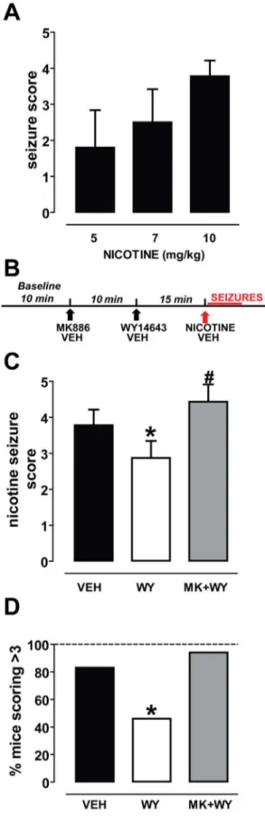
After a first exploratory phase, mice displayed impaired locomotor activity with shakes. A few seconds later they developed strong tachypnea with tremors, back arching, and partial loss of righting reflex. At higher doses, the previous symptoms were followed by increased locomotor activity, with rapid movements of the legs and wild running. Clonic and tonic seizures occurred after complete loss of righting reflex. Fig. 1A shows that nicotine elicited seizures with dose-dependent severity (r2= 0.76, P,0.001). The calculated ED50for nicotine was 5.0860.07 mg/kg, similar to that
already reported in the literature for the same mouse strain [24]. Only the dose of 10 mg/kg was able to induce severe symptoms,
which scored higher than 3. In this case, the number of animals which scored 4 or 5 was 15 out of 18 (83%) and, therefore, we chose this dose for the subsequent experiments.
The PPARa agonist WY14643 Acutely Reduces the Severity of Nicotine-induced Seizures
To test the effect of the PPARa agonist WY14643 (WY, 80 mg/ kg, i.p.) and the antagonist MK886 (MK, 3 mg/kg, i.p.) on nicotine-induced seizures, mice were randomly assigned to three groups: i) the nicotine (VEH group), ii) the vehicle-WY-nicotine group (WY group), and iii) the MK886-vehicle-WY-nicotine group (MK-WY group). Each animal received the three injections spaced 10 and 15 min (Fig. 1B).
When scores were evaluated, non-parametric ANOVA yielded a highly significant difference among groups (P,0.0001, Kruskal-Wallis test) (Fig. 1C). Post-hoc analysis revealed that mice pre-treated with the PPARa agonist WY14643 were significantly protected against nicotine-induced seizures when compared with vehicle pre-treated animals. The difference in the average score was statistically significant (VEH mice: 3.860.4, n = 18; WY mice: 2.960.5, n = 24; P = 0.019, Dunn’s test) (Fig. 1C). Additionally, WY mice which obtained a score of 4 or 5 were 46% of all treated mice (11 out of 24). The difference was statistically significant when compared with the VEH-group (15/18, 83%) (P,0.05, Fisher’s test, Fig. 1D). The protective effect of WY was reverted by the PPARa antagonist MK. Hence, post-hoc analysis indicated that in the MK-WY-group the severity of nicotine-induced seizures was restored, since the average score was significantly higher than that assigned to the WY mice (4.460.5, n = 16, P = 0.001, Dunn’s test, Fig. 1C), but not different from that of VEH-mice (P.0.05, Dunn’s test) (Fig. 1C). Consistently, MK-WY mice receiving a score of 4 or 5 were 15 out of 16 (94%) (P = 0.6 vs. VEH mice) (Fig. 1D).
In vivo electrophysiological recordings revealed that high doses of nicotine elicited seizures that originate in the hippocampus [28] and in the thalamo-cortical pathways [11]. These neural circuits may also be involved in NFLE (for review, see [29]). For this reason, one EEG electrode was placed in the sensorimotor cortex and two in the dorsal hippocampus. Of the two electrodes positioned in the hippocampus the one with the best signal was chosen.
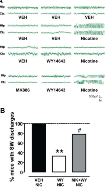
As depicted in Fig. 2, nicotine-induced (10 mg/kg, s.c.) convulsive activity was paralleled by a synchronous spiking discharge pattern both in the hippocampus and in the cortex in 7 out of 7 (100%) treated mice (Fig. 2A, B). Spiking activity had an onset of 13067 s after nicotine injection and a duration of 52610 s (n = 7). No spike/wave discharge events were recorded in vehicle treated mice (Fig. 2 A, B).
Pretreatment with WY significantly attenuated the effects of nicotine, since only 3 out of 9 (33.3%) mice displayed spiking activity when compared with vehicle-treated mice (P,0.05, Fisher’s test, Fig. 2B). WY pretreatment, however, did not change either the onset or the duration of spike-wave activity in those animals not responding to the treatment (114666 s and
Figure 1. The PPARa agonist WY14643 reduces nicotine-induced seizures. (A) Graph displaying the dose-dependency of the severity of nicotine-induced seizures in C57BL/6 mice. Nicotine was administered subcutaneously at 5, 7 and 10 mg/kg (n = 4, n = 5, n = 18, respectively). (B) Schematic representation of the experimental protocol of experiments in (C) and (D). (C) The PPARa agonist WY14643 (WY, 80 mg/kg, i.p., n = 23) reduced the severity of nicotine-induced seizures
(nicotine dose: 10 mg/kg, s.c) (*P,0.05 vs. vehicle, Dunn’s-test). This effect was abolished by the selective PPARa antagonist MK886 (MK, 3 mg/kg, i.p.). (n = 16, # P,0.001 vs. WY, Dunn’s-test). (D) This graph shows that the percentage of mice undergoing severe nicotine-induced seizures (indexed by scores .3) is significantly attenuated after WY14643 pretreatment (*P,0.05 vs. vehicle, Fisher’s test). MK886 reversed this effect (P.0.05 vs. vehicle, Fisher’s test). Data are expressed as mean695% C.I.
32618 s, respectively, n = 3). Consistent with behavioral exper-iments, MK prevented WY-induced protection from seizures, since nicotine’s effect was restored in 7 out of 9 mice (77.8%) (Fig. 2A, B).
Chronic Administration of the Clinically Available PPARa agonist Fenofibrate Attenuates the Severity of Nicotine-induced Seizures
The results of the previous experiments prompted us to assess the efficacy of the clinically available PPARa agonist fenofibrate. Fenofibrate does not cross the blood-brain barrier rapidly enough to allow acute studies in vivo; therefore, we chronically treated the animals with a diet containing 0.2% w/w fenofibrate [30]. Mice were randomly divided into three groups: (i) fenofibrate diet (FBR), (ii) control diet (CTRL) and (iii) fenofibrate washout diet (FBR-WO). The fenofibrate containing diet was administered ad libitum for 14 days. FBR mice consumed a daily average of 3.660.1 g of food pellets, which approximately corresponds to 28–30 mg/kg fenofibrate per day. FBR mice were divided in two additional groups at the end of the treatment: one group received the PPARa antagonist MK886 15 min before the nicotine challenge, and the other received its vehicle. CTRL mice were fed with the control diet, identical and equicaloric but without fenofibrate. FBR-WO mice were fed for 14 days with a fenofibrate-containing diet, as FBR mice, and then fed with a control diet for additional 14 days to assess the effect of fenofibrate washout. In each animal EEG electrodes were implanted 7 days before the beginning of the treatments and nicotine was tested on the 15th day from the beginning of treatment (for FBR and CTRL mice) or on the 30th day (for FBR-WO mice).
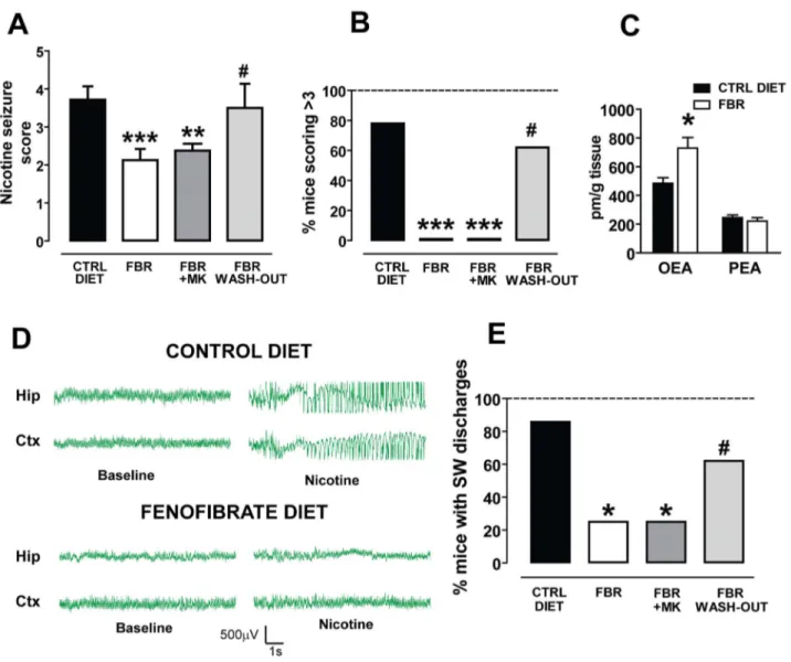
When nicotine-induced seizures were scored, non-parametric ANOVA yielded a highly significant difference among groups (P,0.0001, Kruskal-Wallis test) (Fig. 3A). Post-hoc analysis revealed that FBR mice were significantly protected against nicotine-induced seizures when compared with CTRL animals. The difference in the average score was statistically significant (CTRL mice: 3.460.7, n = 14; FBR mice: 2.160.3, n = 8; P,0.001, Dunn’s test) (Fig. 3A). Additionally, none among the FBR mice obtained a score of 4 or 5 (0 out of 8) (Fig. 3 B). The difference was statistically significant when compared with the CTRL group (11/14, 78%) (P,0.001, Fisher’s test, Fig. 3B). The protective effect of the fenofibrate diet was not reverted by the PPARa antagonist MK. Hence, post-hoc analysis indicated that in the MK-treated FBR mice the severity of nicotine-induced seizures was not restored, since the average score was significantly lower than that assigned to CTRL mice (2.460.4, n = 8, P,0.001, Dunn’s test, Fig. 3A), but not different from that of FBR-mice (P.0.05, Dunn’s test) (Fig. 3A). Consistently, no MK-treated FBR mice received a score of 4 or 5 (Fig. 3B).
The lack of effect by MK could be explained by the fact that PPARa activation induces mitochondrial activity, peroxisomal b-oxidation and the biosynthesis of endogenous PPARa ligands, the N-acylethanolamines OEA and PEA. These combined effects might trigger a feed-forward mechanism to further sustain PPARa activity [31]. To test this hypothesis we measured the levels of OEA and PEA in the FCx from FBR and CTRL mice. As predicted, in FBR mice OEA levels were significantly increased in the FCx when compared to CTRL mice (729.5673.4 pm/g vs. 484.3638.4 pm/g; n = 5; P,0.05, Student’s t-test) (Fig. 3C). No changes of PEA levels were detected (FBR: 221.7623.0 pm/g; CTRL: 245.6618.4 pm/g; n = 5; P = 0.44 Student’s t-test) (Fig. 3C).
Fenofibrate washout for 14 days abolished the protective effects of the drug, since in FBR-WO mice the severity of nicotine-induced seizures was restored (Fig. 3A). FBR-WO mice received a score of 3.560.6 (n = 8), significantly higher that FBR mice (P,0.05, Dunn’s test) but not dissimilar from CTRL mice (P.0.05, Dunn’s test) (Fig. 3A).
Figure 2. The PPARa agonist WY14643 suppresses nicotine-induced spike-wave activity. (A) Representative traces of EEG recordings from hippocampal (Hip) and sensorimotor cortical (Ctx) electrodes chronically implanted in mice. Following the administration of vehicle (VEH) and 10 mg/kg nicotine, bursts of synchronous spike-wave (SW) activity with high-amplitude and low-frequency (most in the delta rhythm range) were recorded. This activity was suppressed when animals were pretreated with the PPARa agonist WY14643 (WY, 80 mg/ kg), which per se did not change baseline EEG activity. The PPARa antagonist MK886 (MK, 3 mg/kg, i.p.) restored nicotine-induced SW discharges. (B) The graph shows the percentage of mice presenting SW discharges following the three treatment protocols. Vehicles treated mice did show SW activity, whereas 100% of nicotine treated mice displayed bursts of SW activity. The effects of nicotine were blocked in the majority of WY treated mice, since SW burst were recorded only in 33% of treated animals (**P,0.01 vs. vehicle, Fisher’s test). Conversely, when MK was administered 15 before WY nicotine-induced SW activity was recorded in 78% of MK+WY treated mice (#P,0.05 vs. WY, Fisher’s test).
EEG recordings revealed that nicotine-induced (10 mg/kg, s.c.) convulsive activity was paralleled by a synchronous spiking discharge pattern in both the hippocampus and the cortex in 6 out of 7 (86%) CTRL mice (Fig. 3D, E). Spiking activity had an onset of 14868 s after nicotine injection and a duration of 5369 s (n = 6).
The effect of nicotine was significantly attenuated in FBR mice, since only 2 out of 8 (25%) mice displayed spiking activity, when compared with CTRL mice (P,0.05, Fisher’s test, Fig. 3E). In
FBR mice, however, neither the onset nor the duration of spike-wave activity changed in those animals not responding to the treatment (179.5625 s and 2762 s, respectively, n = 2).
The effects of nicotine were restored in FBR-WO mice: spiking activity was indeed present in 5 out of 8 mice (P.0.05 vs. CTRL mice, Fisher’s test) (Fig. 3E). Consistent with behavioral exper-iments, MK did not block FBR-induced protection from seizures, since nicotine’s spike-wave activity was assessed in 2 out of 8 mice (25%) (Fig. 3E).
Figure 3. The clinically used PPARa agonist fenofibrate chronically administered with food reduces nicotine-induced seizures and spike-wave activity, and increases the OEA levels in the frontal cortex. (A) Graph displaying that fenofibrate (n = 8, FBR, 0.3% w/w in the diet for 14 days) reduced the severity of nicotine-induced seizures (nicotine dose: 10 mg/kg, s.c) (***P,0.001 vs. control diet, CTRL DIET, Dunn’s-test). This effect was not abolished by the selective PPARa antagonist MK886 (FBR+MK, 3 mg/kg, i.p.) (n = 8, **P,0.01 vs. CTRL DIET, Dunn’s-test) but by withdrawal of fenofibrate treatment for 14 days (FBR WASH-OUT, n = 8, # P,0.05 vs. FBR and P.0.05 vs. CTRL DIET, Dunn’s test). (B) This graph shows that the percentage of mice undergoing severe nicotine-induced seizures (indexed by scores .3) is significantly attenuated after chronic fenofibrate treatment (***P,0.001 vs. control diet, Fisher’s test) and restored after fenofibrate withdrawal for 14 days (#P.0.05 vs. control diet). MK886 did not reverse this effect (***P,0.001 vs. control diet, Fisher’s test). (C) Chronic activation of PPARa by fenofibrate changes oleoylethanolamide (OEA), but not palmitoylethanolamide, levels within frontal cortex. Concentrations of these endogenous PPARa ligands are expresses as pmol per gram of tissue. Error bars depict S.E.M. (*P,0.05, Student’s t-test). (D) Representative traces of EEG recordings from hippocampal (Hip) and sensorimotor cortical (Ctx) electrodes chronically implanted in mice. In mice fed with control diet 10 mg/kg nicotine elicits bursts of synchronous spike-wave (SW) activity with high-amplitude and low-frequency (most in the delta rhythm range). This activity was suppressed in animals fed with fenofibrate in food pellets. (E) The graph shows the percentage of mice presenting SW discharges following the four treatment protocols. 86% of control diet fed mice (6 out of 7) did show nicotine-induced SW activity. The effects of nicotine were fully blocked in all fenofibrate treated mice, since SW burst were recorded only in none of treated animals (***P,0.01 vs. control diet, Fisher’s test). MK, administered 15 before nicotine, did not restore nicotine-induced SW activity (***P,0.001 vs. control diet, Fisher’s test), whereas fenofibrate washout did (#P.0.05 vs. control diet, Fisher’s test). Data are expressed as mean695% C.I.
PPARa Agonists Suppress Nicotine-induced sIPSCs in Layer II–III Pyramidal Neurons in the Mouse and Rat Frontal Cortex
Nicotine-induced seizures, as well as those spontaneously occurring in transgenic mice carrying the NFLE mutations, might be primarily generated within FCx circuits [12,29]. Since excitatory transmission on FCx pyramidal neurons is not affected by NFLE mutations or by nicotine [12], we carried out whole-cell recordings of spontaneous inhibitory postsynaptic currents (sIPSCs) from layer II/III pyramidal cells of mouse and rat coronal slices.
Under voltage-clamp mode (Vholding= 0 mV to isolate sIPSCs
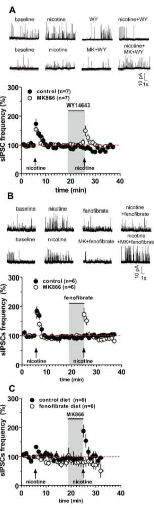
[12]), nicotine (5mM, 30 s) significantly increased both frequency and amplitude of sIPSCs in mouse pyramidal cells. sISPC frequency was enhanced to 133.262.9% of baseline (P,0.0001, n = 23, paired t-test), the amplitude to 116.567.5% of baseline (P,0.05, n = 23, paired t-test) (Fig. 4, Table 1). In marked contrast, pretreatment with the PPARa agonists WY (1mM, 5 min) and fenofibrate (10mM, 5 min) dramatically blunted the effect of nicotine on sIPSCs. During nicotine application in the presence of WY, sIPSC frequency was 91.8610.2% of baseline and amplitude was 91.864.7% of control values (for both parameters, P.0.05 vs. baseline, n = 7, paired t-test) (Fig. 4A, Table 1). Consistently, the structurally different PPARa agonist fenofibrate also suppressed nicotine-induced increase of sIPSC frequency and amplitude (for both parameters, P.0.05 vs. baseline, n = 6, paired t-test) (Fig. 4B, Table 1).
The effects of both PPARa agonists were blocked by the synthetic PPARa antagonist MK. In the presence of MK (0.3mM, 5 min) and either PPARa agonist, nicotine effects on sIPSCs frequency and amplitude were fully restored. Hence, in slices perfused with WY+MK or fenofibrate+MK, nicotine induced an increase in sIPSC frequency to 141.7619.3% and to 172.5620.9% of baseline, respectively (P.0.05vs. nicotine alone, n = 6–7, paired t-test; Table 1, Fig. 4A,B). Table 1 shows that sIPSC amplitude in the presence of nicotine alone was not significantly different from the amplitude in the presence of nicotine+WY+MK or nicotine+fenofibrate+MK(P.0.05, n = 6, paired t-test).
To assess whether suppression of nicotine effects by acute PPARa agonists was species-specific we replicated the experiments described above in Sprague Dawley rats. The results (Supporting Results S1) confirmed that both WY and fenofibrate abolished nicotine-induced effects in both species (Table 1; Figure S1).
Experiments with mice chronically fed with fenofibrate (0.2% w/w for 14 days) confirmed that this drug abolishes
nicotine-induced increases in sIPSC frequency in FCx pyramidal neurons. Hence, nicotine-induced increase in sIPCS frequency was 132.264.6% of baseline (n = 6) in CTRL mice vs. 99.765.6% of baseline (n = 6) in FBR mice (P,0.05, Student’s t-test) (Fig. 4C). Consistent with behavioral experiments, MK was unable to revert the effect of fenofibrate, since in FBR mice MK did not restore the effects of nicotine (sIPSC frequency in control animals was 187.5620.9% vs. 80.7617.6% in FBR mice).
The PPARa Agonist Enhances Phosphorylation of the b2 Subunit of nAChRs
Consistent with our previous reports [13,14], these findings led us to hypothesize that PPARa might regulate the balance between phosphorylated and dephosphorylated b2*nAChRs. To test this hypothesis, we analyzed phosphorylation of b2 subunits with Western blot after in vivo exposure to the synthetic PPARa agonist WY. Rats were administered WY (40 mg/kg, i.p.) and after 15 min, brain were rapidly removed and the FCx were dissected. As expected, in FCx total homogenates an increased immunore-activity for phosphorylated b2 subunits was observed in WY-treated animals (125.067.9% of controls, n = 7, p,0.01, Student’s t-test) (Fig. 5).
Discussion
Our study shows that acute and chronic PPARa agonists, including the clinically available fenofibrate, reduce nicotine-induced behavioral and EEG seizure expression and abolish nicotine-induced enhancement of sIPSCs in FCx pyramidal neurons.
We presume, based on the present results and previous extensive work by our group, that the mechanism whereby PPARa exerts these effects involves phosphorylation of specific nAChR subunits [13,14,22,23]. Indeed, nAChR channel proper-ties depend on its tyrosine phosphorylation status, being the result of a balance between tyrosine kinases and phosphatases [32], which negatively or positively modulates nAChR-mediated currents, respectively, and controls the number of functional surface receptors [33].
We specifically measured the ratio of phosphorylated/dephos-phorylated b2 subunits, which is increased in the FCx by acute PPARa ligand treatment, but it is difficult to quantify this effect in subunit combinations less abundantly expressed in the CNS. Therefore, we cannot exclude that subunits other than the b2 might also be targeted by PPARa. However, it must be pointed out that b2*nAChRs play a major role, yet not exclusive, in Table 1. Table showing frequency and amplitude of spontaneous IPSCs recorded from mice and rat pyramidal neurons in layers II/ III of the frontal cortex.
Baseline Nicotine WY+Nicotine FBR+Nicotine WY+MK+Nicotine FBR+MK+Nicotine C57BL/6J mice Frequency, Hz 46.261.0 61.261.1*** 35.166.6 40.061.6 68.7612.2* 79.7616.7* Amplitude, pA 17.860.8 19.860.8* 18.362.7 18.161.6 21.561.9* 20.361.5* Rats Frequency, Hz 15.363.2 22.264.7*** 15.8611 11.560.6 22.565.7** 22.567.7* Amplitude, pA 16.460.7 21.361.5*** 13.460.8 14.061.3 25.165.4* 22.861.9* Nicotine (5mM) significantly increases both frequency and amplitude of sIPSC (*P,0.05, 01, ***P,0.001 vs baseline, paired t-test). Both WY14643 (WY 1mM) and fenofibrate (FBR, 10mM) suppressed nicotine’s effects on sIPSCs frequency and amplitude. The PPARa antagonist MK886 restored nicotine’s effects on sIPSC frequency (*P,0.05, **P,0.01 vs baseline, paired t-test).
nicotine-induced seizures [10,34]. In fact, the a4b2 subunit combination is the most abundantly expressed in the whole brain and particularly in regions relevant to ictogenesis, such as thalamo-cortical circuits and FCx (see [12] and references therein). Furthermore, it was shown by Klaassen et al. [12] that nicotine-induced sIPSC in layer II/III of the FCx are mediated by a4b2 nAChRs, as they are insensitive to the a7-selective blocker methyllycaconitine but fully blocked by the a4b2-selective antagonist dihydro-b-erythroidine. However, it must be pointed out that other nAChR subunits are implicated in nicotine-evoked seizures. In particular, their severity is reduced in mice lacking a3, a5, and b4 [35,36], but not a7 [24]. The role of a7, however, is still controversial, since pharmacological experiments with
selec-Figure 4. The PPARa agonists WY14643 and fenofibrate suppress nicotine-induced increase of spontaneous inhibitory postsynaptic currents (sIPSC) in frontal cortex (FCx) pyramidal neurons. The graphs illustrate that in mouse FCx slices, nicotine (5 mM perfused at arrows for 30 s) increases sIPSCs frequency in layer II/III pyramidal neurons. Acutely, the PPARa agonists WY14643 (1 mM, WY) (A), and fenofibrate (10 mM) (B) fully suppressed nicotine-induced increase in sIPSC frequency. Similarly, chronic fenofibrate (0.2% w/w for 14 days in food) fully suppressed nicotine-induced increase in sIPSC frequency (C). The gray box represents the time of PPARa agonist (+/2 antagonist) perfusion. The PPARa antagonist MK886 (0.3 mM) (open symbols) blocked the effects of acute WY (A) and fenofibrate (B), but not the one of chronic fenofibrate (C), and restored nicotine-induced increase in sIPSCs. Representative recordings of the effect of nicotine, and of PPARa agonists and antagonist, on spontaneous IPSCs from pyramidal cells at Vh= 0 mV are depicted on the upper part of panels A and B. Symbols represent the average6S.E.M.
doi:10.1371/journal.pone.0064541.g004
Figure 5. The PPARa agonist WY14643 enhances phosphory-lation of b2 subunits of the nicotinic acetylcholine receptors. Graph and representative immunoblots showing that the PPARa agonist WY (40 mg/kg, i.p.) caused an increase in b2-subunit phosphorylation (*P,0.05) in rat frontal cortex homogenates. Tissue lysates were subjected to immunoprecipitation (IP) with anti-b2subunit antibody and were immunoblotted (IB) with anti-phosphotyrosine (PY) antibody. The blots were stripped and reprobed with anti-b2subunit antibody to normalize for protein loading.
tive a7 agonists and antagonists yielded contradictory results [10,34].
Irrespectively of the subunits involved other than b2, the PPARa agonists reduced both the severity of nicotine-induced effects and the number of animals undergoing severe symptoms. These behavioral effects were paralleled by a significant decrease in the number of mice that showed bursts of spike-wave discharges. The pharmacological specificity was confirmed by the finding that pre-treatment with the PPARa antagonist prevented the protection exerted by the agonist, with the exception of chronic fenofibrate treatments. The latter finding might be explained with the finding that PPARa activation induces fatty acid metabolism [31] and NAE biosynthesis with increased production of OEA. Notably, OEA would further sustain PPARa activation by triggering a feed-forward mechanism difficult to be antagonized by a single administration of the PPARa antagonist MK. A second possible explanation is that changes induced by this treatment regimen, possibly on function, number and phosphor-ylation status of nAChRs, are not promptly reversed by an acute treatment with the PPARa antagonist. Indeed, a reversal of fenofibrate-induced protection is observed following a 14-days washout of the drug, achieved by replacing fenofibrate-containing food pellets with control food.
The present data, while confirming the relationship between nAChRs and epileptogenesis, give support to the role played by nuclear receptors PPARa in the modulation of nAChR function in the CNS. Hence, in our previous studies, we demonstrated that both endogenous and synthetic PPARa agonists suppressed nicotine-induced electrophysiological effects on dopamine neurons and prevented behavioral effects of nicotine predictive of its addicting properties by regulating b2* nAChRs specifically [13,14,15,16]. Here we extend those results to the seizure-inducing action of nicotine.
Nuclear and cytoplasmic immunostaining for PPARa is present in neurons distributed in all layers of the FCx [20,21], whereas cholinergic afferents, arising principally from the basal forebrain, innervate neurons in all layers of the rodent FCx [37]. Several subtypes of GABA cortical interneurons express functional a4b2* nAChRs [38,39] and have been involved in nicotine-induced seizures [10,12,34,38] since they innervate adjacent pyramidal cells. Consistently, we show that nicotine induced an increase in the frequency of GABAA-mediated sIPSCs, possibly by
depolar-izing presynaptic GABAergic terminals impinging on pyramidal neurons. According to behavioral observations, these effects were suppressed by PPARa agonists, suggesting that PPARa regulate the functions of nAChRs in cortical interneurons, similarly to dopamine neurons.
Notably, the relevance of our results might extend beyond nicotine-induced seizures or NFLE, since chronic fenofibrate was effective in rats as anticonvulsant in pentylentetrazole-induced seizures and on latencies to the onset of status epilepticus induced by lithium–pilocarpine [30]. Additionally, recent studies demon-strated that the antiepileptic drugs carbamazepine and lamotri-gine, which are employed effectively also in NFLE patients, negatively modulate the activity of a4b2 nAChRs [40,41]. This effect might contribute to their mechanisms of action since these receptors control neuronal excitability and both glutamate and GABA release in the hippocampus and the thalamo-cortical system [42,43,44,45,46].
In conclusion, we provided evidence that PPARa within the CNS might be a key regulator of neuronal activity by the modulation of functional properties of nAChRs. These effects might be therapeutically exploited not only when nicotine addiction is concerned [15], but also for idiopathic or genetically determined forms of epilepsy where nAChRs play a major role.
Supporting Information
Figure S1 The PPARa agonists WY14643 and fenofibrate suppress nicotine-induced increase of spontaneous inhibitory postsynaptic currents (sIPSC) in rat frontal cortex (FCx) pyramidal neurons. The graphs illustrate that in rat FCx slices, nicotine (5mM perfused at arrows for 30 s) increases sIPSCs frequency in layer II/III pyramidal neurons. The PPARa agonists WY14643 (1mM, WY) (A) and fenofibrate (10mM) (B) (n = 6–7; closed symbols) fully suppressed nicotine-induced increase in sIPSC frequency. The gray box represents the time of PPARa agonist (+/ 2 antagonist) perfusion. The PPARa antagonist MK886 (0.3mM) (open symbols) blocked the effects of WY (A) and fenofibrate (B) (n = 6) and restored nicotine-induced increase in sIPSCs. Symbols represent the mean6SEM.
(TIF)
Supporting Results S1 PPAR-a agonists suppress nicotine-induced sIPSCs in layer II-III pyramidal neurons in the rat frontal cortex.
(DOCX)
Author Contributions
Conceived and designed the experiments: M. Puligheddu FM ALM M. Pistis. Performed the experiments: GP MM SL MGM SS SA GC EM ALM M. Pistis. Analyzed the data: M. Puligheddu GP MM SL MGM SS GC EM ALM M. Pistis. Wrote the paper: FM M. Pistis.
References
1. Damaj MI, Glassco W, Dukat M, Martin BR (1999) Pharmacological characterization of nicotine-induced seizures in mice. J Pharmacol Exp Ther 291: 1284–1291.
2. Bertrand D, Elmslie F, Hughes E, Trounce J, Sander T, et al. (2005) The CHRNB2 mutation I312M is associated with epilepsy and distinct memory deficits. Neurobiol Dis 20: 799–804.
3. De Fusco M, Becchetti A, Patrignani A, Annesi G, Gambardella A, et al. (2000) The nicotinic receptor beta 2 subunit is mutant in nocturnal frontal lobe epilepsy. Nat Genet 26: 275–276.
4. Sutor B, Zolles G (2001) Neuronal nicotinic acetylcholine receptors and autosomal dominant nocturnal frontal lobe epilepsy: a critical review. Pflugers Arch 442: 642–651.
5. Steinlein OK, Magnusson A, Stoodt J, Bertrand S, Weiland S, et al. (1997) An insertion mutation of the CHRNA4 gene in a family with autosomal dominant nocturnal frontal lobe epilepsy. Human Molecular Genetics 6: 943–947. 6. McLellan A, Phillips HA, Rittey C, Kirkpatrick M, Mulley JC, et al. (2003)
Phenotypic comparison of two Scottish families with mutations in different genes causing autosomal dominant nocturnal frontal lobe epilepsy. Epilepsia 44: 613– 617.
7. Scheffer IE, Bhatia KP, Lopes-Cendes I, Fish DR, Marsden CD, et al. (1995) Autosomal dominant nocturnal frontal lobe epilepsy. A distinctive clinical disorder. Brain : a journal of neurology 118 (Pt 1): 61–73.
8. Gotti C, Clementi F (2004) Neuronal nicotinic receptors: from structure to pathology. Prog Neurobiol 74: 363–396.
9. Moulard B, Picard F, le Hellard S, Agulhon C, Weiland S, et al. (2001) Ion channel variation causes epilepsies. Brain Res Brain Res Rev 36: 275–284. 10. Dobelis P, Hutton S, Lu Y, Collins AC (2003) GABAergic systems modulate
nicotinic receptor-mediated seizures in mice. The Journal of pharmacology and experimental therapeutics 306: 1159–1166.
11. Oldani A, Zucconi M, Asselta R, Modugno M, Bonati MT, et al. (1998) Autosomal dominant nocturnal frontal lobe epilepsy. A video-polysomnographic and genetic appraisal of 40 patients and delineation of the epileptic syndrome. Brain : a journal of neurology 121 (Pt 2): 205–223.
12. Klaassen A, Glykys J, Maguire J, Labarca C, Mody I, et al. (2006) Seizures and enhanced cortical GABAergic inhibition in two mouse models of human autosomal dominant nocturnal frontal lobe epilepsy. Proc Natl Acad Sci U S A 103: 19152–19157.
13. Melis M, Carta S, Fattore L, Tolu S, Yasar S, et al. (2010) Peroxisome proliferator-activated receptors-alpha modulate dopamine cell activity through nicotinic receptors. Biol Psychiatry 68: 256–264.
14. Melis M, Pillolla G, Luchicchi A, Muntoni AL, Yasar S, et al. (2008) Endogenous Fatty Acid Ethanolamides Suppress Nicotine-Induced Activation of Mesolimbic Dopamine Neurons through Nuclear Receptors. J Neurosci 28: 13985–13994.
15. Mascia P, Pistis M, Justinova Z, Panlilio LV, Luchicchi A, et al. (2011) Blockade of nicotine reward and reinstatement by activation of alpha-type peroxisome proliferator-activated receptors. Biological Psychiatry 69: 633–641.
16. Panlilio LV, Justinova Z, Mascia P, Pistis M, Luchicchi A, et al. (2012) Novel use of a lipid-lowering fibrate medication to prevent nicotine reward and relapse: preclinical findings. Neuropsychopharmacology 37: 1838–1847.
17. Heneka MT, Landreth GE (2007) PPARs in the brain. Biochim Biophys Acta 1771: 1031–1045.
18. Bishop-Bailey D (2000) Peroxisome proliferator-activated receptors in the cardiovascular system. Br J Pharmacol 129: 823–834.
19. Berger J, Moller DE (2002) The mechanisms of action of PPARs. Annu Rev Med 53: 409–435.
20. Moreno S, Farioli-Vecchioli S, Ceru MP (2004) Immunolocalization of peroxisome proliferator-activated receptors and retinoid X receptors in the adult rat CNS. Neuroscience 123: 131–145.
21. Cimini A, Benedetti E, Cristiano L, Sebastiani P, Damico M, et al. (2005) Expression of peroxisome proliferator-activated receptors (PPARs) and retinoic acid receptors (RXRs) in rat cortical neurons. Neuroscience 130: 325–337. 22. Pistis M, Melis M (2010) From surface to nuclear receptors: the
endocanna-binoid family extends its assets. Current Medicinal Chemistry 17: 1450–1467. 23. Melis M, Scheggi S, Carta G, Madeddu C, Lecca S, et al. (2013) PPAR-alpha
regulate cholinergic-driven activity of midbrain dopamine neurons via a novel mechanism involving alpha7 nicotinic acetylcholine receptors. Journal of Neuroscience in press.
24. Franceschini D, Paylor R, Broide R, Salas R, Bassetto L, et al. (2002) Absence of alpha7-containing neuronal nicotinic acetylcholine receptors does not prevent nicotine-induced seizures. Brain Res Mol Brain Res 98: 29–40.
25. Paxinos G, Franklin KBJ (2004) The Mouse Brain in Stereotaxic Coordinates: Elsevier Academic Press.
26. Di Marzo V, Melck D, Orlando P, Bisogno T, Zagoory O, et al. (2001) Palmitoylethanolamide inhibits the expression of fatty acid amide hydrolase and enhances the anti-proliferative effect of anandamide in human breast cancer cells. Biochem J 358: 249–255.
27. Piscitelli F, Carta G, Bisogno T, Murru E, Cordeddu L, et al. (2011) Effect of dietary krill oil supplementation on the endocannabinoidome of metabolically relevant tissues from high-fat-fed mice. Nutr Metab (Lond) 8: 51.
28. Cohen SL, Morley BJ, Snead OC (1981) An EEG analysis of convulsive activity produced by cholinergic agents. Progress in neuro-psychopharmacology 5: 383– 388.
29. Raggenbass M, Bertrand D (2002) Nicotinic receptors in circuit excitability and epilepsy. Journal of Neurobiology 53: 580–589.
30. Porta N, Vallee L, Lecointe C, Bouchaert E, Staels B, et al. (2009) Fenofibrate, a peroxisome proliferator-activated receptor-alpha agonist, exerts anticonvulsive properties. Epilepsia 50: 943–948.
31. Melis M, Carta G, Pistis M, Banni S (2013) Physiological Role of Peroxisome Proliferator-Activated Receptors Type Alpha on Dopamine Systems. CNS Neurol Disord Drug Targets.
32. Charpantier E, Wiesner A, Huh KH, Ogier R, Hoda JC, et al. (2005) Alpha7 neuronal nicotinic acetylcholine receptors are negatively regulated by tyrosine phosphorylation and Src-family kinases. J Neurosci 25: 9836–9849.
33. Cho CH, Song W, Leitzell K, Teo E, Meleth AD, et al. (2005) Rapid upregulation of alpha7 nicotinic acetylcholine receptors by tyrosine dephos-phorylation. J Neurosci 25: 3712–3723.
34. Stitzel JA, Lu Y, Jimenez M, Tritto T, Collins AC (2000) Genetic and pharmacological strategies identify a behavioral function of neuronal nicotinic receptors. Behavioural Brain Research 113: 57–64.
35. Salas R, Cook KD, Bassetto L, De Biasi M (2004) The alpha3 and beta4 nicotinic acetylcholine receptor subunits are necessary for nicotine-induced seizures and hypolocomotion in mice. Neuropharmacology 47: 401–407. 36. Salas R, Orr-Urtreger A, Broide RS, Beaudet A, Paylor R, et al. (2003) The
nicotinic acetylcholine receptor subunit alpha 5 mediates short-term effects of nicotine in vivo. Molecular Pharmacology 63: 1059–1066.
37. Poorthuis RB, Bloem B, Schak B, Wester J, de Kock CP, et al. (2012) Layer-Specific Modulation of the Prefrontal Cortex by Nicotinic Acetylcholine Receptors. Cerebral Cortex.
38. Porter JT, Cauli B, Tsuzuki K, Lambolez B, Rossier J, et al. (1999) Selective excitation of subtypes of neocortical interneurons by nicotinic receptors. J Neurosci 19: 5228–5235.
39. Christophe E, Roebuck A, Staiger JF, Lavery DJ, Charpak S, et al. (2002) Two types of nicotinic receptors mediate an excitation of neocortical layer I interneurons. J Neurophysiol 88: 1318–1327.
40. Zheng C, Yang K, Liu Q, Wang MY, Shen J, et al. (2010) The anticonvulsive drug lamotrigine blocks neuronal {alpha}4{beta}2 nicotinic acetylcholine receptors. J Pharmacol Exp Ther 335: 401–408.
41. Di Resta C, Ambrosi P, Curia G, Becchetti A (2010) Effect of carbamazepine and oxcarbazepine on wild-type and mutant neuronal nicotinic acetylcholine receptors linked to nocturnal frontal lobe epilepsy. Eur J Pharmacol 643: 13–20. 42. Albuquerque EX, Pereira EF, Alkondon M, Rogers SW (2009) Mammalian nicotinic acetylcholine receptors: from structure to function. Physiol Rev 89: 73– 120.
43. Aracri P, Consonni S, Morini R, Perrella M, Rodighiero S, et al. (2010) Tonic modulation of GABA release by nicotinic acetylcholine receptors in layer V of the murine prefrontal cortex. Cereb Cortex 20: 1539–1555.
44. Couey JJ, Meredith RM, Spijker S, Poorthuis RB, Smit AB, et al. (2007) Distributed network actions by nicotine increase the threshold for spike-timing-dependent plasticity in prefrontal cortex. Neuron 54: 73–87.
45. Lambe EK, Picciotto MR, Aghajanian GK (2003) Nicotine induces glutamate release from thalamocortical terminals in prefrontal cortex. Neuropsychophar-macology 28: 216–225.
46. Zolles G, Wagner E, Lampert A, Sutor B (2009) Functional expression of nicotinic acetylcholine receptors in rat neocortical layer 5 pyramidal cells. Cereb Cortex 19: 1079–1091.