Corso di Dottorato di Ricerca in
Genetica e Biologia Cellulare - XXVII Ciclo
Neuronal differentiation processes: lessons from
neuroblastoma models
(BIO/11)
Tesi di dottorato di: Dr. Loredana Guglielmi
Coordinatore del corso Tutore
Prof. Giorgio Prantera Dr. Armando Felsani
Firma Firma
TABLE OF CONTENTS
Pag.
ABSTRACT IV
RIASSUNTO V
AIM VI
Chapter 1 General Introduction
1. Overview of the nervous system 1
2. Basics of the origin of the nervous system 3
2.1 Neurulation 5
2.1.1 Neural crest cells and the origin of the peripheral nervous system 6
2.2 Proliferation, maturation and migration in the nervous system 7 3. Neuroblastoma cell lines as a valid model to study neuronal differentiation 9 3.1 Retinoids: induction mechanism of neuroblastoma differentiation 10
Chapter 2 MYCN gene expression is required for the onset of the differentiation programme in neuroblastoma cells
2.1 Summary 13
2.3 Results 16 2.3.1 Retinoic acid (RA) triggers differentiation in the human neuroblastoma LAN-5 cell line
16
2.3.2 N-Myc expression increases during the early phases of RA-induced differentiation in cells of neural origin
18
2.3.3 N-Myc is necessary to activate the differentiation programme in LAN-5 neuroblastoma cells
18
2.3.4 N-Myc overexpression induces differentiation in poorly differentiating neuroblastoma cells
22
2.3.5 MYCN modulation modifies the expression of miRNAs involved in apoptosis preceding neuronal differentiation
25
2.3.6 Inhibition of miR-20a, miR-9 and miR-92a in the wild type SK-N-AS cells restores apoptosis and their differentiation ability
25
2.4 Conclusions 30
2.5 Materials and Methods 34
2.6 Acknowledgements 40
Chapter 3 Massive change of transcription profile following activation of the differentiation programme by choline acetyltransferase forced expression in N18TG2 neuroblastoma cells
3.1 Summary 41
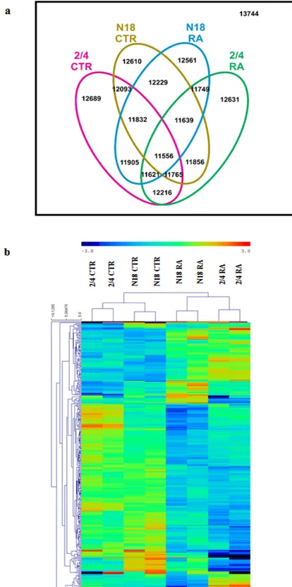
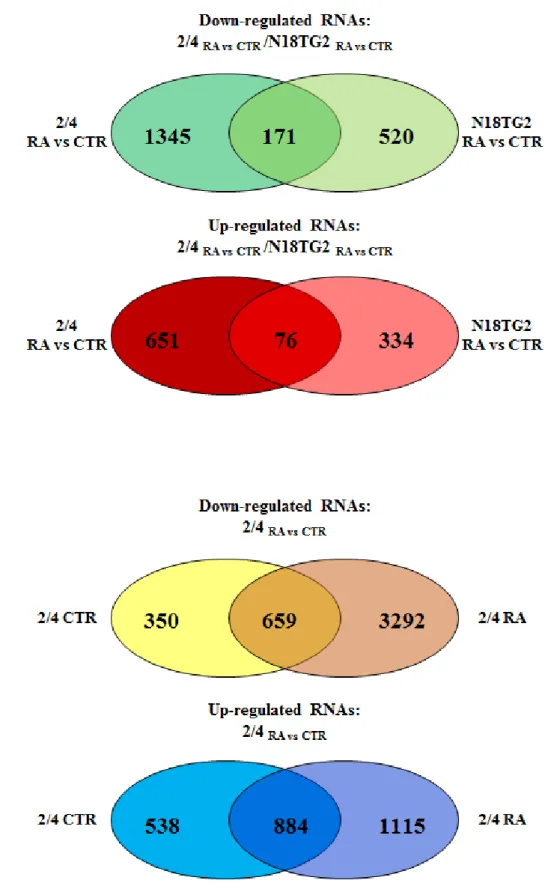
3.3 Results 44 3.3.1 The transcription profile of the 2/4 clone shows extensive modifications by
comparison with the N18TG2 parental cell line
44
3.3.2 ChAT forced exogenous expression modifies cell cycle and neuronal differentiation regulation
46
3.3.3 Lamin A/C is specifically up-regulated after ChAT transfection in 2/4 cells 55 3.3.4 Acetylcholine receptor agonist and antagonist modulates Lmna expression in
N18TG2 and 2/4 cells
58
3.3.5 Lamin A/C interactomics network and forced knock-down suggest an involvement of this nuclear envelope protein in ChAT-dependent differentiation cascade
60
3.4 Conclusions 63
3.5 Materials and Methods 67
3.6 Acknowledgements 72
Chapter 4 General Discussion
4.1 Dying to become a neuron 73
4.2 Neurotransmitters as morphogens 76
REFERENCES i
APPENDIX xix
ABSTRACT
During animal development, a single fertilised egg cell divides and differentiates to produce all of the cells and tissues of the mature organism. Defining the complexity of signals and regulation mechanisms governing the organism's development represents a steep challenge, even nowadays. In this scenario, the nervous system formation and maturation are probably two among the most fascinating fields of developmental biology yet to be completely understood. Fundamental steps of these processes, going from the induction of neural plate to the establishment of rigorous interconnections among billions of nerve cells, are conserved within the Animalia kingdom. In fact, many aspects concerning above all anatomical features of the nervous system development have been clarified thanks to the use of simple model organisms such as, Drosophila, Xenopus and C. elegans. However, many questions remain to be addressed.
A valid replacement of the model organisms, especially for the comprehension of molecular and cellular aspects of the nervous system development, is represented by neuroblastoma cell lines. These systems are extremely suitable to investigate basic, but critical, phases of neuronal differentiation. By treatment with specific substances, able to induce differentiation, neuroblastoma cells display, in vitro, typical characteristics of neurons maturation like the up-regulation of differentiation-related genes, the expression of neurofilaments and the outgrowth of dendrites and axons.
In this thesis work four neuroblastoma cell lines have been employed to examine two known, and still debated, phenomena characterising the initial moments of neuronal differentiation: programmed cell death (PCD) and non-synaptic role of the neurotransmitters. The study has revealed new insights in the regulation of these processes, identifying MYCN as a new regulator of PCD at early stages of differentiation and the nucleoskeletal protein Lamin A/C as a possible effector/intermediate of the acetylcholine receptors (AChRs) molecular cascade.
RIASSUNTO
Durante lo sviluppo animale, una singola cellula uova fecondata é in grado di dividersi e differenziare per produrre tutte le cellule e i tessuti dell’organismo maturo. Definire la complessità dei segnali e dei meccanismi di regolazione che governano lo sviluppo di un organismo rappresenta ancora oggi una sfida di enorme portata. In questo scenario, la formazione e la maturazione del sistema nervoso sono probabilmente due tra i più affascinanti campi della biologia dello sviluppo ancora non completamente chiariti. Momenti fondamentali di questi processi, a partire dall’induzione della placca neurale fino allo stabilirsi di rigorose interconnesioni tra milioni di cellule nervose, sono conservati all’interno del regno animale. Infatti, molti aspetti riguardanti principalmente le caratteristiche anatomiche dello sviluppo del sistema nervoso sono stati messi in luce grazie all’utilizzo di semplici organismi modello quali, Drosophila, Xenopus e C. elegans. Tuttavia, molti quesiti restano da risolvere.
Un valido sostituto degli organismi modello, principalmente per la comprensione degli aspetti molecolari e cellulari dello sviluppo del sistema nervoso, é rappresentato dalle linee cellulari di neuroblastoma. Questi sistemi risultano estremamente utili per studiare fasi basilari, ma allo stesso tempo critiche, del differenziamento. Grazie al trattamento con specifiche sostanze, in grado di indurre il differenziamento, le cellule di neuroblastoma mostrano, in
vitro, caratteristiche tipiche della maturazione dei neuroni come la up-regolazione di geni
coinvolti nel differenziamento, l’espressione dei neurofilamenti e la crescita di dendriti e assoni.
In questa tesi quattro linee di neuroblastoma sono state utilizzate per esaminare due fenomeni noti, ma ancora dibattuti, tipici delle prime fasi del differenziamento neuronale: la morte cellulare programmata (PCD) e il ruolo non-sinaptico dei neurotrasmettitori. Lo studio condotto ha portato a definire nuovi punti riguardanti i due processi, identificando MYCN come nuovo regolatore della morte cellulare programmata durante le fasi precoci del differenziamento e la proteina nucleosheletrica Lamina A/C come un possibile effettore/intermedio della cascata molecolare attivata dai recettori per l’acetilcolina (AchRs).
AIM
The aim of this thesis work was to study neuronal differentiation processes using neuroblastoma cell lines as models.
LAN-5 and SK-N-AS cells were used to study the role of the very well-known oncogene MYCN, which is considered a hallmark for neuroblastoma disease, in the onset of neuronal differentiation programme. Multiple evidences demonstrated how MYCN displays an enigmatic behaviour in defining neuroblastoma phenotype. LAN-5 and SK-N-AS cell lines can be described as two opposites as it concerns MYCN. Indeed, LAN-5 are known to be MYCN-amplified, whereas SK-N-AS are MYCN non-amplified, thus representing an extremely suitable system to investigate MYCN amplification effect on neuronal differentiation.
On the other hand, the N18TG2 and 2/4 cells were chosen to analyse the effect of neurotransmitter synthesis on neuronal differentiation. The production of neurotransmitters and the expression of their specific receptors at early stages during nervous system development, before the formation of synaptic contacts, clearly raise the possibility of a non-synaptic role of neurotransmitters. N18TG2 and 2/4 cells were expressly created to address this question: N18TG2 (parental clone) and 2/4 (ChAT-transfected derived clone) are isogenic cell lines which differ only for the presence of the choline acetyl-transferase (ChAT) enzyme which restores the ability of acetylcholine production and as a consequence of differentiation in 2/4 clone. The manipulation simplicity of this model allowed a genome-wide study to decipher the multiple effects of acetylcholine as morphogen prior to the formation of synaptic connections.
General Introduction
“God may forgive your sins, but your nervous
system won't”
Alfred Korzybski
1. Overview of the nervous system
The nervous system is that part of our body that coordinates both voluntary and involuntary actions thanks to the transmission of specific signals. The nervous system is composed by two main divisions, the Central Nervous System (CNS) and the Peripheral Nervous System (PNS). The brain and the spinal cord are the only components of the CNS, whereas the PNS appears to be more articulated in its structure. Motor nerves and sensory nerves represent the major branches of the PNS; each of these branches is composed by two subgroups, the visceral and the somatic divisions. The first division is associated with involuntary responses, the latter is related to voluntary control of effects. In particular, the involuntary branch of the motor division is known as Autonomic Nervous System (ANS) and is further divided into Sympathetic and Parasympathetic nervous systems. Sympathetic and parasympathetic divisions typically act in opposition to each other. The sympathetic division usually makes the scene when a quick response is required, while the parasympathetic division functions when there is no rush in giving a response. For this reason, the sympathetic system is often referred to as the "fight or flight" system and the parasympathetic system is often considered the "rest and digest" or "feed and breed" system. The nervous system derives its name from nerves, particular fibrous structures innervating every part of our body. Nerves are the constituents of the nervous system and their identification dates back to the ancient Egyptians, Greeks and Romans but their complete structure was not examined in depth until modern time through the use of the microscope (Finger S, 2001). The building blocks of nerves are extremely specialised cells called neurons that form a vast network of billions of units. The typical morphology of a neuron reflects its function: the fibers or dendrites branching from the body of the cell receive "messages" that, after being processed, are transmitted via a long wire-like structure called axon. The nature of these "messages" is essentially electrochemical; electrical signals are converted into chemical ones and vice versa. Neurons communication occurs sequentially from one neuron to the next in charge, like in a domino effect and the junction region between two consecutive neurons is known as synapse. Not all neurons are capable of high-speed transmission signals, only those provided with a proper insulation (myelin) around their axons can reach extreme velocities. Myelin is actually an extension of the cell membrane of a particular type of cells that also populate our nervous system, called glial cells. Glia performs many important tasks within the nervous system; providing nutrients to promote neuron growth, nursing and repairing neurons that are injured and protecting the nervous system from infection. Oligodendrocytes are the glia that
produce myelin in the central nervous system. Glial cells called Schwann cells provide myelin to axons of the peripheral nervous system.
2. Basics of the origin of the nervous system
Specialised cells, tissues, organs, and interconnected systems all originate from a single fertilised egg. Sequential divisions characterize the first phases of development. After the third cleavage a group of loosely arranged blastomeres (embryonic stem cells) called morula is converted to a mass of flattened and tightly interconnected cells. The blastomeres stand against each other at the surface of the morula, maximizing their contact with one another, and a cavity, called blastocoel, starts to appear within the morula. The appearance of this hollow identifies the entire structure as the blastocyst stage and coincides with the formation of two distinct cell types: an outer mass of trophoblasts and an inner mass of blastomeres. The formation of these two cell types constitutes the first lineage restriction that occurs during development. The trophoblasts develop into the placenta, whereas the blastomeres give rise to the embryo. When the embryo reaches the 64-cell stage the blastocoel has expanded and the embryo starts to acquire a sort of polarity with the inner mass cells falling into two subgroups, those facing the trophoblast side and the others facing the blastocoel side and the trophoblasts also divided into those in contact or not with the inner mass cells. At this point, the critical step to properly proceed is represented by the implantation stage. The trophoblasts and the ovaries both participate with specific signals to prepare the wall of the womb for this event. The implantation consists in the physical attachment of the blastocyst to the womb. As soon as implantation occurs, the initial polarity of the embryo become more evident and the gastrulation stage begins. An additional level of commitment is established during this stage with the formation of a three-layered structure: an outer ectoderm, an inner endoderm and a middle mesoderm.
The ectoderm represents the embryonic tissue that gives rise to the skin and the nervous system. The initial step in the formation of the nervous system is called neural induction. When the process starts the embryo is around 16 days old. For years, the nature of the induction has been one of the main issue to be solved in developmental neurobiology. The controversy was based on whether the induction signal acted vertically from the dorsal mesoderm towards the ectoderm or horizontally within the plane of ectoderm. Experiments conducted during the first 30 years of the 20th century by Spemann and Mangold initially proved that the vertical signal was predominating, as the transplant of the dorsal blastopore lip resulted in the formation of a second nervous system in recipient organisms. Interestingly, the duplicated nervous system developed from the ventral region of the recipient’s ectoderm and not from the transplanted tissue. The first experiments performed in Xenopus embryo were then replicated also for higher vertebrates and the “organizer”, i.e. the dorsal blastopore lip, turned out to be able to promote neural induction also across species, suggesting a conserved mechanism in evolution. In 1987, with the discovery of the expression of the N-CAM molecule by the ectoderm cells the alternative explanation of a planar signal was proposed again. Despite all the efforts to find the primitive neural organizer its nature remains elusive still nowadays, but one clear aspect of neural commitment is that neural induction does not depend on signals that promote neural differentiation but on blocking molecules that inhibit neural fate. This fact poses neural commitment as the default state of ectoderm. Every cell that resides in the ectoderm and is not committed to become part of the nervous system has its neural default fate inhibited. Three molecules were isolated from the “organizer” and demonstrated to be the responsible inhibitors of the epidermal-promoting bone morphogenetic proteins (BMPs): noggin, follistatin and chordin. These oversimplified descriptions surely appear slightly more complicated in higher model organisms than in
Xenopus, where the preliminary experiments were performed. It is now evident that others
inhibitors of the BMPs might be present, hence the molecular mechanism of neural induction may definitely turn up to be more complex than that described so far. Studies on other signal cascades paved the way for new insights in the comprehension of neural induction. A direct involvement of the Fibroblast Growth Factors (FGF), Wnts and Insulin-like Growth Factors (IGF) pathways has been proposed, opening a scenario in which the neural fate might not be the default one and active signals may also be required in parallel with BMPs inhibition.
2.1 Neurulation
The primordial consequence of neural induction is the reshaping of those cells destined to become the neural precursors of the whole nervous system. Ectodermal cells that reside medially within the embryo undergo an elongation process acquiring a column-shaped body. These cells define the region of the embryo known as neural plate and they are normally referred to as neuroephitelium or neuroectoderm. Furrowing, folding and bending of the neural plate allow the formation of the neural tube. The bending represents an extremely critical step during which the right and the left tips of the neural plate fuse together; the non-neural ectoderm fuse as well, forming a continuous sheet overlying the newly formed roof plate of the neural tube. A group of cells, initially neighbouring the neural plate cells, remain scattered between the non-neural ectodermal cells and the roof plate; these cells are known as neural crest cells and represent the origin of the peripheral nervous system and other non-nervous body structures.
Neurulation: Formation of the neural tube and origin of the neural crest cells
2.1.1 Neural crest cells and the origin of the peripheral nervous system
Neural crest cells give origin to a surprising number of different specialised tissues in the adult, including cartilage and bone in the head, teeth, endocrine cells, peripheral sensory neurons, all peripheral autonomic neurons (enteric, postganglionic sympathetic, and parasympathetic neurons), all peripheral glia and all epidermal pigment cells. All the aforementioned structures eventually reside in different districts in the developed organism, this fact implies that the neural crest cells must leave their original position in the embryo and reach as well as colonise other zones. One of the first events accompanying the migratory wave is the down-regulation of specific adhesion molecules typically expressed by neuroepithelial cells, such as N-CAM and N-cadherin, and the subsequent epithelial-mesenchymal transition. When these cells accomplish their journey and differentiate, adhesion molecules are normally expressed again according to the specificity of the tissue, which they ultimately populate. As neural crest cells delaminate from the neuroepithelium, they are faced with very different mesodermal environments depending on their axial level. The lineage specification within the neural crest might be explained by two different kind of segregation mechanism: (1) instruction, in which multipotent precursors are instructed by environmental cues to adopt particular fates; (2) selection, in which determined cells, which are only able to adopt one fate, are selected in permissive environments. Data suggest that the migrating population is heterogeneous and characterised by both multipotent and fate-restricted cells. It is important to mention that the neural crest cells form all the PNS, hence neurons and glial cells originate from the same progenitors and it is widely accepted that the sensory–autonomic lineage decision occurs before the neuronal–glial decision.
Neural crest derivatives: adult tissues derived from migrating neural cells http://web.biologie.uni-bielefeld.de
2.2 Proliferation, maturation and migration in the nervous system
The mechanism of generating neurons from a field of neuroectodermal cells is known as neurogenesis. From a cellular point of view, the process of neurogenesis constitutes a gradual progression from multipotent cells to fate-restricted neuronal precursors. Indeed, the very first step of neurogenesis consists in the acquisition of a neural fate by the side of neuroectodermal cells. After neuroepithelial cells initially divide symmetrically to expand the pool of early neural stem cells, neural progenitors then undergo a series of asymmetric divisions to both maintain the stem cell population and become more and more restricted in potential. As the neural tube closes populating cells, organised in a single layer, start to duplicate extensively and they rapidly reach high rate of multiplication with an estimated number of 250,000 new born neurons each minute. This duplication processes paves the way for the formation of three distinct parts of the developing brain: forebrain, midbrain and hindbrain which represent the beginnings of the three major divisions of the brain. The forebrain eventually gives rise to the two cerebral hemispheres, including the thalamus and hypothalamus. The hindbrain corresponds to structures including the cerebellum and, together with midbrain, forms the brain stem. The lumen of the neural tube is known as the
ventricular system, the inner ventricular surface is the site where cells proliferate and this region is generally referred to as Ventricular Zone (VZ). At the same time, also the region next to the VZ, the Subventricular Zone (SVZ), presents diving cells. Early multiplications have been demonstrated to occur perpendicularly to the VZ, securing both daughter cells to be in contact with the ventricular surface after mitosis. This simple trick provides a way for the increase of stem cells before entering commitment and differentiation stages. Later on development, cells start dividing horizontally to the VZ, giving origin to daughter cells that lose contact with the ventricular surface, exit the cell cycle, differentiate and migrate away from the VZ. Despite by histological examination the VZ morphology appears constant in all the regions, it is easily inferable that there must be a difference in its output capacity, since mature structures originating from different parts of the brain are committed to different specialised jobs. Diversity origins from both intrinsic and extrinsic factors. Intrinsic factors are present in the cell itself, they consist of mRNAs and proteins that get inherited asymmetrically by daughter cells; whereas extrinsic factors are essentially environmental influences, determined by the anterior-posterior segmentation as well as by the dorsal-ventral patterning, set in place before neurogenesis actually starts. This ultimately results in neural progenitors expressing a distinct combination of transcription factors that will regulate their differentiation into specific neuronal subtypes. Moreover, the birthdate of a neuron is an important predictor of cell fate; this means that neurons born at a certain time generally follow similar differentiation pathway. Interestingly, neurons of similar phenotype and birthdate cluster together to form layers, nuclei, or ganglia. One more source of variability and diversity is certainly represented by the SVZ that, for example, gives rise to most of glial cells. Moreover, not all the areas of the CNS contain a SVZ and defined subdivisions have been identified which differ not only in location but also in the type of cells they produce. Following the massive cell expansion, newly born neurons must be relocated in those portions of the brain where they are supposed to play their ultimate function. Two main types of migration mechanisms are known in the CNS: radial and tangential. In radial migration, a particular subset of glial cells (radial glia) provide the tool for neurons to span the thickness of the neural tube and reach the surface of the cerebral cortex. On the other hand, tangential migration does not require neurons to interact with glial cells, suggesting the presence of other signals and mechanisms that promote this processes and yet not completely understood. An extraordinary example comes from the neurons populating the olfactory bulb which, closely associated with each other and forming long chains and aggregates, settle in absence of radial glia but thanks to the influence of adhesion molecules and specific signalling
pathways. Similarly, peripheral neurons originating from migrating neural crest cells reach their definitive position under the control of extracellular cues. Semaphorins, ephrin–Eph interactions, glial cell line-derived neurotrophic factor (GDNF) and sonic hedgehog (Shh) are example of factors that cooperate to rule positioning and targeting of peripheral neurons. The terminal location represents for a neuron also the place in which terminal differentiation is achieved. The cell cycle exit and the specification of the fate mark the beginning of the maturation processes for the newly-formed neuron. The final stages will be characterised by the production of the specific neurotransmitters and receptors as well axon guidance and the establishments of innervations. The factors that contribute to the acquisition of the conclusive neuronal function are different and specific for each neuron type, as they depend on the spatial and temporal history of each maturing cell. As an example, POU-homeodomain family genes are known to be expressed during sensory neurons maturation, from worms to mammals. Whereas, LIM transcription factors determine the subtype specificity in CNS motoneurons. The mechanisms that lead to the neuronal differentiation fulfillment is founded on delicate but, at the same time, very well-orchestrated equilibrium. Programmed cell death, termination of migration and inhibition of neuronal differentiation are fine-tuned counteracting proliferation, migration and terminal differentiation, leading to the nervous system structure maturation fulfillment.
3. Neuroblastoma cell lines as a valid model to study neuronal differentiation
Several model organisms have been exploited to study the nervous system development. Going from invertebrates, Caenorhabditis elegans and Drosophila melanogaster, passing through lower vertebrates, Danio rerio and Xenopus laevis, to higher vertebrates such as
Gallus gallus and Mus musculus, important anatomical aspects were clarified, revealing how
the morphogenesis of the nervous system follows conserved rules in the Animalia kingdom. Moreover, the genome sequencing of organisms like the fruit fly Drosophila and the nematode worm C. elegans have been now completed. Therefore, they provide a powerful tool for the identification of genes involved in the regulation of the nervous system development since they can be genetically manipulated with little efforts (Kandel, 2000). On the other hand, also cell lines represent an excellent in vitro model to study molecular and cellular features characterizing neurons differentiation; in particular neuroblastoma cell lines, which maintain their ability to differentiate in culture (Edjö, 2007).
Neuroblastoma is one of the most common cancer in childhood. Neuroblastomas derive from precursors of the sympathetic branch of the PNS, thus primary tumors can be found at any
location of the sympathetic nervous system (SNS), such as adrenal medulla and sympathetic ganglia (Edjö, 2007). Evidences suggest that neuroblastoma originates in utero during sympathoadrenal development and that it goes through an embryonic pre-cancer phase (Marshall, 2014). Sympathoadrenal cells take origin from the neural crest in the trunk region of the embryo and undergo a ventral migratory pathway (Huber, 2006). The expression of the proto-oncogene MYCN is high in the early migratory and post-migratory neural crest cells and it regulates both proliferation and differentiation of neural crest cells (Zimmerman, 1986; Wakamatsu, 1997). When it comes to lineage specification, sympathoadrenal precursors’ development can diverge into neuronal or chromaffin cells (Huber, 2006). Neuroblastoma cell is thought to arise before this commitment, thus specifically from sympathoadrenal precursors (Marshall, 2014). Although the overall incidence of childhood cancer has been slowly increasing since 1975, cancer in children and adolescents is rare, thus the possibility to perform differentiation studies on primary tumor turns out to be barely feasible (Smith, 2014). Therefore, the best way to investigate molecular and morphological characteristics of sympathetic, or generally neuronal, differentiation is to take advantage of neuroblastoma in
vitro models. Neuroblastoma cell lines were established starting from the 1970s thanks to the
pioneering work of June Bielder, Robert Seeger and they have been used so far as model of tumor cell maturation processes and differentiation (Edjö, 2007). In 1981 Påhlman and collaborators succeeded in differentiating SK-N-SH and SH-SY5Y human neuroblastoma cells treating them with 12-O-tetradecanoyl-phorbol-13-acetate (TPA). They demonstrated how TPA induced the development of projections, the production of neurosecretory granules and the increase of neuron-specific markers, as well as the decrease of proliferation rate (Påhlman, 1981). From that moment on, phorbolesters, growth factors, neurotrophins and retinoids have been employed as inducers of neuroblastoma differentiation programme (Edjö, 2007).
3.1 Retinoids: induction mechanism of neuroblastoma differentiation
Retinoids gained particular attention and consideration among the other differentiation-inducing factors, both for promoting differentiation of cell lines in vitro (Sidell, 1982) and as anticancer therapeutic molecules (Matthay, 1999). The term "retinoids" refers to a multitude of substances including vitamin A, its biologically active derivatives and all their synthetic analogues. All-trans-retinoic (ATRA) acid and 9-cis-retinoic acid are examples of naturally occurring retinoids, while 13-cis-retinoic acid is a synthetic form of retinoid. The mechanism of action of retinoids is exerted by binding to specific nuclear receptors and modulating gene
expression (Evans and Kaye, 1999). Six retinoid receptors have been described so far and they belong to two distinct subfamilies: retinoic acid receptors (RARs), α, β and γ, and the retinoid X receptors (RXRs), α, β and γ (Chambon, 1995; Rastinejad, 2001). The RARs and RXRs bind the consensus sequence PuGGTCA, also known as RA responsive element (RARE), and consequently trigger gene expression or suppression (Liu, 2005). Given the ability of RARs and RXRs to form both homodimers and heterodimers complexes, the signalling pathways that can spring from retinoids receptors activation are extremely variable and complicated (Nagpal, 1992; Mader, 1993; Evans and Kaye, 1999). In addition, retinoid binding proteins, such as CRBP-I (cellular retinol binding protein-I) and CRABP-II (cellular retinoic binding protein-II), add a further level of complexity since they may also be involved in the retinoids signalling cascade (Smith, 1991; Durand, 1992). The effects of the activation of the retinoid receptors can be observed both at morphological and molecular levels. Neuron projections development, increasing expression of neuronal markers (GAP-43 - growth associated protein-43; NPY - neuropeptide tyrosine; TH - tyrosine hydroxilase etc.) and the accumulation of neurotrophins receptors, like those belonging to the TRK (tropomyosin receptor kinase) family, represent good indicators that neuronal differentiation is triggered in
vitro (Edjö, 2007). However, outcomes of differentiation protocols might appear slightly
different depending on the neuroblastoma cell line studied. For example, LAN-5 cells are known to display characteristics of cholinergic neurons after treatment with retinoic acid (Hill and Robertson, 1997; Hill and Robertson, 1998) and to express typical cholinergic markers such as ChAT (choline acetyl-transferase) and VAChT (vesicular acetylcholine transporter) (Guglielmi, 2014).
It is not surprising that retinoids can have such an effect on gene expression modulation and in particular on neuronal differentiation stimulation. In fact, they act as very potent morphogens during the early development stages activating Hox genes transcription and determining the caudal patterning of the hindbrain in the embryo (Dupe and Lumsden, 2001). In addition, more recently, it has been proposed a role of RA (retinoic acid) in the coordination of the specification, patterning and alignment of neural and mesodermal tissues that are essential for the organization and function of the neural and skeletal systems (Lee and Skromne, 2014).
MYCN gene expression is required for the onset of the differentiation
programme in neuroblastoma cells
“When cells are no longer needed, they die with
what can only be called great dignity”
Bill Bryson
2.1 Summary
Neuroblastoma is an embryonic tumor of the sympathetic nervous system and is one of the most common cancers in childhood. A high differentiation stage has been associated with a favorable outcome; however, the mechanisms governing neuroblastoma cell differentiation are not completely understood. The MYCN gene is considered the hallmark of neuroblastoma. Even though it has been reported that MYCN plays a role during embryonic development, it is needed its decrease so that differentiation can be completed. We aimed to better define the role of MYCN in the differentiation processes, particularly during the early stages. Considering the ability of MYCN to regulate non-coding RNAs, our hypothesis was that, N-Myc protein might be necessary to activate differentiation (mimicking embryonic development events) by regulating miRNAs critical for this process. We show that MYCN expression increased in embryonic cortical neural precursor cells at an early stage after differentiation induction. To investigate our hypothesis we used human neuroblastoma cell lines. In LAN-5 neuroblastoma cells, MYCN was up-regulated after two days of differentiation induction before its expected down-regulation. Positive modulation of various differentiation markers was associated with the increased MYCN expression. Similarly, MYCN silencing inhibited such differentiation, leading to negative modulation of various differentiation markers. Furthermore, MYCN gene overexpression in the poorly differentiating neuroblastoma cell line SK-N-AS restored the ability of such cells to differentiate. We identified three key miRNAs which could regulate the onset of differentiation programme in the neuroblastoma cells in which we modulated MYCN. Interestingly, these effects were accompanied by changes in the apoptotic compartment evaluated both as expression of apoptosis-related genes and as fraction of apoptotic cells. Therefore, our idea is that MYCN is necessary during the activation of neuroblastoma differentiation to induce apoptosis in cells that are not committed to differentiate.
2.2 Background
Neuroblastoma is an embryonic tumour of the sympathetic nervous system and is one of the most common childhood cancers. The clinical signs and symptoms of neuroblastoma are extremely variable (Brodeur, 2003). A high differentiation stage has been associated with a favourable outcome (Brodeur, 2003); however, the mechanisms governing neuroblastoma cell differentiation are not completely understood. Nevertheless, the ability of many neuroblastoma cell lines to maintain their differentiation capability in vitro has made them suitable models for studying human neuronal differentiation (Hill, 1997).
It is well known that the MYC family member N-Myc, encoded by MYCN, plays a key role during neuroblastoma differentiation. N-Myc has been found to be overexpressed in approximately 25% of primary neuroblastoma tumours (Munoz, 2006). The MYC gene family is composed of three members, MYC, MYCN and MYCL. The Myc proteins acts as transcription factors: they recognise a consensus sequence (CACGTG) known as the E-box sequence and form a heterodimeric complex with their functional partner, Max. The Myc-Max heterodimer recruits other transcriptional co-factors and activates or represses gene expression (Eilers, 2008; Westermark, 2011). Similarly to the other Myc proteins, N-Myc is a transcription factor that controls the expression of many target genes involved in fundamental cellular processes (Malynn, 2000; Murphy, 2011). c-Myc was found to be overexpressed in Burkitt lymphomas; however, during mouse embryogenesis and in highly proliferative adult tissues it is usually expressed, functioning as an inhibitor of differentiation processes and a promoter of cell proliferation (Pelengaris, 2002). Interestingly, MYCN shows a more targeted expression pattern, with temporal and tissue specificity. It is first detected during the seventh day of pregnancy, is observed at high levels during the ninth and eleventh days and rapidly decreases after the twelfth day (Nau, 1986; Hui, 2001). The importance of MYCN expression during the first steps of developmental processes is demonstrated by mutations in the human MYCN gene being associated with birth defects. Mouse embryos defective for MYCN are unable to survive past embryonic stage E11.5 and exhibit hypoplasia in diverse organs and tissues: strongly reduced thickness of the encephalic walls, reduction of mature neurons in the ganglia of the trunk region, hearts underdeveloped often retaining the S-shape typical of 9-day-old embryos, marked underdevelopment in the lung airway epithelium, failure in the organization of the mesonephros of the genitourinary system, absence of a bulging stomach structures (Sawai, 1991; Charron, 1992; Moens, 1992; Stanton, 1992; Moens, 1993; Sawai, 1993). Moreover, in adult tissues, MYCN is expressed at early stages in developing B cells and at low levels in the brain, testis and heart (Squire, 1986; Zimmerman, 1986).
Nevertheless, it is widely accepted that MYCN expression undergoes a necessary decrease during differentiation processes; otherwise, high MYCN levels lead to a neoplastic phenotype (Thiele, 1988).
The aim of this work was to study the role of the N-Myc protein in neuroblastoma differentiation processes, particularly during the early stages. Our hypothesis was that, N-Myc might be necessary to activate neuroblastoma differentiation (mimicking embryonic development events) by regulating certain non-coding RNAs critical for differentiation. Indeed, our data demonstrate that MYCN gene expression is required for neuroblastoma cells to activate the differentiation programme in the early stages. We found that N-Myc expression increased during the early differentiation phases, and its downregulation prevented differentiation in human neuroblastoma LAN-5 cells. Moreover, MYCN gene overexpression in the poorly differentiating neuroblastoma cell line SK-N-AS predisposed the cells to complete the differentiation process. These effects were accompanied by modulation of the apoptotic programme and were mediated by non-coding RNAs, which in turn regulated the expression of various apoptosis-related genes.
2.3 Results
2.3.1 Retinoic acid (RA) triggers differentiation in the human neuroblastoma LAN-5 cell line
First, we performed a comparative western blot analysis of the main proteins studied in the three cell models discussed in the paper (Supplementary Figure 1, Appendix A).
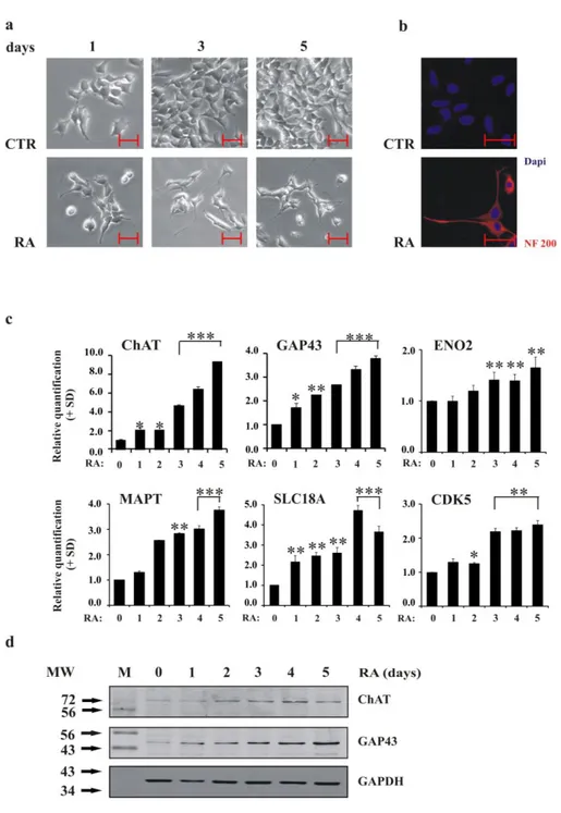
Figure 1a shows neurite outgrowth in LAN-5 cells cultured in a medium supplemented with 10 µM RA. Untreated cells continued expansion without changing morphology or shape; however, in RA-treated cells, neurite appeared after the first day of treatment and increased in number and length after five days. Moreover, neurite interconnections were stimulated by RA: the exposure caused the cells to form new networks. The expression of the differentiation marker neurofilament 200 (NF200), was induced by RA (Figure 1b).
Analysis of a group of typical molecular neuronal markers confirmed that RA treatment activated the differentiation programme in LAN-5 cells. Quantitative PCR (qRT-PCR) revealed increasing levels of GAP43, ChAT, MAPT, SLC18A3, ENO2 and CDK5 (Figure 1c); western blotting analysis of GAP43 and ChAT proteins confirmed the qRT-PCR data (Figure 1d).
Differentiation induction was offset by a reduction in the growth of RA-treated cells; the mean doubling time was approximately 27h in control cells and 54 h in RA-treated ones (Supplementary Figure 2a, Appendix A). Retinoic acid prevented cell growth by arresting cells in the G0/G1 phase of the cell cycle (the percentage of cells in G0/G1 was 50.40% in control cells and 71.48% in RA-treated cells after 3 days of growth; Supplementary Figure 2b, Appendix A). Moreover, a BrdU incorporation assay revealed reduced DNA synthesis after RA exposure. The percentage of cells incorporating BrdU was 53% for untreated control cells and 21% for RA-treated cells (Supplementary Figure 2c, Appendix A). Consistent with the increase in the percentage of cells in the G0/G1 cell cycle phase, Cyclin A was down-regulated, and the kinase inhibitor p27kip1 was up-regulated (Supplementary Figure 2d, Appendix A).
Figure 1. LAN-5 cells differentiate upon RA stimulus a) Inverted light microscopic images of LAN-5 cells
untreated (CTR) or RA-treated for 1, 3 and 5 days. Scale bar, 50 µm. b) Representative fluorescent images of LAN-5 cells untreated (CTR) or RA-treated for 5 days. Red, NF200 immunostaining; blue, DAPI. Scale bar, 50 µm. c) The levels of the indicated mRNA in LAN-5 cells untreated (0) or RA-treated for 1, 2, 3, 4 and 5 days. The data are reported as the level of mRNA relative to the respective untreated cells and are the mean + SD (n = 3). Statistical significance, *p≤0.05; **p≤0.01; ***p≤0.001. d) Representative blots of the indicated proteins in LAN-5 cells untreated (0) or RA-treated for 1, 2, 3, 4 and 5 days. GAPDH expression was used to normalise protein loading. The experiment was repeated three times with similar results. M, molecular weight markers; MW, molecular weight.
2.3.2 N-Myc expression increases during the early phases of RA-induced differentiation in cells of neural origin
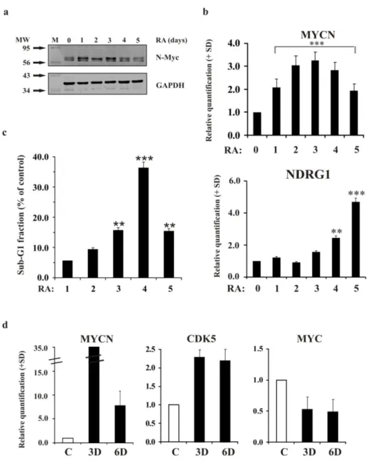
LA5 cells showed an increase in Myc protein when exposed to RA. The maximal N-Myc expression was evident between the first and the third day of treatment (Figure 2a). Afterwards, N-Myc expression levels started to decrease. Analysis of MYCN levels using qRT-PCR confirmed the results observed by western blot analysis, revealing a maximum of an approximately 3-fold-enrichment of its mRNA during the early induction of differentiation (Figure 2b). We also examined the mRNA levels of a well-known down-regulated downstream target of N-Myc, N-Myc downstream-regulated gene 1 (NDRG1) (Melotte, 2010), to verify the functional implications of MYCN up-regulation. As expected, qRT-PCR analysis revealed that the modulation of the N-Myc target NDRG1 inversely paralleled MYCN modulation during differentiation (Figure 2b). The analysis of the sub-G1 fraction, indicative of apoptosis, during the RA-induced differentiation in LAN-5 cells revealed a peak in the sub-G1 region between the third and the fourth day of differentiation, when N-Myc expression was maximal (Figure 2c).
The MYCN expression analysis conducted in mouse cortical embryonic neural progenitor cells induced to differentiate revealed that the MYCN levels in these cells tracked those observed in LAN-5 cells during differentiation (Figure 2d). Evidently, MYCN expression increased during the early phases of differentiation and then decreased as expected. The observed increase in CDK5 expression was consistent with the activation of differentiation. Interestingly, a significant decrease in MYC expression was observed from the early phases of differentiation, further supporting the idea that the MYC and MYCN genes might play different roles in the differentiation programme.
2.3.3 N-Myc is necessary to activate the differentiation programme in LAN-5 neuroblastoma cells
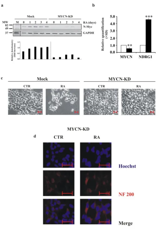
To determine whether and to what extent MYCN plays a determining role in early differentiation, we silenced its expression in MYCN-amplified LAN-5 cells. We introduced an artificial miRNA to interfere with N-Myc expression. We evaluated N-Myc levels after down-regulation, comparing Mock cells and MYCN-KD ones during RA treatment for 4 days (Figure 3a). We obtained a decrease in protein levels of approximately 70%, maintained at similar level during RA-induced differentiation. The down-regulation of the MYCN transcripts was approximately 40%, and the levels of the N-Myc downstream target NDRG1 were significantly up-regulated (Figure 3b).
Phase contrast microscopy revealed a strong reduction of neurite formation in MYCN-KD cells even after RA treatment. In contrast, Mock cells continued to present neurite extensions (Figure 3c). In the same differentiation conditions in MYCN-KD cells we did not observe any significant expression of the neurite marker NF200 (Figure 3d).
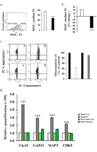
We studied plasma membrane polarisation using flow cytometry and fluorescent dyes responsive to acute changes in the plasma membrane potential to determine functional effects of MYCN down-regulation in LAN-5 cells. As expected, MYCN-KD cells were characterised by a reduced ability to polarise the plasma membrane compared to Mock cells. (Figure 4a). In addition, the membrane hyperpolarisation usually observed in RA-differentiated cells was prevented when the MYCN gene was down-regulated (Figure 4b). We evaluated cell death in Mock and MYCN-KD cells by assessing the mitochondrial membrane potential. We found that the MYCN-silenced cells did not show any cell death after RA treatment, whereas the control Mock cells exposed to RA exhibited apoptosis (Figure 4c), suggesting that MYCN might be necessary in the early phases of differentiation to induce apoptosis in cells not committed to differentiation.
The expression of the molecular neuronal markers previously analysed in the differentiating LAN-5 cells was also studied after MYCN silencing. We observed inhibited expression levels of all four markers analysed at day three after RA treatment (Figure 4d).
Figure 2. MYCN increases during the first days of differentiation in LAN-5 cells and in mouse cortical embryonic neural progenitors a) Representative blots of the N-Myc protein in LAN-5 cells untreated (0) or
RA-treated for 1, 2, 3, 4 and 5 days. GAPDH expression was used to normalise protein loading. The experiment was repeated three times showing similar results. M, molecular weight markers; MW, molecular weight. b) Levels of the indicated mRNA in LAN-5 cells untreated (0) or RA-treated for 1, 2, 3, 4 and 5 days. The data are reported as the level of mRNA relative to the respective untreated cells and are the mean + SD (n = 3). Statistical significance, **p≤0.01; ***p≤0.001. c) Sub-G1 fractions estimated on the DNA content histograms in LAN-5 cells treated with RA for 1, 2, 3, 4 and 5 days. Data are reported as percent of control. Statistical significance, **p≤0.01; ***p≤0.001. d) The levels of the indicated mRNA in mouse cortical embryonic neural progenitor cells undifferentiated (white) or induced to differentiate for 3 and 6 days (black). The data are reported as the level of mRNA relative to the respective untreated cells and are the mean + SD (n = 3). Statistical significance, **p≤0.01; ***p≤0.001.
Figure 3. MYCN gene silencing in LAN-5 cells inhibits neurite outgrowth and NF200 expression after RA induction a) Upper panel, representative blots of the N-Myc protein in LAN-5 cells silenced for MYCN gene
(MYCN-KD) or in control cells (Mock) untreated (0) or RA-treated for 1, 2, 3, 4 and 5 days. M, molecular weight markers; MW, molecular weight. Lower panel, blots densitometry as analysed by ImageJ software. Values are averages + SD of three independent experiments with similar results. GAPDH expression was used to normalise protein loading. b) The levels of the indicated mRNA in MYCN-KD LAN-5 cells (black) or in control cells (white). The data are reported as the level of mRNA relative to the respective control cells and are the mean + SD (n = 3). Statistical significance, **p≤0.01; ***p≤0.001. c) Inverted light microscope images of MYCN-KD LAN-5 cells or in control cells, untreated (CTR) or RA-treated for 3 days. Scale bar, 50 µm. d) Representative fluorescent images of MYCN-KD LAN-5 cells untreated (CTR) or RA-treated for 3 days. Red, NF200 immunostaining; blue, Hoechst. Scale bar, 50 µm.
2.3.4 N-Myc overexpression induces differentiation in poorly differentiating neuroblastoma cells
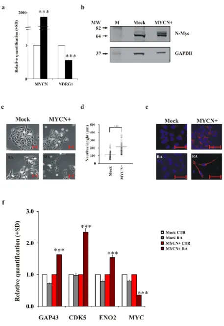
The SK-N-AS neuroblastoma cell line shows a very poor capacity to differentiate after stimulation with RA, and these cells have a single copy of the MYCN gene, thus showing no overexpression of this gene (Suenaga, 2009). We overexpressed the MYCN gene in these cells (Figure 5a) and studied differentiation activation after RA induction. The overexpression of the MYCN gene was also confirmed by the protein level (Figure 5b). Figure 5c shows the micrographs of SK-N-AS Mock and SK-N-AS MYCN+ cells, untreated or treated with RA for three days. In the presence of the MYCN gene, RA activated differentiation in SK-N-AS cells, as indicated by the neurite outgrowth, with most of them showing a bipolar shape. The number of cells with neurites increased by approximately 90% in MYCN+ cells after RA treatment. The neurite length was significantly increased in MYCN+ cells (Figure 5d; Mock cells, mean=121 µM, median=120 µM, range=60-254 µM; MYCN+ cells, mean=213 µM, median=208 µM, range=115-375 µM). NF200 immunostaining confirmed the ability of SK-N-AS MYCN+ cells to differentiate; NF200 was clearly expressed in these cells after RA exposure (Figure 5e). The analysis of GAP43, CDK5 and ENO2 differentiation marker genes corroborated that MYCN overexpression in SK-N-AS cells rendered these cells prone to differentiation after RA exposure (Figure 5f).
Figure 4. MYCN gene silencing produces a depolarisation of the plasma membrane and inhibits the
RA-induced up-regulation of differentiation markers a) FACS analysis of the DiOC5 fluorescence distribution in
MYCN-KD LAN-5 cells (full histogram) or in Mock control cells (empty histogram). Right, FACS histograms; left, median values of the DiOC5 fluorescence distributions in MYCN-KD LAN-5 cells (black) or in Mock
control cells (white). The experiment was repeated three times showing similar results. b) The percent variation of the median values of the DiOC5 fluorescence distributions in MYCN-KD LAN-5 cells (black) or in Mock
control cells (white) after RA treatment. The experiment was repeated three times showing similar results. c) Mitochondrial membrane potential analysed by JC-1 staining in Mock control LAN-5 untreated (A) and RA-treated (B) cells compared to MYCN-KD LAN-5 unRA-treated (C) and RA-RA-treated (D) cells (left panel). In the right panel are shown the ΔΦ values calculated on the FACS cytograms following the formula described in Materials and Methods section. Data are reported as percent of control and are average of at least three separate experiments. Bars represent standard deviation. d) The levels of the indicated mRNA in MYCN-KD LAN-5 cells untreated (light green) or treated for 3 days (green) and in Mock control cells untreated (white) or RA-treated for 3 days (dark grey). The data are reported as the level of mRNA relative to the respective unRA-treated cells and are the mean + SD (n = 3). Statistical significance, ***p≤0.001.
Figure 5. MYCN overexpression in the SK-N-AS cells restores their ability to differentiate after RA a)
The levels of the indicated mRNA in SK-N-AS Mock (white) and SK-NAS MYCN+ (black) cells. The data are reported as the level of mRNA relative to the respective Mock cells and are the mean + SD (n = 3). Statistical significance, ***p≤0.001. b) Representative blots of the N-Myc protein in SK-N-AS Mock and SK-N-AS MYCN+ cells. GAPDH expression was used to normalise protein loading. The experiment was repeated three times showing similar results. M, molecular weight markers; MW, molecular weight. c) Inverted light microscope images of SK-N-AS Mock and SK-N-AS MYCN+ untreated or RA-treated for 3 days. Scale bar, 50 µm. d) Distribution of the neurite outgrowth length as measured in SK-N-AS Mock and SK-N-AS MYCN+ treated with RA for 3 days. Statistical significance, ***p≤0.001. e) Representative fluorescent images of SK-NAS Mock and SK-N-AS MYCN+ untreated or RA-treated for 3 days. Red, NF200 immunostaining; blue, DAPI. Scale bar, 50 µm. f) The levels of the indicated mRNA in SK-N-AS Mock and SK-N-AS MYCN+ untreated (white or light red, respectively) or RA-treated for 3 days (grey or red, respectively). The data are reported as the level of mRNA relative to the respective control cells and are the mean + SD (n = 3). Statistical significance, ***p≤0.001.
2.3.5 MYCN modulation modifies the expression of miRNAs involved in apoptosis preceding neuronal differentiation
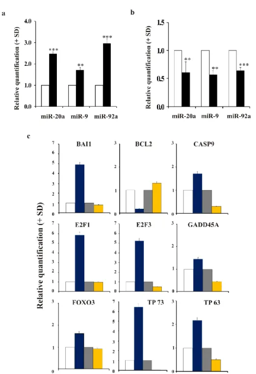
The analysis of specific miRNAs selected by in silico analysis (miRWalk - Dweep, 2011) and reported to be regulated by MYCN and at the same time involved in apoptosis and neuron development, revealed changes in the expression of miR-20a, miR-9 and miR-92a. We found that these miRNAs were up-regulated in MYCN-silenced LAN-5 cells (Figure 6a). In contrast, when MYCN was overexpressed in the SK-N-AS cells, the same miRNAs were down-regulated (Figure 6b). In particular, the p53-family members have been reported to be regulated by these miRNAs. For this reason, we performed PCR Array analysis of the p53 signalling pathway (Figure 6c). As expected, and consistent with the expression of miR-20a, miR-9 and miR-92a, the expression pattern observed in LAN-5 MYCN-KD cells was opposite to that in SK-N-AS MYCN+ cells with regard to genes known to be involved in cell death regulation. We found that pro-apoptotic CASP9 and BAI1 genes were up-regulated in the MYCN overexpressing cells; whereas, the anti-apoptotic BCL2 gene was down-regulated in the same MYCN condition. By contrast, CASP9 and BAI1 were down-regulated and BCL2 up-regulated when MYCN was silenced. As well as, genes known to be involved in the activation of apoptotic programme such as E2F1, E2F3, GADD45A, and FOXO3 were up-regulated in MYCN-amplified and down-regulated in MYCN-silenced cells, respectively. In addition, despite an analysis of validated miRNA targets using miRWalk database reported miR-20a, miR-9 and miR-92a target TP53, TP73 and TP63 mRNAs, the PCR Array analysis did not reveal any modulation of TP53 mRNA levels (data not shown); while an up-regulation of both TP73 (about 7-fold) and TP63 (more than 2-fold) was observed in MYCN overexpressing cells (Figure 6c).
2.3.6 Inhibition of miR-20a, miR-9 and miR-92a in the wild type SK-N-AS cells restores apoptosis and their differentiation ability
We inhibited miR-20a, miR-9 and miR-92a in MYCN-non-amplified SK-N-AS cells avoiding any possible interference by MYCN (Figure 7a).
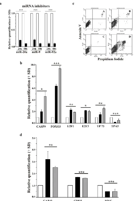
The pro-apoptotic genes (CASP9, FOXO3, E2F1, E2F3 and TP73) were up-regulated by miRNAs inhibition after both 24h and 48h. By contrast, TP63 was not up-regulated suggesting no correlation between miR-20a, miR-9 and miR-92a and TP63 expression (Figure 7b). Interestingly, RA was able to activate apoptosis after miRNAs inhibition in SK-N-AS cells (Figure 7c, panel D). While, control cells did not present any significant induction of apoptosis by RA treatment (Figure 7c, panel B).
Figure 6. miRNAs are inversely regulated in MYCN-silenced and MYCN-up-regulated neuroblastoma cells, and its expression is associated to a different modulation of apoptosis-related genes a) The levels of
the indicated hsa-miR in MYCN-KD LAN-5 cells (black) or in Mock control cells (white). The data are reported as the level of miRNA relative to the respective untreated cells and are the mean + SD (n = 3). Statistical significance, **p≤0.01; ***p≤0.001. b) The levels of the indicated hsa-miR in SK-N-AS MYCN+ cells (black) or in Mock control cells (white). The data are reported as the level of miRNA relative to the respective untreated cells and are the mean + SD (n = 3). Statistical significance, **p≤0.01; ***p≤0.001. (c) The levels of the indicated mRNA in SK-N-AS Mock (white), SK-N-AS MYCN+ (blue), LAN-5 Mock (grey) and LAN-5 MYCN-KD (yellow). The data are reported as the level of mRNA relative to the respective control cells and are the mean + SD (n = 3).
Figure 7. miR-20a, miR-9 and miR-92a inhibition in wild type SK-N-AS cells leads to apoptotic death induction and expression of differentiation-related genes a) The levels of the indicated miRNAs in inhibitor
negative control (white) and miRNA inhibitor (black) cells at 24h and 48h after transfection. The data are reported as the level of mRNA relative to the respective miRNAs negative control cells and are the mean + SD (n = 3). Statistical significance, ***p≤0.001. b) The levels of the indicated mRNA in inhibitor negative control (white), miRNA inhibitor at 24h (black) and miRNA inhibitor at 48h (grey). The data are reported as the level of mRNA relative to the respective miRNAs negative control cells and are the mean + SD (n = 3). Statistical significance, *p≤0.05; **p≤0.01; ***p≤0.001. c) FACS analysis of the Annexin V fluorescence distribution in inhibitor negative control untreated (panel A) and RA-treated (panel B) cells and miRNA inhibitor untreated (panel C) and RA-treated (panel D) at 48h after transfection. The experiment was repeated three times showing similar results. d) The levels of the indicated mRNA in inhibitor negative control (white), miRNA inhibitor at 24h (black) and at 48h (grey) after transfection. The data are reported in RA-treated condition. The data are the mean + SD (n = 3). Statistical significance, **p≤0.01; ***p≤0.001.
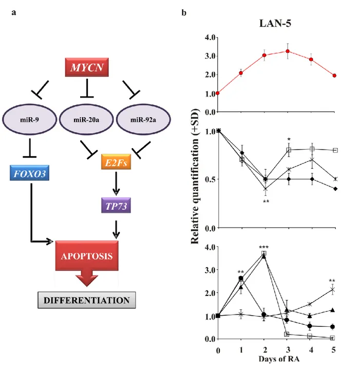
Figure 8. Model of the hypothetical role of MYCN during the early phases of neuroblastoma differentiation a) MYCN overexpression inhibits miR-9, miR-20a and miR-92a. We hypothesise that this
inhibition allows targets of these miRNAs (E2F genes, TP73 and FOXO3) to be up-regulated and in turn to activate apoptosis in those cells not committed to differentiate but still proliferating. In contrast, MYCN down-regulation allows the overexpression of miR-9, miR-20a and miR-92a, preventing the up-down-regulation of apoptosis-related genes, thus inhibiting apoptosis and subsequent differentiation. b) Upper panel, the levels of MYCN gene in LAN-5 cells untreated (0) or RA-treated for 1, 2, 3, 4 and 5 days. Middle panel, the levels of miR-20a (diamond), miR-9 (open square) and miR-92a (star). Lower panel, the levels of E2F1 (closed circle), E2F3 (open square), FOXO3 (triangle), TP73 (star). The data are reported as the level of mRNA relative to the respective untreated cells and are the mean + SD (n = 3). Statistical significance, *p≤0.05; **p≤0.01; ***p≤0.001.
Differentiation markers were up-regulated after the inhibition of the three miRNAs at both 24h and 48h in the presence of RA induction (Figure 7d). As expected, MYC was down-regulated in differentiating cells.
In Figure 8a we summarised our data presenting a hypothetical mechanism by which MYCN could trigger the onset of differentiation programme in neuroblastoma cells. To further validate this hypothesis we analysed the expression kinetics of miRNAs and apoptotic genes presented in the model (Figure 8b, middle and lower panels). To make data reading easier we reported the kinetics of MYCN gene (Figure 8a, upper panel; see also Figure 2b). As MYCN gene is up-regulated during RA treatment the three miRNAs are down-regulated and consequently their targets are up-regulated.
2.4 Conclusions
Several studies of MYC- and MYCN-knockout mice have revealed that embryos of these mice could not survive until gestation (Sawai, 1991; Charron, 1992; Stanton, 1992; Davis, 1993; Knoepfler, 2002), suggesting that the MYC family genes are required for normal development at the beginning of organogenesis. In particular, Knoepfler et al. showed that N-Myc plays an important role in complete nervous system development. In fact, loss of N-N-Myc function during embryogenesis interrupts the ability of neural progenitors to expand, differentiate and populate the brain, causing neurological dysfunction after birth. Unlike c-Myc, which is expressed in all proliferating cells, N-Myc expression is more restricted. Appropriate spatial and temporal expression of N-Myc is important for normal embryonic development (Strieder, 2003). Accordingly, we show that up-regulation of the MYCN gene is restricted to the early stages of differentiation induction in mouse cortical embryonic neural progenitor cells. In addition, as expected, MYC gene expression decreased during differentiation progress. These data suggest that the MYCN gene might play a critical role in the activation of neuronal differentiation. Nevertheless, as also reported by others (Sidell, 1982; Amatruda, 1985; Thiele, 1988), differentiation completion was later associated with a decrease in MYCN expression. It is likely that the N-Myc protein, as a transcription factor, is necessary at the onset of neuronal differentiation to establish the expression of a set of genes essential for subsequent phases.
To examine the role of N-Myc in the neuronal differentiation programme, we used the human neuroblastoma cell line LAN-5, which show MYCN gene amplification (Ribatti, 2002). The LAN-5 cell line, similarly to many others neuroblastoma cell lines, maintains the ability to differentiate in vitro in the presence of specific stimuli, such as RA (Ribatti, 2002; Edjö, 2004). In fact, our results confirm that LAN-5 cells treated with RA form neurites; express the neuronal marker NF200, an intermediate filament that provides structural stability to the axon; and show the induction of a series of neuronal molecular markers. In agreement with previous papers (Sidell, 1982; Påhlman, 1984), GAP43 and MAPT were up-regulated in differentiated LAN-5 cells; in addition, ChAT and SLC18A appeared to have increased expression after differentiation induction in the LAN-5 cell line, which is normally committed to cholinergic differentiation (Hill, 1997). As expected, successful development is associated with cell proliferation inhibition, demonstrated by cell cycle analysis performed using various approaches.
In addition to what has been observed previously (Sidell, 1982; Amatruda, 1985; Thiele, 1988), we found peak MYCN expression during the first days of RA-induced differentiation
in LAN-5 cells. We demonstrated the functionality of the increased N-Myc protein through expression analysis of the N-Myc target NDRG1, which decreased as expected (Melotte, 2010).
Furthermore, differentiation was inhibited by MYCN silencing in the same model; this was indicated by the failure of neurite outgrowth, the decrease in known neuronal molecular markers and the loss of NF200 expression during differentiation. In addition, the plasma membrane of the MYCN-KD LAN-5 cells depolarised and was no longer able to hyperpolarise in response to differentiation stimuli. Plasma membrane polarisation is a parameter known to be essential for differentiated neuronal cells (Sundelacruz, 2009).
In addition, when MYCN is overexpressed in cells with a poor differentiation capacity, developmental processes are restored. This pattern was observed in SK-N-AS MYCN+ cells, in which neurite outgrowth occurred, and the expression of NF200 and known molecular neuronal markers increased after RA treatment.
These data, taken together, demonstrate that N-Myc protein expression is required to activate the differentiation processes in neuroblastoma cells.
To explore the mechanisms by which N-Myc could trigger differentiation in neuroblastoma cells, we studied the expression levels of miR20a, miR-9 and miR-92a which have been reported to be involved in differentiation processes and to be modulated by MYCN (Schulte, 2008; Stallings, 2009). Through the up- or down-regulation of the MYCN gene, we observed that all the three miRNAs were decreased or increased, respectively. Consistently, the inverse relationship between these miRNAs and the cell differentiation status suggests that their down-regulation is needed for differentiation triggering. In fact, according to the miRWalk database, two of the validated targets of these miRNAs are the retinoic acid receptor genes RARA and RARG (Li, 2009; Liu, 2012). The up-regulation of miR-92 following MYCN silencing and its down-regulation by RA treatment are in agreement with Chen and Stallings (2007), who showed that the expression of different miRNAs correlates with neuroblastoma prognosis, differentiation and apoptosis, also in response to RA. Moreover, consistently with Haug et al. (2011) who found that miR-92 inhibits the secretion of the tumour suppressor gene DKK3 in neuroblastoma, our data demonstrated the down-regulation of miR-92a in cells with a restored ability to differentiate, characteristic of a less malignant phenotype. Our findings are also supported by the data of Jee et al. (2012), which showed that inhibition of miR-20a expression induces definitive motor neuron survival and neurogenesis. Consistent with results of Yoo et al. (2009), showing that miR-9 is repressed in neural proliferating
progenitors and is sequentially re-expressed in post-mitotic neurons, is the modulation of miR-9 we observed in LAN-5 cells during the RA kinetics (see Figure 8b, middle panel). In agreement with O' Donnell et al. (2005) and Coller et al. (2007) we demonstrated that miR-20a negatively regulates the E2F factor genes. The transactivating p73 isoforms are transcriptionally induced by E2F and contribute to E2F-mediated apoptosis (Irwin, 2000, Stiewe, 2000; Zaika, 2001). Consistent with these data, up-regulation of TP73 and E2F was observed in MYCN+ cells and after miRNAs inhibition in SK-N-AS cells. Moreover, as expected, when MYCN was down-regulated, and miR-20a and miR-92a were consequently up-regulated, E2F and TP73 were inhibited. The role of p73 in the induction of neuronal differentiation has been previously highlighted by De Laurenzi et al. (2000), who showed an increase in p73 protein levels after RA stimuli in murine neuroblastoma cells. In apparently disagreement with our data, they also showed reduced N-Myc protein levels after RA treatment as a marker of neuronal differentiation (De Laurenzi, 2000). Indeed, they analysed N-Myc expression after 6 days of RA treatment, whereas, although our data demonstrated a peak in N-Myc protein expression between the second and the third day of differentiation induction, we also observed a subsequent decrease at day 5 of differentiation. It is likely that N-Myc, by down-regulating miR-20a and miR-92a, produces the increase in E2Fs gene expression, which in turn induces TP73 transcription. Interestingly, RA treatment in LAN-5 cells confirmed the presence of a hierarchy between E2Fs and TP73 which are overexpressed at day 1 and day 4 of RA treatment, respectively.
In addition, we found FOXO3 up-regulated in differentiating MYCN+ cells and in RA-treated LAN-5 cells. The human FOXOs transcription factor family regulates the expression of genes associated with multiple biological process such as cell cycle arrest and apoptosis (Eijkelenboom, 2013). As demonstrated by Senyuk et al. (2013), miR-9 is able to bind directly to the 3’ UTR of FOXO3. In fact, when we silenced the MYCN gene in LAN-5 cells, an increase in miR-9 and a consequent decrease in FOXO3 mRNA levels were obtained. These data are further demonstrated by the increase in FOXO3 expression achieved after inhibiting miRNAs in SK-N-AS cells.
We reasoned that the miR-20a, miR-9 and miR-92a inhibition in MYCN+, as well as, in RA-treated LAN-5 cells and the subsequent pro-apoptotic genes increase could be a necessary step prior to differentiation programme. In fact, the inhibition of 20a, 9 and miR-92a in the MYCN-non-amplified SK-N-AS cells restored the ability of these cells to respond to RA treatment, as demonstrated by Annexin V assay and differentiation-related genes expression analysis.