1
Università degli Studi di Ferrara
DOTTORATO DI RICERCA IN
"SCIENZE BIOMEDICHE"
CICLO
XXVII
COORDINATORE Prof. Capitani Silvano
Targeting the PI3K/Akt/mTOR signaling
pathway as a new therapeutic strategy for
personalized treatments in acute lymphoblastic
leukemia
Settore Scientifico Disciplinare BIO/16
Dottorando Tutore
Dott.ssa Cani Alice Prof. Neri Luca Maria
(firma) (firma)
2
INDEX
1 Introduction p. 5 1.1 The acute lymphoblastic leukemia
1.2 The PI3K/Akt/mTOR signal transduction pathway 1.2.a The PI3K family
1.2.b Akt 1.2.c mTOR
1.3 Downstream targets of PI3K/Akt/mTOR network 1.3.a GSK-α/β
1.3.b FOXO 1.3.c S6K 1.3.d 4E-BP1
1.3.e PTEN-mediated inhibition of the pathway
1.4 Role of the PI3K/Akt/mTOR network in acute leukemia 1.4.a PI3K/Akt/mTOR signaling in T-ALL
1.4.b PI3K/Akt/mTOR signaling in B-pre ALL
1.5 Therapeutic strategies acting on PI3K/Akt/mTOR network in leukemia 1.5.a Advances in targeting PI3K/Akt/mTOR pathway
1.5.b Akt inhibitors 1.5.c mTORC1 inhibitors 1.5.d mTOR inhibitors
1.5.e Resistance to rapamycin/rapalogs and mTOR inhibitors and the effectiveness to multiple targeting the PI3K/Akt/mTOR signaling pathway
2 Aim of the study p. 29
3 Materials and methods p. 30
3.1 Materials
3.2 Cell culture and patient samples 3.3 MTT test
3.4 Cell cycle 3.5 Western blot 3.6 DAPI staining
3
3.7 SiRNA experiments 3.8 PI/Annexin V assay
3.9 Combined drug effect analysis 3.10 Statistical evaluation
4 Results p. 35 4.1 Treatment with MK-2206 affects T-ALL cells
4.1.a MK-2206 displays cytotoxic pro-apoptotic effects on T-ALL cell lines and induces cell cycle arrest
4.1.b MK-2206 affects PI3K/Akt/mTOR signaling in T-ALL cell lines 4.1.c MK-2206 induces autophagy
4.1.d MK-2206 synergizes with Doxorubicin
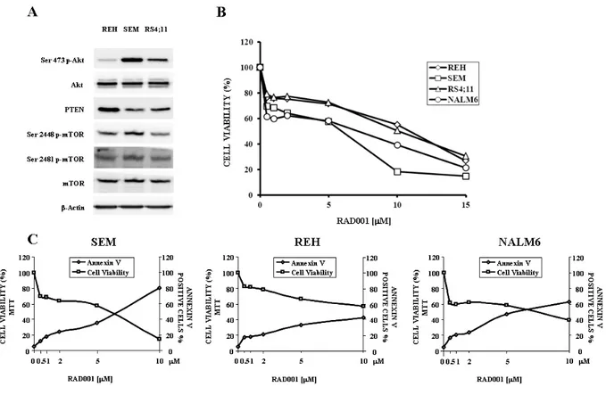
4.2 Treatment with RAD001 affects B-pre ALL cells
4.2.a The PI3K/Akt/mTOR pathway activation status and RAD001 effects in B-pre ALL cell lines
4.2 b RAD001 modulates PI3K/Akt/mTOR signaling in B-pre ALL cell lines 4.2.c RAD001 induces cell cycle arrest, apoptosis and autophagy
4.3 Two hit pathway with RAD001 and MK-2206 leads to synergistic effects 4.3.a Dual targeting of mTOR and Akt results in synergistic inhibition of proliferation in B-pre ALL cell lines
4.3.b B-pre ALL lymphoblasts are sensitive to combined mTOR/Akt inhibition 4.4 Treatment with Torin-2 affects B-pre ALL cells
4.4.a PI3K/Akt/mTOR pathway activation status in B-pre ALL cell lines
4.4.b Torin-2 induces cytotoxicity, blocks cell cycle progression at the G0/G1 phase and induces autophagy
4.4.c Torin-2 causes pro-apoptotic effects on B-pre ALL cell lines 4.4.d Torin-2 affects the PI3K/Akt/mTOR pathway in B-pre ALL cells 4.5 Comparison between RAD001 and Torin-2 in B-pre ALL cells
4.5.a Torin-2 prevents the reactivation of Akt upon mTOR inhibition in B-pre ALL cells
4.6 Comparison between RAD001 and Torin-2 administered in B-pre ALL cells with MK-2206
4.6.a MK-2206 synergizes with RAD001 but not with Torin-2
4
4.7 Triple hit Akt affects T-ALL cells
4.7.a PI3K/Akt/mTOR pathway activation status in T-ALL cell lines
4.7.b Multiple Akt targeting had higher cytotoxic effect and synergized only in Ser 473 p-Akt expressing cells
4.7.c Triple Akt hit increases the inhibition of the PI3K/Akt/mTOR signaling pathway
4.7.d Pre-treatment with Perifosine enhances synergistic effect
4.7.e The triple Akt inhibition induces cell cycle arrest and causes autophagy and pro-apoptotic effects in T-ALL cells
4.7.e The multiple treatment has the capability to inhibit ERK pathway
5 Discussion p. 79 5.1. Treatment with MK-2206 affects T-ALL cells
5.2 Treatment with RAD001 affects B-pre ALL cells
5.3 Two hit pathway with RAD001 and MK-2206 leads to synergistic effects 5.4 Treatment with Torin-2 affects B-pre ALL cells
5.5 Comparison between RAD001 and Torin-2 in B-pre ALL cells
5.6 Comparison between RAD001 and Torin-2 administered in B-pre ALL cells with MK-2206
5
1. Introduction
1.1 The acute lymphoblastic leukemia
Acute lymphoblastic leukemia (ALL) is an acute form of blood cancer, characterized by the uncontrolled clonal proliferation of cancerous, immature lymphoid cells, known as lymphoblasts (Fig. 1). In patients with ALL, lymphoblasts are overproduced and continuously multiply, causing damage and death by inhibiting the production of normal cells -such as red and white blood cells and platelets- in the bone marrow and by spreading (infiltrating) to other organs [1].
In T-cell acute lymphoblastic leukemia (T-ALL), the malignant cells are derived in the thymus from T-cell progenitor cells and express immature T-cell immunophenotypic markers [2, 3]. T-cell neoplastic transformation is a complex process in which multiple lesions, involving both oncogenes and tumor suppressor genes, cooperate to alter the normal signaling pathways that regulate proliferation, differentiation, and survival of developing T-cells [4-7]. T-ALL comprises about 15% of pediatric and 25% of adult ALLs. T-ALL was associated with a very bad outcome, however the introduction of intensified polychemotherapy protocols has improved the prognosis of this disorder and current therapies can achieve 5-year relapse-free survival rates of about 75% in pediatric patients and 40-50% in adults [8, 9].
B precursor cell acute lymphoblastic leukemia (B-pre ALL) is a form of leukemia in which too many B-cell lymphoblasts (immature white blood cells) are found in the blood and bone marrow. It is the most common pediatric malignancy and comprises 85% of childhood ALL [10]. New therapeutic protocols have improved pediatric patient survival rate to approximately 80% at 5 years, however some cases still relapse and are tried by long-term side effects of therapy [11-13]. The overall prognosis of children with relapsed disease remains poor with less than 40% survival at 5 years [14]. B-pre ALL is a heterogeneous disorder including several subtypes with specific cellular and molecular features, that are related to clinical outcome [15]. The Philadelphia (Ph) chromosome is the most common cytogenetic anomaly associated with B-pre ALL. The Ph chromosome results from a reciprocal translocation (t) between chromosomes 9 and 22 (t[9,22][q34;q11]) [16], and results in a fusion gene on chromosome 22, i.e. the breakpoint cluster region-Abelson leukemia (Bcr-Abl) viral proto-oncogene. Bcr-Abl fusion proteins
6
are constitutively active non-receptor tyrosine kinases that alter a myriad of intracellular signaling networks, thus contributing to leukemic cell proliferation and survival.
Figure 1: Scheme of myeloid and lymphoid cells maturation.
1.2 The PI3K/Akt/mTOR signal transduction pathway
1.2.a The PI3K family
The phosphoatidylinositol-3-kinase (PI3K) family consists of a number of serine/threonine and lipid kinases, including those that phosphorylate the 3’-OH of phosphatidylinositols. These enzymes consists of at least nine genes in mammalian systems, corresponding to various isoforms that are grouped into three classes, each one with distinct substrate specificity and lipid products: I, II, and III [17]. In mammalian cells, class I PI3Ks are the best understood PI3Ks and the most widely implicated in human malignancies [18].
7
Class I PI3Ks are further divided into two subgroups: A and B. Class IA PI3Ks contain one of three catalytic subunits (p110α, p110β, p110δ) that form heterodimers with one of the five adaptor (or regulatory) isoforms (p85α, p85β, p55α, p55γ, p50α). In general, class IA PI3Ks are activated downstream of both tyrosine kinase receptors (TKRs) and G protein-coupled receptors (GPCRs). The single class IB PI3K comprises a p110γ catalytic subunit which binds one of two related regulatory subunits, p101, and p87. Class IB PI3Ks mainly act downstream of GPCRs, however they can be stimulated also by TKRs [19]. Only class I PI3Ks have the ability to use phosphatidylinositol-4,5-bisphosphate (PtdIns 4,5P2) to generate the second messenger, phosphatidylinositol- 3,4,5-trisphosphate (PtdIns 3,4,5P3). Once activated by a variety of growth factors and cytokines, class I PI3Ks initiate a cascade of events that promote cancer cell proliferation, survival, and metabolism.
Class II PI3Ks can also be activated by tyrosine-kinase receptors (RTKs), cytokine receptors and integrins; the specific functions in response to these activators are not understood [20].
Class III PI3Ks are heterodimeric enzymes of catalytic (Vps34, 100 kDa) and adaptor (p150) subunits, and use only phosphatidylinositol as a substrate. Class III PI3Ks are implicated in the regulation of mammalian target of rapamycin (mTOR) activity in response to aminoacid availability and the regulation of autophagy in response to cellular stress, indicating the importance of class III PI3K in controlling cell growth and survival [17].
1.2.b Akt
Akt, a 60-kDa serine/threonine kinase, is a key effector of PI3K in carcinogenesis. Akt is a member of the AGC protein kinase family and is the cellular homolog of the v-Akt oncogene. The Akt family includes three highly conserved isoforms: Akt1/α, Akt2/β, and Akt3/γ [21] plus a fourth isoform defined Akt-γ1, have been identified in humans (Fig. 2). They are codified by different genes with 80% sequence homology. Akt-1 is the predominant isoform in the major part of tissues, Akt-2 is present in insulin sensitive tissues and Akt-3 has not been completely localized, but it is absent in central nervous system. Akt is constituted by three distinct modules: the pleckstrin homology (PH) domain in the amino-terminal region able to bind phospho-lipids; the central kinase domain which contains an highly conserved activation loop, called T-loop, with threonine residue important for the enzyme activation; a regulative carboxyl-terminal extension of about 40
8
amino acids containing the hydrophobic F-X-X-F/Y-S/T-Y/F motif. The recruitment of inactive Akt from the cytosol to the plasma membrane, requires that the PH domain of Akt binds to PtdIns 3,4,5P3 or PtdIns 3,4P2 synthesized at the plasma membrane by PI3K. Akt is then phosphorylated at Thr 308 by phosphatidylinositol-dependent kinase 1 (PDK1), and at Ser 473 by mTOR complex 2, resulting in full activation of Akt kinase activity [22]. Threonine 308 is located within the T-loop and is phosphorylated by PDK1, producing a conformational change which promotes the second phosphorylation on serine 473, on the carboxyl-terminal hydrophobic extension of kinase domain. Akt activity is maximal when the kinase is phosphorylated on both residues, increasing substrates affinity and greatly powering the catalytic potential.
Activated Akt is able to translocate from cytoplasm into the nucleus, where signaling events appear independent from those on the plasmatic membrane [23]. Akt phosphorylates a plethora of targets [19, 24, 25] on R-X-R-X-X-S/T consensus motifs [26]. Intriguingly, most of the Akt effects depend on its ability to phosphorylate proteins involved in cell cycle progression, apoptosis, mRNA translation, glycolysis, and angiogenesis, thus unlocking most, if not all, of the critical processes involved in tumorigenesis [27]. The identification of more than 400 different proteins containing the consensus sequence for Akt phosphorylation makes presume that in the future other Akt substrates will be characterized [28]. Thus, the heterogeneity of proteins potentially phosphorylated by Akt supports the key role of this kinase in different fundamental cell processes.
9
1.2.c mTOR
mTOR is a 289-kDa serine/threonine kinase which belongs to the phosphatidylinositol 3-kinase-related kinase (PIKK) family [29]. mTOR encompasses two functionally distinct multiprotein complexes, referred to as mTOR complex 1 (mTORC1) and mTOR complex 2 (mTORC2). mTORC1 is a direct downstream effector of Akt, however its activity is controlled through other signaling networks that include the Ras/Raf/mitogen-activated protein kinase kinase (MEK)/extracellular signal-regulated kinase (ERK) 1/2 signaling network, and the liver kinase B1 (LKB1)/AMP-activated protein kinase (AMPK) cascade [30, 31].
mTORC1 is characterized by the interactions between mTOR and the regulatory associated protein of mTOR (Raptor), which regulates mTOR activity and functions as a scaffold for recruiting mTORC1 substrates. mTORC1 is sensitive to rapamycin and its analogs (rapalogs) that include RAD001, CCI-779, and AP23753. Rapamycin/rapalogs are allosteric mTORC1 inhibitors and do not target the mTOR catalytic site [32, 33]. They associate with the FK506 binding protein 12 (FKBP-12, [34]), and, by doing so, they induce the disassembly of mTORC1, resulting in inhibition of its activity [33].
mTORC2 comprises the rapamycin-insensitive companion of mTOR (Rictor) and is generally described as being insensitive to rapamycin/rapalogs. However, long-term (>24 hours) treatment of about 20% of cancer cell lines (mainly of hematopoietic lineage) with rapamycin/ rapalogs resulted in mTORC2 activity inhibition [35, 36].
mTORC1 controls translation in response to growth factors/nutrients through the phosphorylation of p70S6 kinase (p70S6K) and 4E-BP1. p70S6K phosphorylates the 40S ribosomal protein, S6 (S6RP), leading to active translation of mRNAs [37]. Furthermore, p70S6K phosphorylates the eukaryotic initiation factor 4B (eIF4B) which is critically involved in translation [38, 39]. However, eIF4B is a downstream target also of MEK/ERK signaling [40]. Unphosphorylated 4E-BP1 interacts with the cap-binding protein, eukaryotic initiation factor 4E (eIF4E), and prevents the formation of the 4F translational initiation complex (eIF4F), by competing for the binding of eukaryotic initiation factor 4G (eIF4G) to eIF4E. 4E-BP1 phosphorylation by mTORC1 results in the release of the eIF4E, which then associates with eIF4G to stimulate translation initiation [41]. eIF4E is critical for translating 5’capped mRNAs, that include transcripts mainly encoding for proliferation and survival promoting proteins, such as c-Myc, cyclin-dependent kinase-2 (CDK-2), cyclin D1, signal activator and transducer of transcription-3 (STAT-3), B-cell
10
lymphoma (Bcl) -2, Bcl-xL, survivin, myeloid cell leukemia-1 (Mcl-1), ornithine decarboxylase [35, 41, 42].
Moreover, mTORC1 represses autophagy, a lysosome-dependent degradation pathway which allows cells to recycle damaged or superfluous cytoplasmic content, such as proteins, lipids, and organelles [43]. As a consequence, cells produce metabolic precursors for macromolecular biosynthesis or ATP generation. In cancer cells, autophagy fulfils a dual role, because it can have both tumor-suppressing and tumor-promoting functions. Indeed, the autophagic machinery prevents necrosis and inflammation, that can lead to genetic instability and tumorigenesis. However, autophagy might be important for tumor progression, by providing energy through its recycling mechanism during unfavorable metabolic circumstances, that are very common in tumors [44].
The mechanisms that control mTORC2 activity have only begun to be revealed [45], however mTORC2 activation by growth factors requires PI3K, as pharmacological inhibition of PI3K decreased mTORC2 activity in vitro [46]. mTORC2 phosphorylates Akt at Ser 473 which enhances subsequent Akt phosphorylation on Thr 308 by PDK1.
PI3K, Akt, and mTORC1/2 are linked to each other via regulatory feedback loops, that restrain their simultaneous hyperactivation [35]. A negative regulation of Akt activity by mTORC1 is dependent on p70S6K-mediated phosphorylation of insulin receptor substrate (IRS) -1 and -2 adapter proteins, downstream of the insulin receptor (IR) and/or insulin-like growth factor-1 receptor (IGF-1R) [47-49]. IRS-1 and IRS-2 are normally required to activate class IA PI3Ks after stimulation of IR/ IGF-1R tyrosine kinase activity. When mTORC1 is active, p70S6K phosphorylates the IRS-1 and -2 proteins on Ser residues, targeting them for proteasomal degradation [50, 51]. Therefore, inhibition of mTORC1 signaling by rapamycin/rapalogs blocks this negative feedback loop and activates Akt through PI3K. Recent findings have highlighted the existence of a rapamycin-sensitive, mTORC1/p70S6K-mediated phosphorylation of Rictor at Thr 1135. This phosphorylative event exerted a negative regulatory effect on the mTORC2-dependent phosphorylation of Akt in vivo [52]. Thus, both mTORC1 and mTORC2 could control Akt activation.
PI3K/Akt/mTOR signaling is negatively regulated by lipid and protein phosphatases. Phosphatase and tensin homolog (PTEN) is a lipid phosphatase which removes the 3’-phosphate from PtdIns 3,4,5P3, thereby antagonizing PI3K signaling [53, 54]. Two other lipid phosphatases, Src homology domain-containing inositol phosphatase (SHIP) 1 and 2, remove the 5-phosphate from PtdIns 3,4,5P3 to yield PtdIns 3,4P2 [55]. Protein phosphatase 2A (PP2A) downregulates Akt activity directly, by dephosphorylating it at
11
Thr 308 and several lines of evidence indicates that PP2A is a tumor suppressor [56]. Moreover, Ser 473 Akt is dephosphorylated by the two isoforms (1 and 2) of PH domain leucine-rich repeat protein phosphatase (PHLPP). Decreased PHLPP activity has been linked to specific cancer types [57, 58].
1.3 Downstream targets of PI3K/Akt/mTOR network
1.3.a GSK3-α/β
GSK3 is a serine (S) /threonine (T) kinase. GSK3 is a gene family comprised of two highly conserved members: GSK-3a and GSK-3b. The GSK3A gene encodes a 51 kDa protein while the GSK3B gene encodes a 47 kDa protein [59]. The larger GSK3-α protein has a glycine-rich extension at its amino terminus. GSK3-α and GSK3-β have 98% sequence identity in their highly conserved kinase domains but only 36% identity in their carboxyl termini [60].
GSK3-α and GSK3-β can be active in unstimulated cells. Both GSK3-α and GSK3-β are inactivated by diverse stimuli and signaling pathways. Inactivation of GSK3-α occurs when it is phosphorylated at Ser 21, while inactivation of GSK3-β occurs when it is phosphorylated at the corresponding residue, Ser 9.
GSK3 is believed to be an important regulatory enzyme in many diseases and disorders such as: cancer and aging (cancer stem cells, cellular senescence, control of stem cell pluripotency and differentiation), immune disorders, metabolic disorders (atherosclerosis, diabetes, and heart disease), neurological disorders (Alzheimer’s, amyotrophic lateral sclerosis [ALS], bipolar disorder, mood disorders, Parkinson’s, and schizophrenia), and other maladies. GSK3 may be a key therapeutic target for these and other diseases [61-66]. GSK3 has been implicated to play roles in cancers which are resistant to chemo-, radio-, and targeted therapy [67]. Targeting GSK3 may be a means to overcome the resistance of these cancers to certain chemotherapeutic drugs, radiation and small molecule inhibitors [68-70].
12
1.3.b FOXO
Forkhead box (Fox) proteins are an extensive family of transcription factors, which play a key role in the regulation of crucial biological processes, including cell proliferation, differentiation, metabolism, tissue homeostasis, senescence, survival, apoptosis, and DNA damage repair [71]. The unifying feature of Fox proteins is the “forkhead” box, a sequence of about 100 amino acids that enables binding to specific DNA sequences. The forkhead motif is also known as a “winged-helix” DNA binding domain (DBD) because of its distinct butterfly like appearance. Furthermore the deregulation of the PI3K/Akt signaling cascade has been implicated in the deregulation of almost all the aspects of cell physiology that promotes cell transformation including cell cycle progression, enhanced chemotherapeutic resistance, elevated cell metabolism, increased resistance to hypoxia and tumour metastasis [72, 73]. Many of these processes are controlled by the forkhead (FOXO) transcription family of proteins that bind to a conserved DNA motif (TTGTTTAC) driving transcription of crucial effecter proteins [74, 75]. The FOXO transcription factors are directly phosphorylated by Akt that promotes their export from the nucleus abolishing FOXO-dependent gene transcription, thus ensuring that FOXO activity is suppressed [76]. Given the importance of PI3K signaling in breast cancer and the overwhelming degree of validation for PI3K as a therapeutic target, it is not surprising that the pharmacological inhibition of PI3Ks are considered to be among the most promising strategies in drug development for cancer therapy [77]. Consequently a variety of small molecules with different mechanisms of action (including pan-PI3K, dual PI3K/mTOR, and isoform specific PI3K inhibitors) have been developed and entered a range of clinical trials [17].
1.3.c S6K
mTORC1 controls the hydrophobic motif of p70 ribosomal S6 kinase [32, 78]. Two isoforms of S6K1 are produced from the same transcript by alternative initiation of translational start sites: the shorter form of S6K1, which is largely localized in the cytoplasm, is termed p70S6K. A second isoform, p85S6 kinase, is derived from the same gene and is identical to p70S6 kinase except for 23 extra residues at the amino terminus, which encode a nuclear localizing signal [79]. The functional significance of the differential subcellular localization of the two S6K1 isoforms has not been established,
13
although it is tempting to speculate that the nuclear form is involved in phosphorylation of the nuclear pool of the free, chromatin-bound form of S6 [80].
p70S6K is probably one of the best characterized downstream effectors of mTORC1 [81]. Ribosomal S6 kinase p70 (p70S6K, S6K) is a member of the AGC family of serine/threonine protein kinases. It is a major substrate of mTOR and is a crucial effector of mTOR signaling [82].
The p70 ribosomal protein S6 kinase 1 (S6K1) plays a key role in cell growth and proliferation by regulating insulin sensitivity, metabolism, protein synthesis, and cell cycle. Thus, deregulation of S6K contributes to the progression of type 2 diabetes, obesity, aging, and cancer and will contribute to the ongoing efforts to develop novel drugs that provide effective treatments to combat diseases that are characterized by deregulation of the S6K signaling pathway.
The activity of S6K is regulated by a wide range of extracellular signals including growth factors, hormones, nutrients (glucose and amino acids), and stress. Work from many research groups has revealed the complexity of S6K1 activation via sequential phosphorylation at multiple sites [83]. The best characterized sites are Thr 229 in the activation loop and Thr 389 in a conserved hydrophobic motif [82]. It is known that PDK1 and mTOR can phosphorylate Thr 229 and Thr 389, respectively. The current model for S6K activation under nutrient and energy sufficient conditions is that PI3-kinase and/or Ras signaling converge to suppress the negative regulator of mTORC1 signaling, the tuberous sclerosis complex (TSC1/2). Inhibition of TSC GAP function results in Rheb-G protein and mTORC1 activation. mTORC1 then phosphorylates Thr 389, creating a docking site for PDK1, which is then able to phosphorylate the activation loop Thr 229 [38, 84]. More recently, it has been found that Ser 371, which resides within a turn motif, is essential for Thr 389 phosphorylation and S6K1 activity [82, 84]. However, it remains unclear how S371 phosphorylation is regulated. One report suggested that this site is also regulated by mTOR [85], but did not fully explain how it contributes to the mechanism of S6K1 activation. For example, rapamycin, an mTOR inhibitor, slightly inhibits Ser 371 phosphorylation, whereas it completely inhibits Thr 389 phosphorylation. Serum starvation and insulin treatment also do not substantially affect Ser 371 phosphorylation, whereas Thr 389 phosphorylation is significantly affected by these factors. These examples demonstrate that regulation of these two sites is very different, although it appears that mTOR is involved in regulating both sites through an unknown mechanism.
14
1.3.d 4EBP1
Protein synthesis is controlled primarily at the step of mRNA translation initiation [86]. A critical event in this process is the association of the eukaryotic translation initiation factor 4E (eIF4E) with the mRNA 5′ m7GpppN (where N is any nucleotide) cap structure. eIF4E binding to the cap structure is controlled by the eIF4Ebinding proteins (4E-BPs). Binding of 4E-BPs to eIF4E causes inhibition of cap-dependent translation initiation and is relieved by 4E-BP phosphorylation through the mechanistic target of rapamycin (mTOR) signaling [87].
Protein translation is a fundamentally important process that plays an essential role in maintaining normal homeostasis in cells. Numerous studies have demonstrated that mammalian target of rapamycin (mTOR) plays a critical role in controlling the translation initiation step in protein synthesis [38]. The mTORC1 complex is responsible for controlling protein translation downstream of growth factors, nutrients, and stress signals. Serving as one the major substrates of mTOR, 4E-BP1 directly regulates the rate of translation by affecting the assembly of the translation initiation complex [88]. During the translation initiation step in mammalian cells, the cap structure of the mRNA is recognized by the eIF4F complex, which is comprised of eIF4A, eIF4G, and eIF4E proteins. The hypophosphorylated form of 4E-BP1 binds to the cap-binding protein eIF4E and prevents it from interacting with the scaffolding protein eIF4G, thus suppressing cap-dependent translation. Activation of mTOR leads to phosphorylation of 4E-BP1 and disruption of the binding between 4E-BP1 and eIF4E. As a result, 4E-BP1 is released from the cap structure, which allows the association of eIF4G with eIF4E to form the initiation complex and protein translation to proceed [86, 89, 90]. Given its role in controlling protein translation, mTOR-mediated phosphorylation of 4E-BP1 has been studied extensively [38, 87, 91]. Specifically, two sets of phosphorylation sites have been identified in 4E-BP1 upon mTOR activation, in which mTOR is responsible for directly phosphorylating Thr 37 and Thr 46 and priming for additional phosphorylation at Ser 65 and Thr 70. Furthermore, the phosphorylation status of 4E-BP1 has been identified as a biomarker to indicate the efficacy of anticancer treatments because a complete dephosphorylation of 4E-BP1 is required to effectively inhibit cancer cell growth in vitro and in vivo [92].
15
1.3.e PTEN-mediated inhibition of the pathway
The PI3K/Akt activity is negatively modulated by the Phosphatase and Tensin homolog deleted on chromosome 10 (PTEN) and SH2 Inositol 5-Phosphatase (SHIP) inhibitors. PTEN is a 3’-phosphatase which terminates the PI3K signaling in cells and was found to be inactivated in several human cancers, thus resulting in PI3K/Akt signaling constitutively activated. In particular, PTEN is a dual lipid and protein phosphatase. Its primary target is PIP3 [93], the direct product of PI3K. Since PTEN dephosphorylate PIP3, it acts as negative regulator of the PI3K/Akt pathway [17]. Loss of PTEN function, either in murine embryonic stem cells or in human cancer cell lines, results in accumulation of PIP3 mimicking the effect of PI3K activation and triggering the activation of its downstream effectors. PDK1 contains a C-terminal pleckstrin homology domain, which binds the membrane- bound PIP3 triggering PDK1 activation. Activated PDK1 phosphorylates Akt at Thr 308 activating its serine–threonine kinase activity (100-fold over the basal). Once phosphorylated in Thr 308, further activation of Akt occurs by PDK2 (the complex rictor– mTOR or DNA-PK) phosphorylation at Ser 473. It is known that Akt activation stimulates cell cycle progression, survival, metabolism and other crucial events through phosphorylation of many physiological substrates [94-97]. Activation of Akt results in the suppression of apoptosis induced by a number of stimuli including growth factor withdrawal, detachment of extracellular matrix, UV irradiation, cell cycle discordance and activation of FAS signaling [95, 96, 98]. Hyperactivated Akt has been also shown to promote cell proliferation, possibly through down-regulation of the cyclin-dependent kinase inhibitor p27 as well as up-regulation and stabilization of cyclin D1 [99]. Different genetic approaches have been used to directly assess the role of Akt in PTEN null-induced phenotype. Deleting Akt reversed the cell survival phenotype in PTEN-null cells and reversed its growth advantage [100]. Similarly, inactivation of Akt by dominant-negative mutants inhibits the survival advantage provided by activated class I PI3K [101]. These and other results point out the essential role of PTEN in modulating and turning off the PI3K/Akt network [102-106].
16
1.4 Role of the PI3K/Akt/mTOR network in acute leukemia
Since acute leukemias can still have an extremely poor outcome, at present great interest surrounds the development of novel and less toxic therapeutic strategies that may target aberrantly activated signaling networks involved in proliferation, survival, and drug resistance of leukemic cells [107].One such pathway is represented by the PI3K/Akt/mTOR signaling network. Several lines of evidence, obtained in preclinical settings of acute leukemias, have documented how this network could be targeted by small molecule protein kinase inhibitors (SMIs) [108-112]. Indeed, the PI3K/Akt/mTOR pathway is probably the most easily druggable signaling network in human neoplasias, and an impressive array of inhibitors, targeting critical components of this cascade, have been designed by drug companies [113]. However, optimal therapeutic strategies have yet to be identified for a successful treatment of acute leukemias. Inhibition of critical signaling nodes such as PI3K or mTOR induced cell cycle arrest, apoptosis, and lowered drug-resistance of leukemic cells [108-112]. Several phase I/II clinical trials are now underway, in which PI3K or mTOR inhibitors are being tested in leukemic patients [114-116].
1.4.a PI3K/Akt/mTOR signaling in T-ALL
PI3K/Akt/mTOR signaling up-regulation is very common in T-ALL, being detectable in 70-85% of the patients [117], and portends a poorer prognosis [118]. Similarly to AML, multiple mechanisms could lead to PI3K/Akt/mTOR increased activity in T-ALL cells. Much attention has been devoted to PTEN, since the initial report by Ferrando and coworkers documenting that PTEN gene expression was inactivated in T-ALL cell lines and patients displaying Notch-1 activating mutations, through a repressive mechanism mediated by Hairy and Enhancer of Split homolog-1 (HES-1) [119-121]. In T-ALL cell lines, PTEN loss correlated with resistance to Notch inhibitors, raising concerns that patients with PTEN-negative disease could not respond to Notch inhibitor therapy [120]. However, it has been subsequently demonstrated that PTEN loss did not relieve primary T-ALL cells of their “addiction” to Notch-1 signaling [122]. It has been reported that PTEN down-regulation could be a consequence also of miR-19 overexpression, which resulted in lower expression of several genes controlling the PI3K/Akt/mTOR cascade, including
17
PTEN [123]. Furthermore, in a zebrafish model of T-ALL, c-Myc, which is typically overexpressed downstream of activated Notch-1 in T-ALL [124], caused PTEN mRNA down-regulation [125].
Nevertheless, in most T-ALL clinical samples PTEN is expressed, but is inactivated due to phosphorylation by casein kinase 2 (CK2) and/or oxidation by reactive oxygen species (ROS), which results in overactive PI3K/Akt/mTOR signaling [117].
Mutations in PI3K, Akt, PTEN, and SHIP1 have been described in T-ALL patients. However, their frequency is very low and their functional significance with regard to PI3K/Akt/mTOR activation, has not been thoroughly assessed [126, 127].
IGF-1/IGF-1R signaling plays an important role in the activation of the PI3K/Akt/mTOR cascade in T-ALL cells, as pharmacologic inhibition or genetic deletion of IGF-1R blocked T-ALL cell proliferation and survival [128]. Interestingly, IGF-1R is a Notch-1 target gene and Notch-1 was required to maintain IGF-1R expression at high levels in T-ALL cells. Furthermore, a moderate decrease in IGF1-R signaling compromised T-ALL LIC activity [128].
In T-ALL, cytokines produced by the thymic/bone marrow microenvironment could be involved in up-regulation of PI3K/Akt/mTOR signaling. These include interleukin (IL) -4 [129], and IL-7 [130, 131]. In particular, it has been recently reported that ROS produced by IL-7, are critical for activating PI3K/Akt/mTOR which then mediates proliferation and survival of T-ALL cells [132]. A source for IL-7 could be represented also by thymic epithelial cells [133]. However, increased signaling downstream of the IL-7 receptor (IL-7R) in T-ALL patients, could be a consequence of gain-of-function IL-7R mutations, which are detected in about 9% of T-ALL pediatric patients [134].
Another cytokine with the potential for activating PI3K/Akt/mTOR signaling is the CXC chemokine ligand 12 (CXCL12), referred to as SDF-1a (stromal cell-derived factor 1a), the ligand for the CXC chemokine receptor 4 (CXCR4) [153]. CXCL12 is produced by bone marrow stromal cells in T-ALL patients [135] and has been recently demonstrated to be involved in PI3K/Akt activation and drug-resistance in T-ALL cells [136].
It is not clear whether mTORC1 could be activated by signaling pathways other than PI3K/Akt in T-ALL cells. IL-7 activates MEK/ERK in T-ALL primary cells, however pharmacological inhibition of MEK/ERK did not have any negative effects on cell cycle progression and survival [130]. Thus, the pathophysiological relevance of MEK/ERK activation in T-ALL needs to be further investigated. In any case, MEK/ERK up-regulation is observed in about 38% of adult T-ALL patients [137].
18
1.4.b PI3K/Akt/mTOR signaling in B-pre ALL
In the Philadelphia chromosome, the breakpoint may occur within one of four sites on the Bcr gene to yield three proteins of different sizes: p190, p210, and p230 [138]. The p190 Bcr-Abl fusion protein occurs in about 90% of children and between 50% and 80% of adults with Ph+ B-pre ALL. The p210 Bcr-Abl gene constitutes the rest of the Ph+ B-pre ALL population, while p230 characterizes chronic myelogenous leukemia [15]. Until recently, Ph+ B-pre ALL patients treated with conventional chemotherapy carried a very poor prognosis irrespective of their age (approximately 10% survival at 5 years). However, the outcome for patients with Ph+ B-pre ALL has improved substantially with the introduction of the tyrosine kinase inhibitor (TKI) imatinib in combination with chemotherapy [139]. Second generation TKIs (dasatinib, nilotinib) have displayed a promising activity in Ph+ B-ALL cases that developed resistance to imatinib due to Bcr-Abl mutations, although there are Bcr-Bcr-Abl mutations, such as T315I, that are resistant to these novel TKIs [15, 140].
In Ph+ B-pre ALL, the Bcr-Abl tyrosine kinase is upstream of the PI3K/Akt/mTOR pathway [141-145]. Bcr-Abl associates with a number of proteins (c-Cbl, Shc, GRB-2, and GAB-2) that bind the p85α subunit of PI3K [146], resulting in its activation [147]. Accordingly, the Bcr-Abl inhibitor imatinib down-regulated mTORC1 activity in Ph+ chronic myelogenous leukemia cells [148], while Ph+ B-pre ALL cell lines were hypersensitive to rapamycin [149].
PI3Ks play a key role in Bcr-Abl-dependent models of murine leukemogenesis. Indeed, it was possible to create mice that had Pik3r1 (p85α/p55α/p50α) deleted specifically in the B-cell lineage and Pik3r2 (p85β) deleted in all cells. As a consequence, there was decreased p190 Bcr-Abl-mediated in vitro colony transformation of both α- and α-/β- progenitor B-cells. Moreover, p190+/α-/β- B-cells displayed a severe loss of leukemogenic potential in vivo [150]. However, it was found that either genetic or pharmacological (wortmannin, LY294002) inhibition of PI3K only partially reduced mTORC1 activity, as assessed by phosphorylation of S6RP in these cells. The role of two other potential mTORC1-controlling pathways (MEK/ERK and amino acid sensing) has been investigate in order to explore the mechanism of PI3K/Akt-independentmTORC1 regulation. Basal ERK phosphorylation was consistently elevated in α-/β- leukemic colony forming cells (L-CFCs) and blocked by treatment with a MEK inhibitor [150]. Nevertheless, MEK inhibition did not affect mTORC1 activity, as judged by phosphorylation of 4E-BP1, while
19
p-S6RP levels were modestly reduced in both control and α-/β- L-CFCs, most likely due to stimulatory effects of ERK on p70S6K [151]. When the contribution of amino acid sensing by withdrawal of leucine from the culture media was assessed, mTORC1 activity was rapidly extinguished in α-/β- L-CFCs, as reported in other cell systems [152]. Amino acid sensing by mTORC1 was promoted by class III PI3K (hVPS34), an enzyme whose activity is sensitive to wortmannin [153]. This might explain the partial inhibition of mTORC1 signaling by wortmannin in α-/β- L-CFCs that lack class IA PI3Ks. Therefore, residual mTORC1 activity in α-/β- L-CFCs was MEK/ERK-independent and sustained by amino acid sensing and, perhaps, other pathways that remain to be defined [150].
However, there are some Bcr-Abl-independent mechanisms of PI3K activation that resulted in imatinib resistance [154], but they have not been analyzed thoroughly. Another reason for enhanced PI3K/Akt/mTOR signaling in Ph+ B-pre ALL is due to the fact PP2A is functionally inactivated during the blast crisis of chronic myelogenous leukemia through the inhibitory activity of SET protein, which is regulated by Bcr- Abl [155]. Reactivation of PP2A activity by FTY720 (fingolimod, a PP2A activator which has been approved as an immunomodulator for oral use in patients with multiple sclerosis [156]), led to leukemic cell growth suppression, enhanced apoptosis, impaired clonogenicity, and decreased in
vivo leukemogenesis of imatinib- and dasatinib-sensitive and -resistant Ph+ B-pre ALL
cells, as well as Ph+ B-pre ALL progenitors (CD34+/CD19+). Importantly, healthy CD34+ and CD34+/CD19+ bone marrow cells were unaffacted by FTY720. Moreover, pharmacologic doses of FTY720 suppressed in vivo Bcr-Abl-driven leukemogenesis (including leukemogenesis promoted by the T315I Bcr-Abl mutant which is resistant to imatinib and second generation TKIs) without exerting any toxicity in mice [157].In Ph- B-pre ALL cases, the mechanisms for PI3K/Akt/mTOR up-regulation are unclear, however, they could be dependent on activation of signaling downstream of cytokine receptors, through interactions of leukemic cells with bone marrow stromal cells [158-162]. Interestingly, pediatric B-pre ALL patients with high expression of VLA-4 displayed an adverse outcome, which might be related to activation of PI3K/Akt/mTOR signaling [163]. Moreover, gain-of-function mutations in IL-7R have been identified in pediatric Ph- B-pre ALL cases [164], that could account for pathway activation. Very recently, it has been shown that ETV6/RUNX1 silencing abrogated PI3K/Akt/mTOR signaling in pediatric precursor B-ALL, however, no mechanistic explanation for this phenomenon was presented [165].
20
1.5 Therapeutic strategies acting on PI3K/Akt/mTOR network
in leukemia
1.5.a Advances in targeting the PI3K/Akt/mTOR pathway
The PI3K/Akt/mTOR pathway is also involved in drug resistance, sensitivity to therapy and metastasis [30, 112, 115, 166-172]. PIK3CA mutations may act as driver mutations in certain cancers responsible for metastasis [173]. Novel PI3K-alpha inhibitors have been isolated and they inhibit metastasis [174]. Most PI3K inhibitors are cytostatic rather than cytotoxic and it has been questioned whether treatment with a single PI3K inhibitor will be effective [175].
There have been many recent advances in the development of inhibitors which target this pathway. One of the key developments is in dual PI3K/mTOR inhibitors.
Waldenstrom’s macroglobulinemia proliferates, in part, in response to aberrant PI3K/Akt activity. The dual PI3K/Akt inhibitor NVP-BEZ235 suppresses the growth of the Waldenstrom’s anemia cells as well as has effects on the tumor microenvironment [176]. The PI3K/Akt/mTOR signaling network is activated in acute leukemias of both myelogenous and lymphoid lineage, where it correlates with poor prognosis and enhanced drug-resistance. Treatment of AML and ALL with dual PI3K/mTOR inhibitors has been shown to be more effective than treatment with rapamycin which blocks mTORC1 but not mTORC2 [177]. The dual PI3K/mTOR inhibitors suppressed the rapamycin-resistant phosphorylation of eukaryotic initiation factor 4E-binding protein 1. The novel dual PI3K/mTOR inhibitor NVPBEZ235, an orally bioavailable imidazoquinoline derivative, has entered clinical trials. NVPBEZ235 was cytotoxic to a panel of T-ALL cell lines as determined by MTT assays. NVP-BEZ235 induced cell cycle arrest and apoptosis. A dose- and time-dependent dephosphorylation of Akt and mTORC1 downstream targets was observed after NVP-BEZ235 treatment.
1.5.b Akt Inhibitors
Akt inhibition may represent a potential therapeutic strategy in acute lymphoblastic leukemia. Many attempts to develop Akt inhibitors (Fig. 10) have been performed over the years. In many of the earlier attempts, the various Akt inhibitors either lacked specificity or
21
had deleterious side effects. Part of their deleterious side effects of many “Akt” inhibitors are probably related to the numerous critical functions that Akt plays in normal physiology. Namely some Akt inhibitors will alter the downstream effects of insulin on Glut-4 translocation and glucose transport.
MK-2206 [8-(4-(1-aminocyclobutyl)phenyl)-9-phenyl-[1,2,4]triazolo[3,4-f][1,6]naphthyridin-3(2H)-one] is an allosteric Akt inhibitor which inhibits both Thr 308 and Ser 473 phosphorylation (Fig. 3). It also inhibits the downstream effects of insulin on Glut-4 translocation and glucose transport [178]. MK-2206 decreased T-acute lymphocytic leukemia (T-ALL) cell viability by blocking the cells in the G0/G1 phase of the cell cycle and inducing apoptosis. MK-2206 also induced autophagy in the T-ALL cells. MK-2206 induced a concentration-dependent dephosphorylation of Akt and its downstream targets, GSK3-α/β and FOXO3A.
MK-2206 also was cytotoxic to primary T-ALL cells and induced apoptosis in a T-ALL patient cell subset (CD34+/CD4-/CD7-) which is enriched in LICs [179]. MK-2206 is in at least 43 clinical trials either as a single agent or in combination with other small molecule inhibitors or chemotherapeutic drugs with diverse types of cancer patients.
Figure 3: Chemical structure of MK-2206.
GSK690693 [4-(2-(4-amino-1,2,5-oxadiazol-3-yl)-1-ethyl-7-((S)-piperidin-3-ylmethoxy)-1H-imidazo[4,5-c]pyridin-4-yl)-2-methylbut-3-yn-2-ol] is a pan Akt inhibitor developed
22
by GSK (Fig. 4). GSK690693 is an ATP-competitive inhibitor effective at the low-nanomolar range. Daily administration of GSK690693 resulted in significant antitumor activity in mice bearing various human tumor models including SKOV-3 ovarian, LNCaP prostate, and BT474 and HCC-1954 breast carcinoma. The authors also noted that GSK690693 resulted in acute and transient increases in blood glucose level [180]. The effects of GSK690693 were also examined in 112 cell lines representing different hematologic neoplasia. Over 50% of the cell lines were sensitive to the Akt inhibitor with an EC50 of less than1 µM. ALL, non-Hodgkin lymphomas, and Burkitt lymphomas exhibited 89%, 73%, and 67% sensitivity to GSK690693, respectively. Importantly GSK690693 did not inhibit the proliferation of normal human CD4+ peripheral T lymphocytes as well as mouse thymocytes.
Figure 4: Chemical structure of GSK690693.
Alkylphospholipids and alkylphosphocholines (APCs) are promising antitumor agents, which target the plasma membrane and affect multiple signal transduction networks including Akt.
Perifosine [octadecyl-(1,1-dimethyl-piperidinio-4-yl)-phosphate] (KRX-0401) is a synthetic novel alkylphospholipid (Fig. 5) which inhibits the translocation of Akt to the cell membrane, blocking the growth of several different human cancers [181]. So, via its
23
interference with the turnover and synthesis of natural phospholipids, disrupts membrane-linked signaling pathways at several sites including lipid rafts, thereby inhibiting the PI3K/Akt survival network. The effects of perifosine have been examined on many different tumor types. Perifosine induces caspase-dependent apoptosis and downregulates P-glycoprotein expression in multidrugresistant T-ALL cells by a JNK-dependent mechanism [182]. Perifosine is or has been in at least 43 clinical trials to treat various cancer patients, with either blood cancers or solid tumors, either by itself, or in combination with other agents. It has advanced to phase III clinical trials for CRC and MM. In the USA it has orphan drug status for the treatment of MM and neuroblastoma.
Figure 5: Chemical structure of Perifosine.
1.5.c mTORC1 Inhibitors
Rapamycin (Rapamune, Pfizer) is a macrolide, produced by the microorganism
Streptomyces hygroscopius and showed antifungal properties. Shortly after its discovery,
immunosuppressive properties were detected, which later led to the establishment of rapamycin as an immunosuppressant. In the 1980s, rapamycin was also found to have anticancer activity although the exact mechanism of action remained unknown until many years later [183-185]. In the 1990s there was a dramatic change in this field due to studies on the mechanism of action of rapamycin and the identification of the drug target [186]. It was found that rapamycin inhibited cellular proliferation and cell cycle progression. It was approved by the FDA in 1999 to prevent rejection in organ transplant patients. Rapamycin/rapalogs (Fig. 10) act as allosteric mTORC1 inhibitors and do not directly affect the mTOR catalytic site [115, 177]. They associate with the FK506 binding protein
24
12 (FKBP-12) and by so doing, they induce disassembly of mTORC1, resulting in repression of its activity [33, 34].
The rapalogs have been examined in clinical trials with patients having various cancers including: brain, breast, HCC, leukemia, lymphoma, MM, NSCLC, pancreatic, prostate, and RCC [187, 188]. Furthermore rapamycins are being considered as aging and anti-obestity drugs as well as to prevent diabetic neuropathy [189-192].
The rapalogs includes temsirolimus, and everolimus (RAD001), which are being evaluated in cancer clinical trials [193].
The rapalogs temsirolimus [Rapamycin, 42-[3-hydroxy-2-(hydroxymethyl)-2-methylpropanoate]] (CCI-779) is approved by the U.S. Food and Drug Administration (FDA) and the European Medicines Agency (EMA), for the treatment of
renal cell carcinoma (RCC) and mantle-cell lymphoma. Temsirolimus (Fig. 6) has higher water solubility than rapamycin and is ester analogue of sirolimus that is rapidly converted to the parent compound after intravenous administration therefore administrated by intravenous injection [185, 194].
Figure 6: Chemical structure of CCI-779.
The rapalogs everolimus [23,27-Epoxy-3H-pyrido[2,1-c][1,4]oxaazacyclohentriacontine, rapamycin derive] (RAD001) (Fig. 7) is the second novel rapamycin analog [183]
25
approved by the U.S. Food and Drug Administration (FDA) for the treatment of advanced
renal cell carcinoma, subependymal giant cell astrocytoma (SEGA) associated with Tuberous Sclerosis (TS), and Progressive neuroendocrine tumors of pancreatic origin (PNET), as single agent therapy, and for the treatment of hormonereceptor positive breast cancer as combination therapy with exemestane [195].
Figure 7: Chemical structure of RAD001.
A reason for the limited success of the mTOR inhibitor is that there is a feedback loop between mTORC1 and Akt in certain tumor cells. It seems that mTORC1 inhibition by rapalogs fails to repress a negative feedback loop that results in phosphorylation and activation of Akt [196]. These limitations have led to the development of the second generation of mTOR inhibitors.
1.5.d mTOR Inhibitors
The mTORC1/mTORC2 dual inhibitors (Fig. 10) are the second generation of mTOR inhibitors designed to compete with ATP in the catalytic site of mTOR (ATP-competitive kinase inhibitors). They inhibit all of the kinase-dependent functions of mTORC1 and mTORC2 and therefore, block the feedback activation of PI3K/Akt signaling, unlike
26
rapalogs that only target mTORC1 [196, 197]. This is the most important advantages of these mTOR inhibitors, i.e. the considerable decrease of Akt phosphorylation on mTORC2 blockade and in addition to a better inhibition on mTORC1 [198]. These types of inhibitors have been developed and several of them are being tested in clinical trials. Like rapalogs, they decrease protein translation, attenuate cell cycle progression, and inhibit angiogenesis in many cancer cell lines and also in human cancer. In fact they have been proven to be more potent than rapalogs [197].
Torin-2 [9-(6-aminopyridin-3-yl)-1-(3-(trifluoromethyl)phenyl)benzo[h][1,6]naphthyridin-2( 1H)-one] is a second generation ATP-competitive mTOR inhibitor, with a superior pharmacokinetic profile to previous inhibitors (Fig. 8). It potently target mTORC1-dependent T389 phosphorylation on S6K. Torin-2 also exhibited potent biochemical and cellular activity against PIKK family kinases including ATM, ATR and DNA-PK, the inhibition of which sensitized cells to irradiation. Similar to the earlier generation compound Torin-1 and in contrast to other reported mTOR inhibitors, Torin-2 inhibited mTOR kinase and mTORC1 signaling activities in a sustained manner suggestive of a slow dissociation from the kinase [199].
27
Figure 9: Drugs against the PI3K/Akt/mTOR network.
1.5.e Resistance to Rapamycin/Rapalogs and mTOR inhibitors and the
effectiveness to multiple targeting the PI3K/Akt/mTOR signaling
pathway
The obvious goal of current inhibitor development is to improve the effectiveness of treatments of cancer patients with small molecule signal transduction inhibitors. However, this has proven to be difficult for multiple reasons: first, there is a distinct genetic susceptibility for the success of a signal transduction inhibitor in suppressing cellular growth and proliferation, second, many of the small molecule signal transduction inhibitors are cytostatic as opposed to being cytotoxic and therefore they will need to be combined
28
with a therapeutic modality that induces cell death, and third, more than one signal transduction pathway may be activated in the cancer cells.
Rapalogs have shown objective responses in only a subset of patients, and unfortunately the responses are frequently shortlived. Mechanisms of acquired resistance to rapalogs are unknown. These therapeutic failures have been attributed, in part, to KRAS or BRAF mutations. Since KRAS is frequently mutated in human cancer, many cancers will have constitutive mTOR activity, but may not be sensitive to rapamycin as they will have Raf/MEK/ERK pathway activation. Since rapalogs function by binding FKBP-12, mutations in FKBP12 or the FKB domain of mTOR can suppress binding affinity and lead to rapalog resistance [200-203]. Direct mTOR inhibitors will overcome this resistance. The presence of the IGF1R/PI3K-mediated feedback loop, which results in ERK activation, is another mechanism of resistance to rapamycin rapalogs [115, 171, 177, 204-207].
Resistance to rapamycin has been also associated with rapamycin-induced Akt activation, as a result of inhibition of the S6K/IRS-1 feedback loop. Rapamycin not only inhibits S6K phosphorylation but also induces Akt Ser 473 phosphorylation, hence activating Akt [208, 209]. Therefore, there is a growing interest in multi-component target therapies: the combined delivery of multiple drugs is an attempt to overcome drug resistances and to improve clinical outcome. Approaches to prevent Akt activation, such as the use of specific inhibitors, are being pursued [210]. However, an alternate approach is to target this pathway with mTOR kinase inhibitors that potently inhibit mTORC1 as well as mTORC2, thus inhibiting Akt Ser 473 phosphorylation, and thereby preventing or attenuating the feedback loop activation of Akt and potentially treating PI3K/Akt/mTOR dependent cancers more effectively [45]. Another approaches is targeting simultaneously both Akt and mTOR protein, in order to obtain a more complete inhibition of the pathway, without the feedback loop activation of Akt.
At last, it is important to use SMI (small molecule inhibitors) which are able to inhibits both Raf/MEK/ERK and PI3K/Akt/mTOR pathways, in order to prevent the rebound activation of a second pathway that carry out a cellular escape to overcome Akt inhibition. All these approaches could lead to a synergistic effect in cancer inhibition and could represent a new promising therapeutic strategy for the treatment of acute lymphoblastic leukemia.
29
2. Aim of the study
The acute lymphoblastic leukemia is an aggressive malignant disorder characterized by the abnormal proliferation of cell progenitors. In this pathology chemotherapy resistance and refractory relapses occur, with a poorer prognostic outcome.
The PI3K/Akt/mTOR signaling pathway is a key regulatory cascade controlling cell growth, survival and drug resistance, and it is frequently up-regulated in ALL, where it plays important roles in the etiology, maintenance and progression of acute leukemia. It is very plausible that the oncogenic signature of some acute leukemia cases embrace activation of this key pathway, and that those cases may benefit from tailor-made therapies involving the use of signaling-specific antagonists. Therefore, the analysis of the intracellular signaling profile of leukemia patients could not only serve to reveal novel molecular targets for treatment of this disease, but also to identify critical biomarkers for accurate and clinically relevant diagnosis and prognosis. Moreover, data suggest that inclusion of inhibitors of the PI3K/Akt/mTOR pathway into current leukemia therapeutic protocols may be of particular relevance.
Targeted therapy with small molecule inhibitors (SMIs) could represent a new therapeutic strategy that has been successful for the treatment of multiple tumors (e.g., gastrointestinal stromal tumors, chronic myelogenous leukemia). Hence, this research is aimed at investigating the PI3K/Akt/mTOR signaling pathway in acute lymphoblastic leukemia cells. In particular, the use of specific inhibitory compounds directed against key proteins of the pathway, such as Akt and mTOR proteins, has been analyzed. The approach has employed single compounds as well as combination of inhibitors in order to investigate the use of strategies targeting signaling cascade at specific levels. These pharmacological strategies allow to inhibit the PI3K/Akt/mTOR pathway and could represent new promising and innovative therapeutic treatments in acute lymphoblastic leukemia.
30
3. Materials and Methods
3.1 Materials
Torin-2, RAD001, CCI-779, MK-2206, Perifosine and GSK690693 were purchased from Selleck Chemicals (Houston, TX, USA). For cell viability determination, Cell Proliferation Kit I (MTT) was purchased from Roche Applied Science (Basel, Switzerland). For western blotting, primary Akt-1, Ser 473 p-Akt-1, Tyr 202/204 p-ERK 1/2, ERK 1/2 and FoxO3A primary antibodies were from Santa Cruz Biotechnology (Santa Cruz, CA, USA) while all the other antibodies were from Cell Signaling Technology (Danvers, MA, USA), including the rabbit secondary antibody. The mouse secondary antibody, Bafilomycin A1, Chloroquine, Z-VAD-fmk, 3-Methyladenine (3- MA), 1,4-Diazabicyclo[2.2.2]octane (DABCO) and 4′, 6 diamidino-2-pheny-lindole (DAPI) were from Sigma Aldrich (Milan, Italy). Signals were detected with the ECL Plus reagent purchased from Perkin Elmer (Boston, MA, USA). Alpha-MEM, McCoy’s 5A and RPMI-1640 mediums, fetal bovin serum (FBS), penicillin and streptomycin were from Lonza (Lonza Milano SRL, Milan, Italy). Annexin V/7-AAD detection kit was from Merck-Millipore (Darmstadt, Germany). The kits for magnetic labeling separation of CD4+ or CD34+ cells were obtained from Miltenyi Biotec (Bergisch Gladbach, Germany). Anti-CD34-phycoerythrine, anti-CD4-PC5, anti-CD7- PC7 and anti-Ser 473 p-Akt-AlexaFluor488 were from Beckman Coulter (Miami, FL, USA). Interleukin (IL)-2, -4, -7, -9 and -15 were from Peprotech (Rocky Hill, NJ, USA). SignalSilence control small interfering RNA (siRNA) and Beclin-1 siRNA II were obtained from Cell Signaling Technology.
3.2 Cell culture and patient samples
All the cell lines were obtained from Deutsche Sammlung von Mikroorganismen und Zellkulturen GmbH (Braunschweig, Germany). The T-ALL cell lines, JURKAT, MOLT-4, CEM-S (drug-sensitive) and CEM-R (CEM VBL100, drug-resistant cells overexpressing 170-kDa P-glycoprotein) were grown in RPMI 1640, supplemented with 10% heat-inactivated fetal bovine serum (FBS). PEER and BE-13 were grown in RPMI 1640 with 20% FBS. The B-pre ALL cell lines SEM, REH, NALM6 and BV-173 were grown in RPMI 1640 medium 10% FBS, RS4;11 cells were grown in Alpha-MEM medium 10% FBS, TOM-1 in RPMI 1640 medium with 20% FBS and SUP-B15 in McCoy’s 5A
31
medium with 20% FBS. All the media were supplemented with 100 units/ml penicillin and 100 mg/ml streptomycin. The cells were grown at a density of 0.5 to 2 x 10⁶ cells/ml and were incubated at 37°C with 5% CO2. Primary samples from adult B-pre ALL patients (CD10+/-, CD19+, HLA DR+ and cytoplasmic IgM+) were obtained with informed consent according to institutional guidelines. B-pre ALL patient lymphoblasts were cultured in triplicate in flat-bottomed 96-well plates at 37 1C with 5% CO2 at a density of 2 x 10⁶ cells/ml, using RPMI medium supplemented with 20% fetal bovine serum and 2mM l-glutamine.
3.3 MTT test
The Cell Proliferation Kit (MTT, Roche) is designed to be used for the nonradioactive, spectrophotometric quantification of cell proliferation and viability in cell populations using the 96-well-plate format. It can be used for the measurement of cell proliferation in response to treatment and for the analysis of cytotoxic and cytostatic compounds, such as anti-cancer drugs and other pharmaceutical compounds.
MTT (3-(4,5-dimethylthythiazol-2-yl)-2,5-diphenyltetrazolium bromide) is mostly cleaved to formazan by enzymes of the endoplasmic reticulum. This bioreduction occurs intracellularly in viable cells only, and is related to NAD(P)H production through glycolysis. Therefore, the amount of formazan dye formed directly correlates to the number of metabolically active cells in the culture.
The assay is based on the cleavage of a soluble tetrazolium salt, 3-4,5 dimethylthiazol-2,5 diphenyl tetrazolium bromide (MTT) in the presence of an electron-coupling reagent. Cells (2 x 10⁶ cells/ml), cultured in triplicate in flat-bottomed 96-well plates at 37°C with 5% CO2, are treated with different drugs at scalar concentrations and with the control samples, then are incubated with 10 µl of MTT solution for approximately 4 hours. After incubation, a water-insoluble formazan dye is produced and must be solubilized in an additional step with 100 µl of solubilization buffer, overnight at 37°C. After solubilization, the formazan dye is quantified using a scanning multi-well spectrophotometer (ELISA reader) at the wavelength of 550-600 nm. The measured absorbance directly correlates to cells number.
32
3.4 Cell cycle
Cell cycle analysis was performed using the MuseTM Cell Analyzer (Merck Millipore, Milan, Italy) and/or propidium iodide (PI)/RNase A staining by flow cytometry according to standard techniques. In brief, after 24h of drug treatment, cells were harvested, centrifuged at 300 g for 5 min and washed once with 1X PBS. After fixing them with 70% ethanol at 20°C, cells were centrifuged at 300 g for 5 min and washed once with 1X PBS. Then 200 μl of MuseTM Cell Cycle reagent or 100 μl of propidium iodide (PI)/RNase A staining was added to each tube with an incubation of 30 min at room temperature in the dark. Samples were then analyzed with MuseTM Cell Analyzer or with EPICS XL flow cytometer Beckman Coulter (Miami, FL, USA) with the appropriate software (System II, Beckman Coulter). At least 15000 events/sample were acquired.
3.5 Western blot
For protein extraction 2 × 10⁶ cells were washed twice in PBS and lysed with RIPA buffer (50 mM Tris HCl pH 7.4; 150 mM NaCl; 0.1% SDS and 1% NP40) including protease and phosphatase inhibitors (Roche Applied Science, Mannheim, Germany). Samples were incubated for 30 min in ice. Cell extracts were sonicated and centrifuged at 13000 g for 10 min at 4°C. Total protein concentration of supernatants was determined using the BCA Protein Assay (Euroclone, Milan, Italy). Equal amounts of protein samples were loaded on a polyacrylamid gel for electrophoresis separation (8% or 15%) and transferred to a nitrocellulose transfer membrane. Membranes were blocked in TBS with 5% not-fat dry milk and 0.1% Tween-20 1 h at RT and, after 3 washes in TBS with 0.1% Tween-20, incubated at 4°C overnight with the primary polyclonal antibodies (1:1000 dilutions). After 3 washes in TBS with 0.1% Tween-20, samples were incubated 60 min at RT with secondary antibody and washed as previously described. Specific horseradish peroxidase-conjugated secondary antibodies (anti-mouse or anti-rabbit) were used. Blots were incubated with mouse anti-β-actin antibody (Sigma-Aldrich, St Louis, MO, USA) as a loading control. Signals were detected with ECL Plus reagent (Amersham Biosciences; Buckinghamshire, UK) and a ImageQuant LAS4000 detection system (GE Healthcare Europe GmbH, Freiburg, Germany).
33
3.6 DAPI staining
Cell nuclear morphology was evaluated by fluorescence microscopy following DAPI staining. Cells were treated with Torin-2 for 24 h. The cells were washed with PBS (pH 7.4), cytocentrifuged, fixed with 4% paraformaldehyde/PBS and stained for 3 min with 1 μg/mL DAPI. The cells were then washed with PBS, specimens were embedded in glycerol with antifading agent (DABCO) and examined under Zeiss Axiophot fluorescence microscope (Zeiss, Germany).
3.7
SiRNA experiments
Pre-B and T-ALL cells were transfected with a nucleofection device (Amaxa Inc., Cologne, Germany), using kit C and program X-001, according to the manufacturer’s instructions. The day before transfection, cells were seeded at 5 x 10⁶ cells/well in12-well tissue culture plate. Then, cells were centrifuged at 90 g for 10 minutes at RT and resuspended in 100 µl of Nucleofector® Solution at RT. Then, the 100 μl of cell suspension were combined with 2 μg DNA, 2 μg pmaxGFP® Vector or 30 nM – 300 nM siRNA (3- 30 pmol/sample). After the transfection, cells were analyzed by the appropriate Nucleofector® Program X-001 (X-01 for Nucleofector® I Device) and partly lysed for western blotting.
For silencing Beclin-1 protein, 100 nM of control (scrambled) or Beclin-1-specific siRNA were used.
For the down-regulation of Bcl-XL, Bcl-XL si-Genome duplexes D-003458-01-0010 and D-003458-04-0010 from Dharmacon were used (Chicago, IL, USA). Scrambled siCONTROL Nontargeting siRNA no. 1 D-001210-01-20 from Dharmacon was used as negative, nonsilencing control.
3.8 PI/Annexin V assay
Apoptosis analysis was performed by staining with Annexin V/7-AAD, using the MuseTM Cell Analyzer (Merck Millipore) and with Annexin V-fluorescein isothiocyanate (FITC) (BD Biosciences, Heidelberg, Germany) and propidium iodide (PI) (Sigma Aldrich, St. Louis, USA) using a FC500 flow cytometer by Beckman Coulter with the appropriate software (CXP, Beckman Coulter) for analyses. In the first case, a 100 μl treated cell
34
suspension was labeled for 20 min in the dark with the same volume of the MuseTM Annexin-V & Dead Cell reagent (Merck Millipore). Subsequently, quantitative detection of Annexin-V/7-AAD positive cells was performed with the MuseTM Cell Analyzer. In the second method, 5 × 10⁵ cells were harvested and washed twice (180 g, 5 min, 4°C) with cold PBS at indicated points in time. Each cell pellet was resuspended in 100 μl of binding buffer (1×) and 5 μl Annexin V FITC and 5 μl of PI were added. After an incubation time of 15 min in dark at room temperature, cells were centrifuged and resuspended in 100 μl of binding buffer, and then analyzed by flow cytometry. Unstained and single stained controls were included in each experiment.
3.9 Combined drug effect analysis
To characterize the interactions between RAD001 and MK-2206, Torin-2 and MK-2206 and Perifosine, MK-2206 and GSK690693 the combination effect and a potential synergy were evaluated from quantitative analysis of dose-effect relationship, described by Chou and Talalay [211]. In this method, for every combination of two or more agents tested, dose-response curves are generated for each agent individually, and these data are used to analyze the results obtained from the combination treatment within the same experiment. For each drugs combination experiment, a combination index (CI) number was calculated using the formula:
CI = Ca/Cxa + Cb/Cxb;
where Cxa and Cxb are the concentrations of compound a and b alone, respectively, needed to achieve a given effect (x%) and Ca and Cb are the concentrations of drugs needed for the same effect (x%) when the drugs are combined.
For each combination experiment, a combination index (CI) number was calculated using the BiosoftCalcuSyn software (Biosoft, Cambridge, UK). This method of analysis generally defines CI values from 0.9 to 1.1 as additive, from 0.3 to 0.9 as synergistic and below 0.3 as strongly synergistic, whereas values over 1.1 are considered as antagonistic.
3.10 Statistical evaluation
The data are presented as mean values from three separate experiments ± s.d. Data were statistically analyzed by a Dunnet test after one-way analysis of variance (ANOVA) at a level of significance of P<0.05.
35
4. Results
4.1 Treatment with MK-2206 affects T-ALL cells
4.1.a MK-2206 displays cytotoxic pro-apoptotic effects on T-ALL cell
lines and induces cell cycle arrest
To determine whether MK-2206 could affect viability of T-ALL cell lines, MOLT-4, CEM-R and CEM-S cells were incubated in the presence of increasing concentrations of MK-2206 for either 24 or 48h. Then, the rates of cell survival were analyzed by MTT assays. The experiments documented that all three cell lines were sensitive to MK-2206. After 24h of incubation with the drug, the IC50 was 2.6 µM for MOLT-4, 4.1 µM for CEM-R and 6.9 µM for CEM-S cells (data not shown). After 48h of treatment, the cytotoxic effect was slightly more evident, being the IC50 1.7 µM for MOLT-4, 3.3 µM for CEM-R and 5.1 µM for CEM-S cells (Figure 10a). To establish whether the decreased viability was related to apoptosis, extracts from MOLT-4 and CEM-R cells, treated for 4h with MK-2206 concentrations ranging from 1 to 10 µM, were analyzed by western blotting, which demonstrated cleavage of procaspase-8, procaspase-9, procaspase-3 and of poly(ADP-ribose)polymerase (Figure 10c).
Figure 10. MK-2206 is cytotoxic to ALL cell lines and induces cell cycle arrest and apoptosis. (a) MTT assay of
T-ALL cell lines treated with increased concentrations of MK-2206 for 48h. (b) MOLT-4 cells were treated with increasing concentrations of MK-2206 for 24h. Then cell cycle analysis was performed by flow cytometry. MK-2206 treatment resulted in an increase in cells in the G0/G1 phase and in a decrease in cells in S phase. CTRL, control (untreated) cells.
36 Asterisks indicate significant differences compared with CTRL. In (a) and (b) results are mean of three different experiments ± s.d. (c) Western blot analysis documenting caspase-8, -9, -3 and poly(ADP-ribose)polymerase cleavage in MOLT-4 and CEM-R cells, treated for 4h with MK-2206. Antibody to β-actin served as a loading control.
Given the importance of the PI3K/Akt/mTOR signaling pathway in the regulation of cell proliferation [212], the effects of MK-2206 on cell cycle progression were also investigated. MOLT-4 cells were treated with MK-2206 for 24h. Flow cytometric analysis of propidium iodide-stained cells documented a concentration dependent increase in cells in the G0/G1 phase of the cell cycle and a concomitant decrease in cells in both S and G2/M
phase (Figure 10b).
Overall, these findings demonstrated that MK-2206 potently reduced the growth of T-ALL cell lines and that this effect was due to apoptosis and G0/G1 cell cycle arrest.
4.1.b MK-2206 affects PI3K/Akt/mTOR signaling in T-ALL cell lines
As MK-2206 is an allosteric Akt inhibitor, it was analyzed whether treatment with this drug resulted in down-regulation of Akt phosphorylation. Upon 4h of incubation with MK-2206, a concentration-dependent decrease in both Thr 308 and Ser 473 p-Akt levels was detected in all the cell lines analyzed (Figure 11a). Total Akt levels were unaffected by MK-2206. Akt inhibition had functional consequences on the phosphorylation levels of two well-established Akt substrates, GSK3-α/β and FoxO3A. Both of these proteins displayed dephosphorylation at amino acidic residues (Ser 21/9 for GSK3-α/β and Thr 32 for FoxO3A) that are targeted by Akt. In contrast, expression of total GSK3-α/β and FoxO3A was unaffected by treatment with MK-2206.
MK-2206 affected also mTOR complex 1 (mTORC1) activity, as it dephosphorylated p70S6K on Thr 389 and 4E-BP1 on Thr 37/46. MK-2206 also diminished mTOR complex 2 (mTORC2) activity, as documented by a decrease in the levels of Ser 2481 p-mTOR, a readout for mTORC2 activity (Figure 11b).
37
Figure 11. Effects of MK-2206 on the phosphorylation status of critical components of the PI3K/Akt/mTOR signaling
pathway. (a) Western blot analysis for Akt, GSK3-α/β and FoxO3A. (b) Western blot analysis for mTOR and its downstream targets, p70S6K and 4E-BP1. In (a) and (b), MK-2206 treatment was for 4h, and β-actin served as a loading control.
4.1.c MK-2206 induces autophagy
It has been reported that MK-2206 induces autophagy in human glioma cells and this protected tumor cells against apoptosis [213]. Microtubule-associated protein 1 light chain 3 (LC3A/B) is a structural component in the formation of autophagosomes and is widely used as an autophagic marker, as its lipidated form (LC3A/B-II) monitors the occurrence of autophagy [214]. Therefore, we investigated whether MK-2206-induced autophagy also in T-ALL cell lines. MK-2206 increased the amount of cleaved (14-kDa form) LC3A/B, a well-established autophagy marker (Figure 12a). Interestingly, the increased cleavage was detected by western blot in MOLT-4 and CEM-S, but not in CEM-R cells.