UNIVERSITÀ DEGLI STUDI DI ROMA
"TOR VERGATA"
DIPARTIMENTO DI MEDICINA SPERIMENTALE E SCIENZE BIOCHIMICHE
DOTTORATO DI RICERCA IN
BIOCHIMICA E BIOLOGIA MOLECOLARE
2006/2009 XXII Ciclo
Titolo della tesi:
YAP IS REGULATED BY PHOSPHORYLATION
AT THE G2/M TRANSITION
Dott. Efrem Bertini
Docente Guida/Tutor interno: Prof. Gerry Melino
Correlatore: Dr. Giovanni Blandino
ABSTRACT
La proteina umana Yap è un co-fattore trascrizionale ed è stata di recente oggetto di molti studi per la sua capacità di interagire con molti fattori di trascrizione. Questa capacità ha condotto molti ricercatori a fornire diverse interpretazioni sulla sua funzione, descrivendola sia come una proteina con attività onco-soppressiva, e sia come un fattore oncogenico. Inoltre, recenti studi hanno indicato un suo ruolo nella differenziazione cellulare e nella regolazione della dimensione di organi, in particolare del fegato. Tuttavia, il ruolo di questa proteina nella regolazione del ciclo cellulare è rimasto finora elusivo. Con il lavoro svolto durante il periodo di dottorato e riportato di seguito, mostriamo che Yap viene fosforilata in mitosi e forniamo alcune indicazioni su un suo possibile ruolo in questa fase del ciclo. In particolare, abbiamo notato che la proteina viene de-fosforilata prima dell’uscita dalla mitosi suggerendo un suo ruolo come co-fattore nella trascrizione nell’imminente nuovo ciclo cellulare, e abbiamo quindi fornito le basi per un indagine più approfondita di tale evento.
ABSTRACT
Yap is a small protein that acts as a co-activator of transcription. It has been shown to interact with many and diverse transcription factors and as a result of these promiscuous interactions, Yap has been described to have a role in many cellular events, including apoptosis, proliferation, and differentiation. For this reason, it is described to have a role both in tumor suppression and transformation. However, the function of Yap in the regulation of the cell cycle has not been investigated so far. Here we demonstrate that Yap is phosphorylated at the G2/M, both in physiologic mitotic cells and in cells arrested in mitosis by microtubules-targeting drugs. We show that Yap is not recruited onto the chromatin during mitosis and does not localize to any mitotic organelles. In addition, we have noticed that de-phosphorylation of Yap occurs before the entry into G1. These data give an indication that Yap may have a role in the exit from mitosis, and place a solid foundation for characterizing the function(s) of Yap during this event.
CONTENTS
Introduction
Yap 4
The protein identity card 5 What is the role of this protein? 8 Yap as a pro-apoptotic protein 8 Hippo pathway in Drosophila and Humans 17
The pathway is conserved along evolution: hippo pathway
in mammals 18
Organ size 22
Oncogenetic function 24
Yap in mitosis 27
Regulation of mitosis 29 Proteins implicated in mitosis and related to Yap 30
EXPERIMENTAL PROCEDURES 34
Cell culture 34
Plasmids and transfection 34 RNA interference 34 Synchronization and mitotic harvesting 35 Protein extraction and western blot analysis 35 Immunoprecipitation 36 Immunofluorescence 36 RNA extraction, reverse transcription, and PCR 37 INTRODUCTION TO THE RESULTS 38
RESULTS 39
1.1 The Hippo pathway Yap1 and Lats2 interact 39 1.2 Lats2 and Mob1 cause phosphorylation of Yap1 40 2.1 Effects of nocodazole on PTM of Yap 44 2.2 The shifted form of Yap is linked to mitosis 44 2.3 The shift is caused by phosphorylation 47
2.4 The phosphorylated form of Yap is present in many
cell lines 48 2.5 The phosphorylation is a physiological event 51 2.6 Kinetics of phosphorylation 51 2.7 Localization of the protein 54
DISCUSSION 57
REFERENCES 62
Curriculum Vitae of the candidate 70
INTRODUCTION
Yap
Yap was discovered by Marius Sudol in 1994 during a screening for proteins that interact with the proto-oncogene product of c-Yes, a non-receptor tyrosine kinase that belongs to the Src family. Although c-Src and c-Yes are closely related, there are also evidences that the two proteins have distinct roles, and it was clear that finding distinctive partners may help elucidating these roles. The SH3 domain of c-Yes was able to bind a proline rich stretch in the amino acid sequence of a new protein that for this reason became known as Yes-interacting protein of 65 KDa (Yap65, later named also Yap1) (Sudol, 1994).
During the following year the same group of research deepened their investigation on Yap, and reported interesting findings. They compared the sequence of Yap protein among chicken, human and mouse, and found a new putative protein module, later found to be present also in many other structural, regulatory and signaling molecules. Because one of the prominent features of this motif is the presence of two triptophane residues (W), they named it WW-domain. Their analysis extended also to measuring the expression level of Yap in different human tissues, and found out that it is highly expressed almost ubiquitously, being relatively high in placenta, testis, ovary, but not detectable in peripheral blood leukocytes (Sudol et al., 1995).
The WW domain mediates protein interaction, it is a modular unit similar to SH3 that allows the protein to bind to a proline-rich domain on a different polypeptide. To date, four classes of WW domain have been characterized, distinguishable by the amino acid sequence of the stretch of proline (PY) they recognize on the target protein. The majority of protein-protein interactions described to date for Yap are mediated by its WW domain. The WW domain of Yap belongs to class I, which recognizes the PY domain PPxY. Through alternative splicing, Yap can carry a second WW domain and consequently the isoform has been named Yap2.
The Protein Identity Card
Gene Symbol: YAP1
Alternate Names: Yes Associated Protein, YAP, YAP65 Molecular Class: transcription regulator activity
Alternate variants: YAP1, YAP2
Molecular Weight (Da): 48755 (65-70 KDa on gel electrophoresis) Protein length: 454 aa
Gene Map Locus: 11q13
Localization nucleus-cytoplasm Domains : WW 171-208 Motif: CC 259-292
PTM: SUMOYLAT.: Lys97 and Lys242 (Lapi et al., 2008)
UBIQUITILAT:Lys97 and Lys242 (partial) (Lapi et al., 2008)
PHOSPHOR.:Ser61, Ser109, Ser127, Ser164, Ser381 (Zhao et al., 2007)
PHOSPHOR.: Thy357 (Levy et al., 2007)
Yap1 Protein sequence
MDPGQQPPPQ PAPQGQGQPP SQPPQGQGPP SGPGQPAPAA TQAAPQAPPA GHQIVHVRGD SETDLEALFN AVMNPKTANV PQTVPMRLRK LPDSFFKPPE PKSHSRQAST DAGTAGALTP QHVRAHSSPA SLQLGAVSPG TLTPTGVVSG PAATPTAQHL RQSSFEIPDD VPLPAGWEMA KTSSGQRYFL NHIDQTTTWQ DPRKAMLSQM NVTAPTSPPV QQNMMNSASA MNQRISQSAP VKQPPPLAPQ SPQGGVMGGS NSNQQQQMRL QQLQMEKERL RLKQQELLRQ VRPQELALRS QLPTLEQDGG TQNPVSSPGM
SQELRTMTTN SSDPFLNSGT YHSRDESTDS GLSMSSYSVP RTPDDFLNSV DEMDTGDTIN QSTLPSQQNR FPDYLEAIPG TNVDLGTLEG DGMNIEGEEL MPSLQEALSS DILNDMESVL AATKLDKESF LTWL
Legend:
XXX WW domain The WW domain is a short conserved region
in a number of unrelated proteins, which folds as a stable, triple stranded beta-sheet. This short domain of approximately 40 amino acids, may be repeated up to four times in some proteins (REF). The name WW derives from the presence of two signature tryptophan residues that are spaced 20-23 amino acids apart and are present in most WW domains known to date, as well as that of a conserved Pro. The WW domain binds to proteins with particular proline-motifs, [AP]-P-P-[AP]-Y, and/or phosphoserine- phosphothreonine-containing motifs. It is frequently associated with other domains typical for proteins in signal transduction processes. like SH2, SH3 and PH domains, the WW domain occurs in a variety of structural and signaling molecules with no apparent common functions
____ Coiled coil motif: Coiled coil is a protein domain that forms
in protein interactions but long coiled coil domains which form long rods occur in structural or motor proteins
FLTWL PDZ binding motif. Post-synaptic density, Discs large, Zonula occludens-1 (PDZ)-binding motif that is located at its COOH terminus
PVKQPPPLAP SH3 binding motif. It belongs to class I sequences
S - T- Y Phosphorylated Serine, Threonine, and Thyrosine residues
What is the role of this protein?
The presence of the WW domain but the lack of any domain with catalytic activity suggested that the protein could serve as an adaptor. The PPxY motif recognized by the WW domain is present on many proteins, including transcription factors, and this suggested to the group of Yoshiaki Ito that Yap may be involved in controlling transcription. Following this hypothesis, they found that the C-terminal region of Yap is able to confer transactivation activity when fused to the DNA binding domain of Gal4 in a yeast two hybrid assay. Moreover, Yap binds to the transcription factor PEBP2, and positively regulates its activity. PEBP2 is a member of the Runt domain transcription factor, essential for osteoblastic differentiation and skeletal morphogenesis (Yagi et al., 1999). From that work on, Yap has been addressed as a transcriptional co-activator.
Many of the subsequent proteins shown to bind Yap are transcription factors, and the activity of Yap in this context has always an impact on gene transcription. However, this moved the goal of understanding its role to a higher level of complexity, because the effects that this protein might have in cellular physiology and in pathogenesis heavily depend on the transcription factors partners at the side of Yap. The promiscuity of Yap in choosing its partners has brought the researchers to wandering in the dark, being unable to award it with an explicit place and role in cellular biology. In the following paragraphs we will review the main results that explain this confusion, and that we divided according to the position assign to Yap principally in cancer research.
Yap as a pro-apoptotic protein
P73 is a transcription factor that belongs to the p53 family, along with p63. The three members are prone to alternative splicing and their transcription is driven by alternative promoters, giving rise to a complex expression of proteins. P63 and P73 have two promoters: P1 in the 5’ untranslated region upstream of the non-coding exon 1, and P2 within the 23 kb spanning intron 3. P1 and P2 produce two diametrically opposing classes
of proteins: those containing the TA domain (TAp63 and TAp73) and those lacking it (ΔNp63 and ΔNp73) (Yang et al., 1998). TA-proteins mimic p53 functions in cell culture, including many p53 target genes known to induce apoptosis, whereas the ΔN-proteins act as dominant-negative inhibitors of themselves and of their family members, in-vivo both in mouse and in cultured cell lines. Additional complexity is generated at the C-terminal region by differential splicing of exon 10 to 14. To date, nine isoforms of p73 and three of p63 are known (Yang et al., 1998), and more recently p53 itself has been found to have many isoforms (Bourdon, 2007).
Even though p73 shares many traits with p53, the two proteins differ in many aspects, not last in their upstream regulation. It is known that UV irradiation does not elicit a p73 response, whereas other DNA damaging agents like gamma irradiation and cysplatin do (Agami et al., 1999; Gong et al., 1999). In addition, viral oncoproteins known to inactivate p53 such as large T-antigen, E6, and E1Bp55, are unable to promote the inactivation of p73 (Dobbelstein and Roth, 1998). Moreover, Mdm2, an E3 ubiquitin ligase known to be a key regulator of p53, binds p73 but does not induce its proteasome-mediated degradation as it does with p53. Unlike UV irradiation, cysplatin and gamma irradiation cause p73 tyrosine phosphorylation by c-Abl and protein stabilization (see below for further details). The idea that there might be proteins, that interact specifically with p73 and not with p53 and that could act as upstream regulators, encouraged many groups to search for interactors that can distinguish between the two transcription factors. This has lead to think to the PY domain of p73 as a site where many proteins may dock in the attempt to regulate the activity of p73. Beside the SH3 domains, also the WW domain was known to interact with short stretches of proline residues, and in a pull-down assay Yap, which owns a WW domain, was found to interact with p73 (Strano et al., 2001). All the members of the p53 family were screened, including the various isoforms of p73 and p63, and Yap was found to interact only with the longest isoforms of p73 and p63 (α and β), while it fails to bind p53 and the shortest isoforms of p73 and p63 (Strano et al., 2001). The overexpression of Yap causes an increase of p73α activity, and consequent transcription of pro-apoptotic genes.
During the years that followed, this interaction has continued to be the object of studies for many groups of research. In order to find new protein
substrates for the kinase Akt that might explain more profoundly its effect on cell behavior, the group headed by J. Downward set an affinity purification method to analyze proteins that bind to the cytosolic protein 14-3-3. In fact, Akt is known to add a phosphate group to a target sequence that in turn becomes an optimal binding motif for 14-3-3 proteins. From their analysis, Yap was found to bind to 14-3-3, and this binding was modulated by the activity of Akt. The phosphorylation motif to be phosphorylated by Akt and that would explain the binding to 14-3-3 was found to be around Ser-127 (HxRxxSxP). Adding the notion that Yap and p73 interact, they followed the activity of p73 upon activation of Akt, and showed that as a consequence of phosphorylation by Akt at Ser-127, Yap is retained in the cytosol lowering the transcription activity of p73 as a result (Basu et al., 2003).
The focus then moved to study in more detail the large protein complex set up by Yap and p73, and that regulate transcription. In particular, following cisplatin treatment, p73 was shown to recruit Yap into the nucleus and to assemble a large complex of proteins known to be regulators of transcription, namely p300 and PML (Strano et al., 2005).
The PML tumor suppressor gene was originally identified cloning the (15;17) chromosomal translocation specific of acute promyelocytic leukemia (APL), a distinct subtype of acute myeloid leukemia (AML), where as a consequence of this translocation, PML fuses to the retinoic acid (RA) receptor alpha (RARα) gene. The PML-RARα oncoprotein was found to inhibit RARα transcriptional function, and also to associate physically with PML, thus potentially interfering with its function (Piazza et al., 2001). PML has therefore become the object of intense research on the basis of this premise. This hypothesis was further corroborated by the discovery that while PML is typically concentrated in subnuclear structures variably named PML-nuclear bodies (PML-NBs), or PML oncogenic domains (POD). Bernassola et al., 2004 describes a partial colocalization of p73 with the PML protein. Moreover, cells with a genetic deletion of PML are defective for p73-induced apoptosis. The authors suggest a mechanism for this phenomenon: p73 first needs to be phosphorylated through a p38-dependent kinase pathway, in order to be recruited to the PODs. Conversely, POD-association is a prerequisite for the acetylation of p73 by histone
acetyltransferase p300. Finally, p73 acetylation inhibits the conjugation of p73 to ubiquitin, thereby stabilizing p73 and increasing p73-induced transcription and apoptosis (Bernassola et al., 2004).
With the work of Strano et al 2005, Yap comes to have a role in the regulation of this p73-PML-p300 regulatory complex. In response to DNA damage, Yap requires PML to co-activate p73 dependent transcription, and it localizes to PML-nuclear bodies. They show that silencing of Yap by specific siRNA impairs the recruitment of the complex and acetilation of the p53AIP1 gene promoter by p300, an event required for initiating transcription (Strano et al., 2005).
The relationship among the members of this regulatory complex has been further investigated in a recent work, where PML is shown to bind Yap –the binding mediated through the a PY motif on PML and the WW domain of Yap – causing PML-mediated sumoylation of Yap. Yap protein degradation occurs through the ubiquitin-proteasome pathway, however, the sumoylation mediated by PML protects Yap from being polyubiquitinated and degraded, thus stabilizing the protein. The authors identified Lysine 97 and 242 as good target for SUMO addition, and mutation of these residues causes lower ubiquitination, increase protein stability, and lower response to cisplatin treatment –as expected since it lost its SUMO mediated regulation. Moreover, they show that PML is a transcriptional target of p73-Yap complex, thus proving the existence of a pro-apoptotic autoregulatory feedback loop, activated in response to DNA damage (Lapi et al., 2008).
In response to DNA damage the protein kinase c-Abl is activated, binds p73 via its SH3 domain and phosphorylate the transcription factor at Tyr99. This induce a stabilization of p73 and activation of a transcription program of pro-apoptotic genes. Unlike p53, p73 is not ubiquitinated by Mdm2, but by another E3 ubiquitine ligase protein called Itch. Itch binds p73 on the C-terminal PY motif (PPPPY) via its WW binding domain. Once bound to its target, the E3 ligase add ubiquitin moieties to p73 marking the transcription factor to a rapid proteasome-mediated degradation. On the other hand, the activation of c-Abl in response to DNA damage causes also the phosphorylation of Yap on Thy357. The phosphorylation of Yap induce stabilization of the protein and increased affinity for p73. Given that Yap binds p73 on its PPPPY motif, it enters in competition with Itch, causing the
displacement of the latter and the consequent stabilization of p73, now free to operate the activation of pro-apoptotic genes (Levy et al., 2007). These results were confirmed by an independent work, where Yap is shown to attenuate the binding between Itch and p73 (Danovi et al., 2008). The work that soon followed added the intriguing notion that the thyrosine phosphorylation on Yap influences the selectivity for promoters: following DNA damage and subsequent phosphorylation on Thy357 by c-Abl, the association of Yap with the Bax promoter (a member of the Bcl2 family pro-apoptotic genes) was increased; on the other hand, the association of Yap with the promoter of p21 (whose product induce cell cycle arrest) was completely abolished following DNA damage (Levy et al., 2008). With the work of Basu et al 2003, the regulation of Yap seemed to be achieved by the control of its intracellular location, however, the result of Levy et al 2008 moves the regulation of Yap and its contribution to transcription to a higher level of regulation, where phosphorylation of sensitive residues can finely tune the transcription profile of the p73-Yap complex with the choice of promoters for pro-apototic versus growth arrest genes (Basu et al., 2003; Levy et al., 2008) (see fig 1).
Figure 1 Phosphorylation appears to be a means to regulate the activity of Yap as
co-activator of transcription not only by controlling its nucleo-cytoplasmic shuttling (see figure 2) but also by having a direct control in the choice of the promoters. DNA damage induces c-Abl to phosphorylate Yap on Tyrosine 357. This new protein make-up guides Yap and its transcription-factor partner p73 in choosing to preferentially activate promoters that will transcribe for genes involved in mounting an apoptotic response to the DNA insult.
Following cisplatin –induced DNA damage, c-Jun is able to stabilize p73 and induce the transcription of pro-apoptotic genes. c-Jun mediated stabilization of p73 requires Yap, as expected from what we know from the previous literature; they also report that Yap can stabilize the longer isoforms of p73 but not the short p73γ, which lack the PY motif. This short report indicates Yap as a downstream effector of c-Jun mediated apoptosis, showing that Yap is a direct transcriptional target of c-Jun. Therefore, when activated by the cisplatin genotoxic activity, c-Jun elevates the levels of Yap by transcription and contribute to stabilize p73 by Yap-mediated displacement of Itch (Danovi et al., 2008).
Starting from the data of previous works that show that the 11q22-23 locus is a site of frequent loss of heterozygosity in breast cancer, and given that this locus hosts YAP gene, the group headed by S. Basu has undertaken a screening of breast cancer specimens of various grade of malignancy, and found that the level of Yap protein is frequently decreased or lost in breast cancer (Yuan et al., 2008). Moreover, according to Yuan et al.2008, depletion of Yap from breast cancer cells by siRNA confers a protection from anoikis, a program of detachment-induced cell death, and increase colony formation once the cultured floating cells were placed back on plates. They also assessed the effect of Yap knock down on cellular invasion using the matrigel invasion assay with MCF-7 cell line, and found that loss of Yap significantly increased cell invasion.
An interesting and yet peculiar contribution of Yap to cell death induction comes from the field of neurobiology. Studying neurodegenerative disorders, a group in Japan has stumble upon Yap and identified also new isoforms that seem to mediate an atypical slow neuronal death induced by transcriptional repression, called TRIAD. The expression of truncated isoforms of Yap (YAPΔC), lacking the transcription activation domain, are shown to attenuate p73-mediated cell death, while the repression of these isoforms enhanced the neuronal cell death (Hoshino et al., 2006).
Following the concept that the pattern of intracellular protein-protein interaction changes as a consequence of different stimuli, Matallanas and colleagues followed the behaviour of Ras Association Domain Family 1A (RASSF1A) after triggering apoptosis. They triggered Fas receptor to fire and this induces RASSF1A to take the place of Raf1 in binding to Mst2,
promoting a subsequent formation of a complex with Lats1. The effect was the translocation of Yap1 from the cytosol into the nucleus (Matallanas et al., 2007) (see figure 2, right panel). The proposed mechanism however, goes in opposite direction to those described before in literature and that we will review shortly. Their analysis show that the formation of a complex of Lats1-RASSF1A-Mst2 would stimulate the phosphorylation of Yap by Lats1, and the modified Yap to relocate itself into the nucleus. According to this scenario Lats1 would serve as a cytosolic anchor for Yap, and the phosphorylation would free Yap from its clench. The data they show does indeed prove that nuclear Yap1 is phosphorylated but it is not informative on the nature of this phosphorylation. It is still plausible that, as a consequence of the induction of apoptosis, Yap1 migrates into the nucleus where it might undergo phosphorylation by other kinases, perhaps by c-Abl, as proposed by the group of Y. Shaul, making it an indirect effect of RASSF1A activation (Levy et al., 2008). In light of the new findings regarding the relation between Lats1/Lats2 and Yap (see below) we could also draw a picture where the activation of the RASSF1A complex reroutes Lats1 from phosphorylating Yap1 and as a consequence would produce more un-phosphorylated Yap, now free from the binding to 14-3-3; this eventually would make Yap a candidate for other kinases whose phosphorylation events would cause its translocation into the nucleus, and transcription of genes involved in mounting apoptosis via p73. To make things more complicated, a recent report show that Mst1 and Mst2 are responsible for phosphorylation and cytoplasmic sequestration of Yap, via a Lats1/2 independent mechanism. Mst1/2 inactivation in liver resulted in deregulation of Mob1 and Yap1 but not Lats1 and Lats2, and protected hepatocyte cells from Fas-induced apoptosis (Zhou et al., 2009).
Figure 2 The shuttling between nucleus and cytoplasm was one of the first mechanism
that was proposed as a means to control the activity of Yap. Phosphorylation of Yap emerged as an important switch for controlling the shuttling. Left panel: Phosphorylation of Yap on Ser-127 by the kinase Akt causes the binding of Yap to the 14-3-3, a cytosolic proteins that anchor Yap in the cytoplasm. Excluded from the nucleus, Yap can no longer enhance the transcription of pro-apoptotic genes via the p73 transcription factor. Right panel: The phosphorylation of Yap by Lats1/2 in a cellular context where RASSF1A has been activated by the Fas receptor, stimulates the entrance of the modified Yap into the nucleus, causing an increase in transcription of pro-apoptotic genes via p73.
Hippo pathway in Drosophila and humans
Developmental genetics has aimed for many years to dissect the molecular machinery that regulate the development of a functional organ, and eventually it has revealed many signaling pathways that mediate the majority of cell fate decisions. However, the development of a functional organ and the control of its final size requires also that the growth of each cell is under strict regulation. For a long time the control of the size of cells and ultimately of an organ has been neglected and received little attention. One of the main reasons was the lack of tools that allow to take growth control under analysis. In fact, developmental biologists make large use of Drosophila embryos, creating genetics screens of homozygous mutant animals, but the size of an embryo is essentially pre-established by the mother, making it a difficult environment to study cell growth. It was essential to have an organ that increases its mass not by cell division but by increasing the size of its cells. The imaginal discs of Drosophila provided the researchers with the appropriate tool. It is a larval structure that is able to increase its mass by about 1000 folds before differentiating into the adult organ. The second problem that these researchers had to face, was the difficulty in making homozygous mutations, because the complete deletion of many genes implicated in growth control was deleterious for the imaginal discs. The solution came from the field of yeast biology with the introduction of FRT/FLP recombination system, which enabled the researchers to analyze homozygous mutant clones in an heterozygous environment, thus creating genetic mosaics that overcome early lethality (Pan, 2007). With this tool in hand, it was possible to find a gene, named
warts (wts), whose deletion lead to cell-autonomous overgrowth without
affecting the fate of cells (Justice et al., 1995). After a few years, other proteins have been found to share Wts’ duty; the first was Salvador (sav), a WW domain-containing protein, whose mutation had effects similar to Wts’ (Tapon et al., 2002). The work went on indicating that the loss of the two proteins, wts and sav, is associated with an increase of the cell cycle regulator Cyclin E, and of Diap1 an inhibitor of apoptosis (Tapon et al., 2002). The next year, more reports came out adding another component, a kinase named Hippo (Hpo), and drawing the first draft of a new pathway,
which took the name from this kinase: the Hippo pathway (Harvey et al., 2003; Wu et al., 2003). In 2005, Lai et. al, identified Mats (Mts), a Mob1 related protein, as an interacting partner of Wts. The latter is a member of the NDR family of protein kinases, known to function in combination with members of the Mob1 family also in yeast and mammals, and indeed Mats was able to potentiate the kinase activity of Wts also in Drosophila (Lai et al., 2005).
An important aspect that has been emphasize by the work of Wu et al. 2005, was that the protein levels of Cyclin E and Diap1 are controlled at the transcriptional level, and they are not a consequence of post translational modifications of the two proteins (Wu et al., 2003). This meant that there should have been a protein that could link the two central protein kinases of the Hippo pathway, to the machinery that control the transcription of genes. This gap was filled by the work of Huang et al. 2005, who identified Yorkie (Yki) as a transcriptional coactivator, able to take the message of the Hippo pathway to the machinery in control of gene transcription. When the Hippo pathway is active, Yorkie gets phosphorylated and becomes inactive, with the result that the genes for Cyclin E and Diap1 are no longer transcribed (Huang et al., 2005).
The pathway is conserved along evolution: hippo pathway in mammals
As soon as the findings of this new pathway in Drosophila came out, the attention of the researchers was focused on those proteins that might be involved in a mammalian Hippo pathway. Soon it became clear that the proteins described in Drosophila are members of conserved protein families, and therefore each have a counterpart in mammals (Fig. 3). For Hippo, Salvador, Warts, Mats, and Yorkie, we can find the mammalian orthologs in Mst1/2, hWW45, Lats1/2, Mob1, and Yap respectively. Most of these protein were already object of study and here we are going review the key achievements in the field.
Mst1-2 was reported by the group of O’Neill in 2004 to cause apoptosis when overexpressed or activated by stress signals, and to be inhibited by binding to Raf-1 (O'Neill et al., 2004). In their work, Chan et al. 2005
investigated the kinase activity of Mst2, and showed that it is able to phosphorylate both Lats1 and Lats2. Moreover, they observed a direct interaction between Mst2 and hWW45 (Chan et al., 2005). The dimerization of Mst2 and hWW45 has been confirmed in another report, which also showed that hWW45 is directly phosphorilated and stabilized by Mst1 (Chan et al., 2005).
Soon, the association of Mob1 and Lats2 was demonstrated (Yabuta et al., 2007), while phosphorylation of Mob1 on Thr-74 by Mst2 has been proved to be essential to make a complex of Mob1, Mst2 and NDR1 (another member of the Ste20 family to which Lats1/2 belong), and to fully activate NDR1 (Hirabayashi et al., 2008). The phosphorylation of Mob1 by Mst1/2 was also confirmed by another report, and the consequence of this phosphorylation is the inhibition of cell proliferation (Praskova et al., 2008).
In addition to these signals that point toward the existence of a mammalian Hippo pathway, other data existed regarding these proteins, giving them a role in cell cycle control, development and tumorigenesis. For example, knock-out mice have been generated recently in order to investigate the physiological function of Mst1 and Mst2. A characterization of heterozygous and homozygous combinations of Mst1 and Mst2 mutant mice showed that mice containing a single copy of either gene underwent normal organ development; however, Mst1(-/-);Mst2(-/-) mice lacking both Mst1 and Mst2 genes started dying in utero at approximately embryonic day 8.5, granting Mst1/2 a role in embryonic development (Oh et al., 2009).
The role of Mst1/2 and Lats1/2 in tumorigenesis has been the focus of many investigations, where both Mst1/2 and Lats1/2 cover the role of tumor suppressors. The analysis of the CpG island in promoters found that MST1/2 gene promoter is hypermethylated in many sarcomas (Seidel et al., 2007), while LATS1/2 gene promoter has been found hypermethylated in astrocytomas and in breast cancer (Jiang et al., 2006; Takahashi et al., 2005). Other reports describe a role for Lats1/2 in mitosis, in particular it is worth noting that Lats2 has been described to reside at centrosomes and to have a role in mounting a p53 dependent response upon insults to the mitotic splindle/microtubules machinery (Abe et al., 2006; Aylon et al., 2006; Toji et al., 2004).
Although the field was ready to demonstrate the existence of a Hippo pathway in mammals, it wasn’t until recently that this has been proved to exist, mainly due to the lack of a specific readout that measures pathway activity. The study of Yorkie in Drosophila has revealed the presence of a few serine and treonine residues where Yorkie undergoes phosphorylation by the Hippo pathway kinases, and in particular Ser 168 a conserved motif present also in the orthologs protein Yap: Ser 127, a 14-3-3 docking site known already for its description in the work of Basu et al 2003. A phosphospecific antibody was raised against this residue, and by transient transfection experiments it was demonstrated that overexpressing Hippo pathway components can induce phosphorylation of Yap on this residue (Dong et al., 2007).
On the same line, the data in Zhao et al, show a correlation between cell density and Yap localization (Zhao et al., 2007). In particular this study shows that Mst2 and Lats2 have a synergistic effect on Yap2 phosphorylation, and thus results in cytoplasmic confinement of Yap2. The model they propose sees cell surface proteins –still to be identified- that upon cell to cell contact activate downstream effectors that fire the Hippo pathway which eventually leads to phosphorylation and inhibition of Yap (Figure 3 – right panel). In addition, they give a list of five target sites along Yap protein sequence that can be phosphorylated by Lats2 in vitro, with S127 being the most important among the five. The importance of this target site is given by data collected in Drosophila m., where point mutations nearby this key Serine residue have important effects on the morphology of some organs of the fly (Zhao et al., 2007).Other studies, done in parallel, show similar results and testify that Lats1 can also phosphorylate Yap as a result of the Hippo pathway activation (Hao et al., 2008).
It is worth noting that none of these works identify Cycline-E as a product of the transcriptional activity of Yap in mammals, although Yap still induces transcription of negative regulators of apoptosis, like Birc2/cIAP1 and Birc5 also known with the name of surviving (Dong et al., 2007), as well as genes involved in cell migration, cell adhesion, EMT, and control of cell fate (Hao et al., 2008; Zhang et al., 2008).The only exception is a work were
cyclin-E has been found induced by elevated levels of Yap in protein lysates that derive from mouse hepatoma cells (Zender et al., 2006).
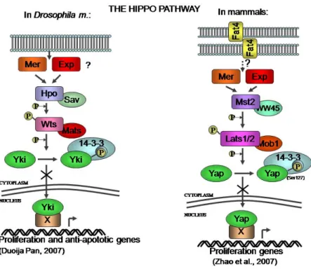
Figure 3. The Hippo pathway was first described in Drosophila m. (left panel), and more
recently it has been described in mammalian cells as well (right panel). Both mammalian Yap and fly Yki, the two orthologous proteins, were proposed to share the same mechanism of action, which sees the inactivation of Yki/Yap proteins by the Wts/Lats kinase upon stimulation by some still not fully understood signals coming from the membrane. In both cases the proposed result is the inhibition of transcription of genes that drive proliferation.
Organ size
In Drosophila, the Hippo pathway limits organ size by limiting cell proliferation and promoting apoptosis.
Pan’s laboratory showed that the activation of the Hippo pathway in Drosophila leads to cytoplasmic sequestration of Yorkie upon phosphorylation at Ser168 by Wts, presenting this even as the mechanistic basis for cell growth suppression by the Hippo pathway (Dong et al., 2007). In addition, their work established the conservation of this pathway in mammals, showing the phosphorylation of Yap on ser127 by Lats2 as a key achievement in suppressing the growth effect of Yap (Dong et al., 2007). Finally, what came out with this work, and concomitantly with another group of research, was the new vision of Yap being able to control the organ size in mammals (Camargo et al., 2007; Dong et al., 2007). Conditional overexpression of Yap in mouse liver caused the organ to reach 25% of total body weight in 4 weeks of continuous induction, almost 5 fold increase compared to control. Surprisingly, the liver of these mice returned to normal size after blocking Yap expression, implying that the increase in organ size is dependent on continuous Yap expression. The same authors investigated whether this increase in organ size is also correlated with the appearance of tumors, and found that as a consequence of a prolonged expression of Yap, 8 weeks, the mice developed numerous discrete nodules throughout their livers that displayed many characteristics of hepatocellular carcinoma (Dong et al., 2007). To support this role of Yap in HCC, they show that increase nuclear abundance of Yap1 is present in about 50 % of human HCC. This dramatic and reversible changes in organ size through manipulation of Yap protein levels alone gives to Yap and the upstream Hippo pathway a leading position in the control of organ size.
A similar approach was taken in parallel by the group of Brummelkamp, that generated transgenic mice in which Yap1 could be activated in a doxycycline-inducible manner. After 35 days of expression these mice had a liver 4 folds bigger in size, which presented dysplastic hepatocytes with irregular and enlarged nuclei, and increase proliferation rate. The increase in liver mass was completely reversible, as indicated by the fact that interruption of YAP1 expression for 5 weeks resulted in a normally sized
liver without any gross abnormalities (Camargo et al., 2007). In addition, they created transgenic mice overexpressing ubiquitously Yap1. The most dramatic phenotype observed in these mice was a severe dysplasia located along the entire intestinal epithelium. Yap1 is expressed in the crypt compartment where stem cells are located, and its overexpression leads to an expansion of stem cell population. When analyzed for alkaline phosphatase activity, a marker of differentiated enterocytes, they noticed a complete absence of staining in the small intestine 5 days after Yap1 induction. Moreover, Yap overexpression correlates with elevated levels of cyclin D and BclXL, which are found to be elevated also in many human colon carcinomas. Therefore, these results indicate for the first time that activation of YAP1 leads to a loss of differentiated cell types in the small intestine. They suggests that this is not the effect of a de-differentiation process but the result of undifferentiated cells derived from the crypt that expand and replace mature cell types following YAP1 activation. In this instance, Yap is described as a “stemness gene”, i.e. its activity is required to maintain the multipotent undifferentiated progenitor cells, and has to be lost for these cells to progress to more differentiated stages. Mesenchymal stem cells (MSCs) are pluripotent precursor cells with ability to differentiate into several distinct lineages. A recent study showed that TAZ, a paralog of Yap, functions as a transcriptional modulator of MSC differentiation by promoting osteoblast differentiation while repressing adipocyte differentiation (Hong et al., 2005). In addition, upon TGFβ stimulation TAZ binds heteromeric Smad2/3-4 and plays an essential role in Smad nuclear accumulation to maintain human embryonic stem cell (hESC) pluripotency (Varelas et al., 2008).
A recent work took under analysis other components of the Hippo pathway, Mst1 and Mst2, the central kinases. According to the phosphorylation scheme applied to the Hippo pathway, Mst1/2 should phosphorylate Lats1/2-Mob1 and this complex in turn phosphorylates Yap1. Here however they show that Mst1/2 can indeed induce phosphorylation of Yap in liver, but via some an unknown kinase different from Lats1/2. Interestingly, the role of Yap in the control of liver size and the induction of HCC, was seen not as the effect of altered expression levels, but as a consequence of loss of Mst1/2-driven phosphorylation on Ser 127. Using
anti-phospho(S127)-antibody they found a loss of p-Yap1 in 30 % of HCC specimens, and this was related to loss of Mst1/2 signaling in the same specimens. Moreover, loss of both Mst1 and Mst2 leads to Yap activation and is sufficient to initiate hepatocyte proliferation, resulting in a dramatic liver growth, resistence to apoptosis and development of hepatocellular carcinoma (Zhou et al., 2009). This work add two important concepts to the growing story of Yap. First, it shifts the control of Yap activity from a direct intervention in the hands of Lats2 to a less defined but strong involvement of Mst1/2. Second, it underlines the importance of Yap in the control of Liver size and induction of HCC, with the remarkable suggestion that PTMs of Yap, and phosphorylation on Ser-127 in particular, are more important in controlling its activity than the level of the protein itself are.
Oncogenetic function
A positive role for Yap in tumorigenesis was first proposed by a couple of works that found its gene in human chromosome 11q22 amplicon, present in many human cancers. Zender et al. began their investigation from the creation of mouse models for liver cancer. It consisted in transplanting genetically altered liver progenitor cells (hepatoblastomas) in recipient livers, and then to analyze the liver of these animals for tumor onset. Many of the hepatoblastoma cells were found to be able to form in situ liver carcinomas, and from a genome wide analysis for spontaneous alteration in gene copy number, it was found a focal amplicon of mouse chromosome 9qA1, which contain many metallo-proteinases, cIAP1, cIAP2 and Yap. The synthenic region of this locus is human chromosome 11q22. To test the activity of the genes of this locus, they suppressed the expression of cIAP1 and cIAP2 (Inibitors of Apoptosis Protein), and of Yap and found that tumor progression was slower compared to control. Interestingly, the two genes have a synergistic effect in inducing cancer formation when overexpressed (Zender et al., 2006).
The other important contribution comes from the group of Haber, which also started from the fact that amplification of 11q22 region was found in many cancer, and in particular in glioblastoma, oral squamous carcinoma,
cancer of pancreas, lung, ovary and cervix (see Overholtzer et al. 2006 for specific references). They screened the whole genome of mouse mammary carcinoma, which were engineered to have tissue-specific knock-out of Brca-1 and Trp-53 heterozygosity, by Comparative Genomic Hybridization (CGH) analysis and restricted the identity of the amplificated locus to the region containing only YAP. Next, they spent time proving that this gene is indeed responsible for the phenotype of mammary cancerous cells. They demonstrate that the human breast cancer cell line MCF10A show an aggressive phenotype when Yap is overexpressed following retroviral infection. In fact, Yap was able to induce an Epithelial Mesenchimal Transition (EMT), a program of aggressive behavior link to motility and metastatic events. Moreover, these cells showed reduce sensitivity to apoptotic stimuli and increased ability of colony formation in soft agar, another important parameter for the evaluation of oncogenic transformation (Overholtzer et al., 2006).
These new results paved the way for a series of works that soon came out, especially from those groups that were interested in the fly Hippo pathway and that began looking at the mammalian field with interest.
The oncogenic function of Yap was further supported by the description of the proteins that belongs to the Hippo pathway as tumor suppressors. Lats1 and Lats2 were elegantly described to negatively regulate the co-transcription activity of Yap by its phosphorylation and sequestration in the cytoplasm (Hao et al., 2008; Zhao et al., 2007), and recently Mst1 and Mst2 seem to have gained a key position in the control of Yap phosphorylation and activity (Zhou et al., 2009). Moreover, it was known that Lats1 know-down leads to soft-tissue sarcoma and ovarian tumor development (St John et al., 1999). Finally, as we have commented in the paragraph above, Yap overexpression or presence in the nucleus of hepatocytes leads to organ overgrowth and eventually to HCC (Camargo et al., 2007; Dong et al., 2007; Zhou et al., 2009).
Even though proofs for a confident identification of upstream regulators for the Hippo pathway are still elusive, the gene NF2 (Neuro-Fibromatosis type 2) and its product Merlin, are good candidate for the position. This gene has been shown to be mutated in many cancers, including mesothelioma, and its product was reported to interact with Yap and cause Ser-127
phosphorylation, leading to a reduction of nuclear localization. RNA interference of Yap in a mesothelioma cell line induced suppressed growth (Yokoyama et al., 2008). Another work ,on the same line of research, reports that merlin controls the cell cycle of meningioma cells, and that its loss was associated with increase expression of Yap, concluding that Merlin regulates Yap protein expression and Yap nuclear localization (Striedinger et al., 2008).
Among the large numbers of transcription factors that had come to play a part along with Yap, the TEAD family of transcription factors has recently gained some attention. To this family belong four homologous proteins sharing a conserved DNA binding TEA domain in human and mouse, that have been shown to interact with Yap extensively (Vassilev et al., 2001). More importantly, TEAD was shown to play a crucial role in Yap function. TEAD is shown to be crucial for YAP-induced overgrowth, epithelial-mesenchymal-transition (EMT), and oncogenic transformation in MCF10A cells (Zhao et al., 2008). The interaction between TEAD and Yap was shown to be compromised and suggested to be at the basis of a human genetic disease called Sveisson’s chorioretinal atrophy, caused by a heterozygous mutation of a highly conserved tyrosine in the Yap binding domain of TEAD1 (Fossdal et al., 2004).
Yap has been shown to promote the survival of neuronal progenitor cells during neural tube development in chicks (Cao et al., 2008). In line with these findings comes a work done in zebrafish, where Yap has been knocked-down by Morpholino oligonucleotides. Zebrafish Yap shares significant sequence similarities with representative proteins from Drosophila, chicken, mouse, and human: 31%, 59%, 76%, and 62% respectively. The result of knocking down Yap in zebrafish was the impairment of the development of brain and eye caused by apoptosis in the zebrafish brain (Jiang et al., 2009).
TAZ is a YAP paralog initially identified as a 14-3-3 binding protein, and share approximately 50 % sequence identity with YAP (Kanai et al., 2000). Both Yap and Taz contain a 14-3-3 binding site, a coil-coiled region, one or more WW domains, and a PDZ binding motif at the C-terminal region.
In the work of Zaidi et al. 2004, it was shown that c-Src/Yes kinases can bind and phosphorylate Yap on Tyrosine residues, affecting the regulation of the functional interaction of transcription factor with the chromatine. In particular, Yap becomes a negative regulator of Runxs2-driven gene expression by recruiting the transcription factor Runx2 to sub-nuclear site, playing an important role in osteoblast differentiation (Zaidi et al., 2004). Shortly thereafter, TAZ was also described to have a role as a transcriptional modulator in mesenchimal stem cell differentiation by activating Runx2 driven gene transcription during terminal osteoblast differentiation (Hong et al., 2005). Similar to Yap, overexpression of TAZ in human breast MCF10A cell line promotes cell transformation, EMT, and invasion and is shown to be overexpressed in approximately 20% of breast cancer samples (Chan et al., 2008).
Yap in Mitosis
Many of the proteins described to interact with Yap are known to have a role in regulating the cell cycle and in particular the events occurring during mitosis. However, until now Yap has not been reported to have a role or to be regulated in any phase of the cell cycle. With the work of my doctorate thesis comes the first data of Yap involvement in cell cycle, and in particular in mitosis. In the following paragraph we will briefly outline the main characteristic of mitosis, and the most relevant information collected to date about the proteins known to interact with Yap and that have also a role in mitosis.
Each eukaryotic cell goes through the cell cycle, a precisely programmed series of events that enable the cell to duplicate its contents and to generate two daughter cells. The cell cycle can be divided roughly in two parts, interphase - when cells duplicate their contents - and mitosis –when cells divide and distribute the genetic and biochemical material between the two daughter cells. The cell cycle regulation relies on two post-translational modifications: phosphorylation and proteolitic degradation. Protein kinases are enzymes ideally suited for this task, because they can target multiple
substrates and create small and reversible covalent modifications that serve to switch on or off the activity of their substrate. Members of the most important family of kinases known to regulate cell cycle progression are the cyclin-dependent-kinases (CDKs) to indicate that these proteins require a partner to work, a regulatory subunit named Cyclin. The complex Cdk1/CyclinB1 is active in mitosis and it is also known with the name of mitosis promoting factor (MPF). When the complex is active it phosphorylates many proteins causing the dramatic event of mitosis, from nuclear envelope break down to chromatin condensation. Mitosis begins and ends with the activity of Cdk1/cyclin-B1, whose regulation is under strict control.
Mitosis is divided in four distinct phases, each clearly distinguishable from the others under the microscope. These are named prophase, metaphase, anaphase, and telophase. During the prophase of mitosis, the chromosome, which were invisible microscopically during interphase, begin to condense and become visible under the microscope, while the centrosome at the poles of the cell begin to assemble. During metaphase, the chromosomes align along a plane that bisect the cell and become attached to the microtubule fibers of the mitotic spindle. At the same time, the nuclear membrane has disappeared. During anaphase, the two chromatids of each chromosome are pulled apart by the mitotic spindle to the opposite poles of the cell. During telophase, shortly after the chromosomes cluster together into two sets, the chromatids de-condense, and a new nuclear membrane forms around each set of chromatids, which from now on are called chromosomes again. These four subphases together constitute mitosis. During the following phase – the process of cytokinesis – the cytoplasm of the mother cell divides, yielding two daughter cells. Some people tend to consider cytokinesis as part of mitosis, but it should be kept in mind that the two events are distinct and can occur separately. For instance, in lower eukaryotes and in the first stages of fly embryo development, the chromosomes can divide without subsequent division of cytoplasm.
Like virtually all machinery, the machine that execute the various steps of mitosis is subject to malfunction. To contrast this fault, the cell deploys a series of surveillance mechanisms that monitor each step of mitosis and
cytokinesis. Proper allocation of the duplicated DNA is vital for the wellness of the daughter cells. Indeed, misdistribution of even a single chromosome can lead to a fatal imbalance of genetic material, that most of the times can be recognized by the checkpoint machinery which would invite cells to suicide. The importance of cell’ self-destruction as a consequence of improper mitosis has become clear in the study of tumor formation. In the case that cells exit mitosis without dividing –partially or entirely- their genetic contents, and escape suicide, the daughter cells will find themselves in a condition of polyploidy (in case of doubling a complete set of chromosomes) or aneuploidy (partial doubling). The latter condition is characterized by having deletion or loss of entire chromosomes, or acquisition of parts or whole chromosomes. This means that cells, in one shot, will lose or gain large sets of genes –among which are tumor suppressors and proto-oncogenes.
Proofs for the importance of aneuploidy for cancer cells come from the analysis of many karyotypes on specimens taken from different cancers in various patients. Following this analysis it is possible to see that many cancer cells display the most disparate combinations of chromosomes. It is therefore paramount for all cells to complete mitosis without errors
Regulation of mitosis
During pro-metaphase, the cell is under a “wait” signal until all chromosomes are attached to the centrosomes, a process known as the spindle check-point. When all chromosome are properly attached, the Anaphase Promoting Complex (APC) becomes activated. This is an E3 ubiquitin ligase, which began to target many cell cycle regulators for degradation, thus promoting the separation of the chromosomes and the onset of cytokinesis. Errors in the choreography of these events may lead to chromosome missegregation and aneuploidy. To prevent errors, the cell employ a
Proteins implicated in mitosis and related to yap
Aurora-A, an important protein kinase known to regulate mitosis progression, was shown to phosphorylated Lats2 on ser-83, directing its localization to centrosomes (Toji et al., 2004). Cells from Lats2-deficient mice present mitotic defects associated to centrosomes fragmentation (Yabuta et al., 2007).. The mechanism of how Lats2 can regulate centrosomes integrity is still elusive, though it was shown that Lats2 can physically interact with Ajuba, whose activity is known to regulated G2/M transition. Depletion of both Lats2 and Ajuba results in depletion of γ -tubulin at centrosomes, and this might explain at least partially the molecular mechanism of Lats2 activity at this location (Abe et al., 2006).
Lats1 and Lats2 were reported to control the cell cycle at specific check points. Overexpression of Lats2 in NIH3T3/v-ras cells by retroviral transfection induced cell cycle arrest at the G1/S transition, which seems to be achieved by down-regulation of the kinase activity of cyclin E/cdk2 (Li et al., 2003). A similar work showed that ectopic expression of Lats1 decrease the protein levels of cyclin A and cyclin E, and consequently reduce the activity of the complex with cdk1 (Xia et al., 2002). On the other hand, another report shows how overexpression of Lats1 in LATS(-/-) MEF cells suppressed the growth of human tumor in vitro and its tumorigenicity in vivo by inducing cell cycle arrest in G2/M or apoptosis (Yang et al., 2004). In conclusion, it seems likely that Lats1 and Lats2 share control over cell fate in many situations, especially during G2 and mitosis, while keeping some individuality that put them on duty separately in different conditions.
The Ras Association Domain Family 1A (RASSF1A) has been shown to interact with the Mst2 following Fas receptor activation (Matallanas et al., 2007). Another report confirms the interaction between RASSF1A and Mst2, and show also that Mst2 enhances the interaction between RASSF1A and WW45, which requires the C-terminal SARAH domain of both proteins. Components of this complex RASSF1A, Mst2, WW45, and Lats1 were found to be localized at centrosomes and midbody. Both RASSF1A and WW45 activate MST2 by promoting MST2 autophosphorylation and LATS1 phosphorylation. Loss of RASSF1A prolonged mitosis and
frequently results in cytokinesis failure or stimulate creation of binucleated cells. RASSF1A, MST2, or WW45 can rescue this defect. This work suggests that the complex made by RASSF1A, MST2, WW45, and LATS1 appears to be involved in controlling mitotic exit (Guo et al., 2007).
Centrosome duplication during S phase, the delicate event that allow the creation of the two spindle poles for subsequent chromosome separation in mitosis, was reported to be regulated by hMob1/Mst1/NDR1 signaling pathway (Hergovich et al., 2009). In this recent work, the activity of Mst1 is shown to be essential for centrosomes duplication, while the complex between Mst1 and hSav or RASSF1A seems to be dispensable.
P53 is well known to patrol the integrity of the genome and to act in DNA damage checkpoints. In addition, many reports have shown a role for p53 in preserving the genomic stability, the control of ploidy, and prevention of tetraploidization, which make p53 a key protein also to avoid that cells with a compromised chromosome assortment can continue proliferating (for references and more information see review: (Tomasini et al., 2009) ). We have encountered above a case where Lats2 can shuttle to the nucleus from the centrosomes to deliver the message of the presence of damaged mitotic spindle and dysfunction of centrosomes. Lats2 does so by binding to Mdm2, which then leaves p53 free to mount a response that ultimately stops cell from proceeding with tetraploid DNA content.
Although p53 has always played the champion character, many have wondered whether p73 has also an important part on the scene or merely a role of secondary actor. In recent years, p73 has been demonstrated to have indeed a role in genomic maintenance, has shown by the following reports. P73 was described to be a target of cyclin-B1/cdk1 complex activity, being hyperphosphorylated both in physiological mitosis and in mitotic cells arrested by microtubules targeting drugs. In this state, p73 is unable to bind the chromatin and the protein relocates to the mitotic cytoplasm (Fulco et al., 2003). The same group went on showing that this phosphorylation on Thr-86 is lost during mitotic exit and is accompanied by p73 relocalization to telophase nuclei and recovery of transactivating ability. Loss of p73 by RNA interference produced an accumulation of aberrant mitotic figures and generation of abnormal and fragmented nuclei. They show that the
cyclin-dependent kinase inhibitor p57/Kip2 gene is a specific target of p73 during mitotic exit, thus granting p73 a role in the mitotic exit (Merlo et al., 2005b).
The analysis of Trp-p73 -/- mice reveals that loss of p73 causes mis-localization of spindle assembly checkpoint components. Subsequently, a physical interaction of p73 with BubR1 and Bub1 was demonstrated, showing also that p73 can potentiate the function of BubR1. They assert that in the absence of TAp73, MEFs and human cells have a reduced ability to initiate and maintain a proper mitotic spindle checkpoint with the effect of augmented genomic instability (Tomasini et al., 2009). The correct function of protein kinase Bub1 and Bub3 is required for the accurate organization and correct localization of kinetochore proteins, a complex that supervises chromosome alignment at the metaphase plate and prevents erroneous divisions. This is the core of the molecular machinery that makes the mitotic spindle checkpoint, and Vernole and co-workers reported that overexpression the TAp73α isoform can bind to Bub1 and Bub3 thus preventing proper assembly of the mitotic checkpoint complex. In their report they show that only the longest isoform of p73 (both TA and ΔN ) can interact, a clear indication that p73 needs a protein region on its C-terminal (Vernole et al., 2009). Taking in consideration the other works that showed that downregulation of p73 can induce aneuploidy, and considering the fact that in this work the overexpression of one isoform is shown to cause chromosome mis-distribution, we can see how much important is the correct steady state level of the protein p73. A preceding report showed that p73 is important in suppression of polyploidy and aneuploidy only in the absence of p53. In this work p73 played as substitute of p53 in maintenance of normal ploidy, acting on a G2 checkpoint and causing a failure of premitotic mechanism in case of absence. In fact, they observed that in absence of p53, p73 can prevent cells carrying damaged DNA or polyploid and aneuploid cells to re-enter mitosis through the activation of a G2 checkpoint (Talos et al., 2007).
Even though many of the proteins that are known to interact with Yap have been shown to have a role in regulation and maintenance of mitosis, the function of Yap in this regards has remained obscure. Unfortunately, the creation of knock-out mice for YAP gene is not of big help in elucidating
this role. In fact, loss-of function study in mice has shown that Yap mutant mice are arrested at around E8.5 with widespread defects, therefore preventing an assessment of the in-vivo function of Yap during embryogenesis (Morin-Kensicki et al., 2006).
EXPERIMENTAL PROCEDURES
Cell culture
Human U2OS (osteosarcoma), H1299 (lung carcinoma), MCF-7 (breast carcinoma), HeLa (cervix carcinoma), HCT-116 and SW480 (colon carcinoma), cells were all cultured in Dulbecco’s modified Eagle’s medium (DMEM) containing 10% fetal bovine serum (FBS) plus of 100 units/ml penicillin and 100 µg/ml streptomycin at 37°C in a humidified atmosphere containing 5% CO2.
Plasmids and transfection
Overexpression of was achieved by transfection of the plasmids pEGFP-YAP, pEGFP, pCDNA3-flagYap1. The empty vectors pEGFP and pCDNA3 were used to keep the amount of the transfected DNA constant among samples, and in control transfections. Transient transfections were done by the calcium phosphate method in the presence of BES (Sigma), or the Lipofectamine 2000 reagent from Invitrogen, according to the manufacturer’s instructions.
RNA interference
Knock down of Yap protein levels was achieved by use of small interfering RNAs. The day before oligos transfection, cells were plated in number sufficient to cover 40-50 % of the plate the day after. Just before transfection, the DMEM-10%FBS medium is replaced with OptiMEM (Gibco) without Serum and with no antibiotics. Transfection was carried out by incorporating the oligonucleotides into a cationic lipid complex (Lipofectamine 2000 from Invitrogen) following the manufacturer’s instructions. Five hours after the transfection, the culture medium is changed
back to DMEM-10% FBS with antibiotics. Cells are harvested 24-48 hrs after the transfection.
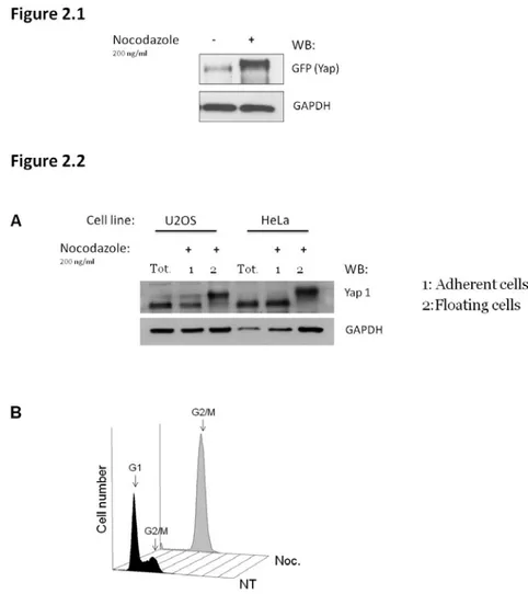
Synchronization and mitotic harvesting
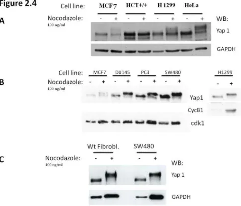
Cells are plated the day before synchronization, which is achieved by the addition of Nocodazole (100 ng/ml final concentration) to the medium. This drug interfere with the polymerization of microtubules, disrupting the functionality of the cytoskeleton and causing the cells to stop in mitosis. Mitotic cells are round and weakly attached to the plate, making them easy to be collected by shaking the plate vigorously (Mitotic shake off). Mitotic cells are collected 16 hrs after the addition of nocodazole (overnight treatment), then harvested for protein or RNA extraction, or washed twice with PBS and placed in fresh medium without Nocodazole, for different time-points needed to study the mitotic exit.
Protein extraction and Western blot analysis
For protein extraction, cells are harvested either in an NP-40 based lysis buffer, or in Urea buffer. The NP-40 buffer is mild buffer, used for a gentle lyses of cells, which is needed for the immunoprecipitation analysis. It is composed as follows: Nonidet P-40 1%, Tris-HCl pH7.5 50 mM, NaCl 150 mM, EDTA 1mM, PMSF 1mM, NaF 3 mM, NaVO4 1mM, DTT 1mM (DTT is not used in IP analysis), and protease inhibitors. Cells extracts must be kept on ice, sonicated for 20-30 seconds, and centrifuged at 13’000 rpm for 20 min to eliminate cell debris. Alternatively, the Urea buffer is a strong and denaturating buffer, able to dissolve all the components of the cell and to denature all proteins, among these are proteases and phosphatases. For this reason it is a good choice for the extraction and maintenance of phosphorylated proteins. The Urea buffer is composed as follows: 8M Urea, Chaps 2%, 5 mM DTT. Cells extracts are kept at room temperature, and sonicated for 20-40 seconds before western blot analysis. Protein concentration was determined by a colorimetric assay (Bradford reagent from BioRad). Usually, 30 micrograms of proteins were used for western
blot analysis, SDS-Protein Loading buffer 4X (Glycerol 40%, TrisHCl pH 6.8 250 mM, SDS 8%, Beta-mercaptoEt. 10%) is added to the protein extracts before loading. Protein extracted in NP-40 buffer require 10 min at 95°C to denature. Proteins are separated by gel electrophoresis and blotted onto a nitrocellulose membrane by using the mini-protean III apparatus from BioRad. Primary antibodies purchased from Santa Cruz Biotechnology, Inc., Santa Cruz (CA) are: Yap (H125), GAPDH, Cdk1. All were used at 1:1000 dilution in TBS-tween 0.5% BSA 5%.
Immunoprecipitation
Cells were harvested in NP-40 buffer with protease inhibitors and without DTT, and the extracts were sonicated for 10 sec and centrifuged at 13’000 rpm for 15 min in order to eliminate cell debris. The protein concentration was determined by a colorimetric assay (Bradford from BioRad). After pre-clearing for 1 h at 4°C, immunoprecipitations were performed by incubation 1-2 mg of whole cell exctract with 1.5 µg/sample of antibody conjugated with protein A/G-agarose beads, rocking at 4°C for 2 hrs. The immunoprecipitates were washed three times with 1 ml of NP-40 buffer (Nonidet P-40 1%, Tris-HCl pH7.5 50 mM, NaCl 150 mM, EDTA 1mM, PMSF 1mM, NaF 3mM, NaVO4 1mM, and protease inhibitors). After the last wash the excess liquid was aspirated and 60 µl of SDS-protein sample buffer 1X was added (TrisHCl pH 6.8 62.5 mM, Glycerol 10%, SDS 2%, BetaMercaptoEt 5%, traces of bromophenol blue). Immunoprecipitates as well as 30 µg of whole cell exctract were resolved by SDS-Page.
Immunofluorescence
Cells to be analysed by immunoflorescence were grown directly onto sterile coverslid glasses. HeLa cells were washed and fixed with 4 % PFA for 20 min at room temperature (RT). Cells were washed twice with PBS, permeabilized by adding PBS-triton 0.5 % for 10 min at RT; washed twice with PBS, and blocked with PBS-BSA 5% for 40-60 min at 4°C. Cells were
then incubated with primary antibody - Yap1-H125 from SantaCruz Biotechnology, Inc, Santa Cruz (CA) – used at 1:300 dilution for 1-2 hrs at 4°C. After washing cells twice with PBS for 5 min each, the secondary antibodies were added (anti-rabbit-Cy3 1:400), along with DAPI, 1:1000. After 1 hour at 4°C, cells are washed twice in PBS and mounted on microscope slides using Moviol (SIGMA). The analysis was carried out using a Zeiss confocal microscope LSM 510.
RNA extraction, reverse transcription, and PCR
Cells were harvested in TRIzol reagent (Invitrogen) and total RNA was isolated according to the manufacturer's instructions. Five micrograms of total RNA was reverse-transcribed at 37 °C for 45 min in the presence of random hexamers and Moloney murine leukemia virus reverse transcriptase (Invitrogen).