FACULTY OF MEDICINE AND DENTISTRY
DEPARTMENT OF ORAL AND MAXILLOFACIAL SCIENCES
Director Prof. Ersilia Barbato
DOCTOR OF PHILOSOPHY IN INNOVATIVE TECHNOLOGIES IN THE DISEASES OF SKELETON, SKIN AND ORO-CRANIOFACIAL DISTRICT
-ODONTOSTOMATOLOGIC DISEASES- XXX CYCLE
Director Prof. Antonella Polimeni
VAPORIZATION TECHNIQUE BY CO
2LASER AS A
TREATMENT OF THE TRUE ORAL LEUKOPLAKIA:
CLINICAL STUDY
Tutor Candidate
Prof. Umberto Romeo Dr. Mohamed Mohsen
Supervisor N. Matricola 1506557
Dr. Gaspare Palaia
“A writer is a person for whom writing is more difficult than it is for other people”
Acknowledgements
I would like to extend my gratitude to the many people who helped bring this research project to fruition.
First, I would like to thank Professor Umberto Romeo for giving me the opportunity to be a part of this PhD program. I am deeply grateful for his help, professionalism, valuable guidance and support throughout this research and through my entire program of study that I do not have enough words to express my deep and sincere appreciation.
Special appreciation goes to my supervisor, Doctor Gaspare Palaia, for his supervision and constant support. His invaluable help of constructive comments and suggestions throughout the experimental and thesis works has contributed to the success of this research. Not to forget to thank Doctor Amelia Bellisario; as her collaboration, support, and perpetual enthusiasm were of a great help for me.
Finally, I must express my profound gratitude to my parents, my wife, and my brother for providing me with unfailing support and continuous encouragement throughout my years of study and through the process of researching and writing this thesis. This accomplishment would not have been possible without them, Thank you.
Abstract
Aim
To determine the sufficient safety margins during laser vaporization of oral leukoplakia as a trial to reduce the recurrence.
Introduction
Definitive treatment of oral leukoplakia is essential because of its recurrence and potentiality to the malignant transformation.
CO2 laser vaporization is characterized by being with minimal damage to the
adjacent tissues, limited scarring, little wound contraction, and low post-operative complications.
Materials and Methods
This study was conducted on 36 true leukoplakia lesions and diagnosed in 34 patients (20 Females and 14 Males). The range of the patients age was between 39 and 79 years.
The lesions were divided into three groups; Group A: 11 lesions in 11 patients, in which the laser vaporization was done for the entire lesion adding a maximum of 1 mm of safety margins; Group B: 9 lesions in 7 patients, in which the laser vaporization was done for the lesion adding at least 3 mm of safety margins; and finally the Control Group: consists of 16 lesions in 16 patients.
During six months after the laser vaporization, four follow-up visits were performed in order to evaluate the healing course and to evaluate the recurrence rate and its degree.
Results
Among all the completely healed lesions, 75% of which were in groups A and B while 25% were in the Control Group.
In this study, it was observed that some of the vaporized lesions which showed partial or complete recurrence after 6 months of follow-up, have shown the initial recurrence after 3 weeks of laser vaporization.
The best results were obtained in patients with no history of smoking habits as the complete healing was 87.5% (7 of 8 lesions) and the complete recurrence was 12.5% (1 of 8 lesions). However, in ex-smokers, the complete healing was 41.5% (5 of 12 lesions), the partial recurrence was 41.5% (5 of 12 lesions), and complete recurrence was 17% (2 of 12 lesions).
Discussion
The primary treatment of oral leukoplakia focuses on the elimination of associated risk factors (smoking, alcohol, and local irritating factors).
In the literature, the recurrence rate varies between 13.6 and 40.7%, while in our study, after 6 months of follow-up, it was 45% in Group A and 33% in Group B.
Conclusion
The recommended optimal safety margins should be at least 3 mm in width; in addition, deep surgical margins may be related to the recurrence of oral leukoplakia.
Contents
Acknowledgements ...ii
Abstract ...iii
Contents ...iv
Statement of Original Authorship ...v
1.Chapter 1 : Introduction ...1
1.1. Definition of Oral Leukoplakia ...1
1.2. Differential Diagnosis ...2
1.3. Oral Leukoplakia and Level of Certainty ...9
1.4. Staging and Classification of Oral Leukoplakia ...13
1.5. Prognosis of Oral Leukoplakia ...15
1.6. Treatment of Oral Leukoplakia ...17
1.7. Recurrence of Oral Leukoplakia ...29
1.8. Malignant Transformation of Oral Leukoplakia ...31
1.9. Laser Tissue Interactions ...33
1.10.Types of Laser ...38
1.11. The CO2 Laser ...41
1.12.Objectives of the Study ...43
2.Chapter 2 : Materials and Methods ...44
3.Chapter 3 : Results ...49 3.1. Statistical Results ...49 3.2. Results ...50 3.3. Clinical Results ...57 4.Chapter 4 : Discussion ...60 5.Chapter 5 : Conclusions ...68 6.Chapter 6 : Bibliography ...70
Statement of Original Authorship
The work contained in this thesis has not been previously submitted to meet requirements for an award at this or any other higher education institution. To the best of my knowledge and belief, the thesis contains no material previously published or written by another person except where due reference is made.
Signature: _________________________
1.
Chapter 1 : Introduction
1.1. DEFINITION OF ORAL LEUKOPLAKIA
The term leukoplakia was defined by the World Health Organization (WHO) as “white patch or plaque that cannot be characterized clinically or histologically as any other disease”. [1, 2]
WHO in collaboration with the center for oral cancer and pre-cancer in the United Kingdom, in May 2005, replaced the term “pre-cancerous lesions” with “Potentially Malignant Disorders” (PMD) which include Oral Leukoplakia (OL) among other diseases. [3]
In 2012, a new definition was proposed which seems more opportune as it includes the histological confirmation “A predominantly white lesion or plaque of questionable behaviour having excluded, clinically and histopathologically, any other definable white disease or disorder”. This one hasn’t been assessed yet by WHO, but it has good chances for acceptance.
In 2017, Villa [4] used the term leukoplakia for describing a white lesion that is pre-cancerous while recently WHO defined it as “a white plaque of questionable risk having excluded (other) known diseases or disorders that carry no increased risk for cancer.”
It is one of several potentially malignant oral lesions, including erythroplakia and submucous fibrosis. As such, it is essential to be recognized because of its premalignant potentiality and managed accordingly and differently from other white lesions.
There is continuous confusion on the use of the term leukoplakia, especially on how to manage leukoplakia in a patient whose diagnosis shows ‘hyperkeratosis with no evidence of dysplasia’.
There is also no consensus on the guidelines for the management and treatment of dysplastic lesions, much less leukoplakias without
1.2. DIFFERENTIAL DIAGNOSIS
Oral white lesions, including leukoplakia, are commonly encountered in daily practice by oral health care providers, especially oral and maxillofacial surgeons.
They are often investigated by biopsy examination to rule out the presence of dysplastic changes or cancer. [1]
Most white lesions are benign frictional keratosis or keratosis from inflammatory conditions, (e.g. Lichen Planus (LP)) and the diagnosis is usually evident from the histopathology.
Taking a careful history, clinical and histopathological examinations of all white lesions by an oral and maxillofacial pathologist is required to achieve the final diagnosis.
It is extremely important to accurately diagnose and differentiate between reactive or inflammatory keratotic conditions and True Leukoplakia (TL).
Because of its pre-cancerous nature, the management of TL should be performed through a close follow-up or complete removal. [4]
Not all white keratotic lesions on the oral mucosa are OL, as noted in the WHO definition. The oral mucosa becomes white for the following reasons: [4]
• Excess production of keratin as a response to injury (e.g. friction or biting).
• Excess production of keratin intrinsically from benign keratotic diseases (e.g. genodermatoses) or dysplasia.
• Thickening of the epithelium (acanthosis).
• Damage of epithelial cells from direct and/or identifiable contact injury.
These changes can occur because of a genetic dyskeratotic disease (Cannon white sponge nevus, a very rare condition), immune-mediated disease (LP), bite, trauma, or oncogenic mutations (e.g. Leukoplakia with dysplasia). (Table 1)
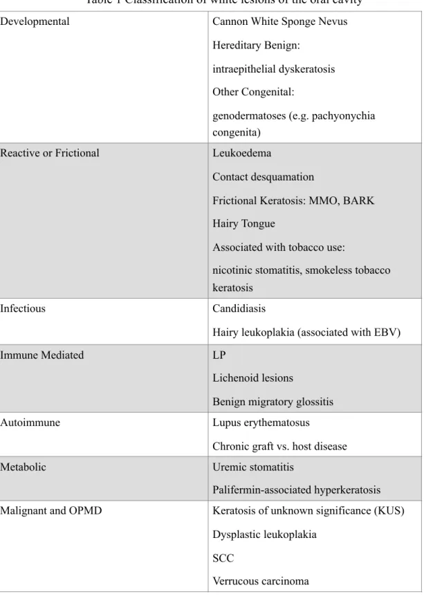
Table 1 Classification of white lesions of the oral cavity
Developmental Cannon White Sponge Nevus Hereditary Benign:
intraepithelial dyskeratosis Other Congenital:
genodermatoses (e.g. pachyonychia congenita)
Reactive or Frictional Leukoedema
Contact desquamation
Frictional Keratosis: MMO, BARK Hairy Tongue
Associated with tobacco use:
nicotinic stomatitis, smokeless tobacco keratosis
Infectious Candidiasis
Hairy leukoplakia (associated with EBV)
Immune Mediated LP
Lichenoid lesions
Benign migratory glossitis
Autoimmune Lupus erythematosus
Chronic graft vs. host disease
Metabolic Uremic stomatitis
Palifermin-associated hyperkeratosis Malignant and OPMD Keratosis of unknown significance (KUS)
Dysplastic leukoplakia SCC
I. Developmental
These lesions are extremely uncommon and all have specific and distinctive histopathologic features.
- White Sponge Nevus:
Present as diffuse bilateral white plaques of the oral mucosa; could involve the buccal mucosa in particular, tongue; esophageal and genital mucosa but not the skin. (Fig. 1)
- Hereditary Benign Intraepithelial Dyskeratosis:
Present as bilateral thick white plaques of the oral mucosa and as gelatinous plaques of the conjunctiva without involvement of the skin.
- Pachyonychia Congenita:
Oral plaque, but always accompanied by the presence of thickened skin lesions. Congenital dyskeratosis causes leukoplakias and oral cancer at a young age. [4]
II. Reactive or frictional - Leukoedema:
Occurs in up to 90% of the population and can occur after exposure to mildly irritating substances (e.g. mouthwash, toothpaste, or tobacco and marijuana smoke).
It is presented as delicate gray-white lacy lines on the buccal mucosa or ventral tongue that disappear with stretching of the mucosa and it shows histopathologically edema of epithelial cells.
These are rarely submitted for biopsy examination because they are readily recognized and no treatment is necessary except for stopping the habit.
Figure 1 White sponge nevus
- Morsicatio Mucosae Oris (MMO): Is usually self-induced and manifests as white plaques and papules with poorly demarcated ‘fading’ margins. (Fig. 2)
Affected sites are those that are easily traumatized by teeth, such as the lower lip
mucosa, lateral or ventral surface of the tongue, and buccal mucosa. Patients are usually unaware of this parafunctional habit, especially if the habit is nocturnal.
- Benign Alveolar Ridge Keratosis (BARK):
Is due to constant trauma to the edentulous alveolar ridge; it is commonly seen on the retromolar pad and underneath ill-fitting dentures.
This is a common traumatic frictional keratosis that constitutes approximately 75% of all biopsy results of white lesions and has distinct and readily recognized histopathological features. (Fig. 3)
The diagnosis on the pathology report should be ‘benign frictional keratosis’. [4]
- Hairy Tongue:
It is a benign retention keratosis caused by decreased exfoliation of keratin and the development of elongated filiform papillae ‘hairs’. (Fig. 4)
The dorsum of the tongue is presented as a white, coated, or a hairy appearance. It can become pigmented from intrinsic bacteria or food;
Figure 4 Hairy Tongue Figure 3 Benign Alveolar
Ridge Keratosis Figure 2 Morsicatio
Patients might also complain of sticky and mucinous saliva with an associated pasty, metallic taste and gagging (when the coating is localized in the posterior third of the tongue).
The most common cause of this condition is dehydration and hyposalivation. This is seen in patients who have had recent illness (often associated with antibiotic therapy), use of alcohol-containing rinses, or smoking.
Conditions that cause dry mouth, such as polypharmacy, chronic anxiety, radiotherapy, and Sjögren syndrome, may also cause the hairy tongue.
This condition is also seen in patients with poor diet for the consumption of mainly soft foods (common in hospitalized patients). [4]
III.Infectious - Oral Candidiasis:
Is the most common opportunistic fungal infection. It is usually caused by Candida-albicans, a commensal present in 20 to 30% of patients.
Oral lesions occur when the normal flora is altered (as in patients with hyposalivation, wear
dentures, smokers or are on immunosuppressive agents).
Additional contributing factors include anaemia, endocrine dysfunction, immunosuppression (e.g. acquired immunodeficiency syndrome, human immunodeficiency virus (HIV)), prolonged antibiotic intake, diabetes mellitus, infancy, or advanced age.
Candidiasis also can develop overlying dysplastic lesions. Pseudomembranous candidiasis, the most common form, is characterized by thick white plaques and papules that can be rubbed off, which often leave a raw and bleeding surface. (Fig. 5)
Figure 5 Acute pseudomembranous
Other forms include erythematous and hyperplastic candidiasis. The latter is a rare chronic variant that manifests as white plaques that cannot be rubbed off, mimicking leukoplakia.
Treatment consists of topical and systemic anti-fungal agents. The most commonly used topical medications
include: nystatin suspension (100,000 U/mL) swished in the mouth 4 to 5 times a day, clotrimazole troches (10 mg) dissolved in the mouth 4 to 5 times a day for 7 to 10 days, or systemic therapy with fluconazole 100 to 200 mg/day for 7 days. (Fig. 6)
- Epstein-Barr virus:
Is mostly seen in immunocompromised patients, in particular those with HIV and a low CD4+ T-cell count and those after undergoing organ transplantation, although it can also be seen in healthy older individuals likely from immune senescence. In this condition, the term leukoplakia is not related to malignancy or to dysplastic changes.
IV. Immune-Mediated Keratotic Lesions [4] - Oral Lichen Planus (OLP):
Is an immuno-mediated chronic condition present in 1 to 2% of the population, usually middle-aged women. 10 to 15% of patients with OLP have cutaneous lesions.
OLP can be idiopathic or may be associated to local or systemic conditions and in particular to medication intake, such as antihypertensive and hypoglycemic drugs.
Oral lesions are typically symmetric and bilateral and there is a controversy regarding the clinical types.
Figure 6 Chronic hyperplastic candidosis,
Although 6 distinct forms, reticular, atrophic, erosive, papular, plaque, and bullous, have been described, this is not universally accepted. (Fig. 7- 8)
The 3 most recognizable forms are reticular, erosive, and ulcerative. Bullous (rarely seen), atrophic, and erosive forms are likely better considered clinical entity because they present a spectrum of disease and the most common appearance is an erythematous or erosive lesion because of ruptured bullae or thin, atrophic red appearing mucosa.
The reticular form is characterized by classic white (keratotic) reticular lesions. The plaque type of LP may not be readily distinguishable from a leukoplakia. Lupus erythematous and chronic graft-versus-host disease are 2 conditions that can clinically resemble OLP.
Biopsy and histopathological examinations are usually indicated. Treatment includes topical corticosteroids (e.g. 0.05% fluocinonide or clobetasol gel) or 0.1% tacrolimus ointment, and for refractory cases, treatment may include systemic corticosteroids.
Figure 8 Oral lichen planus, Plaque form Figure 7 Oral lichen
1.3. ORAL LEUKOPLAKIA AND LEVEL OF CERTAINTY
- Epidemiology
The estimated reported prevalence of OL, worldwide, is approximately 2%. However, when viewed in relation to an annual malignant transformation rate of 1%, this prevalence figure would result in development of oral cancer in 20 per 100,000 population per year.
Obviously, this cancer incidence figure, based on the malignant transformation of OL alone, is much too high. Probably, the prevalence of OL has to be set at a more realistic figure of less than 0.5%. There are some geographical differences with regard to the gender distribution.
Leukoplakia is six times more common among smokers than non-smokers. Alcohol is an independent risk factor, regardless of the beverage type or drinking pattern. There are conflicting results of studies related to the possible role of human papilloma virus infection. [5]
- Clinical Aspects
Leukoplakia may affect any site of the oral and oropharyngeal cavity. Clinically, leukoplakias are divided into homogenous and non-homogeneous lesions.
The homogeneous type is usually a thin, flat, and uniform white plaque with at least 1 area that is well demarcated with or without fissuring. (Fig. 9)
The non-homogeneous type has been defined as a mixed white and red lesion, that may either be irregularly flat (speckled) or nodular ‘erythroleukoplakia’. (Fig. 10)
Erythroleukoplakia can be misdiagnosed as OLP because of its white and red components.
Figure 9 Homogeneous Leukoplakia
Figure 10 Non-homogeneous, nodular
clinician to the correct diagnosis. Erythroleukoplakia does not possess the typical white reticular changes and is usually unilateral and associated with well-demarcated white plaques.
Verrucous leukoplakia is yet another type of non-homogeneous leukoplakia. Although verrucous leukoplakia usually has a uniform white appearance, its verrucous texture is the distinguishing feature from homogeneous (flat) leukoplakia. Verrucous leukoplakia is clinically indistinguishable from the clinical aspect of verrucous carcinoma.
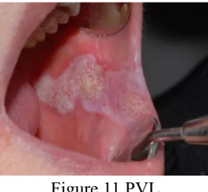
Proliferative Verrucous Leukoplakia ( P V L ) i s a s u b - t y p e o f v e r r u c o u s leukoplakia, characterized by multifocal presentation, resistance to treatment, and a high rate of malignant transformation. (Fig. 11)
PVL seems more prevalent among elderly female. There may or may not be a history of tobacco use. [1]
Areas of firmness or induration should always be submitted for periodic biopsy examination.
PVL is usually multifocal or affects contiguous areas and is characterized by relentless progression and spread with the gingiva, being the most frequently affected site. [5, 6]
Most cases of PVL are non-homogeneous with a verrucous, nodular, or erythroleukoplakia-like appearance. Similar to erythroleukoplakia, the erythroleukoplakia form of PVL can be misdiagnosed as LP because it is multifocal and bilateral.
Multiple biopsy examinations show no evidence of cytologic dysplasia but often exhibit verrucous hyperplasia, hyperkeratosis, or parakeratosis with epithelial atrophy. There are many differences between the localized lesions of OL.
- Level of Certainty
Leukoplakia is mostly used as a clinical term. Provisional diagnosis is made when a predominantly white area at clinical examination cannot be clearly diagnosed as any other disease in the oral mucosa, its definition is usually modified after the histopathological evaluation.
“For example, a clinical impression of leukoplakia at biopsy examination might show candidiasis, bite keratosis, or LP; being aware of the diagnostic criteria of the other lesions can help in recognizing the type of the lesion”. [5] (Table 2)
Table 2 The most common white or predominantly white benign diseases of the oral mucosa and their main diagnostic criteria
Lesion Main diagnostic criteria
Aspirin burn History of local application of aspirin tablets Candidiasis,
pseudomembranous
Clinical aspect (pseudomembranes, often symmetrical pattern)
Frictional lesion Presence of mechanical irritation (e.g. habit of vigorous tooth brushing)
Hairy leukoplakia Clinical aspect (bilateral localization on the tongue); histopathology (EBV)
Leukoedema Clinical aspect (symmetrical pattern)
Linea alba Clinical aspect (location on the line of occlusion in the cheek mucosa)
Lupus erythematosus History of skin lesions; clinical appearance (bilateral pattern); histopathology
Morsicatio (habitual chewing or biting of the cheek, tongue, lips)
History of habitual chewing or biting; clinical aspects
Papilloma and allied lesions
Clinical aspect; histopathology Syphilis, secondary
‘mucous patches’
Clinical aspect; demonstration of T. pallidum; serology Smoker’s palate (nicotinic
stomatitis)
Clinical aspect; history of smoking Snuff induced lesion Clinical aspect; site where snuff is placed
The term leukoplakia can be used at different levels of certainty (C-factor). According to Van der Waal (2015), four steps of certainty may be useful in the diagnosis of OL: C1 and C2 which are clinical terms and C3 and C4 which are clinicopathological terms. [5] (Table 3)
The diagnosis of OL is thus made after the exclusion of other disorders and a biopsy is recommended when other disorders cannot be identified. The disorder is diagnosed as an OL with or without epithelial dysplasia.
It has been recommended to make a distinction between a provisional clinical diagnosis of OL and a definitive one.
Apparently, the recommendation to use a certainty factor has not been widely accepted in the recent literature, although it is a common practice to use such factor in cancer registries. [5]
Table 3 Certainty (C) factor for diagnosis of oral leukoplakia [5]
C 1 Evidence from a single visit, involving inspection and palpation, including a
clinical picture of the lesion, ‘provisional clinical diagnosis’.
C2 No evidence of resolution following elimination of etiological factors (eg.
mechanical irritation) over 2- 4 weeks of follow-up or in the absence of any other etiological factors. ‘definitive clinical diagnosis’.
C3 As C2, but complemented by pretreatment incisional biopsy in which, histopathologically, no definable lesion is observed, ‘provisional histopathological diagnosis’.
C4 Evidence from histopathological examination of completely excised lesions, ‘definitive histopathological diagnosis’.
1.4. STAGING AND CLASSIFICATION OF ORAL LEUKOPLAKIA
For the purpose of a standardized reporting system of OL management, staging and classification is highly recommended.
The size, age, gender, causative factors, histopathology, and localization of OL should be taken in consideration at the time of diagnosis.
According to the treatment plan, if the management of OL will be performed only through clinical follow-up (observation), the size, colour, and/or texture of the individual lesion should be taken in consideration.
The clinical sub-types of OL may not be useful, because the clinical appearance of OL may vary and does not always allow a clear classifications.
Considering assessment after surgical intervention, biopsy reveals that the staging system is an important consideration; and it is mainly dependent on size and pathology.
The system takes into account the highest pathological score in case of multiple biopsies of single or multiple lesions of OL and the lowest size if there is any uncertainty in considering size category.
For classification and staging purposes it is recommended to record the size of a single OL, and to add together the sizes of multiple OL.
The oral subsite should be specified according to the International Classification of Diseases Application to Dentistry and Stomatology (ICD-DA) codes for the oral cavity
In this system, three size categories have been proposed, analogous to the TNM system of oral cancer:
I. L represents the size of a single or multiple OL as follows: L1 < 2cm
L2 = 2-4cm L3 > 4cm
Lx size not specified.
II. P represents the pathology of OL as follows: P0 No epithelial dysplasia
P1 Mild or moderate dysplasia P2 Severe dysplasia
Px Absence or presence of dysplasia not specified
III.Accordingly, four stages have been proposed in this system: Stage I L1 P0
Stage II L2 P0
Stage III L3 P0 or L1 L2 P1 Stage IV L3 P1 or any L P2
1.5. PROGNOSIS OF ORAL LEUKOPLAKIA
Various oral mucosal lesions, have a potential for malignant transformation; the most common white lesions have the lowest risk of malignant transformation. Practitioners will see many oral white lesions, but a few carcinomas.
However, they must be able to recognize lesions at particular risk; several features help to assess the likelihood of malignant transformation.
The accuracy of such prediction is low, but the process of identifying the 'at risk' lesions is fundamental for diagnosis and treatment planning. Important factors are listed and information on each of these should be sought.
The best predictor of the potential for malignant transformation is the degree of dysplasia seen histologically. For this reason, few lesions will already be malignant, biopsy of white patches is mandatory.
The term dysplasia (literally, abnormal growth) is given to cytological abnormalities seen in both malignant and premalignant cells.
Premalignancy is distinguished from malignancy only by the invasiveness and release of metastases.
It must be emphasized that the implications of epithelial dysplasia are quite different from fibrous dysplasia, in which there is no risk of carcinomatous change.
Deep cell keratinization (dyskeratosis) refers to individual cells which start to keratinize before the surface is reached and show eosinophilic change deeply within the epithelium.
With loss of intercellular adherence, the cells become separated. A lymphoplasmacytic infiltrate of highly variable intensity is usually present in corium.
Dysplasia is usually graded as mild, moderate or severe as a guide for patient management. (Fig. 12- 17)
‘Carcinoma in-situ’ is a term sometimes used for the most severe dysplasia where the abnormalities extend throughout the thickness of the epithelium, a state sometimes graphically called ‘top-to-bottom change’.
In such a lesion, all the cellular abnormalities characteristic of malignancy may be present, only invasion is absent.
The histological assessment of oral epithelial dysplasia is notoriously unreliable because it is subjective and changes are not reliably correlated with behaviour. [1]
10X 20X
Figure 12- 13 Mild dysplasia (10X- 20X)
10X 20X
Figure 14- 15 Moderate dysplasia (10X- 20X)
10X 20X
1.6. TREATMENT OF ORAL LEUKOPLAKIA
Clinicians treated OL with vitamin A, vitamin E and beta-carotene. However, the toxicity of vitamin A and the unsatisfactory response to vitamin E and beta-carotene caused them to discontinue to use such drugs. [9, 10]
Other treatment modalities for OL are scalpel excision or electrocautery and cryosurgery, for which there is a recurrence rate of approximately 33%. [9, 11] Studies on the clinical usefulness of laser surgery in treatment of OL have shown that the laser surgery prevents not only the recurrence and malignant transformation, but also post-operative dysfunction. [9, 12]
I. Non-surgical treatment
In order to carry out a treatment for OL, the degree of epithelial dysplasia should be assessed. However, OL presenting low to moderate malignant transformation risk may or may not be completely removed and the decision should consider other factors, such as location, size in the case of smokers, and smoking cessation. [13, 15, 16]
In the presence of moderate or severe epithelial dysplasia, surgical treatment is recommended. [15] Surgical treatment of OL may be performed either through conventional surgery [13, 14, 17], electrocauterization, laser vaporization, [12, 18] or cryosurgery. [19, 20] The recurrence rate of OL after surgical treatment has been reported to vary between 10 and 35%. [21, 22]
Non-surgical treatment may also be considered for the management of OL. This modality offers minimal adverse effects to patients, especially for patients with widespread OL that involves a large area of the oral mucosa or patients with medical problems and consequently high surgical risks. [19, 20, 23, 24]
Additionally, potential advantages of the non-surgical treatment of OL include an easy application that does not require treatment at a medical center and a relatively low cost. [25]
- Carotenoids
Beta-Carotene. The carotenoids are a group of extremely
hydrophobic molecules with little or no solubility in water. [26] Beta-carotene is a carotenoid commonly found in dark green, orange or yellowish vegetables, such as spinach, carrots, sweet potato, mango, and oranges. [34]
Beta-carotene is a vitamin A precursor. The only known effect of excessive beta-carotene intake is a state in which the skin becomes strongly yellowish, the so-called ‘carotenodermy’, which disappears in a few weeks after the reduction of consumption. [26, 27, 28]
In other studies, the supplement diet based on beta-carotene caused headaches and muscle pain in some of the patients. Patients with OL can be treated with beta-carotene in oral doses of 90 mg/day, for three cycles of 3 months. The use of beta-carotene has been recommended in order to prevent OL and possibly oral cancer. [29, 40]
The potential benefits and protective effects against cancer are possibly related to its antioxidizing action. [30, 31] This function is accomplished through a ligation between beta-carotene and oxygen, which is an unstable reactive molecule; thus, diminishing the damaging effects of free radicals. [31, 32]
A diet supplemented with beta-carotene can prevent changes in the oral mucosa, especially in smoking patients, who present low serum levels of vitamin C and beta-carotene when compared to non-smokers.
It has also been shown that beta-carotene has a better therapeutic clinical response in the prevention of OL lesions in smoking patients than in the non-smoking ones. [33]
Lycopene. is a carotenoid without provitamin A action. This is a
fat-soluble red pigment found in some fruits and vegetables; the greatest known source of licopene is tomatoes. [35] There is a positive relationship between lycopene consumption and a reduction in the risk of the development of degenerative diseases caused by free radicals, such as cancer and cardiovascular diseases. [36, 37]
The supplementation of lycopene (8 mg/day and 4 mg/day) in a three month time period reduces hyperkeratosis. [40] No systemic significant toxic effect of lycopene has been observed and there is no evidence of side effects from the treatment with lycopene. [36]
- Vitamins
L-Ascorbic Acid (L-AA) (Vitamin C): so-called vitamin C, is found
in citrus fruits, such as kiwi, strawberries, papaya, and mango. [41] It is suggested that a daily intake of at least 140 mg/day is required for smokers because they usually present a reduction of the L-AA concentration in serum leukocytes. [42]
L-AA has antioxidizing properties and reacts with superoxide produced as a result of the cells normal metabolic processes. This inactivation of superoxide inhibits the formation of nitrosamines during protein digestion and helps avoid damage to DNA and cellular proteins. [43]
L-AA toxicity does not occur, since vitamin is water-soluble, and a decrease in absorption efficiency occurs when the consumption exceeds 180 mg/day. [44] There are no studies regarding the efficacy of the use of
L-AA alone for OL treatment.
α-Tocoferol (AT) (Vitamin E): is the most common and most active
form of vitamin E. Tocoferol is an effective antioxidant at high levels of oxygen, protecting cellular membranes from lipidic peroxidation. [45, 46, 47, 48, 49]
Retinoic Acid 13-cis-retinoic acid (13- cRA): the current definition
of retinoid includes all the natural and synthetic compounds with an activity similar to that of Vitamin A.
Retinoic acid is obtained from carotene and animal products, such as meat, milk, and eggs, which are converted in the intestine respectively into retinal and retinol. [33, 48]
The use of systemic retinoids is not indicated in cases of: pregnancy or probability of pregnancy, non-compliance with the use of contraceptives, breastfeeding, and hypersensitivity to parabeno.
It is relatively contraindicated in cases of: leukopenia, hypothyroidism (patients using bexarotene), high levels of cholesterol and triglyceride, hepatic malfunction, and renal malfunction. [48, 50]
This treatment is not widely accepted due to its side effects: hypervitaminosis, toxicity, teratogenic effects, and alterations in various organic systems. [51]
13-CRA is the retinoid recommended for OL treatment. However, the high recurrence rate after short periods of discontinuance together with its side effects are limiting factors. [30, 51, 52, 53] OL recurred upon the discontinuance of medication; some patients ceased treatment due to its side-effects.
Fenretinide (4-HPR) or N-(4-hydroxyphenyl) Retinamide is a
vitamin A analogue that was synthesized in the United States during the late 1960s.
This retinoid shows a preferential accumulation in breast instead of liver. [54] Systemic use of 4-HPR with 200 mg/day for 3 months demonstrated partial clinical resolution of OL.
- Bleomycin
Bleomycin is a cytotoxic antibiotic used for the treatment of
squamous cell carcinoma (SCC) of the head and neck region, esophagus, and skin. [55]
After 12 to 15 applications, the white patch peeled off and the resultant raw surface was epithelialized over the following 14 days. Repeated biopsies showed a significant reduction of dysplasia and keratinisation. [33]
Topical bleomycin in treatment of OL was used in dosages of 0.5% /day for 12 to 15 days or 1% /day for 14 days.
- Photodynamic Therapy
Photodynamic therapy (PDT) is a non-invasive method for the treatment of premalignant lesions of head and neck cancers. [56, 57, 144]
The principle of PDT is a non-thermal photochemical reaction, which requires the simultaneous presence of photosensitizing drug (photosensitizer), oxygen, and visible light.
After a period of allowing the photosensitizer to be collect in the target tissue, the photosensitizer is activated by the exposure to low-power visible light of a drug-specific wavelength.
Mainly, the light source consists of portable diode laser and the light is transmitted via laser fibers to or into the tumor. Illumination of the tumor by light at the activating wavelength results in the destruction of cells by a non-free radical oxidative process.
These reactive oxygen species may damage crucial cell components, such as structural proteins, enzymes, DNA, and phospholipids. PDT is a cold photochemical reaction and the photosensitizing agents are of inherently low systemic toxicity.
PDT damage heals mainly by regeneration rather than scarring. Due to the organ preserving principle of PDT, important structures are maintained by good functional and cosmetic outcome. [57, 58] (Fig. 18- 23)
Several photosensitizers have been developed during the past years, haematoporphyrin and haematoporphyrin derivatives were the first photosensitizer.
Four photosensitizers have been approved so far:
• Photofrin has been approved in many countries for the treatment of esophagus and lung cancers.
• 5-Aminolaevulinic acid (5-ALA) is also approved in several countries for the treatment of skin cancer.
• Verteporfin for the treatment of macular degeneration.
• Foscan is the only photosensitizer that has been approved for the treatment of advanced SCC of the head and neck in Europe in the year 2001. [59]
The 5-ALA is a naturally occurring compound in the haem biosynthetic pathway, which is metabolized to photosensitive product ‘protoporphyrin IX’.
The major advantage of 5-ALA when compared to synthetic photosensitisers is the rapid metabolism, which significantly reduce the period of cutaneous photosensitivity; mostly indicated in head and neck surgery.
The photosensitizer is administered systemically by intravenous injection. [58, 145] Only for very superficial skin lesions or premalignant lesions of the oral mucosa, the 5-ALA can be applied topically. For all other indications, an intravenous application is mandatory. [59]
II. Surgical Treatment
Although removal of a lesion still seems to be the predominant method of treatment by the majority of relevant health care professionals, no randomised controlled trials have been undertaken to test the hypothesis that excision either by scalpel or laser greatly influences the potential for later malignant transformation.
Surgical treatment have a beneficial effect. Theoretically, this effect may be reduced by the elimination of risk factors, such as tobacco usage and alcohol consumption. However, further studies are needed to prove such theory. [60]
The first aim in the management of OL is to avoid malignant transformation; and as a consequence, it might be appropriate to consider the outcomes of therapy of OL in terms of later expected Oral Squamous Cell Carcinoma (OSCC) incidences. [61]
Elimination or reduction of the size of the lesion (extent) cannot be
Figure 19 Application of 5-ALA Figure 20 Laser application (635 nm) Figure 18 PVL, first session Figure 21 PDT, second session Figure 22 PDT, third session Figure 23 PDT, fourth session
a common event (can be as high as 30% in the same or another oral mucosal site) and it cannot be excluded as the recurring lesions represent a high-risk group.
In addition, histopathological change (i.e. lessened or resolution of dysplastic features) cannot be considered a robust and useful outcome. [62] Although surgery is the first choice in the management of OL by most relevant specialists [63, 64], the hypothesis that removing potentially malignant oral lesions by different surgical techniques (scalpel, laser, and cryosurgery) can prevent the onset of oral cancer remains unproven.
Till now, there are a few reported randomized controlled trials evaluating surgical treatment of OL that underwent surgical (e.g. scalpel excision) or laser treatment.
Results are hardly comparable because of the differences in diagnostic and inclusion criteria, follow-up time intervals, patient characteristics, and surgical techniques employed.
The selection of patients may have been determined by the site, size, nature, or histopathology of the lesion as well as the medical history and desires of the patients.
There is no evidence that the incidence of oral carcinoma can be diminished by surgical removal of OL. This does not mean that surgical removal should be abandoned mainly for histologic diagnosis. [69]
In fact, an important issue in discussing the role of surgical removal in the management of OL is the potential relevance of excisional biopsy as a diagnostic tool. Even if excisional biopsy of OL is not effective as an intervention of primary prevention (i.e. to prevent malignant transformation), it may have a role as an intervention of secondary prevention. [65, 66, 67]
- Scalpel surgical excision
The surgical removal of OL may not reduce the risk of malignant transformation. [68, 69, 70]
Nevertheless, surgical excision does allow the opportunity for examination to ensure that all areas of dysplasia have been identified and excised.
- Cryotherapy
It is well received by patients due to a relative lack of discomfort; cryotherapy is the deliberate destruction of tissue by application of extreme cold, with minimal scarring and an absence of bleeding. [78]
Clinical advantages include the ease of application, preservation of inorganic structure of bone, and a very low incidence of infection. It can be repeated without permanent side effects. [79, 80]
Perhaps its greatest advantage is its usefulness in candidates for whom surgery is contraindicated due to either age or medical history.
Disadvantages of cryotherapy include an unpredictable degree of swelling and lack of precision with the depth and area of freezing. It is also highly dependent on the operator’s skills and experience.
The value of histopathological examination of frozen sections to ensure adequate clearance of the disease at surgical margins as with OSCC may not be helpful as it will not detect the ploidy areas (if relevant) and surgeons may have to be more aggressive, than their usual practice, in the excision of the disease. [71, 72]
The basic technique of cryotherapy stresses rapid cooling, slow thawing, and repetition of the freezing process to maximize tissue destruction. [80] The two recognized methods are a closed system with the use of probes and nitrous oxide or an open system with the use of a
The nitrous oxide technique is useful for the treatment of various benign and malignant lesions of the oral cavity where the depth of necrosis is necessary.
Liquid nitrogen sprays and cotton swabs are more accessible to clinicians but are not suitable for use in the oral cavity. Their disadvantage is a lack of control over the temperature achieved within cells and the area of freezing, which makes them hazardous for the intraoral use.
During the freeze cycle as the temperature drops, it is believed that extracellular water undergoes crystallization. In addition, membrane lipids harden at low temperatures decreasing the cell resistance to shrinkage. As extracellular stores of water diminish, the electrolyte concentration increases.
In order to counteract this concentration gradient, intracellular water moves out of the cell and becomes involved in the crystallization process. Intracellular electrolytes reach toxic levels, which become lethal to the cell. Re-epithelialization occurs within 7- 12 days in the mouth and 10- 20 days on the skin. [81]
Cryotherapy is not considered to be the first line of treatment for OL, the reported recurrence rate varies from 20 to 71.4% and the malignant transformation varies from 7 to 25%; therefore, cryotherapy does not seem to be of a particular benefit. [12, 73]
There is a lack of widespread clinical availability because of the risk of post-operative scarring, tissue contraction, and importantly the resultant inability to observe signs of clinical recurrence. [73, 75, 76]
At the present time, no physical method of local excision of OL guarantees long-term resolution of relevant OL or the possibility of OSCC development. [77]
- Laser treatment
Laser beam differs from white light in its coherence. A high density of power is produced when the laser beam is focused by a lens because it has properties that neither diverge nor interfere. [84]
Several kinds of laser beams have been developed since the ruby laser was clinically used for skin lesions for the first time. The absorbed energy causes vaporization of intra and extracellular fluid with a minimal destruction of cell membrane and its surrounding tissue. [87, 88]
Laser surgery has a haemostatic effect, [82, 83] which makes it easier to maintain the operative field; it has the ability to remove lesions accurately. [84, 86]
The damage to the adjacent tissue is minimal, which reduces acute inflammatory reactions and post-operative pain. Also, wound healing after laser surgery is excellent because of the limited contraction. [83, 84, 85]
There have been reports over the past 30 years that among the different surgical techniques proposed for the treatment of OL, laser surgery has received the greatest attention.
Unfortunately, as with other surgical techniques, most studies have major methodological flaws and are very low in the hierarchy of evidence.
CO2 and KTP lasers have been employed with various vaporization
or excision techniques for the treatment of OL.
The main advantages of laser therapy are the potential haemostatic effects, limited tissue contraction, and post-therapy scarring; which may permit the treatment of lesions of large dimensions.
In addition, laser therapy may include reduced post-operative pain, swelling, and infection.
Wounds may take longer to re-epithelialize, small granulomas can complicate healing, and histopathological confirmation of the nature of the excised lesion will not be possible if ablation techniques have been employed. [89]
The excision technique can be performed by either the CO2 laser or
KTP laser in patients with a localized OL occurring in the field on the non-keratinized epithelium.
In the excision technique, both the CO2 and KTP lasers were used in
a non-contact application. As a role, the excised edges of the wound made by the excised technique shall be unsutured.
The vaporization technique can be performed using either CO2 laser
or Nd:YAG laser in patients who had comprehensive OL, or OL on the gingiva and hard palate. In this technique, CO2 laser should be used in a
non-contact mode using an average power output between 4 and 10 Watts, while Nd: YAG laser should be used in a contact mode using an average power output between 3 and 12 Watts. [18]
1.7. RECURRENCE OF ORAL LEUKOPLAKIA
It has been suggested that a recurrence of OL should be defined when a lesion subsequently occurs at the primary lesion site after it was confirmed that the condition had been reached with no evidence of a lesion for a definite period.
Actually, a thin white plaque can remain in the surrounding tissue of the primary lesion and subsequently increase its size and thickness.
Frame et al. [85] reported that the new epithelium that migrates from the periphery to cover the wound may originate from an area of potentially unstable mucosa; that may explain why new patches of OL develop adjacent to the margins of previously treated areas.
The recurrence rate differed significantly among the types of treatment procedures. [90] It was reported, from previous studies, that the recurrence after laser surgery was 7.7- 38.1%. [85, 86, 90, 91] Variation of recurrence is attributed to the difference in the variety, conditions of the laser beams, race, and the follow-up period.
Chiesa et al. [92] reported that the probability of developing recurrence and new occurrences was 23% within the first year of surgery and 40% after 3 years of follow-up.
On the basis of these findings, it is important to ascertain the border of the lesion. It is strongly recommended that vital tissue staining, such as iodine [94, 95] or toluidine blue staining immediately preoperatively, is necessary to reduce local recurrence and malignant transformation, because multi-centric or micro-invasive cancer may develop from OL. [18, 91]
It was also reported that all dysplastic and malignant oral mucosal lesions were detected as unstained by the iodine staining method [95] and that only 1.9% false negatives were defined by toluidine blue staining. [96]
It was found that the recurrence of OL is irrespective of complete excision by laser or conventional surgery.
This suggests that the origin of the cells in recurrence of OL as potentially premalignant lesions is located in adjacent epithelium of the OL visualized as normal oral mucosa before treatment. Despite complete removal or laser vaporization, the adjacent or peripheral epithelium may proliferate in the recurrence phenomenon and it is proposed that these epithelial tissues, that show clinically normal features, consist of highly active cells which are probably abundantly widespread in the basal cell layer.
It has been accepted that the ‘field of cancerization’ (FC) or ‘field change’ of oral mucosal cancer is very important in explaining the presence of dysplastic cells adjacent to SCC and recurrence following complete laser vaporization. [97]
It is suggested to include a wide FC for the lateral side of the tongue since it is characterized by a marked proportion of dysplastic changes and also a high recurrence rate.
Potentially, premalignant cells that are present adjacent to OL or oral carcinoma usually reveal normal clinical features under inspection, irrespective of pathologic and immunohistochemical abnormalities.
As asserted by the concept of FC or widespread carcinogenesis in oral mucosa, when a recurrence following laser surgery is visible, microscopic examinations in those lesions should permit a detailed diagnosis for the management of such patient and for long-term predictive assessments.
1.8. MALIGNANT TRANSFORMATION OF ORAL LEUKOPLAKIA
Oral cancer is the sixth most frequent leading cause of cancer death worldwide; as a result, the early detection of malignant events is of a high priority.
OSCC is the most frequent form for 90% of oral cancer, with 5-year survival rate of 50% despite various management in the past 3 decades. [96, 97]
It is widely accepted that development of OSCC in potentially malignant lesions evolve through a multi-step process followed by varying grades of epithelial dysplasia and invasive carcinoma.
Thus, biopsy is considered as the standard for detecting dysplasia and carcinoma in oral lesions, and histologic assessment of oral dysplasia is currently the gold standard for determining the risk of malignant transformation.
The grade of oral dysplasia significantly associated with malignant transformation remains a considerable source of debate. [100, 101] Therefore, further studies are necessary to improve the histological assessment of the dysplasia.
OL is the most common OPMD, with a higher tendency of malignant transformation increased with follow-up years. Unfortunately, the risk of OL malignant transformation is difficult to assess.
The role of tobacco and alcohol use as carcinogens related to OL malignant transformation remains controversial. Hence, assessment of these potential risk factors for oral cancer development in patients with OL is still needed.
For this reason Napier and Speight [99] recently reviewed the clinical risk factors (e.g. gender, age, lesion type, and location) for the
malignant transformation of OL, and the study was done on different populations.
The literature focusing on a longitudinal observational study of a comprehensive analysis of the clinicopathological factors predictive of outcome in patients with OL is not robust; whereas, a large cohort of patients are required to produce meaningful data supported by robust statistical analysis. [102, 103, 104]
There are six alterations and levels of OL: I. Hyperkeratosis
II. Parakeratosis
III. Acanthosis which is increase of thickness of the spinous layer with handling the limit between the epithelium and lamina propria.
IV. Dysplasia
V. Carcinoma in-situ lesion with cancer characteristics, which does not exceed the basement membrane.
VI. Invasive carcinoma
The first three alterations are benign as in the majority of the cases of OL, only a small percentage of alterations are dangerous.
1.9. LASER TISSUE INTERACTIONS
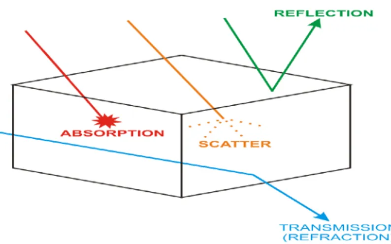
In clinical dentistry, laser light is used to perform controlled and precise changes in the target tissue through the transfer of electromagnetic energy. [105]
Light energy interacts with a target medium (e.g. oral tissue) in one of four ways: [106] (Fig. 24)
I. Transmission
Laser beam enters the medium and emerges distally without interacting with the medium. The beam exits either unchanged or partially refracted.
II. Reflection
When either the density of the medium or angle of incidence is less than the refractive angle, total reflection of the beam will occur. The incident and emergence angles of the laser beam will be the same for true reflection or some scatter may occur if the medium interface is non-homogenous or rough.
III.Scatter
There is an interaction between the laser beam and the medium. This interaction is not intensive enough to cause complete attenuation of the beam.
The result of light scattering is a decrease of laser energy with distance, together with a distortion in the beam (rays travel in an uncontrolled direction through the medium).
IV. Absorption
The incident energy of the laser beam is attenuated by the medium and converted into another form. With the use of dental diode lasers, the most common form of conversion of laser energy into heat or in case of very low energy values, biomodulation of receptor tissue sites seems to occur. [107, 108] Heat transfer mediated physical change in target tissue is termed ‘photothermolysis’.
In any desired laser-tissue interaction, the goal is to achieve the maximum absorption of laser light by the target tissue, as this will allow the maximum control of the resultant effects.
Absorption is determined by matching incident laser beam energy (wavelength) to the electron shell energy in the target atoms.
Absorption of laser energy in the target tissue leads to generation of heat. Rising heat levels leads to dissociation of covalent bonds (in tissue proteins), phase transfer from liquid to vapour (in intra and inter-cellular water), onto phase transfer to hydrocarbon gases and production of residual carbon. [109]
Secondary effects can occur because of heat generation (through conduction). When predicting the conversion of electromagnetic energy to heat effects in target tissue, unwanted change through conductive thermal spread must be taken into account and reduced to the lowest possible level.
The ability to control a progressively increasing heat loading of target tissue is termed ‘thermal relaxation’. [110]
Thermal relaxation rates are proportional to the area of tissue exposed and inversely proportional to the absorption coefficient of the tissue, assuming fixed values of thermal and light diffusivity for the tissue in question.
There are many different mechanisms by which laser light can interact with tissue; these have been categorized in a number of different ways.
For the purposes of these facts, the most common interaction mechanisms for therapeutic and surgical applications will be divided into five broad classes:
- Photochemical reaction
A molecule absorbs a photon of sufficient energy, the energy can be transferred to one of the molecule's electrons.
An electron with higher energy can more easily escape the nuclear forces, keeping it close to the nucleus and so excited molecules (which are molecules with an electron in a higher energy state) are more likely to undergo chemical reactions (exchange or share electrons) with other molecules.
In photodynamic therapy, for instance, a photosensitizing drug (a concoction of molecules when they absorb light, causing reactive oxygen species to form) is used to cause necrosis (cell death) and apoptosis (‘programmed’ cell death). Photodynamic therapy is increasingly widely used in oncology to destroy cancerous tumors.
- In photothermal interaction
The energy of the photons absorbed by chromophores (a term used to refer to any light-absorbing molecules) is converted into heat energy via molecular vibrations and collisions, which can cause a range of thermal effects from tissue coagulation to vaporization.
A denaturation of proteins occurs for temperatures ranging from 42°C to 60°C; the water inside the tissues vaporizes at 100°C. Over 200°C, the carbonization with vaporization of the tissues takes place. It represents the last stage of the laser tissue interaction. (Fig. 25)
Applications include tissue cutting and welding in laser surgery and thermal effects which are explained as:
T = 40- 60°C: edema, functional imbalance of metabolism (up to 50°C reversible)
T = 60- 100°C: denaturation of the proteins, coagulation, and contraction because of dehydration
T > 100°C : disruption, evaporation of water, and ablation
T > 150°C : vaporization, and ablation T > 300°C : carbonisation
- In photoablation interaction
Ultraviolet (UV) photons are absorbed by electrons, raising them from a lower energy 'bonding' orbital to a higher energy 'non-bonding' orbital; thereby, causing virtually immediate dissociation of the molecules.
This naturally leads to a rapid expansion of the irradiated volume and ejection of the tissue from the surface.
This is used in eye (corneal) surgery, among other applications. - In plasma-induced photo ablation
A free (sometimes called ‘lucky’) electron is accelerated by the intense electric field which is found in the vicinity of a tightly focused laser beam. When this very energetic electron collides with a molecule, it gives up some of its energy to the molecule. When sufficient energy is transferred to free a bound electron, a chain reaction of similar collisions is initiated, resulting in a plasma: a soup of ions and free electrons.
One application of this is in lens capsulotomy to treat secondary cataracts.
- The final set of related mechanisms
Grouped under the term ‘photo disruption’, are the mechanical effects that can accompany plasma generation, such as bubble formation, cavitation, jetting, and shockwaves. These can be used in lithotripsy (breaking up kidney or gall stones). [111]
1.10.TYPES OF LASER
Various laser devices are available in the market, all of them have particular features in relation to different parameters: the type of cut to be performed in the tissue, the lapse of time in which to operate, the depth of surgical wounds, and the absorption of laser wavelengths by the tissue. (Table 4)
Table 4 Common laser types used in dentistry
- Nd:YAG laser
(Neodymium: Yttrium Aluminum Garnet) is surely one of the most versatile devices for the wide range of emission frequencies it allows (corresponding to the values of 1064 nm and 1320 nm).
The light ray is mainly absorbed by melanin and haemoglobin. The using pattern can be pulsing or continuous with variable time.
This device is particularly indicated for the thermal destruction of vascularized or sessile lump tumors and in the surgery of vascular lesions.
Laser type Construction Wavelength(s) Delivery system(s) KTP laser Solid state 532 nm Optical fiber Argon laser Gas laser 488, 515 nm Optical fiber Helium-neon laser Gas laser 633 nm Optical fiber
Diode laser Semiconductor 635, 670, 810,
830, 980 nm Optical fiber Nd: YAG laser Solid state 1064 nm Optical fiber
Er: YAG laser Solid state 2940 nm
Optical fiber, Waveguide, Articulated arm
CO2 laser Gas laser 9600, 10600 nm Waveguide,
The employment of this type of device is not suitable for lesions located in thermic sensible anatomical district (as periosteum) because of the heat release by deep (in depth).
- Er: YAG laser
(Erbium: Yttrium Aluminum Garnet) the emission frequencies are of 2940 nm. It is greatly absorbed by water and used in a pulsed pattern. The possibility to work using very short impulses (ranging between 50 and 100 microseconds) allows the mechanical removal of superficial vascularized or sessile lumps and the recovery of wide oral lesions in extremely temperature sensible zones as well.
The higher the water content of the tissue, the greater the laser removing effect. On the contrary, regarding the hemostasis, its effect is minimal, except when using longer impulses and high frequencies. The laser can be transmitted by different devices.
- KTP laser
(Potassium, Titanium, Phosphate) It is a solid-state laser that emits a green light with the wavelength of 532 nm and is used in oral surgery, in the dental bleaching, and the dentine hypersensitivity treatment. [112]
- He-Ne laser
(Helium- Neon) It is a gas laser whose active medium consists of a mixture of helium and neon inside a small-bore capillary tube. Its wavelength is of 633 nm and it emits in the infrared light.
- Argon laser
Its active medium is gas and it emits at 13 wavelengths through the visible ultraviolet and near-visible spectrum, including: 351.1 nm, 363.8 nm, 454.6 nm, 457.9 nm, 465.8 nm, 476.5 nm, 488.0 nm, 496.5 nm, 501.7 nm, 514.5 nm, 528.7 nm, and 1092.3 nm.
- CO2 laser
(Carbon Dioxide) laser is considered the best laser from the surgical point of view. It is the most used laser because of its high absorption by water. Its emission frequency is 10600 nm and it is possible both to choose two use patterns, pulsing or continuous and to diminish the impulse range down to few nanoseconds. [119]
This type of laser is particularly indicated for surgical procedures (in those regions extremely difficult to reach), for a good superficial hemostasis and thermal therapy in some solid tumors. Since the thermal effect could be important, during the laser treatment, extreme caution is needed in those highly sensible zones to the temperature.
- Diode laser
The nucleus of a diode laser consists of a semi-conductive material (Indium Gallium Arsenic). [114]
The most common laser used in oral dental surgery, has a wavelength of 810 nm or 980 nm and shows a high affinity for the hemoglobin.
This type of laser is particularly devoted to the treatment of vascular lesions both by direct removal or by means of lesion clotting. Moreover, all the other surgical procedures, concerning both the major and minor oral surgeries, can be performed with diode laser. [120]
1.11. THE CO2 LASER
The carbon dioxide laser (CO2) was one of the first
models of gas lasers to be invented by Kumar Patel of Bell Labs in 1964. This type of laser device emits in the infrared light and its principal wavelength is between 9.4 and 10.6 µm. (Fig. 26)
The active medium is represented by a discharge tube gas-cooled air or with water in high power applications.
The gas is constituted by: carbon dioxide (CO2), about 10- 20%,
nitrogen (N2) about 10- 20%, hydrogen (H2), and/or xenon (Xe) 1- 2%
and helium (He) (the remaining part of the gas mixture).
The necessary inversion of population is obtained by passing an electric discharge in the gaseous mixture, which causes the following chain of events: [113]
• The impacts of the electrons excite the vibrational modes of the nitrogen molecule. This molecule cannot rid the energy acquired by emitting a photon and its excited state is metastable and persists for a long time.
• The collisions between gas molecules transfer the energy from the excited molecules of nitrogen to those of CO2, with the sufficient
efficiency to produce the desired inversion of population.
• The excited CO2 molecule comes back to the ground state
emitting a photon and contributing to the establishment and the emission of the laser beam.
Medical and dental researchers soon began to study different types of lasers for extra and intraoral surgical procedures. Because of its affinity for water-based tissues, the CO2 laser has become a favourite
Figure 26 The CO2 laser
instrument for oral surgeons for treatment of pathologic conditions of the oral mucosa. [115]
The CO2 laser has been recommended to treat benign oral lesions,
such as fibromas, papillomas, hemangiomas, gingival hyperplasia with different causes (idiopathic or due to side effects of medications), aphthous ulcers, mucosal frenula, or tongue ties (ankyloglossia), as well as premalignant lesions, such as OLs. [115, 116]
Some reports on the use of the CO2 laser also support the possibility
of treating malignant oral diseases in early stages (e.g. T1N0 carcinomas) with excisional biopsies. [117]
A study demonstrated dissemination of cancer cells into the blood circulation upon incisional biopsies with the scalpel, resulting in an increased risk of metastasis.
The CO2 laser, with its sealing effect on vessels smaller than 500
µm in diameter, could be an advantage and therefore prevent occult micrometastasis. [118]
1.12.OBJECTIVES OF THE STUDY
Determining the anatomical distribution of the lesions to realize the sites attacked by OL.
Recognizing the effects of associated risk factors.
Dividing large lesions into multiple sections for laser vaporization to observe if it will reduce the post-operative symptoms and malfunctions.
Determining the sufficient safety margins during laser vaporization of OL as a trial to reduce the recurrence.
Trying to improve the management of OL which may prevent not only the recurrence rate and the malignant transformation, but also post-operative dysfunction.
2.
Chapter 2 : Materials and Methods
Laser technology has made rapid progress over the past few decades. Because of its many advantages, it has been widely used in oral and maxillofacial surgeries.
Soft tissue laser is a state of the art tool that creates predictable esthetic results within a general dental practice. Ease of use and affordability makes it simple to incorporate in general practice.
All the patients in this study were treated by the same surgeon, they were informed about the advantages and disadvantages of laser surgery; also, side effects of laser surgery were explained for the patients and they signed an informed consent prior intervention.
Questions concerning carbonization, coagulation, cutting speed, pain, swelling, bleeding, need for drugs, functional reduction, and fibrin layer on wounds during treatment were discussed with every patient.
Full clinical examination, consisted of medical and dental history, were performed and the clinical treatment was documented by photos and questionnaires for patients and surgeons.
Elimination of associated risk factors was done for all the selected patients in the three groups.
An oral swab was done for all the lesions to exclude any fungal infection.
Patients were always photographed with the same equipment (Nikon D200, Nikon Corporation, Tokyo, Japan)
Local anesthesia was performed by 1.8 ml of Mepivacaine solution (Mepivacaina Pierrel, 30 mg/ml, injection solution 1.8 ml, Pierrel S.p.A., Milan, Italy).