Getting insights into the multi-faceted role of
Notch3 in different tumor contexts
PhD Programme in Molecular Medicine
XXXII CYCLE
Candidate
Maria Valeria Giuli
Tutor Supervisor
Saula Checquolo Isabella Screpanti
SUMMARY...5
1. INTRODUCTION...9
1.1 Notch receptors...10
1.1.1 Notch structure...11
1.1.2 Canonical Notch signalin pathway...14
1.2 The other side of the coin: Notch dysregulation...17
1.2.1 Notch signaling and genetic disease...17
1.2.2 Notch signaling and cancer...17
1.2.3 Highlights on anti-Notch therapies...19
2. AIMS OF THE WORK...22
- Crosstalk between Notch3 and other signaling pathways (Project n°1 and Project n°2) - Post-translational modifications (PTMs) of Notch3 (Project n°3) 3. RESULTS...26
3.1 CROSSTALK BETWEEN NOTCH3 AND OTHER SIGNALING PATHWAYS...27
3.1.1 Notch3 and EGFR in Triple Negative Breast Cancer...27
3.1.1a Glance at Project n°1...27
3.1.1b Paper (Diluvio G et al., Oncogenesis 2018)...29
3.1.2 Notch3 and Unfolded Protein Response in T-cell Acute Lymphoblastic Leukemia...52
3.1.2a Glance at Project n°2...52
3.1.2b Paper (Giuli MV et al., Submitted)...54
3.2 POST-TRANSLATIONAL MODIFICATIONS OF NOTCH3...102
3.2.1 Introduction...102
3.2.2 Material & Methods...105
3.2.3 Results...112
3.2.4 Discussion...122
3.2.5 Conclusion...126
Figures & Figure legends...127
4. CONCLUSION...142
REFERENCES...146
APPENDIX I...161 Review: Notch signaling and Triple Negative Breast Cancer (Giuli MV et al., Journal of Oncology 2019)
SUMMARY
ABSTRACT
Notch receptor family comprises evolutionary conserved single-pass transmembrane proteins which are involved in several cellular processes during embryogenesis and in adult tissues. Given their pleiotropic effects, their de-regulation is associated to the development of several diseases including cancer. Since Notch receptors are differentially implicated in essentially all of the hallmarks of cancer, dissecting every facets of each receptor in different tumor contexts could help to foster effective and specific targeted therapies.
In keeping with this consideration, during my PhD project I specifically focused my attention on Notch3 receptor in three different tumor contexts: Triple-Negative Breast Cancer (Project n°1), T-cell Acute Lymphoblastic Leukemia (Project n°2), and Ovarian Cancer (Project n°3).
The main objective of this work, performed in the laboratory of Prof. Isabella Screpanti (at the Department of Molecular Medicine of Sapienza University) under the direct supervision of Dr. Saula Checquolo, was to broaden the knowledge of Notch3 receptor, trying to puzzle out its role in cancer, mainly focusing on how it is specifically regulated at the post-translational level, which still represents an unknown field of Notch3 regulation process.
Indeed, Project n°1 and Project n°2 report two different crosstalk between Notch3 and other signaling pathways, the EGFR signaling and the Unfolded Protein Response (UPR), respectively. Specifically, on the one hand, we demonstrate that Notch3 regulates EGFR localization in Triple-Negative Breast Cancer, making the receptor unavailable to be targeted by the anti-EGFR agents, such as tyrosine kinase inhibitors, thus highlighting how Notch3 could be crucial in promoting drug-resistance. On the other hand, we document that Notch3 is involved in the activation of pro-survival UPR by directly interacting with an UPR “effector”, thus sustaining cancer cell growth under ER stress conditions.
Interestingly, in both tumor contexts Notch3 fulfils its function in a transcription-independent manner, paving the way for the study of a mostly untouched aspect of Notch3 function in cancer.
Moreover, in Project n°3 we cover a largely unstudied but crucial layer of fine-tuning and regulation of Notch3: its post-translational modifications (PTMs). To date, little is known about the potential different PTMs of Notch3, mainly regarding the glycosylation of its extracellular domain and the acetylation of its intracellular domain. Here we focus our attention on the study of the phosphorylation status of Notch3 intracellular domain showing how it influences its longevity and who are the actors of this regulation, thus finally fostering a novel therapeutic approach to target Notch3-dependent tumors through the modulations of these specific Notch3 protein regulators.
All in all, there are still lots of gaps in the puzzle of “Notch3 world” but some small pieces were falling neatly into place.
ORGANIZATION OF THE WORK
This thesis will start with a First Chapter providing an overall introduction to Notch receptors concerning their structure, their canonical signaling pathway and how their de-regulation is associated to several diseases.
The Second Chapter will be devoted to a brief explanation of the major objectives of each project in order to show the golden thread that runs through them.
The Third Chapter will cover the results obtained in each project. The results of the Project n°1 were reported in one publication (Diluvio G et al., Oncogenesis 2018) and the ones of Project n°2 are currently under revision (Giuli MV et al., manuscript submitted to Haematologica Journal). Therefore, in the Section 3.1, I will insert these two papers preceded by a brief introductory paragraph which will summarize the state-of-the-art of the studied tumor context, the role of the
involved pathways and the novelty of each project. The manuscript on the Project n°3 is in preparation so the Section 3.2 will be more detailed and it will be divided in several paragraphs.
Since each project has its own conclusion included in the Third Chapter, in the Conclusion Section of the present PhD Thesis (Forth and last Chapter), I will briefly discuss the obtained results focusing on their potential therapeutic application based on anti-Notch3 strategies.
Finally, in the Appendix I, I will insert a recent published review on the role of Notch receptors in Triple-Negative Breast Cancer (Giuli MV et al., Journal of Oncology 2019).
1. INTRODUCTION
1.1 NOTCH RECEPTORS
The Notch receptors are single-pass transmembrane proteins involved in several cellular processes during development and in adult tissues (Louvi A et al., 2012). For instance, Notch signaling is implicated in several organ and tissue development programs. On the one hand, Notch pathway impedes differentiation and secures a pool of stem or progenitors cells; on the other hand, Notch signaling promotes differentiation and it is involved in cell commitment. Moreover, increasing evidence suggests that Notch receptors orchestrate tissue homeostasis, both under normal conditions and during repair and regenerative processes
(Siebel C and Lendhal U, 2017).
Notch signaling is evolutionary conserved across the metazoan spectrum and occurs via short-range cell-cell communication between transmembrane ligands on one cell and transmembrane receptors on the neighboring one
(Artavanis-Tsakonas et al., 1999).
The first observations of Notch receptors derived from genetic studies of mutants of the fruit fly Drosophila melanogaster. The fruit fly genome encodes one Notch protein whereas two receptors (LIN-12 e GLP-1) with redundant roles were discovered in Caenorhabditis elegans (Fitzgerald K et al., 1993). Moreover, in mammals four Notch paralogs are synthesized and they have only partly shared functions (Wu J and Bresnick EH, 2007).
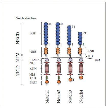
1.1.1 Notch structure
Figure 1. Notch family receptors in mammals. Schematic representation of the Notch receptors
structure in mammals (Giuli MV et al., 2019).
The Notch locus encodes a large (~300 kDa) multidomain protein which is expressed on the plasma membrane as processed heterodimer. It is possible to distinguish a large extracellular domain (NECD), a transmembrane region (TM) and a large intracellular domain (NICD) (Hori K et al., 2013).
The NECD comprises several tandem Epidermal Growth Factor (EGF)-like repeats followed C-terminally by a negative regulatory region (NRR) (Gordon W et al.,
2008).
The EGF-like domain is an evolutionarily conserved structure and it is formed by nearly 40 amino acid residues, among them 6 cysteines that are responsible for 3 characteristic disulphide bonds (de Celis JF et al., 1993). Calcium-binding EGF repeats are functionally important to interact with the ligands (de Celis JF et al.,
1993) and, as a result, it has been proposed that extracellular Ca2+ concentration might influence Notch signaling (Raya A et al., 2004). Non-calcium-binding EGF repeats are not able to coordinate Ca2+ ions and they are present in flexible regions of the ECD (Hambleton S, 2004). The number of EGF-like repeats is not conserved across the metazoan: 11-14 in C. elegans and 29-36 in D. melanogaster and in mammals (Rana NA and Haltiwanger RS, 2011). Despite the large number of EGF-like repeats, only two, 11 and 12, are essential for mediating interactions with the ligands (Rebay I et al., 1991; Xu A et al., 2005).
The NRR comprises 3 Lin-12-Notch repeats (LNRs) and an heterodimerization domain (HD). Structural studies revealed that in the resting state, the LNRs masks the heterodimerization domain, thus preventing ligand-independent proteolysis
(Gordon WR et al., 2009). This region contains two proteases sites, known as S1
and S2, which are involved in the processing and signaling of Notch receptors
(Gordon WR et al., 2008).
The ECD undergoes several processing events during their intracellular routing to the cell surface (Kadesch T, 2000).
These processes comprise N- and O-glycosylation and occur in the Endoplasmic Reticulum (ER) and in the Golgi apparatus (Jafar-Nejad H et al., 2010). Early work on the Notch receptors documented that the EGF-like repeats are decorated with N-glycans (Kornfeld K et al., 1981). In addition to N-glycosylation, several O-linked glycosylation (O-fucosylation, O-glucosylation and O-GlcNAcylation) occurred in the ECD (Moloney DJ et al., 2000; Matsuura A et al., 2008). A growing piece of evidence highlighted that glycosylation affects Notch signaling and it is crucial for their functions (Stanley P, 2007; Luther KB and Haltiwanger RS, 2009). Besides glycosylation, Notch receptors are cleaved at the S1 site in the Golgi apparatus by a furin-like convertase and reach the plasma membrane as heterodimers (Logeat F et al., 1998).
The NICD is formed by several regulatory motifs, including RAM (RBP-jk-Associated Molecule), TAD (Transcriptional Activation Domain) and PEST (P: Proline; E: Glutamic Acid; S: Serine; T: threonine) degron sequence. Between RAM and TAD there are 7 tandem Ankyrin repeats (ANK) flanked by 2 Nuclear Localization Signal (NLS) sequences (Rana NA and Haltiwanger RS, 2011). The RAM and the ANK domains are essential to recruit transcriptional co-activators whereas PEST domain is involved in NICD degradation (Hori K et al., 2013).
The 4 Notch receptors in mammals display subtle structural differences. For instance, Notch1 and Notch2 have 36 EGF-like repeats while Notch3 and Notch4 have 34 and 29 repeats, respectively (Previs RA et al., 2015). These differences correlate with diverse affinity for their ligands (Rebay I et al., 1991). Moreover, another source of diversity rests within TAD resulting in differential transactivation activity: it is absent in Notch4 (Kurooka H et al., 1998) and Notch3 TAD is shorter with respect to Notch1 and Notch 2 (Beatus P et al., 2001).
Furthermore, the NICD may differ between Notch paralogues (Chillakuri CR et
al., 2012) since amino acid sequence identity is partial across the 4 Notch receptors (Bellavia D et al., 2008). For instance, Beatus and colleagues identified a novel
region located C-terminally to ANK domain known as RE/AC (REpression/ACtivation) which is necessary for N1ICD’s ability to activate and for N3ICD’s ability to repress a HES promoter (Beatus P et al., 2001).
Taken together, these structural differences amplify and diversify the signaling output.
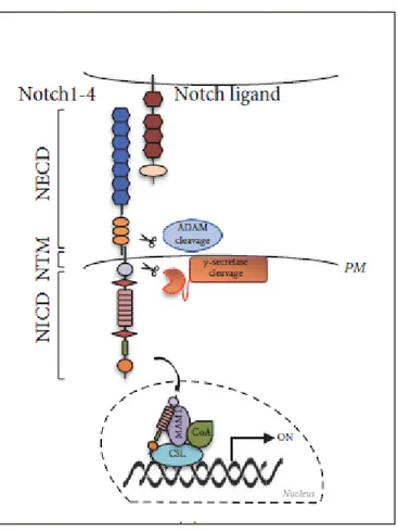
1.1.2 Canonical Notch signaling pathway
Figure 2. The canonical Notch signaling pathway. Ligand binding triggers two sequential
proteolytic cleavages (by ADAM and γ-secretase complexes), resulting in the release of NICD which translocates to the nucleus where interacts with transcriptional regulators (CSL, MAM1 and CoA) (Giuli MV et al., 2019).
Notch signaling is made by 3 key steps: 1. ligand recognition; 2. conformational exposure of the S2 protease site and 3. assembly of transcriptional complexes. Activation of the canonical pathway requires the trans-interaction of a Notch receptor in signal-receiving cell with a ligand on signal-sending neighboring cell
(D’Souza B et al., 2010). Cis-interactions between Notch receptors and ligands
expressed in the same cell also occur and they inhibit Notch1 signaling (Sprinzak D et al., 2010) while activating Notch3 signaling in a subset of T-cell acute lymphoblastic leukemia (Pelullo M et al., 2014).
Several genetic studies revealed that Notch ligands have been conserved across metazoan. Mammalian Notch ligands fall into two classes, depending on their
homologies with the ones found in D. melanogaster (Delta and Serrate). Mammals have three Delta-like proteins, called Delta-like 1 (DLL1), Delta-like 3 (DLL3), and Delta-like 4 (DLL4), and two homologues of Serrate, called Jagged-1 (JAG1) and Jagged-2 (JAG2) (D’Souza B et al., 2008). Canonical Notch ligands are single-pass transmembrane proteins and exhibit a modular domain arrangement. The extracellular domain comprises an N-terminal MNNL (Module at the N-terminus of Notch ligands) domain and a cysteine-rich Delta–Serrate–LAG2 (DSL) domain
(Kovall RA and Blacklow SC, 2010). The DSL domain is evolutionary conserved and it is crucial for the interaction with Notch receptors. Moreover, all ligands contain several EGF-like repeats followed by the transmembrane domain and a short C-terminal cytoplasmic tails (D’Souza B et al., 2008). Despite the similar overall modular organization, a number of structural differences exists among the DSL ligands. For instance, Serrate family (Serrate, JAG1 and JAG2) proteins and DLL1 have specific EGF-like repeats which form the DOS (Delta and OSM-11-like proteins) domain. Furthermore, Serrate family proteins are characterized by a large number of EGF-like repeats and a cysteine-rich region, partially homologous with the von Willebrand Factor C (VWFC) (D’Souza B et al., 2010). DLL1, DLL4, and JAG1 contain PDZL motifs (PSD-95/Dlg/ZO-1 ligand) that recognize PDZ domain of several cytoskeletal proteins responsible for cell-cell junctions (Popovic
M et al., 2012).
Notch receptors have distinct ligand affinities, thus resulting in differential downstream transcriptional outcome (Shimizu K et al., 2002).
Upon ligand binding, the key step in the activation of the pathway is a conformational change of the NRR domain, following ligand endocytosis within signal-sending cell, which unmasks the S2 protease site. Notch receptor is further
cleaved by two metalloproteases: ADAM10 (A Disintegrin And Metalloprotease 10) e ADAM17/TACE (A Disintegrin And Metalloprotease 17/TNFα Converting Enzyme) (Gordon WR et al., 2008). This cleavage creates a truncated membrane-bound form of Notch known as NEXT (Notch EXtracellular Truncation) which is progressively cleaved by γ-secretase complex (Presenelin/Nicastrin/APH1/PEN2) at S3 site and S4 to release NICD in the cytoplasm (Gordon WR et al., 2008). Subsequently, NICD translocates to the nucleus where it forms a binary complex with DNA-binding factor CSL (CBF1/Suppressor of Hairless/Lag1), converting it from a repressor to an activator of transcription (Wang H et al., 2015). Indeed,
prior to pathway activation, CSL directly interacts with transcriptional co-repressor proteins and histone deacetylases (Wang H et al., 2015), which are displaced after NICD binding. The binary complex CSL-NICD is then recognized by co-activators belonging to the Mastermind (MAM) family. The formation of the CSL-NICD-MAM ternary complex leads to the recruitment of various transcriptional co-activators (CoA) and chromatin remodeler (Wilson JJ and
Kovall RA, 2006), finally resulting in the transcription of several Notch target
genes, among them transcriptional repressors of the HES and HEY families, MYC, NF-κB, cyclinD1 (Bray SJ, 2006). The target genes activated by Notch are differential depending on ligand-receptor interaction and cell-type-specific transcriptional programs (Bray SJ, 2016), thus partially explaining Notch pleiotropic effects.
1.2 THE OTHER SIDE OF THE COIN: NOTCH DYSREGULATION
Consistent with the crucial role of Notch pathway in many aspects of cellular processes, increasing evidence links perturbations in the pathway to various diseases, including several inherited syndromes and cancer.
1.2.1 Notch signaling and genetic disease
Some monogenic disease are related to mutations in the Notch signaling pathway, among them Alagille syndrome, Hajdu-Cheney syndrome and CADASIL (Cerebral Autosomal Dominant Arteriopathy with Subcortical Infarts and Leukoencephalopathy) disease.
Given the role of Notch pathway in organogenesis, Alagille syndrome affects multiple organs and clinical features may differ significantly. The initial diagnosis is based on the presence of intrahepatic bile duct paucity and at least of 3 other clinical features: chronic cholestasis, cardiac disease, ocular abnormalities, skeletal abnormalities, and peculiar facial features (Reyes-de la Rosa ADP et al., 2018). Alagille-bearing patients are characterized by mutations in JAG1 and rarely in Notch2 (Grochowski CM et al., 2016).
In the Hajdu-Cheney syndrome, the excessive bone resorption is caused by losso ofo Notch2 PEST domain (Simpson MA et al., 2011).
Furthermore, CADASIL displays degenerated vascular smooth muscle cells in the brain and recurring small brain infarcts. This disease is associated with frequent missense mutations in Notch3 which alter the number of cysteine residues in the N3ECD (Rutten JW et al., 2014).
1.2.2 Notch signaling and cancer
Since this conserved pathway is very dosage-sensitive, too much or too little can lead to cancer. Indeed, functional studies highlighted that Notch signaling is
implicated in essentially all of the hallmarks of cancer, playing oncogenic or tumor suppressive role depending on cell type (Aster JC et al., 2017).
Notch receptors as oncoproteins
One of the first evidence that dysregulation of Notch signaling is linked to carcinogenesis derived from studies on the genetic signature of T-cell acute lymphoblastic leukemia (T-ALL) cells when Ellisen and colleagues described a chromosomic translocation involving Notch1 locus (Ellisen LW et al., 1991).
From that moment on, numerous reports have associated alterations in the Notch pathway with the pathogenesis of T-ALL, since activating Notch1 mutations are found in approximately 55–60% of T-ALL cases (Weng AP et al., 2004). Indeed, Notch1 is a well-characterized oncoprotein in this type of cancer. Interestingly, functional studies suggested also a pro-tumorigenic effect of Notch3 in T-ALL. For more details on the relationship between Notch3 and T-ALL see Section 3.1.2. Extensive research over the past decades highlighted that dysregulation of Notch signaling is implicated also in the pathogenesis of B-cell malignancies (Mirandola
R et al., 2011).
Furthermore, up-regulated Notch signaling has been found in multiple solid malignancies such as breast cancer, prostate cancer, lung cancer and ovarian cancer.
The possibility that Notch could function as proto-oncogene in human breast cancer development came from studies on mouse mammary tumor virus-induced cancer (Gallahan D et al., 1987). Indeed, Notch signaling is frequently up-regulated, either for activating mutations of Notch receptors or for high levels of Notch ligands such as JAG1 (Dickson BC et al., 2007; Reedijk M et al., 2005;
Stylianou S et al., 2006). Moreover, Notch receptors are implicated in tumor
initiation and progression of the most aggressive breast cancer subtype, the
negative breast cancer (TNBC), as we described in a recently published review (see Appendix I - Giuli MV et al., Journal of Oncology 2019). In particular, for more details on the relationship between Notch3 and TNBC see Section 3.1.1. Despite the fact that further investigations are needed, high levels of Notch1 and JAG1 correlate to prostate cancer progression and recurrence (Carvalho FL et al.,
2014). Similarly, the aberrant activation of the Notch pathway is a very common
feature of non-small cell lung cancers (NSCLC) (Westhoff B et al., 2009). For instance, high expression of Notch1 increases lymph- and tumor-node metastasis
(Yuan X et al., 2015).
Deregulated expression of all Notch receptors and their ligands has been associated with high grade ovarian carcinoma and carcinogenesis (Rose SL, 2009). For instance, Notch1 is involved in ovarian cancer (OC) proliferation (Hopfer O et al., 2005) and chemoresistance (Rose SL et al., 2010) whereas Notch3 is implicated
in OC growth, drug resistance and OC stem cell maintenance. For more details on the relationship between Notch3 and Ovarian cancer see Section 3.2.
Notch receptors as tumor suppressors
Notch receptors can act also as tumor suppressors since a number of mutations loss-of-function have been unexpectedly linked to cancer development.
Loss of Notch1 signaling causes skin cancer (Stransky N et al., 2011) and myeloid malignancies (Lobry C et al., 2014). Moreover, Viatour and colleagues proposed a novel tumor suppressor role for Notch signaling in hepatocellular carcinoma (HCC) (Viatour P et al., 2011).
1.2.3 Highlights on anti-Notch therapies
Collectively, there is mounting evidence that the level of Notch signaling is critical within the cells and dramatic up- or down-regulation of this pathway can lead to
disease (Guruharsha K et al., 2012). Indeed, it is necessary to precisely fine-tune it with ad-hoc therapies.
A number of interesting avenues have been tested rendering the development of Notch-based therapies a very active area of research (Braune EB and Lendhal U,
2016).
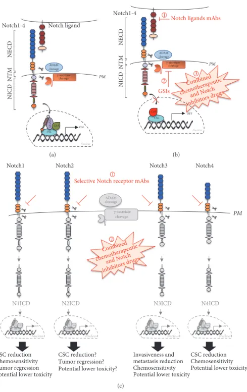
Briefly, the therapeutic strategy that has been explored more extensively is based on the use of γ-secretase inhibitors (GSIs). GSIs block the release of NICD in the cytoplasm by inhibiting the final proteolytic cleavage. Nevertheless, GSIs prevent the activation of all Notch receptors thus contributing to the onset of several adverse side effects in vivo, likely dependent on the fact that Notch receptors are involved in tissue homeostasis (Shih IeM and Wang TL, 2007). For instance, Notch signaling plays a homeostatic role in the intestine where it not only secures the stem cell pool in the crypts but also it sustains enterocytes differentiation at the expense of secretory lineages (Siebel C and Lendhal U, 2017). Therefore, GSIs-dependent blockade of Notch signaling results in secretory cells differentiation and excessive mucus production which explain the intestinal toxicity observed in many clinical trials (van Es JH et al., 2005). Moreover, the use of GSIs is further complicated by the fact that more than 40 other proteins are cleaved by the γ-secretase complex (Selkoe DJ and Wolfe MS, 2007).
In spite of these potential shortcomings, a number of clinical trials of GSIs have been proceeded. However, it would be desirable to modulate Notch in more specific ways to avoid the issues related to pan-Notch inhibition.
Receptor- or ligand-specific antibodies have been recently developed. On one hand, anti-Notch antibodies block individual Notch receptors by binding NRR and preventing the proteolytic processing by ADAM10 and ADAM17/TACE, thus reducing the cleavage upon ligand binding (Wu Y et al., 2010). On the other hand,
anti-ligand antibodies impair the trans-interaction between receptors and ligands which is required for the proper activation of the signaling (Hoey T et al., 2009). A third potential avenue is to target Notch pathway together with another signaling mechanism. Combination therapy might be particularly suitable since several studies documented the cross-talk between Notch signaling and other pathways (Braune EB and Lendhal U, 2016). Moreover, it is noteworthy that in combination therapies it is possible to use lower doses of each inhibitor circumventing some of the high-dosage-dependent side effects.
Up to date, any anti-Notch therapies are in routine clinical use, so dissecting new facets of Notch signaling in different cancer contexts becomes a paramount issue in order to ameliorate the current treatments.
2. AIMS OF THE WORK
Decades of detailed studies revealed that Notch receptors are deeply involved in several processes, from cell commitment during embryogenesis to homeostasis maintenance in adult tissues.
Such pleiotropy is unexpectedly based on a simple core pathway where few proteins are involved (Palermo R et al., 2014) without any step of amplification such as phosphorylation cascades or second messengers (Braune EB and Lendhal
U, 2016). A noteworthy source of diversity relies on the fact that in mammals there
are four Notch paralogs characterized by variable structural homology (Bellavia
D et al., 2008) which contributes to partial shared functions (Wu J and Bresnick EH, 2007). Nevertheless, it is only the tip of the iceberg since the structural
variability cannot be an exhaustive answer. This consideration hints at a complex underlying network circuitry that diversifies the signaling output. Indeed, the downstream diversity may be achieved by multiple mechanisms derived from the crosstalk between Notch and other signaling pathways also involving different post-translational modifications (PTMs) of Notch receptors (Braune EB and
Lendhal U, 2016).
Despite a lot of effort has been put in dissecting every facets of each receptor, there is a long way to go before they are fully understood. Therefore, in the development of my PhD project, I focused my attention on the Notch3 receptor and how the afore-mentioned mechanisms influence its function in different tumor contexts.
Crosstalk between Notch3 and other signaling pathways
Triple-negative breast cancer (TNBC) is characterized by the poorest prognosis
(Dawson SJ et al, 2009). Since the tyrosine kinase receptor EGFR overexpression
accounts for 45–70% of TNBC (Hoadley KA et al., 2007), anti-EGFR therapies (such as tyrosin-kinase inhibitors, TKIs, or monoclonal antibodies) have been
developed in the recent years but the activation of compensatory pathways led to disappointing results (Masuda H et al, 2012). Therefore, effective therapeutic strategies for overcoming drug-resistance are urgently required.
As a result, the main aim of the research Project n°1 (where I participated to the last part of the project and during the revision process) was to evaluate the Notch3-EGFR interplay in TNBC and its involvement in the resistance to TKIs. All the data obtained were included in a recent published paper where I am a co-author (Diluvio G et al., 2018) and it is inserted in this PhD Thesis at Section
3.1.1b.
Cells adapt to stress and re-establish of ER homeostasis by activation of an integrated signal transduction pathway called Unfolded Protein Response (UPR)
(Ron D and Walter P, 2007). Since malignant cells could rely on UPR for their survival (Papaioannou A and Chevet E, 2018), understanding how oncogenes regulate these pathways might help in searching for a novel therapy. To date, no relevant data are known about the possible involvement of Notch proteins and UPR system in cancer.
The main aim of the research Project n°2 was to get new insights into the role of Notch3 in T-cell Acute Lymphoblastic Leukemia (T-ALL) regarding the adaptive response of Notch3-dependent cancer cells to further stimulate the development of Notch3-targeted therapies. All the data obtained were included in a new paper in submission, where I am the first author (Giuli MV et al., Submitted paper) and it is inserted in this PhD Thesis at Section 3.1.2b.
Post translational modifications (PTMs) of Notch3
Since PTMs are known to modulate protein activity, localization and stability inside a cell (Walsh CT, 2005), the characterization of these modifications plays an important role in understanding their function, thus predicting cell behavior. Among PTMs, phosphorylation (the addition of phosphate groups from ATP to specific Serine, Threonine, and Tyrosine residues) is one of the key mechanisms for tight dynamic regulation of protein activity in eukaryotic cells as one third of all eukaryotic proteins undergo reversible phosphorylation (Antfolk D et al.,
2019).
Several proteins, such as the peptidyl-prolyl cis/trans isomerase PIN1, recognize and bind specific phosphorylated residues affecting the protein function of their targets (Zannini A et al., 2019). Studies documented that Pin1 enhances the oncogenic potential of Notch receptors by phosphorylation-dependent prolyl isomerization in breast cancer context (Rustighi A et al., 2009; Rustighi A et al.,
2014). In this scenario we have recently demonstrated a specific functional
Pin1-Notch3 crosstalk in T-cell leukemia (Franciosa G et al., 2016). Based on these observations, the main aim of the research Project n°3 (see Section 3.2) is to investigate in more detail the Notch3 – Pin1 relationship by focusing our attention on: 1. the underlying molecular mechanism; 2. the possibility of exploiting this relationship in order to develop a novel therapeutic strategy for the treatment of Notch3-overexpressing tumors which rely on Notch3 protein function to survive and spread to secondary organs (Giuli MV et al., Manuscript in preparation).
3. RESULTS
3.1 CROSSTALK BETWEEN NOTCH3 AND OTHER SIGNALING PATHWAYS
3.1.1 Notch3 and EGFR in Triple Negative Breast Cancer
3.1.1a Glance at Project n°1
● General features of Triple Negative Breast Cancer (TNBC)
Breast cancer (BC) is one of the most commonly diagnosed cancer in women worldwide (Siegel R et al., 2017; Torre LA et al., 2016). BC is classified into different subtypes according to the presence or absence of estrogen receptors (ERs), progesterone receptors (PRs) and the human epidermal growth factor receptor 2 (Her2/neu) (Carlson RW et al., 2009). Triple Negative Breast Cancer (TNBC) is characterized by the lack of expression of ER, PR and, Her2 (Abramson VG et al., 2015), and it accounts for 15-20% of diagnosed breast cancer (Lehmann BD et al., 2014). TNBC is an heterogeneous tumor and displays aggressive phenotype and high relapse rates (Dent R et al., 2007), resulting in shorter overall survival when compared to other subtypes (Prescott JD et al., 2007). Since the inter-tumoral and intra-inter-tumoral heterogeneity is one of the major issue to deal with, the development of targeted-therapies, less toxic and more effective than chemotherapy, is urgently needed.
● Overview of the role of EGFR in TNBC
The Epithelial Growth Factor Receptor (EGFR) belongs to Tyrosine Kinase (TK) Receptors (Schlessinger J et al., 2000). Several studies reported that EGFR is frequently overexpressed in TNBC and this is due principally to gene amplification (Rakha EA et al., 2009) and, to a lesser extent, to gene mutation (Teng YH et al., 2011).
Furthermore, TNBC cases with EGFR expression are associated with a poor prognosis (Nicholson RI et al., 2001), thus rendering EGFR a hallmark of this BC subtype. Despite the reliance of TNBC on EGFR downstream signaling, anti-EGFR therapies are ineffective mainly because of the activation of compensatory pathways that induce drug resistance (Bernsdorf M et al., 2011). Indeed, this complicates matters further and cell re-sensitization to anti-EGFR therapies becomes a paramount issue.
● Overview of the role of Notch3 in TNBC
Recently, we provided a detailed overview of the specific role of all four Notch receptors in TNBCs (see Appendix I, Giuli MV et al., Journal of Oncology 2019). Regarding Notch3, several studies provide evidence of the correlation between Notch3 signaling and TNBC: in particular, Notch3 activating mutations (Wang K et al., 2015) and gene amplification (Turner
N et al., 2010) are quite recurrent in TNBC context. Moreover, Notch3
displays an oncogenic role by sustaining TNBC cells proliferation (Hirose
H et al., 2010; Choy L et al., 2017), the acquirement of the metastatic
phenotype (Zhang Z et al., 2010; Leontovich AA et al., 2018) and chemo-resistance (Boelens MC et al., 2014).
To sum up, Notch3 is known to be associated with TNBC tumor initiation and progression.
● Novelty of the Project n°1
We reported a non-canonical role of Notch3 in TNBC: it is involved in the resistance to TKI-gefitinib (Tyrosine Kinase Inhibitor - gefitinib) by regulating EGFR intracellular localization. Therefore, Notch3 depletion
makes the cells sensitive to TKI-gefitinib, thus suggesting a potential novel combined therapeutic approach in the treatment of TNBC-bearing patients.
3.1.1b Paper
“NOTCH3 inactivation increases triple negative breast cancer sensitivity to gefitinib by promoting EGFR tyrosine dephosphorylation and its intracellular arrest”
G. Diluvio, F. Del Gaudio, M. V. Giuli, G. Franciosa, et al. Oncogenesis. 2018;7:42
Diluvio et al. Oncogenesis (2018) 7:42
DOI 10.1038/s41389-018-0051-9 Oncogenesis
A R T I C L E O p e n A c c e s s
NOTCH3 inactivation increases triple
negative breast cancer sensitivity to
ge
fitinib by promoting EGFR tyrosine
dephosphorylation and its intracellular
arrest
Giulia Diluvio1, Francesca Del Gaudio2, Maria Valeria Giuli1, Giulia Franciosa3, Eugenia Giuliani1, Rocco Palermo1,4, Zein Mersini Besharat 1, Maria Gemma Pignataro5, Alessandra Vacca6, Giulia d’Amati5, Marella Maroder7, Claudio Talora1, Carlo Capalbo1, Diana Bellavia1and Saula Checquolo 7
Abstract
Notch dysregulation has been implicated in numerous tumors, including triple-negative breast cancer (TNBC), which is the breast cancer subtype with the worst clinical outcome. However, the importance of individual receptors in TNBC and their specific mechanism of action remain to be elucidated, even if recent findings suggested a specific role of activated-Notch3 in a subset of TNBCs. Epidermal growth factor receptor (EGFR) is overexpressed in TNBCs but the use of anti-EGFR agents (including tyrosine kinase inhibitors, TKIs) has not been approved for the treatment of these patients, as clinical trials have shown disappointing results. Resistance to EGFR blockers is commonly reported. Here we show that Notch3-specific inhibition increases TNBC sensitivity to the TKI-gefitinib in TNBC-resistant cells. Mechanistically, we demonstrate that Notch3 is able to regulate the activated EGFR membrane localization into lipid rafts microdomains, as Notch3 inhibition, such as rafts depletion, induces the EGFR internalization and its intracellular arrest, without involving receptor degradation. Interestingly, these events are associated with the EGFR tyrosine dephosphorylation at Y1173 residue (but not at Y1068) by the protein tyrosine phosphatase H1 (PTPH1), thus suggesting its possible involvement in the observed Notch3-dependent TNBC sensitivity response to gefitinib. Consistent with this notion, a nuclear localization defect of phospho-EGFR is observed after combined blockade of EGFR and Notch3, which results in a decreased TNBC cell survival. Notably, we observed a significant correlation between EGFR and NOTCH3 expression levels by in silico gene expression and immunohistochemical analysis of human TNBC primary samples. Ourfindings strongly suggest that combined therapies of TKI-gefitinib with Notch3-specific suppression may be exploited as a drug combination advantage in TNBC treatment.
Introduction
Triple-negative breast cancer (TNBC), which lacks estrogen receptor (ER), progesterone receptor, and human epidermal growth factor 2 receptor (HER2), accounts for about 15–20% of breast cancers and repre-sents the most aggressive breast cancer (BC) subtype1. To date, no molecularly targeted agents have been approved for TNBC, leaving to the conventional chemotherapy the © The Author(s) 2018
Open Access This article is licensed under a Creative Commons Attribution 4.0 International License, which permits use, sharing, adaptation, distribution and reproduction in any medium or format, as long as you give appropriate credit to the original author(s) and the source, provide a link to the Creative Commons license, and indicate if changes were made. The images or other third party material in this article are included in the article’s Creative Commons license, unless indicated otherwise in a credit line to the material. If material is not included in the article’s Creative Commons license and your intended use is not permitted by statutory regulation or exceeds the permitted use, you will need to obtain permission directly from the copyright holder. To view a copy of this license, visithttp://creativecommons.org/licenses/by/4.0/.
Correspondence: Carlo Capalbo ([email protected]) or Diana Bellavia ([email protected]) or Saula Checquolo ([email protected])
1
Department of Molecular Medicine, Sapienza University, Rome, Italy
2Department of Cell and Molecular Biology, Karolinska Institutet, 17177
Stockolm, Sweden
Full list of author information is available at the end of the article These authors contributed equally: Giulia Diluvio, Francesca Del Gaudio.
Oncogenesis 1234567890() :,; 1234567890( ):,; 30
role of primary option for systemic treatment. Although TNBC-bearing patients better respond to current che-motherapy than do non-TNBC ones, patients with TNBC experience a more rapid relapse evolving as metastatic disease. For this reason, this BC subtype suffers from the poorest prognosis1. Therefore, targeted therapeutic stra-tegies for TNBC are urgently needed.
The overexpression of the tyrosine kinase receptor epidermal growth factor receptor (EGFR) is a hallmark of TNBC (45–70%) and exhaustive gene expression profiling has identified several EGFR-associated poor prognostic signatures2. Anti-EGFR therapies, including tyrosine kinase inhibitors (TKIs) and monoclonal antibodies, have been developed and are already available for treatment of different cancers such as non-small cell lung cancer (NSCLC) and colorectal cancer, making EGFR inhibitors an attractive option for TNBC therapy3. Unfortunately, no EGFR inhibitory therapies are currently approved for BC treatment, including TNBC, as results from clinical trials are disappointing4. This limited clinical activity is often due to the existence of compensatory pathways that confer resistance to EGFR inhibition, thus allowing con-tinued cancer cell growth and survival5–7.
Notch signaling dysregulation is often associated with tumor transformation8, including the TNBC pathogenesis and progression9–11. In particular, TNBCs show Notch3 amplification and overexpression12,13, and Notch3 knockdown has been shown to reduce the proliferation of ErbB2-negative breast tumor cells9,14. More recently, these data have been strongly supported by Choy et al.15 who demonstrated that constitutive Notch3 signaling can drive an oncogenic program in a subset of TNBCs, thus suggesting that Notch3 activity (and not others Notch paralogues) may be clinically relevant in this BC subtype. There is a growing body of evidence that Notch hyper-activation or mutation results in several events that enable BC cells to become resistant to targeted treatments through different mechanisms16,17, thus suggesting that the inactivation of Notch signaling could be a potential
therapeutic approach for overcoming resistance to drugs7. Interestingly, more recently, it has been demonstrated that Notch3 pathway is strongly involved in the stroma-mediated expansion of therapy-resistant TNBC cells18.
Notch-EGFR interplay occurs in different cellular con-texts19,20, including BC16, raising the possibility that Notch signaling could be involved in the above mentioned resistance to EGFR inhibition. Arasada et al.21 first reported that the EGFR inhibition by erlotinib treatment is able to activate Notch signaling in human lung cancer, resulting in an enriched stem cell-like populations in a Notch3, but not Notch1-dependent manner. In TNBC, it has been demonstrated that combined Notch-EGFR pathway inhibition is a rational treatment strategy for this type of tumors22. Pan-Notch inhibition using γ-secretase inhibitor (GSI) treatment supports this conclu-sion. Unfortunately, the use of GSIs fails to distinguish the particular Notch receptor driving growth, besides eliciting severe side effects.
Here we analyze the effects of a selective Notch3 inhi-bition in the response to gefitinib (GEF) treatment of resistant TNBC cells. We show that Notch3 (but not Notch1) depletion enhances the therapeutic target activity of the EGFR, by inducing its dephosphorylation via pro-tein tyrosine phosphatase H1 (PTPH1),finally leading to an increased TNBC sensitivity to TKI-GEF.
Results
Notch3-EGFR correlation in primary TNBC samples
To deepen the understanding of the possible Notch3-EGFR crosstalk in TNBC context, wefirst performed an in silico analysis of the NOTCH3 and EGFR gene expression levels in two cohorts of TNBC patients, collectively con-sisting of 777 individuals23–26 (Fig.1a). The summary of the obtained results (Fig. 1a, upper panel) highlights a direct correlation between EGFR and NOTCH3 gene expression levels in both datasets analyzed, while a weaker correlation between EGFR and NOTCH1 is observed. This is also evident by the graphs included in the Fig.1a (lower
Fig. 1 Notch3 and EGFR levels correlate in TNBC primary samples. a Upper panel: summary of the NOTCH3-EGFR and NOTCH1-EGFR gene expression levels correlation obtained by an in silico analysis from two TNBC tissue arrays (GSE76124 and GSE31519). Lower panels: representative graphs showing correlation between NOTCH3 (left) or NOTCH1 (right) and EGFR gene expression levels from GSE31519 dataset in a cohort of 579 TNBC patients. In both graphs, each dot corresponds to one patient and the expression value of NOTCH3, NOTCH1, and EGFR is given in log2 scale after normalizing data with justRMA algorithm normalization. The X–Y axis represent NOTCH3 (left) or NOTCH1 (right) and EGFR (both) expression levels, respectively. The index Pearson’s R indicated expresses the linear relation between paired samples and P-values were calculated using Student’s T-test, as described in Material and Methods section. b Upper panel: heatmap representing the protein levels of EGFR, Notch3, and Notch1 obtained by immunohistochemical analysis (IHC) in a cohort of 18 TNBC patients. The colors represent positive (red) or negative (blue) protein levels according to protein expression cutoff (see Materials and Methods section). Lower panel: summary of the Notch3-EGFR and Notch1-EGFR protein expression levels correlation showing percentage of each category calculated on the precedent category of patients. c Pattern of immunostaining in two different cases of TNBC. In case 1 (upper panels), there is a strong and diffuse staining of neoplastic cells both for EGFR (A) and Notch3 (B), whereas Notch1 is completely negative (C). In case 2 (lower panels), the neoplastic cells are negative for both EGFR (D) and Notch3 (E), whereas Notch1 (F) shows a weak positivity in about 20% of the cells
Diluvio et al. Oncogenesis (2018) 7:42 Page 2 of 15
Fig. 1 (See legend on next page.)
Diluvio et al. Oncogenesis (2018) 7:42 Page 3 of 15
panels), representative of the larger dataset. These data indicate that in a consistent proportion of TNBC-bearing patients (about 23%) the presence of EGFR coexists with NOTCH3 gene expression, allowing us to hypothesize a possible direct relationship between EGFR and Notch3 at the protein level in TNBC. To test this hyphotesis, we then analyzed the pattern of immunohistochemical expression of Notch3, Notch1, and EGFR in tissue sam-ples of 18 human TNBCs. In the majority of cases (15/18), we found EGFR positivity in neoplastic cells. Notch3 is expressed in a higher percentage of EGFR-positive tumors as compared with Notch1 (93% vs. 53%) (Fig. 1b, lower panel). Figure 1c (case 1) shows an example of TNBC tumor expressing both EGFR (panel a) and Notch3 (panel b) but not Notch1 (panel c), representative of 6 out of 15 TNBC EGFR+tissue samples analyzed. Interestingly, two out of three TNBC samples not expressing EGFR are also Notch3 negative but express Notch1 (Fig.1c, case 2), thus reinforcing the relevant Notch3-EGFR direct correlation in this cancer subtype.
Notch3 inhibition by siRNA sensitizes TNBC cells to EGFR-TKI-GEF treatment
To examine whether Notch3 could be involved in the mechanism of resistance to EGFR TKI, wefirst selected a group of TNBC cells expressing EGFR at various levels and known to be EGFR-TKI-resistant cells27,28, and then
we analyzed the expression of both Notch3 and Notch1 proteins (Supplementary Figure S1a). Almost all TNBC cells expressed activated Notch1 and/or Notch3 protein (N3IC), thus confirming the hyperactivation of Notch signaling observed in this BC subtype14, mainly involving the upregulation of N3IC expression, as it appears at undetectable levels in MCF10A, a normal immortalized mammary epithelial cell line. This also occurs in MDA-MB-453 cells, which express lower EGFR expression (data not shown) (Supplementary Figure S1a).
As drug resistance commonly involves several mechanisms that are often closely interconnected with their genetic profile, for our next analysis we chose the MDA-MB-468 and BT-549 cells, as they show a“similar” genetic background (i.e., phosphatase and tensin homolog (PTEN), RB1, and P53 mutations)29, which could help us to predict a“similar” sensitivity to TKIs27. Wefirst eval-uated whether the knockdown of Notch3 (siN3) or Notch1 (siN1) by small interfering RNA (siRNA) could affect cell growth or viability in such cells (Fig. 2a, d). Notably, both MDA-MB-468 (Fig. 2a) and BT-549 (Fig.
2d) cells display a more significant cell growth reduction
after the selective depletion of Notch3 with respect to Notch1, measured by counting cell number until 6 days from the starting point, day 0 thus confirming previous data14. This effect could be due to the growth arrest of the cells, as the absence of Notch3 in both cell lines correlates
Fig. 2 Notch3 downregulation by siRNA affects TNBC cells survival. a, d Analysis of cell growth after 0–3–6 days of Notch3 and Notch1 silencing in a MDA-MB-468 and d BT-549 cells. b, c Whole cell extracts from a MDA-MB-468 or d BT-549 cells at 6 day of silencing were used for western blot against Notch3 (N3IC) and Notch1 (N1IC), to control the efficiency of the b, e Notch3 and c, f Notch1 silencing, respectively. Extracts were then
immunoblotted with anti-p27, anti-cyclin D1, and anti-cyclin D3 antibodies. Anti-β-actin was used as a loading control. b, c, e, f are representative of three separate experiments. The statistical analysis associated is available in the Supplementary Figure S2
Diluvio et al. Oncogenesis (2018) 7:42 Page 4 of 15
with a significant upregulation of the cyclin-dependent kinase inhibitor p27Kip1 and downregulation of both the cyclins D1 and D3, known to be important protein reg-ulators that exhibit dynamic changes during the cell cycle (Fig.2b, e and Supplementary Figure S2a and c). Notably, the Notch1 silencing does not correlate with any sig-nificant changes of the same cell cycle regulators analyzed (Fig. 2c, f and Supplementary Figure S2b and d). These results demonstrate a specific role of Notch3 in the reg-ulation of TNBC cell growth, as confirmed by the absence of viability of MDA-MB-468 clones stably deleted for NOTCH3 (but not for NOTCH1), generated by using genome-editing CRISPR/Cas9 technique (data not shown).
Previous studies suggested that selective Notch3 inhi-bition (rather than pan-Notch inhiinhi-bition) combined with EGFR TKI therapy should be explored as a novel strategy in the treatment of lung cancer patients21. In keeping with these data, we observed that Notch3 silencing significantly enhances the gefinitib (GEF)-induced growth inhibition in both MDA-MB-468 and BT-549 cells (Fig. 3a, c, left panels: compare siCTR+ GEF vs. siN3 + GEF), in a similar or even more extensive way observed after com-bined treatment with GSI plus TKI-GEF (Supplementary Figure S3a and b, left panels: compare siCTR+ GEF vs. GSI+ GEF). These data thus strongly suggests that Notch3 depletion rather than pan-Notch inhibitor is sufficient to sensitize TNBC to TKI-GEF (Fig. 3a vs. Supplementary Figure S3a; Fig. 3c vs. Supplementary Figure S3b, left panels: compare siN3+ GEF vs. GSI + GEF). The quality of Notch(s) silencing were monitored until 6 days by evaluating the expression of both Notch3 and Notch1 proteins (Fig.3and Supplementary Figure S3, all the right panels).
In addition, although Notch1 silencing does not induce any significant changes in BT-549 GEF-treated cells with respect to control cells (Fig.3d, compare siCTR+ GEF vs. siN1+ GEF), it seems to paradoxically increase the MDA-MB-468 cell growth in response to GEF (Fig. 3b, left panel: compare siCTR+ GEF vs. siN1 + GEF). These data suggest a potential different role of the different Notch receptors expressed in the same TNBC context relative to the TKI-response, which remains to be fully elucidated.
Dual targeting of EGFR and Notch3 increases both EGFR internalization and dephosphorylation, and decreases the EGFR nuclear localization
To understand how the Notch3-dependent TKI resen-sitization observed above could occur in TNBC cells, we initially examined whether EGFR turnover could be influenced by the absence of Notch3 rather than Notch1. To this purpose we focused our next studies on MDA-MB-468 cells, by evaluating both the EGFR subcellular localization and its tyrosine phosphorylation status, which
is essential for EGFR to activate downstream mitogenic pathways and represents the basis for targeted therapy with TKIs30.
The MDA-MB-468 cells were treated with GEF, alone or in combination with Notch3 or Notch1 silencing (siN3 + GEF or siN1 + GEF, respectively) for 6 days, followed by the analysis of the following: (1) the EGFR surface expression (EGFREC) by fluorescence-activated cell sort-ing (FACS) analysis (Figs. 4a) and (2) the tyrosine-phosphorylated EGFR expression at 1173 residue (pEG-FRY1173) in both whole cell (Fig.4b) and nuclear extracts (Fig. 4c). Notably, the absence of Notch3 amplifyies the GEF-dependent decrease of EGFREC surface-expressing cells (siN3+ GEF: 54,4% vs. GEF: 66,9%) (Fig. 4a, left panels) whereas Notch1 silencing does not (siN1+ GEF: 69% vs. GEF: 70%) (Fig. 4a, right panels). Similarly, Notch3 depletion leds to a significant decrease of pEG-FRY1173 expression, which appears rarely detectable in both total and nuclear extracts of GEF-treated Notch3-silenced cells (Fig.4b, c, left panels, respectively). On the contrary, Notch1 silencing does not induce important alterations of pEGFRY1173expression neither in whole cell nor in nuclear extracts from GEF-treated cells (Figs.4b, c, right panels, respectively). It has been reported that the full-length form of nuclear EGFR is involved in several mechanisms including cell proliferation31. Consistent with this, by measuring bromodeoxyuridine (BrdU)-positive cells during the combined experiments (after 4 days), we observed a significant decrease in the per-centage of cells entering the S phase in GEF-treated Notch3-silenced cells with respect to GEF-treated cells (Fig.4d, compare siCTR+ GEF vs. siN3 + GEF). Notably, in keeping to what is shown above (Fig.3b), we observed an increase in the percentage of proliferative GEF-treated Notch1-silenced cells with respect to their counterpart treated with GEF alone (Fig.4e, compare siCTR+ GEF vs. siN1+ GEF).
All these data suggest an important correlation between EGFR behavior and Notch3 receptor in TKI-response of TNBC cells, thus providing a rationale for a combined therapy approach with TKI-GEF and Notch3 inhibition.
Rafts depletion correlates with EGFR dephosphorylation by PTPH1 phosphatase in TKI-resistant TNBC cells
In order to further understand the molecular mechan-ism underlying the EGFR-Notch3 crosstalk in TNBC, we investigated in more detail whether and how Notch3 may be involved in the regulation of the above described processes of EGFR subcellular localization and its phos-phorylation/activation status.
It has been shown that EGFR localizes within lipid rafts in different cell lines32and this specific localization could induce different functional effects33,34. More recently, Irwin et al.35have shown that EGFR localization to lipid
Diluvio et al. Oncogenesis (2018) 7:42 Page 5 of 15
Fig. 3 (See legend on next page.)
Diluvio et al. Oncogenesis (2018) 7:42 Page 6 of 15
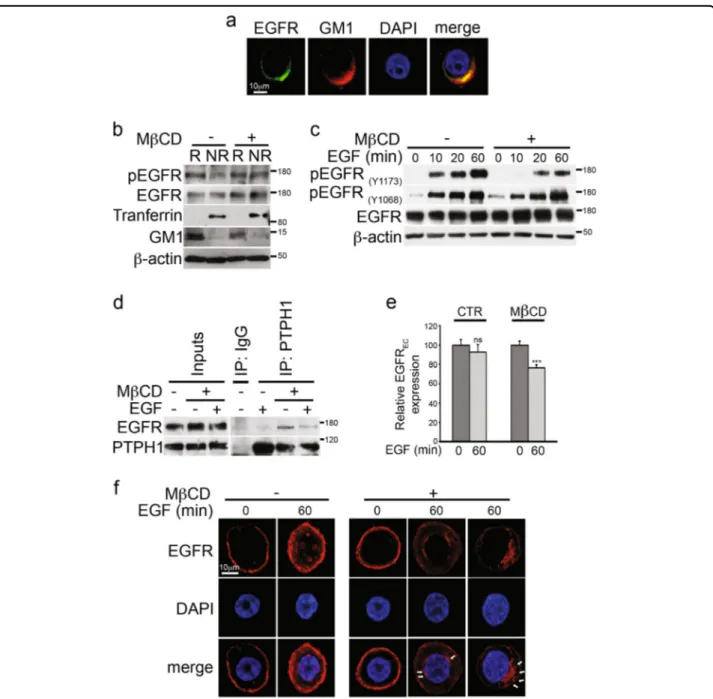
rafts of TNBC cells may correlate with their resistance to EGFR TKI-induced growth inhibition. First, we confirmed the presence of EGFR within lipid rafts by using bio-chemical and confocal microscopy analyses: Fig.5a shows that EGFR (green) strongly colocalizes with GM1 (red), a lipid raft glycosphingolipid specifically recognized by the Cholera toxin subunit B. Biochemical rafts isolation shown in the Fig. 5b confirms these data. Notably, the
tyrosine-pEGFR expression, essential for its functional activity36and predictive for target therapy efficiency with
TKIs30appears to be exclusive of raft compartment, as it
moved to the non-rafts fractions in the presence of Methyl-β-cyclodextrin (MβCD), a drug which removes cholesterol from the plasma membrane, thus disrupting the integrity of membrane rafts microdomains (Fig. 5b). Interestingly, after MβCD treatment, we observe a clear defect in the increase of EGF-induced tyrosine phos-phorylation of EGFR at 1173 residue (pEGFRY1173) but not at 1068 residue (pEGFRY1068) (Fig.5c), thus suggest-ing the presence of potential different roles between EGFR phosphorylation pattern and function of different tyrosine phosphorylation sites30. These data indicate a
Fig. 3 Notch3 downregulation (but not Notch1) sensitizes TNBC cells to TKI-gefitinib. a–d Left panels: inhibition of a, b MDA-MB-468 and c, d BT-549 cell growth was observed after gefitinib (GEF) treatment combined with Notch3 silencing in a, c but not with b, d Notch1 silencing. All the right panels showed in thefigure represent western blotting of total extracts from cells described above against Notch3 (N3IC) and Notch1 (N1IC), to
control the efficiency of the a, c Notch3 and b, d Notch1 silencing, respectively. Anti-β-actin was used as a loading control. All data are representative of at least three independent experiments, each in triplicate. Results shown in a, b, c, and d are expressed as the means average deviations and P-values were calculated using Student’s T-test (i.e., ns not significant; P > 0.05, *P ≤ 0.05, **P ≤ 0.01, ***P ≤ 0.001)
Fig. 4 Combined Notch3 and EGFR targeting induces EGFR internalization but defect the nuclear-activated EGFR localization. a FACS analysis of the EGFR surface expression (EGFREC) in MDA-MB-468 cells treated with gefitinib (GEF) alone or in combination with Notch3-silencing
(siN3+ GEF) or Notch1 silencing (siN1 + GEF) for 6 days. b Whole cell extracts (WCE) and c nuclear extracts from the same cells used in a were immunoblotted with anti-EGFR and anti-pEGFR(Y1173)antibodies, to evaluate the EGFR expression and phosphorylation, and with anti-N3ICor
anti-N1Val1744antibody to control the efficiency of Notch3 (left panels) or Notch1 (right panels) silencing, respectively. Anti-lamin B and anti-tubulin were
used as fraction markers; anti-β-actin was used as a loading control. d, e Proliferation analysis by BrdU assay (see Matherials and Methods section): compared with control cells (siCTR+ GEF), the percentage of BrdU+cells is lower after Notch3 silencing plus d GEF (siN3+ GEF) and not after Notch1 silencing plus e GEF (siN1+ GEF). All data are representative of at least three independent experiments, each in triplicate. Results are expressed as the means average deviations and P-values were calculated using Student’s T-test (i.e., *P ≤ 0.05)
Diluvio et al. Oncogenesis (2018) 7:42 Page 7 of 15
Fig. 5 Rafts depletion induces endogenous EGFR-PTPH1 interaction, EGFR dephopshorylation, and its intracellular arrest in MDA-MB-468 TNBC cells. a Immunofluorescence assay (IF) was performed by using anti-EGFR (green) and anti-GM1 (red) antibodies to reveal the endogenous EGFR-rafts colocalization, shown in yellow (merge). Nuclei were DAPI labeled (blue). b Raft (R) and non-raft (NR) fractions derived from Methyl- β-cyclodextrin (MβCD)-treated and untreated cells were used for immunoblot assay with anti-pEGFR(Y1173)(indicated as pEGFR) and anti-EGFR
antibodies, to test activated and total EGFR expression in rafts compartment, respectively. Anti-transferrin and anti-GM1 antibodies were used as a fraction markers. c Cells have been activated with EGF ligand for the times indicated, in the presence or absence of MβCD: the expression of phospho-EGFR at tyrosine 1173 and 1068 residues and total EGFR was determined in whole cell extracts by immunoblot analysis using the specific indicated antibodies. d–f MDA-MB-468 cells were treated with MβCD and stimulated with EGF for 60 min: control or anti-PTPH1 antibody immunoprecipitates were probed with anti-EGFR, to detect the EGFR-PTPH1 binding, and with the anti-PTPH1 antibody, to show PTPH1 immunoprecipitated protein levels. The inputs indicated in the panel shows 5% of each total lysate d. Relative EGFR extracellular expression (EGFREC)
was evaluated by FACS e. IF assay was performed by using anti-EGFR (red) antibody to reveal the endogenous EGFR intracellular localization. Nuclei were DAPI labeled (blue). White arrows indicated peri-nuclear EGFR localization in EGF stimulated MβCD-treated cells (f). a, f Representative single plane confocal IF images captured using a × 60 oil objective. Scale bar: 10μm. In both b and c, western blotting against the anti-β-actin was used as a loading control. All data are representative of at least three independent experiments, each in triplicate. Results shown in e are expressed as the means average deviations and P-values were calculated using Student’s T-test (i.e., ns, not significant P > 0.05, **P ≤ 0.01)
Diluvio et al. Oncogenesis (2018) 7:42 Page 8 of 15
possible relationship between rafts compartment integrity and EGFR/Y1173 dephosphorylation, which is known to have an important role in the therapeutic activity of EGFR TKI inhibition through the involvement of the tyrosine phosphatase H1 (PTPH1)37.
Several protein tyrosine phosphatases (PTPs) depho-sphorylate EGFR at Y1173 (alone or together with other residues)38,39. Among them, the PTPH1 specifically cata-lyzes EGFR/Y1173 dephosphorylation (and not EGFR/ Y1068 dephosphorylation), thus finally increasing non-TNBC BC sensitivity to TKIs, including GEF37. The EGFR/PTPH1 direct interaction is closely required to favor the therapeutic targeting of EGFR itself37. In agreement with this, we observed that MβCD-treated TNBC cells showed high levels of endogenous EGFR-PTPH1 interaction (Fig.5d), thus suggesting the possible PTPH1 involvement in the observed decreased levels of EGFR/Y1173 phosphorylation after rafts depletion (Fig.
5c). Interestingly, the EGFR-PTPH1 interaction dis-appears after MβCD plus EGF ligand (Fig.5d), probably due to the EGF-dependent endocytic events of ligand-activated EGFRs which may influence the kinetics of EGFR availability to PTPs-mediated dephosphorylation40. In keeping with this, we observed a decreased extra-cellular EGFR expression (EGFREC) in MβCD-treated cells with respect to untreated cells, upon stimulation with EGF (Fig.5e), despite the natural slowdown of EGFR endocytic trafficking in MDA-MB-468 cells due to their known saturated endocytic machinery41. In agreement with previous data42, our results suggest that rafts depletion may allow the internalization of ligand-occupied EGFR. Following ligand binding and receptor phosphorylation/activation, pEGFR is endocytosed and commonly transported to lysosome where it is degra-ded43. In our experiments, we do not observe decreased levels of total EGFR expression after rafts depletion (Fig.
5c, d), thus suggesting that removal of EGFR from the cell surface observed in MβCD-treated cells may be correlated to a different mechanism of EGFR downregulation, not involving receptor degradation. Since it has been demonstrated that many tumor cells which overexpress EGFR, including the MDA-MB-468 cells, have limited ligand-stimulated EGFR degradation44 and that tyrosine dephosphorylation of EGFR is correlated with an increased EGFR stability37, we wanted to know where the EGFR accumulated after MβCD treatment, in order to completely understand how signaling by the EGFR is terminated. To this purpose, cells were treated with or without MβCD and stimulated with EGF ligand for 60 min, followed by the immunostaining with anti-EGFR antibody (Fig. 5f): confocal analysis shows that rafts depletion correlates with the accumulation of EGFR at a peri-nuclear level (white arrows) whereas the majority of MβCD-untreated cells (EGF stimulated) show spots of
nuclear EGFR, which represents a specific localization known to be associated with resistance to EGFR-targeted therapies31. In addition, the same control cells stimulated with EGF ligand show persistent high levels of EGFR cell surface expression (Fig. 5f), thus confirming the
saturation of the endocytic machinery previously mentioned41.
Together, these data indicate that EGFR trafficking is retained outside the nucleus in MDA-MB-468 TNBC cells in response to the rafts-disrupting agent, MβCD.
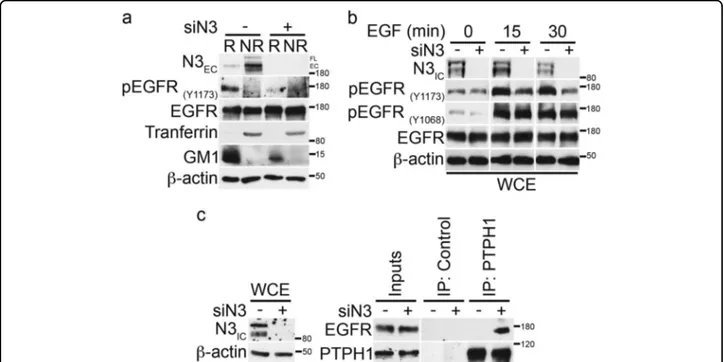
Notch3 inhibition by siRNA mimics rafts depletion effects on EGFR in TKI-resistant TNBC cells
We have previously shown that Notch3 receptor con-stitutively localizes to lipid rafts of Notch3 overexpressing lymphocytes, thus contributing to sustain the signaling pathways responsible of the T-cell leukemia develop-ment45. Here wefirst hypothesized that both Notch3 and EGFR receptors could share the same localization to directly interact, leading to the observed EGFR-TKI resistance process in TNBC cells. Surprisingly, both confocal analysis (Supplementary Figure S4a, upper panels) and biochemical rafts isolation with or without MβCD treatment (Supplementary Figure S4b) show that in MDA-MB-468 cells Notch3 receptor (N3EC) is widely expressed in all the cell surface, whereas Notch1 receptor appears to be restricted to lipid rafts microdomains (Supplementary Figure S4a, lower panels, and S4b). Thus, Notch3 and EGFR do not completely colocalize (Supple-mentary Figure S4c, upper panels), whereas Notch1 shows a strong rafts colocalization with EGFR (Supplementary Figure S4c, lower panels). Notably, by in situ proximity ligation assay (PLA), we still observed the endogenous Notch3/EGFR complex all around the cell membrane (Supplementary Figure S4d) while Notch1/EGFR complex seems to be mainly restricted to a limited portion of the membrane (Supplementary Figure S4e), reflecting their strictly shared localization (Supplementary Figure S4c, lower panels).
These results suggest the existence of a different rela-tionship between Notch3 or Notch1 and EGFR in TNBC. However, in order to deepen inside the molecular mechanism related to the TKI-GEF resensitization of TNBC cells observed only when Notch3 (and not Notch1) is depleted (Fig. 3), here we further investigated how the EGFR-rafts localization could be influenced when Notch3 is depleted. Similar to what happens in MβCD-treated cells (Fig. 5b-d), in the absence of Notch3 the tyrosine phosphorylation of EGFR at 1173 residue (pEGFRY1173) disappears and this event does not involve receptor degradation, as EGFR total levels remain unchanged (Fig.
6a). Notably, after Notch3 depletion, we observed a clear defect in the increasing levels of EGF-induced tyrosine phosphorylation of EGFR at 1173 residue (pEGFRY1173)
Diluvio et al. Oncogenesis (2018) 7:42 Page 9 of 15
but not at 1068 residue (pEGFRY1068) (Fig.6b), as already shown after rafts depletion (Fig.5c). For this reason, we further investigated whether Notch3 could influence the EGFR/Y1173 dephosphorylation by the phosphatase PTPH1, by using co-immunoprecipitation assay. In agreement with the above results (Fig. 5d), we observed that the absence of Notch3 is able to induce the endo-genous EGFR/PTPH1 interaction (Fig.6c), thus suggest-ing a possible link between Notch3, EGFR-rafts localization and EGFR dephosphorylation event by PTPH1.
In addition, we also observed that the absence of Notch3 correlates with a rapid and persistent EGFR downregulation from the cell surface, as revealed by the decrease of EGFRECmeanfluorescence intensity (MFI) in Notch3-depleted cells (siN3) with respect to control cells (siCTR) after EGF stimulation (Fig. 7a, upper panel). As expected, treatment of MDA-MB-468 cells with EGF until 270 min results in an increased EGFR surface expression (Fig. 7a, upper panel), also supported by the unchanged levels of total EGFR protein (Fig. 7a, lower panels), as previously reported (Fig. 5f and41). Interestingly, despite the increased EGFR internalization observed in the
absence of Notch3, although the pEGFRY1173expression decreases, the EGFR total levels does not change, thus suggesting that Notch3 depletion (such as rafts depletion) could correlate with an increased dephopshorylated EGFR endocytosis followed by its intracellular shuttling block-ade rather than sorting for intracellular degradation. Using immunofluorescence staining, we obtained addi-tional evidence in support of the Notch3-depletion dependence of EGFR intracellular fate. As shown in the Fig. 7b, after 2 h of EGF stimulation combined with Notch3 silencing, we observed that EGFR localizes pre-ferentially at a peri-nuclear level, similarly to what observed after MβCD treatment (Fig.5f). Interestingly, a few cells show a similar EGFR staining also without EGF stimulation (Fig. 7b, see white arrows), thus suggesting that Notch3, alone, may influence the EGFR internaliza-tion also through ligand-independent mechanisms (data not shown).
Together these results demonstrate that Notch3 depletion mimics the effects of rafts depletion on EGFR, as the Notch3 silencing correlates with EGFR depho-sphorylation (by PTPH1) and its persistent internaliza-tion, followed by intracellular arrest.
Fig. 6 Notch3 downregulation induces EGFR dephosphorylation by promoting the endogenous EGFR/PTPH1 interaction. a Raft (R) and non-raft (NR) fractions derived from 6 days of Notch3-silenced cells were used for immunoblot assay with anti-N3EC, anti-pEGFR(Y1173), and anti-EGFR
antibodies, to test the effect of Notch3 downmodulation on EGFR-rafts localization. Anti-transferrin and anti-GM1 were used as a fraction markers. b Cells have been activated with EGF ligand for the times indicated, combined or not with Notch3 silencing for 3 days: the expression of phospho-EGFR at tyrosine 1173 and 1068 residues and total phospho-EGFR was determined by immunoblot analysis using the specific indicated antibodies. c Control or anti-PTPH1 antibody immunoprecipitates from control and Notch3-silenced cells were probes with anti-EGFR, to detect the EGFR-PTPH1 binding, and with the anti-PTPH1 antibody, to show PTPH1 immunoprecipitated protein levels. The inputs indicated in the panel shows 5% of each total lysate (right panels). Whole cell extracts (WCE) were incubated with anti-N3ICantibody to control the efficiency of Notch3 silencing (left panels). In all panels
a, b and c, western blotting against the anti-β-actin was used as a loading control. The results are representative of three independent experiments
Diluvio et al. Oncogenesis (2018) 7:42 Page 10 of 15