Scuola doctorale in Biologia (XXV Ciclo)
Sezione “Scineze Biomoleculari e Cellulari”
The involvement of Arabidopsis thaliana
polyamine oxidases in plant development
and defence responses
Dr. Abdellah Ahou
Docente Guida: Prof.ssa Paraskevi Tavladoraki
Coordinatore: Prof. Paolo Mariotini
Scuola doctorale in Biologia (XXV Ciclo)
Sezione “Scienze Biomolecolari e Cellulari”
The involvement of Arabidopsis thaliana
polyamine oxidases in plant development
and defence responses
Coinvolgimento delle poliammino ossidasi di
Arabidopsis thaliana nei processi di sviluppo
e di difesa delle piante
Dr. Abdellah Ahou
Docente Guida: Prof.ssa Paraskevi Tavladoraki
Coordinatore: Prof. Paolo Mariottini
I
TABLE OF CONTENTS
ABBREVIATIONS ... III ABSTRACT ... IV
INTRODUCTION: ... 1
Polyamines: General characteristics ... 1
Polyamine biosynthesis ... 2
Polyamine catabolism... 5
Copper-containing amine oxidases ... 5
Polyamine oxidases... 6
Arabidopsis polyamine oxidases ... 8
Physiological roles of polyamines ... 13
Physiological roles of polyamine catabolism ... 16
Plant polyamine catabolism under abiotic and biotic stress conditions ... 21
RESULTS ... 23
Studies on the physiological roles of AtPAO1 ... 23
Polyamine levels in atpao1 loss-of-function mutant ... 23
Physiological studies on atpao1 mutant ... 25
Studies on the physiological roles of AtPAO2-4 gene family ... 26
AtPAO4 space-temporal expression pattern ... 27
Characterization of single and multiple mutants for the peroxisomal AtPAO2-4 subfamily. ... 28
Polyamine content in double and triple mutants for AtPAO2-4 gene family ... 29
Stomatal closure in mutants for AtPAO2-4 gene family... 30
Water loss in atpao243 triple knockout mutant ... 32
Effect of dehydration on atpao243 triple mutant growth ... 33
Growth of atpao243 triple mutants in the presence of ABA ... 34
Germination of atpao243 triple mutants in the presence of sucrose .. 35
Studies on AtPAO5 physiological roles ... 36
AtPAO5 sequence analysis ... 36
Phylogenetic relationship of AtPAO5 with other plant PAOs ... 37
Purification of recombinant AtPAO5 from Arabidopsis transgenic plants ... 39
Substrate specificity of recombinant AtPAO5 ... 41
II
Subcellular localization of AtPAO5... 42 Effect of the proteasomal inhibitorMG132 on AtPAO5 accumulation ... 44 Polyamine levels in AtPAO5 over-expressing Arabidopsis plants and
atpao5 knock out mutant ... 47
Effect of cytokinins on Arabidopsis root growth and differentiation . 47 DISCUSSION ... 50 PA levels in the atpao mutants and AtPAO over-expressing plants ... 50
AtPAO physiological roles ... 50
AtPAO5 substrate specificity, reaction products and post-translational regulation ... 52 REFERENCES ... 55 SUPPLEMENTARY DATA ... 70
III
ABBREVIATIONS
ABA Abscisic acid
ACL5 ACAULIS5
ADC Arginine decarboxylase
ADH Aldehyde dehydrogenase
Arg Arginine
ATAO Arabidopsis thaliana copper-containing amine oxidase
AtPAO BAP BSAO
Arabidopsis thaliana polyamine oxidase
6-benzylaminopurin Bovine serum amine oxidase
Cad Cadaverine
CMV Cucumber mosaic virus
CoA Coenzyme A
CuAO Copper-containing amine oxidase
Dap 1,3-diaminopropane
GABA γ-aminobutyric acid
HDL Hydrolase
HR Hypersensitive response
HvPAO Hordeum vulgare polyamine oxidase
JA Jasmonic acid
MdPAO Malus domestica polyamine oxidase
NO Nitric oxide
Nor-Spd Norspermidine Nor-Spm Norspermine
NtPAO Nicotiana tabacum polyamine oxidase
ODC Ornithine decarboxylase
Orn Ornithine
OsPAO Oryza sativa polyamine oxidase
PAO Polyamine oxidase
PCD Programmed cell death
Pro Proline
Put Putrescine
SAM S-adenosylmethionine
SAMDC S-adenosylmethionine decarboxylase
SMO Spermine oxidase
Spd Spermidine SPDS Spermidine synthase Spm Spermine SPMS Spermine synthase SSAT Spd/Spm N1-acetyltransferase Ther-Spm Thermospermine
TPQ 2,4,5-trihydroxyphenylalanine quinone cofactor TYMV Turnip yellow mosaic virus
IV
ABSTRACT
The polyamines (PAs) putrescine (Put), spermidine (Spd), and spermine (Spm) are small aliphatic polycations found in all living cells. They are involved in several cellular processes and play important roles in morphogenesis, growth, differentiation and senescence. In plants, they are also implicated in defence responses to various biotic and abiotic stresses. PA homeostasis is strictly regulated through anabolic and catabolic
processes, but also through conjugation, transport and
compartimentalization.
Polyamine oxidases (PAOs) are FAD-dependent enzymes involved in PA catabolism. PAOs from monocotyledonous plants, such as the apoplastic maize PAO (ZmPAO), oxidize spermine (Spm) and spermidine (Spd) to produce 1,3-diaminopropane, H2O2 and an aminoaldehyde and are
considered involved in a terminal catabolic pathway of PAs. Conversely, animal PAOs and spermine oxidases (SMOs) oxidize Spd, Spm and/or their acetyl-derivatives to produce Put and Spd, respectively, in addition to H2O2
and 3-aminopropanal and are thus considered involved in a PA back-conversion pathway.
In Arabidopsis thaliana, five PAO genes (AtPAO1-5) are present with a varying amino acid sequence homology to ZmPAO and subcellular localization (putative cytosolic for AtPAO1 and AtPAO5 and peroxisomal for AtPAO2, AtPAO3 and AtPAO4). Furthermore, following heterologous expression in bacteria it was shown that AtPAO1 oxidizes Spm but not Spd, whereas AtPAO2, AtPAO3 and AtPAO4 oxidize both Spm and Spd. Conversely, AtPAO5 substrate specificity has not been determined so far since production of the recombinant protein in various heterologous systems has not been successful. The four characterized AtPAOs are also active towards the uncommon PAs thermospermine (Ther-Spm) and norspermine (Nor-Spm). In particular, AtPAO1 shows a higher catalytic activity towards Ther-Spm and NorSpm than towards Spm, which suggests that these two uncommon PAs may be the physiological substrates of this enzyme. This is of particular interest because it has been recently shown the existence in
Arabidopsis of an enzyme able to synthesize Ther-Spm and a
loss-of-function mutant for this gene shows a severely dwarfed phenotype. Another important characteristic of the four Arabidopsis PAOs is their involvement in a PA back-conversion pathway, producing Spd from Spm and Put from Spd, similarly to the animal PAOs / SMOs and contrary to ZmPAO.
Studies on the tissue- and organ-specific expression pattern of AtPAO1,
AtPAO2, AtPAO3 and AtPAO5 using AtPAO::GFP-GUS transgenic
V
the four AtPAOs, such as in the transition region between cell division and elongation zones of roots and anther tapetum for AtPAO1, in columella, stipules and pollen for AtPAO2, in hypocotyls and roots, stipules, columella, trichomes, guard cells and pollen for AtPAO3, and in the vascular system of roots and hypocotyls for AtPAO5. These studies also evidenced increased expression of AtPAO1 in roots and of AtPAO2 in guard cells following treatment with the stress-related plant hormone abscisic acid (ABA).
In the present work, the study on the tissue- and organ-specific expression pattern of the five AtPAOs was completed analysing AtPAO4 promoter activity. In particular, histochemical GUS staining of
AtPAO4::GFP-GUS transgenic Arabidopsis plants evidenced that AtPAO4
is expressed in the roots (from the meristematic/elongation transition region up to the hypocotyl–root junction site), in the guard cells, in the base of very young and completely closed flower buds, in anther tapetum and in mature pollen grains. These data together with data from promoter analysis of the other four AtPAOs indicate distinct physiological roles for the various AtPAOs during seedling growth and flower development and suggest functional diversity inside the AtPAO gene family.
To determine the physiological roles of the various AtPAOs, loss-of-function T-DNA insertional mutants (atpao1, atpao2, atpao3, atpao4 and
atpao5) have been previously obtained from the NASC collection of
Arabidopsis seeds and homozygous mutant lines have been selected. In the present study, double (atpao2/atpao4 and atpao3/atpao4, atpao3/atpao2) and triple (atpao2/atpao4/atpao3) mutants for the peroxisomal AtPAOs as well as the double atpao1/atpao5 mutant for the two AtPAOs with predicted cytosolic localization were also obtained through sexual crossings.
The atpao1 single mutant was analyzed for the levels of the common PAs Put, Spd and Spm as well as of the uncommon PA Ther-Spm, evidencing no statistically significant variation comparing to the wild-type plants. This may be due either to gene redundancy or to the activation of homeostatic mechanisms and may exclude the possibility that Ther-Spm is the physiological substrate of AtPAO1. The atpao1 single mutant was also analyzed for germination and growth rate under physiological and stress conditions, but also in these cases no variation was observed as compared to the wild-type plants. Similar studies on the atpao1/atpao5 double mutant are in progress.
Analysis of PA levels in the single and triple mutants for the three peroxisomal AtPAOs showed some alterations in the atpao2/atpao4/atpao3 triple mutant. Furthermore, since all three AtPAO2, AtPAO3 and AtPAO4 are highly expressed in the guard cells, specialized cells surrounding stomata pores, stomata movements were evaluated evidencing reduced
VI
ABA- and PA-mediated stomata closure in the corresponding mutant plants as compared to wild-type plants. Furthermore, the atpao2/atpao4/atpao3 triple mutant appeared more tolerant to dehydration and ABA treatment. Altogether, these data suggest the involvement of the three peroxisomal AtPAOs in the ABA-mediated signaling network. On the other hand, germination and growth rate of the atpao2/atpao4/atpao3 triple mutant in the absence of sucrose was shown to be delayed comparing to the wild-type plants. The underlying mechanisms in these phenotypical alterations in the
atpao2/atpao4/atpao3 triple mutant are currently under investigation.
In the present study, it was also possible to express AtPAO5 in
35S::AtPAO5-6His transgenic Arabidopsis plants, to partially purify the
corresponding recombinant protein and to determine substrate specificity and reaction products. In particular, it was shown that AtPAO5 has indeed PAO activity, catalyzing the oxidation of Spm, N1-acetyl-Spm, Ther-Spm and Nor-Spm through a PA back-conversion pathway. Furthermore, confocal analysis of 35S::GFP-AtPAO5 and 35S::AtPAO5-GFP transgenic Arabidopsis plants indicated that AtPAO5 is a cytoplasmic protein undergoing proteasomal control. It was also shown cytokinin-inducible expression of AtPAO5 as well as AtPAO5 involvement in the control of xylogenesis by cytokinins. Experiments are in progress to determine the physiological significance of AtPAO5 regulation by the proteasomal pathway as well as to unravel the mechanisms by which AtPAO5 is involved in the cytokinin-mediated pathways.
This study represents the starting point to understand the distinct physiological roles of the different PA catabolic pathways in plants during development and defense responses which may permit the application of biotechnological strategies to transfer increased yield and stress-tolerance traits to crops of agronomical relevance.
1
INTRODUCTION:
Polyamines: General characteristics
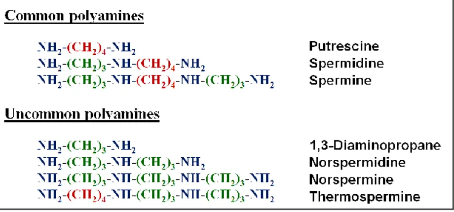
Polyamines (PAs) can be considered as one of the oldest group of substances known in biochemistry (Cona et al., 2006). They are small aliphatic polycation molecules having variable hydrocarbon chains and two or more primary and secondary amino groups. The di-amine putrescine (Put), the tri-amine spermidine (Spd) and the tetra-amine spermine (Spm) (Fig. 1) are the most common PAs in eukaryotes (Galston and Sawhney, 1990). In addition, other PAs, such as 1,3-diaminopropane (Dap), cadaverine (Cad), thermospermine (Ther-Spm), norspermidine (Nor-Spd) and norspermine (Nor-Spm) (Fig. 1) are found in many organisms as minor components of the cellular PA pool and are referred to as uncommon PAs (Tavladoraki et al., 2011).
Fig. 1. Structures of common and uncommon PAs
PAs may be present in a free soluble form, but also bound to macromolecules such as proteins and nucleic acids or conjugated to phenolic compounds (Bakhanashvili et al., 2005; Groppa and Benavides, 2008). In particular, in plants, PAs can be further conjugated to hydroxycinnamic acid forming hydroxycinnamic acid amides (HCAAs; Bagni and Tassoni, 2001). Indeed, coumaroyl-Put, feruloyl-Put, coumaroylagmatine, dicoumaroyl-Spd, diferuloyl-Spd, and diferuloyl-Spm are present in a wide range of plant species (Martin-Tanguy, 1997; Grienenberger et al., 2009; Luo et al., 2009). Although these compounds were discovered many years ago, their physiological role(s) remain largely
2
unknown. HCAAs have been associated with a wide range of growth and developmental processes, including cell division, flowering, cell-wall cross-linking, as well as responses to environmental challenge (Bouchereau et al., 1999; Luo et al., 2009; Bassard et al., 2010). Furthermore, data suggesting turnover and translocation of PA conjugates as well as interconversion between free and conjugated precursors have been reported (Bassard et al., 2010).
The overall intracellular concentration of PAs is in the range of several hundred micromolars to a few millimolars and is tightly regulated, as higher levels of PAs are toxic to cells and lead to cell death. PA homeostasis in plants correlates with several important physiological functions, including the control of the N:C balance (Mattoo et al., 2006; Moschou et al., 2012), stress responses (Alcázar et al., 2011), xylem differentiation (Muñiz et al., 2008; Tisi et al., 2011), pollen tube growth (Wu et al., 2010), membrane fluidity, and protein regulation(Baron and Stasolla, 2008; Takahashi and Kakehi, 2010). The intracellular pool of free PAs depends not only on its synthesis, but also on conjugation, transport, degradation and back-conversion (Tiburcio et al., 1997; Angelini et al., 2010; Moschou et al., 2012).
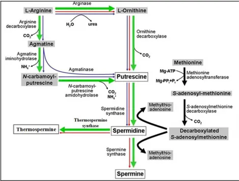
Polyamine biosynthesis
The pathways of PA biosynthesis have been established for many organisms (Bagni and Tassoni, 2001; Wallace et al., 2003) . The general mechanism of biosynthesis is conserved from bacteria to animals and plants (Tabor and Tabor, 1984) and begins from the synthesis of the precursor Put, followed by successive additions of aminopropyl groups to produce Spd and Spm (Fig. 2). It has been suggested that plants have acquired a part of the PA biosynthetic pathway from an ancestral cyanobacterial precursor of the chloroplast (Illingworth et al., 2003). Therefore, it can be assumed that this is an ancient metabolic route, which is also present in all organisms (Minguet et al., 2008).
Put is formed directly by the decarboxylation of ornithine (Orn), via ornithine decarboxylase (ODC; EC 4.1.1.17), or indirectly from arginine (Arg) by arginine decarboxylase (ADC; EC 4.1.1.19) via agmatine (Agm) (Tabor and Tabor, 1985). The biosynthesis of Put from Arg requires the activity of three consecutive enzymes: ADC, agmatine iminohydrolase (AIH; EC 3.5.3.12) and N-carbamoylputrescine amidohydrolase (CPA; EC 3.5.1.5; Alcázar et al., 2011).
In Arabidopsis thaliana, Put is produced exclusively through the ADC pathway, since no ODC gene has been identified in the sequenced genome
3
of this plant and the corresponding enzyme activity has not been detected (Hanfrey et al., 2001; Alcázar et al., 2010b). In particular, two different genes encoding ADC (ADC1 and ADC2) have been described (Soyka and Heyer, 1999). Although ADC1 and ADC2 show 80% homology in amino acid sequence to each other, they exhibit a different expression pattern:
ADC1 is expressed in all tissues, whereas ADC2 is mainly expressed in
cauline leaves and siliques, and is induced by different abiotic stresses (Soyka and Heyer, 1999; Perez-amador et al., 2002; Urano et al., 2003). In animals, Put is mainly synthesized through the ODC pathway, the ADC pathway being just a minor pathway in specific mammalian tissues (Gilad et
al., 1996) (Fig. 2). In bacteria, in addition to ADC and ODC, another
enzyme is present involved in Put biosynthesis, agmatinase, which directly produces Put from agmatine. Furthermore, in bacteria Spm is not synthesized, since no SPMS gene is present (Wortham et al., 2007).
Put is converted into Spd and Spm through two sequential reactions catalyzed by two closely related but distinct enzymes, the Spd synthase (SPDS; EC 2.5.1.16) and Spm synthase (SPMS; EC 2.5.1.22), respectively, which add aminopropyl groups. These aminopropyl groups are donated by decarboxylated S-adenosylmethionine (dcSAM), which is formed by decarboxylation of S-adenosylmethionine (SAM) by SAM decarboxylase (SAMDC; EC 4.1.50) (Fig. 2; Alcázar et al., 2011). Spd can be also converted to Ther-Spm by Ther-Spm synthase (TSPMS or ACL5) which add an aminopropyl group at the N1-(aminopropyl)end of Spd, differently from SPMS which adds the aminopropyl group at the N8-(aminobutyl) end of Spd. In Arabidopsis, Spd synthase is encoded by two genes (SPDS1 and
SPDS2) whereas SPMS and TSPMS are encoded by single genes (Hanzawa et al., 2002; Knott et al., 2007). SPDS1 shows high sequence similarity to
SPDS2 (82.7% amino acid identity), whereas SPMS shows only 56% identity with both SPDS1 and SPDS2, respectively. Exon structure is conserved between SPDS1, SPDS2, and SPMS whereas TSPMS has a completely different genomic organization. SPMS interacts with SPDS1 and SPDS2, to form ‘‘metabolon’’ complexes, while TSPMS does not interact with SPDS (Panicot et al., 2002).
The Arabidopsis genome carries at least four genes coding for SAMDCs (SAMDC1-4) (Urano et al., 2004). They have an unusually long 5’-UTR where two uORFs are well conserved (Franceschetti et al., 2001) which control the PA levels. The first uORFs called tiny uORFs which aredistal to the 5’ end are 3–4 codons long, while the second one termed small uORFs consists of 50–54 codons. The small uORF-encoded peptide is responsible for translational repression of the main ORF under conditions of excess PA concentration; while the tiny uORF is required for induced translation of the
4
main ORF during conditions of low PA concentration (Hanfrey et al., 2002; 2005). In Arabidopsis, the sequences of the uORFs of SAMDC1 and SAMDC2 are highly conserved, while the uORFs of SAMDC3 and SAMDC4 are not complete which affects their expression (Urano et al., 2003).
SAMDC has an important role in the regulation of PA homeostasis in all organisms. Particularly in plants, it is considered to be the rate-limiting enzyme for the synthesis of Spd and Spm(Kusano et al., 2008). The activity of SAMDC is positively regulated by Put and negatively regulated by Spd and Spm, thus making the cellular levels of dcSAM responsive to the demands of the PA biosynthetic pathway (Pegg, 1986; Kameji and Pegg, 1987; Xiong et al., 1997).
Fig. 2. PA biosynthetic pathways. Plant pathway is indicated by green bold arrows. Blue and
red arrows indicate bacterial and animal pathways, respectively. Figure Modified from Kusano
5
Polyamine catabolism
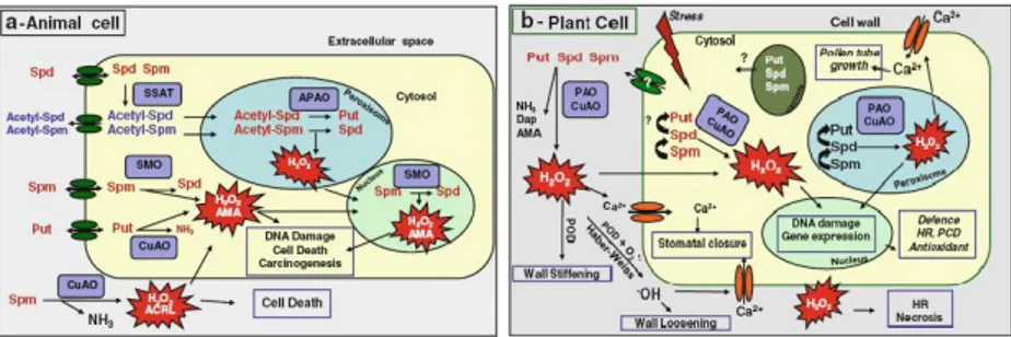
Two classes of amine oxidases are implicated in PA catabolism, the copper-containing amine oxidases (CuAOs) and the FAD-dependent amine oxidases (PAOs) (Tavladoraki et al., 2011).
Copper-containing amine oxidases
CuAOs are homodimeric enzymes; each subunit of 70-90 kD contains a copper ion and a 2,4,5-trihydroxyphenylalanine quinone cofactor generated by a post-translational autocatalytic modification (Medda et al., 1997; Angelini et al., 2010). They catalyze the oxidation of Put and Cad at their primary amino groups. The reaction products from Put are H2O2, NH4+ and
4-aminobutanal (Fig. 3). The latter spontaneously cyclises to generate Δ1 -pyrroline and can be further converted to γ-aminobutyric acid (GABA) by an aldehyde dehydrogenase. GABA is subsequently transaminated and oxidized to succinic acid, which is incorporated into the Kreb’s cycle, ensuring the recycling of carbon and nitrogen from Put (Tavladoraki et al., 2011; Moschou et al., 2012) CuAOs are also able to oxidize Spd and Spm, although with a lower affinity than Put, producing 4-aza-8-amino-octan-1-al and 4,9-diaza-dodecan-1,12 dialdehyde, respectively, in addition to H2O2,
and NH4+. In particular, the activities of pea CuAO (PsAO;Tipping and
McPherson, 1995) with Put, Spd and Spm are at a ratio of 100:35:0.3, while those of the lentil enzyme at a ratio of 100:42:20 (Sebela et al., 2001). Only the animal serum CuAOs, such as bovine serum amine oxidase (BSAO), oxidize preferentially Spd and Spm. The two aminoladehydes produced from CuAO-mediated Spd and Spm oxidation may undergo spontaneous degradation, when not previously further oxidized by aldehyde dehydrogenases, forming Put and Spd, respectively, and the highly toxic aldehyde acrolein (Fig. 3; (Tavladoraki et al., 2012).
Plant CuAOs occur at high levels in the extracellular space of several
Fabaceae species (Federico and Angelini, 1991; Cona et al., 2006),
reaching levels as high as 2 to 10 U/gfw in etiolated young seedlings (Rea et
al., 1998). These CuAOs have a quite high catalytic activity, the kcat with the best substrate being about 260 s-1, conversely to the animal CuAOs which have a lower catalytic activity; for example the kcat of the bovine serum CuAOs is 2 s-1 (Pietrangeli et al., 2007). In A. thaliana, sevenCuAOs
have been identified by database search. Only one of them (At4g14940;
AtCuAO1) was partially characterized and is active with Put and Spd
6
Arabidopsis CuAOs are targeted to the secretory pathway but their final subcellular sorting has still to be determined.
In plants, a class of CuAOs is also present which preferentially oxidize
N-methyl-Put although they oxidize also Put and Cad. In particular, in Nicotiana tabacum, two N-methyl-Put oxidase genes (NtMPO1 and NtMPO2) are present (Heim et al., 2007; Katoh et al., 2007) which are
specifically expressed in the roots and are up-regulated by the plant hormone jasmonate (JA). The two NtMPOs share essential structural motifs with the other CuAOs and have high sequence homology to AtCuAO1 and to PsAO (Heim et al., 2007; Katoh et al., 2007). A putative N-methyl-Put oxidase is also present in Arabidopsis (At2g42490; AtMPO), which has a high sequence homology with NtMPO1 and NtMPO2. Phylogenetic tree shows that NtMPO1, NtMPO2 and AtMPO form a distinct clade and are separated from PsAO, CuAO1 and the other Arabidopsis CuAO-like proteins (Katoh et al., 2007; Moschou et al., 2012) . In contrast to the CuAOs, NtMPO1, NtMPO2 and AtMPO are predicted to be localized to peroxisomes (Heim et al., 2007). The oxidation of N-methyl-Put by this class of CuAOs produces 4-methylaminobutanal, which spontaneously cyclises to give rise to the N-methylpyrrolinium cation, a precursor of the pyridine and tropane alkaloids, thus driving the flow of N away from PA biosynthesis towards alkaloids (Fig. 3).
The expression of some plant CuAOs has been shown to be modulated during development, pathogen attack, wound healing and salt stress. Plant hormones, for example JA and abscisic acid (ABA), were also shown to regulate expression of plant CuAOs ( Møller and McPherson, 1998; Cona et
al., 2006; Quinet et al., 2010; Toumi et al., 2010). Moreover, CuAO activity
is higher, and increases to a greater extent upon infection, in chickpea cultivars resistant to the fungus Ascochyta rabiei compared with the susceptible ones was shown to be strongly impaired by in vivo CuAO inhibition (Angelini et al., 1993). Moreover, infection of Arabidopsis plants with nematodes also induces differential expression of AtCuAO1 (Møller and McPherson, 1998).
Polyamine oxidases
PAOs are monomers of 50-60 kDa bearing a non-covalently bound FAD molecule ( Tavladoraki et al., 1998; Binda et al., 1999). They catalyze the oxidation of Spm, Spd and/or their acetylated derivatives at the secondary amino groups. PAO reaction products depend on the mode of substrate oxidation, which in turn depends on the mode of substrate binding inside the catalytic site resulting in the oxidation of a different carbon atom. On
7
the basis of the reaction products, PAOs can be classified in two families; those which terminally oxidize PAs and those catalyzing PA back-conversion. PAOs of the first family have been until now detected only in plants and bacteria. From the plant species, they are present at high quantities in particular tissues of plants belonging to Gramineae, such as maize (Zea mays), barley (Hordeum vulgare), oat (Avena sativa), wheat (Triticum aestivum) and rye (Secale cereale) (Federico et al., 1989; Federico and Angelini, 1991; Sebela et al., 2001; Stránská et al., 2007; Maiale et al., 2008; Angelini et al., 2010). In particular, in maize three genes (ZmPAO1, ZmPAO2 and ZmPAO3) have been identified encoding identical proteins (ZmPAO; (Cervelli et al., 2000), while in barley two genes (HvPAO1 and HvPAO2) have been cloned (Cervelli et al., 2001). The PAOs of this family oxidize the carbon at the endo-side of the N4 of Spd
and Spm, producing 4-aminobutanal and
N-(3-aminopropyl)-4-aminobutanal, respectively, in addition to 1,3-diaminopropane (Dap) and H2O2 (Fig. 3). The aminoaldehydes produced in the reaction spontaneously
cyclise to ∆1-pyrroline and 1,5-diazabicyclononane, respectively (Federico and Angelini, 1991; Sebela et al., 2001), while Dap can be converted to β-alanine by a Dap-aminotransferase, reported in bacteria but not yet in plants, and an aminoaldehyde dehydrogenase (AMADH; Fig. 3). β-Alanine in turn might be metabolized to the osmoprotectant alanine betaine by β-alanine N-methyltransferase (Fig. 3). Dap is also a precursor of the uncommon polyamines Nor-Spd and Nor-Spm (Fig. 3) which in plants are associated with stress tolerance (Cona et al., 2006). These PAOs have a cleavable N-terminal signal peptide which targets them to the apoplast. Only the barley PAO isoform HvPAO2, which has also a signal peptide for secretion, is localized to the vacuoles (Cervelli et al., 2004). The pH optima for the oxidation of the substrates vary among different species, but for most of the enzymes and for both substrates they are in the range of 5.5 to 6.8 (Federico and Angelini, 1991), which probably reflects their extracellular localization. Only HvPAO2, shows two different pH optima for the two substrates (5.5 for Spm and 8.0 for Spd; Cervelli et al., 2001). The so far characterized PAOs from the Gramineae are almost equally active with Spd and Spm. Furthermore, these PAOs are characterized by high specific activity (kcat in the range of 50-100 s-1) and affinity (Km in the range of 1-10 μM) for the two PAs, which may correlate to the low PA levels in the cellular compartment of enzyme accumulation. ZmPAO also cleaves N1-acetylSpd, N1-acetylSpm and N8-acetylSpd at the same C atom site and at the same optimal pH as it does with non-acetylated Spd and Spm (Federico et al., 1996). However, the enzyme is quickly inactivated during the reaction (Federico et al., 1996). ZmPAO is additionally active with
Nor-8
Spm and Ther-Spm, though with kcat values 10- to 30-fold lower than those towards Spm (Tavladoraki et al., 2006; Fincato et al., 2011). It has been demonstrated that Oat PAO is also active with Nor-Spd (Maiale et al., 2008).
The PAOs catalyzing back-conversion of PAs have been so far detected in animals, yeasts and in plants. Animal PAOs and yeast Saccharomyces
cerevisiae Spm-oxidase (Fms1) oxidize N1-acetyl-Spm, N1-acetyl-Spd, and
N1, N12-bis-acetyl-Spm at the carbon on the exo-side of N4-nitrogen to produce Spd, Put, and N1-acetyl-Spd, respectively, in addition to 3-acetamidopropanal and H2O2 with a pHoptimal of around 8.0 (Landry and
Sternglanz, 2003; Vujcic et al., 2003; Wu et al., 2003; Cona et al., 2006). In this catabolic pathway, PA acetylation is catalysed by the tightly regulated Spd/Spm N1-acetyltransferase (SSAT), which is the rate-limiting enzyme of this pathway (Wallace et al., 2003). Similarly, animal Spm oxidases (SMOs) and Fms1 oxidize Spm at the carbon on the exo-side of N4-nitrogen to produce Spd, 3-aminopropanal and H2O2 with a pHoptimal of around 8.0
(Wang et al., 2001; Vujcic et al., 2002; Cervelli et al., 2003; Landry and Sternglanz, 2003). 3-Aminopropanal and 3-acetamidopropanal can be further metabolized by an aminoaldehyde dehydrogenase (AMADH) to form β-alanine and N-acetyl-β-alanine, respectively which may be converted to the toxic acrolein (Fig. 3). The best so far characterized plant PAOs involved in PA back-conversion are those of Arabidopsis.
Arabidopsis polyamine oxidases
In A. thaliana, five PAO genes are present: AtPAO1 (At5g13700),
AtPAO2 (At2g43020), AtPAO3 (At3g59050), AtPAO4 (At1g65840) and AtPAO5 (At4g29720). AtPAO1, which has a predicted cytosolic
localization, shares with the extracellular ZmPAO a 45% homology at the amino acid level and a similar intron/exon organization (Tavladoraki et al., 2006). AtPAO1 oxidizes Spm but not Spd (Table 1), differently from ZmPAO but similarly to the animal SMO. It oxidises also the uncommon PAs Ther-Spm and Nor-Spm (Tavladoraki et al., 2006) with high efficiency which suggests that these two PAs may be its physiological substrates. In contrast, AtPAO1 has a low catalytic activity with N1-acetyl-Spm (Table 1).
AtPAO2, AtPAO3, and AtPAO4 display low sequence homology (23%-24%
homology) with ZmPAO and the other two AtPAOs, but a high sequence homology to each other (85% between AtPAO2 and AtPAO3, 58% between
AtPAO2 and AtPAO4, 50% between AtPAO3 and AtPAO4). Furthermore, AtPAO2, AtPAO3 and AtPAO4 have a very similar intron/exon organization
9
Fig. 3. Schematic representation of PA catabolic pathways and related matabolites in animals and plants. Green arrows indicate PA catabolic pathways in plants, blue arrows
indicate PA catabolic pathways in animals and black arrows indicate metabolic pathways related to PA catabolism. ALDH aldehyde dehydrogenase, ADC arginine decarboxylase, AMADH aminoaldehyde dehydrogenase, AMT β-alanine N-methyltransferase, DAT 1,3-diaminopropane-aminotransferase, GABA, HDL N-acetyl-β-alanine amidohydrolase, ODC
ornithine decarboxylase, PMT putrescine N-methyltransferase, SSAT spermidine-spermine N1
-acetyltransferase, SPDS spermidine synthase, SPMS spermine synthase, SRD Schiff-base reductase/decarboxylase, TSPMS thermospermine synthase. From Tavladoraki et al., 2012.
10
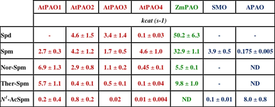
This, together with the elevated sequence homology to each other, suggest that these three Arabidopsis genes are recent derivatives from a common ancestor, thus forming a distinct PAO subfamily (AtPAO2–AtPAO4 subfamily). Interestingly, all the three members of this subfamily have a peroxisomal localization(Kamada-Nobusada et al., 2008; Moschou et al., 2008a). AtPAO2, AtPAO3 and AtPAO4 oxidize both Spd and Spm (Takahashi et al., 2010; Fincato et al., 2011). In particular, while AtPAO2 is equally active with Spm and Spd, AtPAO3 is 2-fold more active with Spd than with Spm and AtPAO4 is 10-fold more active with Spm than with Spd. AtPAO2, AtPAO3 and AtPAO oxidize also Nor-Spm and Ther-Spm though less efficienly than Spd and/or Spm (Table 1). Furthermore, the catalytic activity of all three peroxisomal AtPAOs towards N1-acetyl-Spm is very low (Table 1). The fifth Arabidopsis PAO gene (AtPAO5) has low sequence homology with the other four AtPAOs and a predicted cytosolic localization. However, information about its catalytic properties has not been still obtained.
AtPAO1, AtPAO2, AtPAO3 and AtPAO4 have different optimum pH (7.0–8.0) than the extracellular ZmPAO (optimum pH of 6.0; Polticelli et
al., 2005). In particular, the optimum pH for AtPAO1 catalytic activity is 8,
for AtPAO2 and AtPAO3 catalytic activity 7.5 and for AtPAO4 8.0 towards Spd, and is 7.0 towards Spm (Tavladoraki et al., 2006; Moschou et al., 2008b; Fincato et al., 2011). These differences in optimum pH among the various enzymes may reflect differences in subcellular localization and/or physiological role(s).
AtPAO1 AtPAO2 AtPAO3 AtPAO4 ZmPAO SMO APAO
kcat (s-1) Spd - 4.6 ± 1.5 3.4 ± 1.4 0.1 ± 0.03 50.2 ± 6.3 - - Spm 2.7 ± 0.3 4.2 ± 1.2 1.7 ± 0.5 4.6 ± 1.0 32.9 ± 1.1 3.9 ± 0.5 0.175 ± 0.005 Nor-Spm 6.9 ± 1.3 2.9 ± 0.8 1.1 ± 0.2 0.45 ± 0.1 5.5 ± 0.1 - ND Ther-Spm 5.7 ± 1.1 0.4 ± 0.1 0.5 ± 0.1 0.1 ± 0.04 9.8 ± 1.0 - ND N1-AcSpm 0.2 ± 0.4 0.8 ± 0.2 0.02 0.01 ± 0.004 ND 0.1 ± 0.01 8.0 ± 0.8
Table 1. Catalytic activity of recombinant AtPAOs, ZmPAO SMO and APAO. Data were
taken from Cervelli et al., 2003; Wu et al., 2003; Polticelli et al., 2005 and from Fincato et al., 2011. ND: not determined.
11
Analysis of the AtPAO reaction products evidenced that all characterized A. thaliana PAOs are involved in PA back-conversion (Tavladoraki et al., 2006; Kamada-Nobusada et al., 2008; Fincato et al., 2011), similarly to the animal PAOs/SMOs and in contrast to the extracellular PAOs from monocotyledonous plants characterized thus far, which are involved in a terminal PA catabolic pathway (Fig. 3). In this regard, the information so far available allows to propose the terminal catabolic pathway of PAs as specifically active in the extracellular compartments, while the PA back-conversion pathway as mostly intracellular (Fincato et al., 2011).
Analysis of promoter activity for AtPAO1, AtPAO2, AtPAO3 and
AtPAO5 using AtPAO::β-glucuronidase (GUS) Arabidopsis transgenic
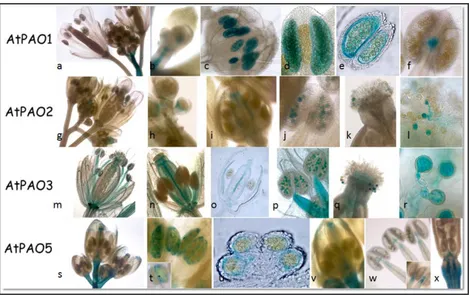
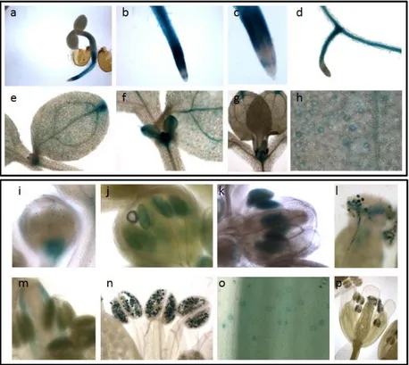
plants evidenced distinct expression patterns during seedling and flower development (Fincato et al., 2012). In particular, AtPAO1 is highly expressed in the transition region between the meristematic and the elongation zone of the root (Fig. 4a, b), while AtPAO2 and AtPAO3 are expressed in the root cap (Fig. 4h, n). Interestingly, at the root cap differences exist between AtPAO2 and AtPAO3, although they belong to the same PAO subfamily (Fincato et al., 2011). Indeed, while AtPAO2 is expressed only near the quiescent center and columella initials, AtPAO3 is expressed in lateral root cap and in the whole columella (Fig. 4h, n). Furthermore, while all four genes are expressed in the maturation zone of the roots, AtPAO5 is specifically expressed in the vascular system of this zone (Fig. 4t, u), the other three being present both in vascular and cortical tissues (Fig. 4c, i, o)
In hypocotyls, AtPAO2 is expressed along the whole organ but only at the early developmental stages of the seedlings (Fig. g), AtPAO3 is expressed only in the region adjacent to the hypocotyl-root junction site (Fig.4m) and AtPAO5 is highly and specifically expressed in the vascular system of the whole hypocotyl (Fig. 4s), whereas there is no expression of
AtPAO1 (Fig. 4a). As far as the shoot apex is concerned, AtPAO1 and AtPAO2 are expressed in both the shoot apical meristem (SAM) and the
stipules (Fig. 4d, e, j, k), AtPAO3 is expressed only in the stipules (Fig. 4p, q), while AtPAO5 is not expressed stipules but expressed in SAM (Fig. 4v, w). In addition, while AtPAO1- and AtPAO5-related GUS-staining is observed in the young cotyledons (Fig. 4f, w), both AtPAO2- and AtPAO3-related GUS staining are absent in this organ (Fig. 4k, q), AtPAO2 being expressed only at the cotyledonary tips (Fig. 4k, l). Specific expression pattern for each AtPAO gene is also observed both in the newly emerging leaves and the expanded ones. Indeed, while AtPAO1, AtPAO2 and AtPAO3 are characterized by a quite localized expression pattern, AtPAO5 shows a
12
Fig. 4. Histochemical GUS staining of seedlings from AtPAO::GUS transgenic Arabidopsis plants for AtPAO1, AtPAO2, AtPAO3, AtPAO5. Modified from Fincato et al.,
2012.
Fig. 5. Histochemical GUS staining of flowers from AtPAO::GUS transgenic Arabidopsis plants for AtPAO1, AtPAO2, AtPAO3, AtPAO5. Modified from Fincato et al., 2012.
13
rather diffused pattern (Fig. 4v). More specifically, AtPAO1 and AtPAO2 are expressed in the leaf hydathodes (data not shown for AtPAO1 and Fig. 4k for AtPAO2) and AtPAO3 in the guard cells (Fig. 4q, 6) and the trichomes (Fig. 4r). Staining of trichomes is also observed in the
AtPAO5::GUS transgenic plants which is however restricted to the base of
the trichomes (Fig. 4x), differently from the AtPAO3-related staining which is present throughout the whole area of the trichomes.
Distinct expression pattern of AtPAO1, AtPAO2, AtPAO3 and AtPAO5 is also additionally evident in inflorescences. In particular, AtPAO1 seems to be specifically expressed in the microspores and the tapetum (Fig. 5c, d, e) while AtPAO2 and AtPAO3 are specifically expressed in pistils (Fig. 5h, n) and pollen grains (Fig. 5j, p). Interestingly, expression of both AtPAO2 and AtPAO3 in pollen persisted during pollination and pollen tube growth (Fig. 5k, l, q, r). AtPAO5 exhibits an overlapping expression pattern with that of AtPAO1 and AtPAO2 but with some differences. In particular, although AtPAO5 is initially expressed in anther tapetal cells (Fig. 5u) and then in the anther-filament junction site similarly to AtPAO1. AtPAO5 is also expressed in the upper part of the filament (Fig. 5w), in sepals and in petals (Fig. 5s) where AtPAO1 expression is not found. Furthermore, while both AtPAO1 and AtPAO5 are expressed in receptacles at the early stages of flower development (Fig. 5a, s), AtPAO5 expression in receptacles is observed also at the level of the siliques (Fig. 5x). Similarly to AtPAO2 and
AtPAO3, AtPAO5 is also expressed in pistils but only in the stigma and the
septum and not in the ovary wall (Fig. 5v) as do AtPAO2 and AtPAO3 (Fig. 5i, o). Furthermore, AtPAO5 is not expressed in pollen grains, in contrast to
AtPAO2 and AtPAO3. All these data together support different
physiological role(s) of each of the members of the AtPAO gene family.
Physiological roles of polyamines
Since PAs are protonated at physiological pH, they have the capability to interact with negatively charged macromolecules, such as DNA, RNA, proteins and phospholipids thus altering the physical and chemical properties of numerous cellular components, stabilizing nucleic acid structures and modulating enzyme activities (Galston and Sawhney, 1990). In this way PAs are involved in the regulation of several fundamental cellular processes, including DNA replication, regulation of gene expression, RNA modification, translation, cell proliferation, cell cycle regulation, ion-channel regulation, modulation of cell signaling, membrane stabilization (Kusano et al., 2008; Tavladoraki et al., 2011). However, PAs are associated with several cellular processes not only through their
14
interaction with anionic macromolecules, but also through their metabolic products (Alcázar et al., 2010b).
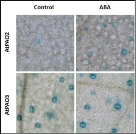
Fig. 6. ABA-inducible expression of AtPAO2 in guard cells. AtPAO2::GUS and
AtPAO3::GUS transgenic Arabidopsis plants were treated or not with 10 µM ABA for 4 h and
then analyzed for GUS activity. From Fincato et al., 2012.
In animals, PAs play an important role in cell differentiation and proliferation. Indeed, mice with specific inactivation of SPMS have severe developmental defects (Pegg and Michael, 2010). PA synthesis is down-regulated as cells become senescent in many tissues of adults. Administration of Spd markedly extends the longevity of yeast, flies and worms and human immune cells (Eisenberg et al., 2009). On the other hand, dysregulated PA metabolism has been associated with neoplastic transformation and cancer cell growth (Pegg and Feith, 2007). PAs affect numerous processes in carcinogenesis. In fact, PAs are often present at increased concentration in both tumor cell cultures and solid tumors, as determined in breast and colon cancer (Heby and Persson, 1990), while PA depletion leads to inhibition of tumor growth (Averill-Bates et al., 2005). It has been demonstrated that PAs can also induce programmed cell death (PCD) in various animal cell types ( Wallace et al., 2003; Igarashi and Kashiwagi, 2010), thus indicating a bivalent function for these molecules, promoting both cell growth and cell death, likely depending on their concentration and other developmental and environmental signals (Wallace
et al., 2003, Toninello et al., 2006).
In plants, PAs have been suggested to play important roles in regulation of cell proliferation, somatic embryogenesis, differentiation and morphogenesis (Kusano et al., 2007, 2008), dormancy breaking of tubers and in seed germination, development of flowers and fruits (Kusano et al.,
15
2007) and senescence (Takahashi et al., 2010). Indeed, A. thaliana double mutant for ADC1 and ADC2, which cannot produce PAs, died at the embryo stage (Urano et al., 2005) while embryo development of Arabidopsis double mutant for SPDS1 and SPDS2 is arrested at the heart stage indicating a requirement for Spd during the course of embryogenesis (Imai et al., 2004a). On the other hand, it has been demonstrated that organisms deficient in Spm are viable, but show different degrees of dysfunction. This indicates that Spm, although not essential, must also play very important roles in growth and development (Imai et al., 2004b; Alcázar et al., 2010). Furthermore, Ther-Spm has been shown to be involved in the regulation of vascular differentiation. Indeed, acl5 mutants of A. thaliana which do not synthetise Ther-Spm shows a severely dwarfed phenotype with over-proliferation of xylem tissues (Vera-Sirera et al., 2010) . Exogenous application of Ther-spm but not Spm to the acl5 mutant partially rescues plant phenotype (Kakehi et al., 2008).
In plants, PAs are also known to enhance plant tolerance to environmental stresses such as salinity, chilling, drought, potassium deficiency (Martin-Tanguy, 2001; Alcázar et al., 2010a), and defence signalling against pathogens (Walters, 2003). Indeed, exogenous applications of PAs have frequently been shown to affect plant growth and response against various stress factors (Groppa et al., 2007; Kusano et al., 2008; Vera-Sirera et al., 2010). Genetic studies using either transgenic plants overexpressing PA biosynthetic genes or loss-of-function mutants support their protective role in plant response to abiotic stress (Alcázar et
al., 2006; Kusano et al., 2008; Gill and Tuteja, 2010), and provide a major
advance in the understanding of PA functions. In addition, analysis of expression profiles of PA biosynthetic genes evidenced stress– responsiveness for several of them (Table 2). It has been shown that drought, dehydration, salt and ABA treatments (Urano et al., 2003; Alcázar
et al., 2006) induce ADC2 expression. In particular, following cold
treatment (Table.2), induced expression of ADC genes was in parallel with the increase in Put levels and constant or even decreased levels of free Spd and Spm (Cuevas et al., 2008) while, other studies showed that higher levels of Spd and Spm and lower levels of free Put could improve the adaptability of plants to salt stress (Duan et al., 2008). Furthermore, significant accumulation of SAMDC2 mRNA is observed in Arabidopsis under stress conditions (Urano et al., 2003). Overexpression of SAMDC has been shown to enhance tolerance to different abiotic stress in different transgenic plants. Indeed, it enhances tolerance to salt stress in Oryza sativa (Roy and Wu, 2002), Nicotiana tabacum (Waie and Rajam, 2003), Pyrus
16
2005). It enhances also tolerance to drought and fungal wilts (caused by
Verticillium dahliae and Fusarium oxysporum) in N. tabacum (Waie and
Rajam, 2003), to high temperature in Lycopersicon esculentum (Cheng et
al., 2009) and to cold in M. sylvestris (Table. 2; Hao et al., 2005) . On the
other hand, spms Knock-out mutant appears to be more sensitive to drought stress than the wild-type plants (Yamaguchi et al., 2007). This phenotype is believed be related to the fact that inward potassium currents across the plasma membrane of guard-cells are blocked by intracellular PAs (Liu et
al., 2000). It has been also evidenced that acl5/spms double mutant is
hypersensitive to high levels of KCl but not to high levels of MgCl2 and
mannitol (Yamaguchi et al., 2006).
On the other hand, PAs play a role as mediators in defence signalling against plant pathogens (Takahashi et al., 2003). In particular, ‘Spm signalling pathway’ involves transport of Spm in the apoplast, upregulation of a subset of defence-related genes, such as those encoding pathogenesis-related proteins and mitogen-activated protein kinases, and a type of programmed cell death (PCD) known as the hypersensitive response. This response is triggered by Spm-derived H2O2, produced through the action of
PAOs localized in the apoplast (Cona et al., 2006; Kusano et al., 2008; Moschou et al., 2008c).
Physiological roles of polyamine catabolism
In animals, PA catabolism contributes to important physiopathological processes such as cell proliferation and differentiation, apoptosis, amine detoxification and cell signalling through both regulation of PA levels and their oxidatively deaminated reaction products, mainly H2O2,
aminoaldehydes or dialdehyde and acrolein (Sharmin et al., 2001; Averill-Bates et al., 2008; Agostinelli et al., 2009). As evidenced by the complex role of PAs in cell growth and proliferation, optimal PA levels are necessary for mammalian health. In fact, an important difference between normal and tumor cells is PA content. To ensure optimal PA levels, PA homeostasis is tightly regulated at various steps of PA metabolism and transport (Wallace
et al., 2003), thus becoming difficult to be perturbed through inhibition of a
single biosynthetic or catabolic reaction. Despite this tight control, it has been shown that an altered PA catabolism can cause changes in PA homeostasis. Indeed, constitutive or inducible over-expression of SSAT in animal cells brought a substantial reduction in Spd and Spm pools as well as a large increase in Put and N1-acetyl-Spd intracellular levels and export of acetylated PAs (Jänne et al., 2005; Zahedi et al., 2007). Furthermore, over-expression of SMO in mouse neuroblastoma cells and HEK293 cells caused
17
a statistically significant decrease in Spm levels and an increase in Put levels (Vujcic et al., 2002; Amendola et al., 2005; Zahedi et al., 2007). Notably, the changes in PA levels through PA catabolism were often accompanied by increased DNA damage and changes in cell proliferation (Zahedi et al., 2007). These data suggest that PA catabolism has an important role in controlling PA content and thus can be used as a therapeutic target for several diseases.
The other catabolic product, H2O2, which can get converted into the
highly reactive hydroxyl radical through Fenton-like-catalysis (Fig. 7), is able either to impair cell growth and proliferation or to regulate signal transduction and gene expression, depending on its concentration. Indeed, it has been demonstrated that, in human breast cancer cells, the SMO-derived H2O2 in response to treatment with the PA analogue bis(ethyl)norspermine
(BENSpm) is cytotoxic (Fig. 7a; Pledgie et al., 2005; Casero and Pegg, 2009). Furthermore, the H2O2 produced by purified BSAO and Spm
exogenously supplied to human colon adenocarcinoma and melanoma cells has been also shown to cause cytotoxicity (Calcabrini et al., 2002; Agostinelli et al., 2009). However, it is still an open question whether or not H2O2, formed by PA catabolism, is always pathologic, or has a role in cell
signalling (Wang and Casero, 2006). The aminoaldehydes produced through PA catabolism have been shown to be cytotoxic on animal cells (Fig. 7a), probably due to the inhibition of nucleic acid and protein synthesis (Nocera
et al., 2003; Wallace et al., 2003). Indeed, it has been reported that
3-aminopropanal and acrolein produced from PA catabolism (Fig. 3) are intimately involved in cell damage during ischemia in rats (Igarashi and Kashiwagi, 2010). It was also observed that renal failure patients had increased levels of SMO activity and both free and protein conjugated acrolein (PC-Acro). Furthermore, PC-Acro increased at the locus of infarction after induction of stroke in mice (Igarashi and Kashiwagi, 2010). Moreover, acrolein has been shown to have an inhibitory effect on cell growth. In particular, it has been determined that the toxicity of acrolein on cells in culture medium containing fetal bovine serum with amine oxidase activity is greater than that caused by H2O2 (Sharmin et al., 2001).
However, it was observed that acrolein is not formed under normal conditions, likely due to the fact that PAs mainly exist as RNA-PA complexes, rather than as free molecules. It was therefore hypothesized that when cells are damaged, PAs are released from RNA and acrolein is produced from PA catabolism, especially from Spm by SMO, so that the aldehyde might be used as a biochemical marker for pathologies involving cell damage (Igarashi and Kashiwagi, 2010; Saiki et al., 2011).
18 Ta b le 2 . Tr a n sg e n ic p la n ts e n g in e e r e d to s y n th e si z e P A s fo r e n h a n c e d a b io ti c s tr e ss to le r a n c e . M o d ifi e d fro m G ill a n d T u te ja , 2 0 1 0 a n d A lc á z a r e t a l., 2 0 1 0
19
Fig. 7. Physiological roles of PA catabolism in animals and plants. (a) PA catabolism in
animals. (b) PA catabolism in plants. AMA, aminoaldehyde; ACRL, acrolein; Dap, 1,3 diaminopropane; HR, hypersensitive response; PCD, programmed cell death; POD, peroxidase. From Tavladoraki et al., 2012.
Also in plants, PA catabolism has been shown to have important roles in plant development and stress responses through both regulation of PA levels and their reaction products. Although a key role of the PA biosynthetic pathways in PA homeostasis has been highlighted, recent evidences suggest that the PA catabolic pathways equally play an important role in the regulation of PA levels. In particular, it has been shown that increased PA levels are accompanied by a concomitant increase in their catabolism (Bhatnagar et al., 2002). Furthermore, recently, it has been shown that it is possible to induce changes in the levels of specific PAs through manipulation of the catabolic pathways (Kamada-Nobusada et al., 2008; Moschou et al., 2008a; Fincato et al., 2011). A tight regulation of PA levels in plants is very important not only because PAs have a direct role in several physiopathological processes as discussed above, but also because PA metabolism has a central role in cellular metabolism (Mattoo et al., 2010; Mohapatra et al., 2010).
Similarly to animals, PA catabolism in plants plays an important role through the production of H2O2 (Fig. 7b), which is necessary for several
plant developmental processes. In plants, H2O2 produced via apoplastic
degradation of PAs drives peroxidase-mediated oxidative cross-linking of structural cell wall components contributing to cell-wall strengthening during development and under stress conditions, such as wound-healing and pathogen attack (Fig. 7b; Cona et al., 2006; Angelini et al., 2008, 2010). H2O2 has also been identified as an important second messenger in signal
transduction networks. Indeed, in A. thaliana H2O2 produced by
PAO-mediated Spd oxidation triggers the opening of hyperpolarization-activated Ca2+-permeable channels in pollen, thereby regulating pollen tube growth (Wu et al., 2010), a process important for sexual plant reproduction. Plant
20
PA catabolism is also involved in the regulation of gene expression as shown in AtPAO4-deficient Arabidopsis mutants altered in the expression of genes related to abiotic stress responses and flavonoid and/ or lignin metabolism (Kamada-Nobusada et al., 2008). It has been reported that PA catabolism is involved in the regulation of gene expression also under stress conditions (Fig. 7b). H2O2 produced by PA catabolism has been also
proposed to activate PCD associated with developmental differentiation. Indeed, the presence of an A. thaliana CuAO (ATAO1; (Moller et al., 1998) and ZmPAO in developing tracheary elements and root cap cells suggests their involvement in PCD which both cell types eventually undergo (Cona
et al., 2006). H2O2 produced by PA catabolism has been shown to induce
PCD also as a defence response to abiotic and biotic stresses.
PA catabolism contributes also to the formation of GABA, an important cellular metabolite which is also synthesized by cytosolic glutamate decarboxylase (Yu and Sun, 2007). GABA is rapidly produced in plants in response to biotic and abiotic stresses (Petrivalský et al., 2007; Dittami et
al., 2011). Furthermore, PA catabolism contributes to the formation of
β-alanine, which in turn can be further converted in plants to the osmoprotectant β-alanine betaine (Fig. 3). Dap is also a precursor of the uncommon PAs Nor-Spd and Nor-Spm which in plants are associated with stress tolerance (Cona et al., 2006). However, the exact contribution of PA catabolism to plant development and defence responses through production of these metabolites has still to be evaluated. Further studies are necessary to verify whether, similar to what is reported in animals, aminoaldehydes derived from PA catabolism in plants have cytotoxic activity and whether acrolein is also formed.
In the yeast Saccharomyces cerevisiae, it was shown that β-alanine produced from Spm oxidation by the spermine oxidase FMS1 is necessary for the production of pantothenic acid (vitamin B5), a metabolic precursor to coenzyme A (CoA) which is a cofactor of a large number of metabolic enzymes (White et al., 2001). Indeed, overexpression of FMS1 caused excess of pantothenic acid to be excreted into the medium, whereas deletion mutants required β-alanine or pantothenic acid for growth. Conversely, in bacteria, the β-alanine necessary for pantothenic acid production was shown to be derived by the decarboxylation of L-aspartate. The difference between yeast and bacteria in β-alanine biosynthesis questions as to how other organisms, such as fungi and plants, make β-alanine. At the present time in the public sequence data bases there are over a dozen identifiable aspartate-1-decarboxylase genes from different prokaryotic species, whereas this enzyme does not appear to be present in eukaryotic species.
21
Plant polyamine catabolism under abiotic and biotic stress conditions Numerous studies in different plant species have shown that PA catabolism contribute to plant defence responses to several biotic and abiotic stresses. However, this contribution has been mainly shown for extracellular PA catabolic enzymes and interestingly it is linked to PA transport to the apoplast where only limiting amounts of PAs are present under normal growth conditions (Moschou et al., 2008a; Kusano et al., 2008; Takahashi et al., 2010). Stress-related factors that have been shown to induce PA transport in the apoplast are: incompatible and compatible plant-pathogen interactions, salt stress and treatment with the stress-related hormone ABA (Yoda et al., 2003; Yoda et al., 2006; Marina et al., 2008; Moschou et al., 2009; Toumi et al., 2010). This suggests that PA catabolism in the apoplast is a general defence response against several stresses.
Several data based on the use of PAO-specific inhibitors and transgenic plants evidenced that PA catabolism in the apoplast contributes to stress defence responses through H2O2 production. Indeed, it has been shown that
the H2O2 produced by PA catabolism in the apoplast contributes to the
second phase of ROS production during TMV-induced HR, a plant response which is developed during an incompatible plant-pathogen interaction and consists of rapid ROS production, PCD and induction of defence responses aiming to restrict pathogen expansion (Yoda et al., 2003). Similar approaches showed that H2O2 produced by PA catabolism in the apoplast
contributes to the synthesis of the ROS that accumulate under abiotic stress conditions (Moschou et al., 2008a) or following treatment with ABA, an hormone which plays a crucial role in plant responses to abiotic stresses (Xue et al., 2009; Toumi et al., 2010). The H2O2 produced by PA
catabolism in the apoplast under stress conditions and/or the apoplastic PAs themselves trigger a downstream signal cascade pathway leading to increased expression of specific genes, such as of superoxide dismutase, ascorbate peroxidase, pathogenesis-related proteins, protein kinases, transcriptional factors and several other stress responsive genes (Yamakawa
et al., 1998; Moschou et al., 2008a; Moschou et al., 2009; Xue et al., 2009).
Interestingly, exogenous application of Spm to tobacco leaves, which mimics the apoplastic accumulation of PAs upon an incompatible plant pathogen interaction, increased expression of HR marker genes (Kusano et
al., 2008). Furthermore, ABA-inducible generation of H2O2 by Put
catabolism in the apoplast of guard cells signals stomatal closure through a mechanism involving Ca2+ as a second messenger (An et al., 2008).
22
The H2O2 produced by PA catabolism in the apoplast upon stress may
also lead to PCD. In particular, it has been shown that accumulation and further oxidation of free PAs in the apoplast induce PCD during tobacco defence against infection by microorganisms with diverse pathogenesis strategies, i.e. microorganisms establishing host and non-host incompatible interactions, such as TMV and Pseudomonas cichorii in tobacco,
Pseudomonas syringae in Arabidopsis and Magnaporthe grisea in rice
(Yoda et al., 2003, 2006, 2009). PCD was shown to be induced by oxidation of extracellular Spd also under abiotic stress conditions. Indeed, under salt stress conditions the levels of H2O2 and PCD were higher in transgenic
plants over-expressing the apoplastic ZmPAO than in the wild-type plants (Moschou et al., 2008a). Accumulation and further oxidation of free PAs in the apoplast has been also shown to enhance necrotic cell death, and thus increase disease severity, following infection of N. tabacum plants with the necrotrophic pathogen Sclerotinia sclerotiorum, an effect that was blocked by PAO- and CuAO–specific inhibitors (Marina et al., 2008). Interestingly, when the biotrophic bacterial pathogens Pseudomonas viridiflava,
Pseudomonas syringae pv tabaci or hemibiotrophic pathogen oomycete Phytophthora parasitica var nicotianae were tested in N. tabacum host
plants PA oxidation in the apoplast strongly decreased bacterial growth in
planta and caused a reduction in the oocyte induced necrosis (Marina et al.,
2008; Moschou et al., 2009). These data suggest that increased PA catabolism in the apoplast may have opposing effect against pathogens with different pathogenic strategies
Although the data described above strongly support the contribution of the apoplastic amine oxidases involved in the terminal catabolism of PA to plant defence responses, more studies are still necessary to determine in detail the concerned mechanism(s), to comprehend in depth the pleitrophic effects of the PA catabolic pathways and to unravel co-interacting metabolic and signalling pathways. More studies are also necessary to understand the contribution of the newly identified PA back-conversion pathways to plant defence responses.
23
RESULTS
Studies on the physiological roles of AtPAO1
Polyamine levels in atpao1 loss-of-function mutantIt has been recently shown that recombinant AtPAO1 has high catalytic activity towards Spm but not at all towards Spd (Tavladoraki et al., 2006). Recombinant AtPAO1 has elevated catalytic activity also towards the uncommon PA Therm-Spm (Tavladoraki et al., 2006), which is of particular interest because it has been recently shown the existence in
Arabidopsis of an enzyme (ACL5) able to synthesize Therm-Spm and the
loss-of-function mutant acl5 shows a severely dwarfed phenotype (Rambla
et al., 2010; Vera-Sirera et al., 2010). To determine whether AtPAO1 is
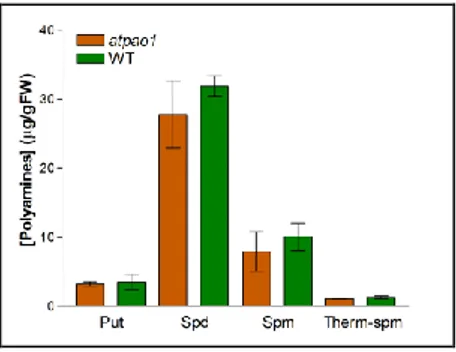
involved in the regulation of PA levels, an Arabidopsis insertional T-DNA knockout mutant for AtPAO1 (atpao1), recently obtained from the SAIL (Syngenta Arabidopsis Insertion Library) collection of Arabidopsis seeds (Fig. 8) and characterized (Dr L. Pomettini, graduation thesis), was analyzed for PA levels. Since the classical methods for PA determination, based on high-performance liquid chromatography or thin-layer chromatography, do not distinguish between Therm-Spm and Spm being isomers, this analysis was also performed by a gas chromatography–mass spectrometry method (Rambla et al., 2010) in collaboration with Prof. Juan Carbonell (Universidad Politécnica de Valencia-CSIC, Spain). Results obtained both by HPLC (data not shown) and gas chromatography–mass spectrometry (Fig. 9) showed no significant difference in the levels of the common PAs Put, Spd and Spm as well as in the level of Therm-Spm between atpao1 and wild-type seedlings. Since it has been recently shown that AtPAO1 is highly expressed in roots (Fig. 4; Fincato et al., 2012), PA levels were also analysed in this specific organ, but also in this case no statistically significant difference was observed between atpao1 mutant and wild-type plants (data not shown). Similarly, HPLC analyses did not evidence altered PA levels in flowers and leaves of atpao1 mutant (data not shown). The lack of differences in PA levels between atpao1 mutant and wild-type Arabidopsis plants may be due either to gene redundancy and/or to activation of homeostatic mechanisms and may exclude the possibility that Ther-Spm is the physiological substrate of AtPAO1.
24
Fig. 8. Schematic representation of the AtPAO gene structures with T-DNA insertion sites in the corresponding atpao1, atpao2, atpao3, atpao4 and atpao5 mutants. Black triangles
indicate the T-DNA insertion site. The black lines represent introns and boxes represent exons. Exons are numbered in Roman numerals. Colored and light grey boxes indicate shared and unshared exons, respectively. Dark grey boxes are unshared exons. Gene analyses were done using FGENESH available on Softberry website and protein domain structures were done using DOG 2.0 software.
Fig. 9. Polyamine content in atpao1mutant. PA levels of atpao1 and wild-type seedlings
were determined by gas chromatography-mass spectrophotometry. Statistical analysis was performed by one way ANOVA test (p < 0.001). Bars indicate standard error.
To evaluate whether homeostatic mechanisms have been activated in the
atpao1 mutant, the expression levels of biosynthetic genes (ADC1, ADC2, SAMDC) were examined in this mutant by semi-quantitative RT-PCR but