ORIGINAL ARTICLE
One-year outcomes of 27-gauge
versus 25-gauge pars plana vitrectomy
for uncomplicated rhegmatogenous retinal
detachment repair
Giancarlo Sborgia
1, Alfredo Niro
2*, Luigi Sborgia
1, Maria Oliva Grassi
1, Samuele Gigliola
1, Mario R. Romano
3,
Francesco Boscia
4, Alessandra Sborgia
1,2and Giovanni Alessio
1Abstract
Background: 27-gauge (27G) and 25-gauge (25G) transconjunctival sutureless vitrectomy (TSV) were considered equal about safety, effectiveness and vitrectomy time for the treatment of rhegmatogenous retinal detachment (RRD), although larger and long-term comparative studies are needed to confirm previous knowledge. Furthermore, a bined comparison of time duration of surgery and vitreous removal was never performed. Our purpose was to com-pare the safety and efficacy of 27G versus 25G TSV for the treatment of uncomplicated RRD over a 1-year follow-up. Methods: A 12-months single-center prospective, randomized, interventional study of 92 consecutive patients was performed. 46 patients underwent 27G TSV (Group 1) and 46 underwent 25G TSV (Group 2). Primary outcomes were primary and final reattachment rate, and final functional success (visual acuity ≥ 20/200, 1 LogMar). Secondary out-comes were the surgical and vitrectomy time. Complications were recorded.
Results: All functional and morphologic data at baseline and at all follow-up time points up to 12 months after surgery were available for only 88 patients. Four patients in Group 1 dropped out of the study after surgery. There was no significant difference in baseline characteristics between the two groups. Primary and final reattachment rates were 90.5% and 100% in Group 1, and 95.6% and 100% in Group 2, respectively (p > .05, p > .05, respectively). Visual acuity improved from 1.5 ± 1.09 LogMar to 0.38 ± 0.55 LogMar in Group 1 (p < .001) and 1.2 ± 0.9 LogMar to 0.49 ± 0.53 LogMar in Group 2 (p < .001), without significant difference between the groups (p > .05). The surgical time was 73.2 ± 11.3 min with 27G TSV and 64.4 ± 9.5 min with 25G TSV (p = .0001). The vitrectomy time was 19.9 ± 3.8 min with 27G TSV and 20.8 ± 3.8 min with 25G TSV (p > .05). One single case of choroidal detachment occurred.
Conclusions: Reattachment rates, functional success and vitrectomy time were comparable between 27G and 25G TSV for RRD. Surgical time was significantly longer using 27G vitrectomy.
Keywords: 27-Gauge, 25-Gauge, Transconjunctival sutureless vitrectomy, Rhegmatogenous retinal detachment, Surgical time, Vitrectomy time
© The Author(s) 2019. This article is distributed under the terms of the Creative Commons Attribution 4.0 International License (http://creat iveco mmons .org/licen ses/by/4.0/), which permits unrestricted use, distribution, and reproduction in any medium, provided you give appropriate credit to the original author(s) and the source, provide a link to the Creative Commons license, and indicate if changes were made. The Creative Commons Public Domain Dedication waiver (http://creat iveco mmons .org/ publi cdoma in/zero/1.0/) applies to the data made available in this article, unless otherwise stated.
Open Access
*Correspondence: [email protected]
2 Eye Clinic, Hospital “S. G. MOSCATI”, ASL TA, Via per Martina Franca,
74010 Statte, Taranto, Italy
Background
Since the introduction of pars plana vitrectomy in early 1970 by Machamer et al. [1], as alternative to the scleral buckling surgery to treat rhegmatogenous retinal detach-ment (RRD), the innovation trend has been moving in the direction of smaller instrument calibers. About this surgical technique, Chen et al. [2] proposed a sutureless approach to decrease surgical trauma and postoperative inflammation. Fujii et al. [3, 4] introduced the 25-gauge transconjunctival sutureless vitrectomy (TSV). Three years later, Eckardt [5] developed the 23-gauge TSV in order to reach a compromise between the more trau-matic 20-gauge and too flexible 25-gauge. Recently, inno-vations like more powerful light sources, rigid materials for small gauge instruments and more efficient vitrec-tomy machines with better fluidics and controls, allowed the progression to the 27-gauge (27G) vitrectomy system [6–8].
The progressive reduction of the size of the probes associated with high-performance vitrectomy allowed better postoperative comfort and earlier visual recovery for patients affected by RRD.
Oshima et al. [9] first reported a preliminary study regarding the safety and practicality of the 27G system for the vitreo-retinal surgery. Several reports have sug-gested the clinical outcomes and short-term safety profile of 27G TSV for many vitreo-retinal surgical indications, including RRD [10–13].
In two prospective 6-months follow-up studies, Romano et al. [14] and Rizzo et al. [15], comparing 27G and 25G TSV, reported that these two systems were equal about safety, effectiveness and vitrectomy time for treat-ment of RRD but a combined comparison of time dura-tion of surgery and vitreous removal was not performed.
Furthermore, larger and long-term comparative studies would be needed to confirm suggested data.
We conducted a 12-months prospective randomized interventional study to compare clinical outcomes, surgi-cal and vitrectomy time, and complications between 27G and 25G TSV to repair RRD.
Methods
A single-center prospective randomized comparative study was performed on 92 consecutive patients with RRD between April 2016 and June 2017.
Patients who met all of the following criteria were considered for inclusion in the study: RRD with one or more retinal breaks and the ability to give informed consent. Patients who met any of the following criteria were excluded from study entry: a history of any previ-ous vitreoretinal surgical procedures or penetrating ocular trauma, proliferative vitreoretinopathy (PVR) of grade C or higher, significant ocular comorbidities such
as uveitis, uncontrolled glaucoma, amblyopia, severe non proliferative or proliferative diabetic retinopathy, and inability to maintain postoperative posturing. All patients underwent preoperative evaluation included complete medical, surgical, and ophthalmic history. Preoperative data included age, sex, axis length, lens status, macular status, extent of retinal detachment, number of breaks, best-corrected visual acuity (BCVA), days from visual loss to surgery and intraocular pressure (IOP). Patients were divided in two groups. In Group 1, 46 patients were treated with 27G TSV and in Group 2, 46 patients under-went 25G TSV. All surgeries were carried out by one sur-geon G.S. in Eye Clinic, Department of Ophthalmology, University of Bari, Italy.
A postoperative examination was carried out on 2 weeks and 1, 3, 6 and 12 months after surgery, and included visual acuity measurement (BCVA), biomi-croscopy, intraocular pressure (IOP) measurement, funduscopy and optical coherence tomography (OCT) analysis. IOP measurement was also performed at 1 day after surgery.
Main outcomes were: the primary anatomical suc-cess as a complete reattachment of the retina following a single procedure when all the gas in the eye had disap-peared; the final anatomical success including the pri-mary anatomical success as well as those patients for whom a reattached retina was achieved with subsequent surgery over 1 year follow-up; the functional success con-sidering BCVA ≥ 20/200 (1 LogMar).
Secondary outcomes were the surgical time (the time period between trocars positioning and control of scle-rotomies after trocars removing) and the vitrectomy time (the time period when the cutter was activated for removing the vitreous) recorded by the vitrectomy machine. At the end of surgery intraoperative complica-tions were recorded.
Visual acuity was recorded after gas bubble got smaller not limiting central vision. BCVA was recorded as a Snel-len visual acuity and converted to logarithm of minimal angle of resolution (logMar) units for statistical analysis. Counting finger (CF) vision was defined as 2.0 logMar and hand movements (HM) were defined as 3.0 logMar. OCT scans using 6 × 6 radial scans protocol by OCT AVANTI RTVUE XR (OPTOVUE, Fremont, CA, USA) were recorded starting from the third month. IOP was measured using Goldmann applanation tonometry, and severe postoperative ocular hypotony and hypertony were defined as IOP < 6 mmHg and IOP > 30 mmHg, respectively.
After each patient signed the informed consent form, their identification number was recorded and patients were randomly assigned into Group 1 or Group 2 using a computer-generated randomization list. A total of 92
patients underwent surgery and met inclusion criteria. All functional and morphologic data at baseline and at all post-baseline time points up to 12 months after surgery were available for only 88 patients. 4 patients in Group 1 dropped out of the study after surgery.
The study followed the tenets of the Declaration of Hel-sinki and was approved by the institution’s review board. Surgical technique
Before starting surgery, the eyelid and periorbital skin were prepared with 5% povidone-iodine (Betadine; Purdue Fredrick Co, Norwalk, CT). All surgeries were performed under a retrobulbar block (a mixture of 2% Lidocaina and 2% Mepivacaina), using the Constellation® vitrectomy system (Alcon, Fort Worth, TX, USA). For this study, the machine was set with an initial aspiration of 0 mmHg moving linearly to 650 mmHg when the foot pedal is fully depressed, maintaining a fixed cut rate of 7500 cuts per minute (cpm) in both vitrectomy systems (27+ and 25+ Total Plus Pak). During surgery, IOP was controlled to 25 mmHg. For posterior visualization, sur-gical microscope Resight 700 (Carl Zeiss Meditec AG, Oberkochen, Germany) was used. Both the 27G and 25G procedures were performed using a three-port transcon-junctival pars plana approach.
After the conjunctiva was displaced slightly, the tro-cars were placed through the conjunctiva and the sclera 3.5 mm from the limbus using a 30 degree angled biplanar approach to create a self-sealing incision (as suggested
by Shimada) [16]. Phacoemulsification was performed
in all phakic eyes to maximize the complete removal of the anteroperipheral vitreous. After core vitrectomy, if needed, posterior vitreous detachment was induced and complete removal of the vitreous gel was performed. Triamcinolone acetonide was routinely injected to facili-tate visualization of the vitreous base which was meticu-lously shaved circumferentially with scleral indentation. Endolaser photocoagulation was performed to treat any detected tears or suspicious retinal lesions.
Transvitreal diathermy was used, at need, to mark retinal breaks and achieve intraocular hemostasis. If needed, intraoperative use of perfluorocarbon liquids (PFCL) was chosen during the procedure. After air-fluid exchange, 22% sulfur hexafluoride (SF6) gas or 14% per-fluoropropane (C3F8) gas were used as tamponade. At the end of the surgery, the microcannulas were removed and a gentle massage of the sclerotomy with a cotton-tipped applicator was applied to facilitate self-sealing and avoid leakage. If persisting leakage was detected, 8-0 vicryl sutures were placed in the wound and the overly-ing conjunctiva. Either 3 days face down positionoverly-ing or one side pose depending break positioning were assigned to the patient. All patients were placed on antibiotic and
betamethasone eye drops therapy with tapered frequency during the 5 weeks after surgery. During the follow-up period if IOP was higher than 25 mmHg, antiglaucoma eye drops were prescribed.
Statistical analysis
The qualitative variables are presented as frequencies and percentages, while quantitative data as means ± standard deviations. Effectiveness of the treatment was assessed with a t test on morpho-functional changes (paired 12 months post-surgery value minus pre-surgery value).
Differences between the groups were assessed using Pearson’s Chi squared for categorical variables and Kruskal–Wallis test or One-way Analysis of Variance (ANOVA) followed by post hoc Tukey’s HSD test for quantitative ones.
The sample-size was determined considering a con-fidence level of 95% and a concon-fidence interval of 10%. p value < 0.05 was considered statistically significant. All statistical analyses were conducted using R (v 3.3.1) and Rstudio (v 1.0.153).
Results
Preoperative Characteristics
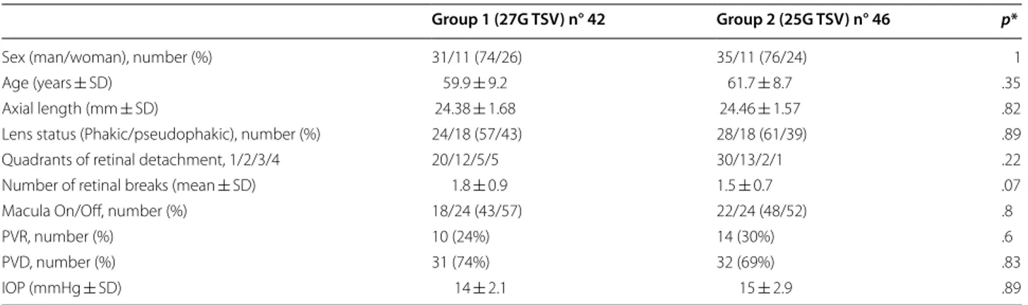
Table 1 summarizes patients’ demographic and
base-line clinical data. Forty-two eyes underwent 27G TSV (Group 1) and forty-six eyes underwent 25G TSV (Group 2) were considered in the analysis. The age of the patients ranged from 39 to 75 years in the Group 1 and 38 to 78 years in the Group 2. There were 31 men and 11 women in the Group 1 and 35 men and 11 women in the Group 2 (p = 1). Mean time from visual loss to surgery was 8.5 ± 4.4 days (range: 2–21 days) in the Group 1 and 7.9 ± 4.9 days (range: 3–22 days) in the Group 2 (p = .56). There was no statistical difference in lens status between both groups (p = .89). A posterior vitreous detachment (PVD) was detected intraoperatively in 31 eyes and 32 eyes in Group 1 and 2, respectively (p = .83). The mean number ± SD of retinal breaks was 1.8 ± 0.9 (range: 1–5) in Group 1 and 1.5 ± 0.7 (range: 1–4) in Group 2 (p = .07). Thirty-two eyes had RRD involving one or two quad-rants and ten eyes had more extensive RRD involving three or four quadrants in Group 1. Forty-two eyes had RRD involving one or two quadrants and four eyes had more extensive RRD involving three or four quadrants in Group 2 (p = .22). The macula was attached preoper-atively in 42.8% of eyes in Group 1 and 47.8% in Group 2 (p = .8). PVR grade ≤ B was preoperatively reported in 10 (24%) eyes in Group 1 and 14 (30%) eyes in Group 2 (p = .6). Preoperative mean IOP was 14 ± 2.1 mmHg and 15 ± 2.9 mmHg in Group 1 and 2, respectively (p = .89).
Main outcomes
Anatomical Results
The primary and final anatomical success rates were 90.85% and 100% in Group 1 and 95.7% and 100% in Group 2, respectively (p = .59, p = 1, respectively). In the 27G TSV group, one eye developed a retinal redetach-ment within 1 month, one eye within 3 months and 2 eyes within 6 months after first surgery. In the 25G TSV group, two cases detached within 3 and 6 months after primary vitrectomy, respectively. The redetachments were due to PVR in 2 eyes (1 eye in the Group 1 and 1 in the Group 2), a new retinal break in 3 eyes (2 eyes in the Group 1 and 1 in the Group 2), and the opening of an original retinal break in one eye in the Group 1. All these eyes were retreated using 25G TSV approach. Silicone oil tamponade was used in case of redetachment due to PVR, in the other cases gas tamponade was used. Silicone
oil was removed after an average of 3 months after sec-ond surgery.
Visual acuity
Table 2 summarizes BCVA at each examination.
Preop-erative BCVA was 20/630 (1.5 ± 1.09 LogMar) in Group 1 and 20/320 (1.2 ± 0.9 LogMar) in Group 2 (p = .15). Six eyes underwent reintervention for redetachment, because of the use of 25G TSV in these cases, the sili-cone oil tamponade in some cases, and in general, the postsurgical condition, were not considered in meas-uring of visual recovery. Therefore, the change of post-operative visual acuity was compared between the two groups, considering 38 and 42 patients with primary anatomical success, respectively. Their postoperative BCVA at the last visit improved to 20/48 (0.38 ± 0.56 LogMar) in Group 1 (p < .001) and 20/62 (0.49 ± 0.53 Table 1 Demographic and baseline clinical data
PVR proliferative vitreoretinopathy of grade ≤ B, PVD posterior vitreous detachment, IOP intraoculare pressure p* values, comparison between Group 1 and Group 2; p values < 0.05 were considered significantly
Group 1 (27G TSV) n° 42 Group 2 (25G TSV) n° 46 p*
Sex (man/woman), number (%) 31/11 (74/26) 35/11 (76/24) 1
Age (years ± SD) 59.9 ± 9.2 61.7 ± 8.7 .35
Axial length (mm ± SD) 24.38 ± 1.68 24.46 ± 1.57 .82
Lens status (Phakic/pseudophakic), number (%) 24/18 (57/43) 28/18 (61/39) .89
Quadrants of retinal detachment, 1/2/3/4 20/12/5/5 30/13/2/1 .22
Number of retinal breaks (mean ± SD) 1.8 ± 0.9 1.5 ± 0.7 .07
Macula On/Off, number (%) 18/24 (43/57) 22/24 (48/52) .8
PVR, number (%) 10 (24%) 14 (30%) .6
PVD, number (%) 31 (74%) 32 (69%) .83
IOP (mmHg ± SD) 14 ± 2.1 15 ± 2.9 .89
Table 2 Best corrected visual acuity over 12 months follow-up
p values < 0.05 were considered statistically significant BCVA best corrected visual acuity
p* values, comparison between Group 1 and Group 2
p** values, comparison between Baseline BCVA and Postoperative BCVA within single groups
a patients with primary anatomical success (38 eyes, Group 1; 42 eyes, Group 2)
Group 1 (27G TSV) Group 2 (25G TSV) p*
Baseline BCVA (logMAR) (mean ± SD) 1.5 ± 1.09 1.2 ± 0.9 .15
Postoperative BCVAa (1 month) 0.54 ± 0.59 0.75 ± 0.50 .08
p** < .001
Postoperative BCVAa (3 months) 0.45 ± 0.68 0.51 ± 0.54 .68
p** < .001
Postoperative BCVAa (6 months) 0.43 ± 0.56 0.49 ± 0.54 .50
p** < .001
Postoperative BCVAa (12 months) 0.38 ± 0.56 0.49 ± 0.53 .38
LogMar) in Group 2 (p < .001), without significant dif-ference between the groups (p = .38).
Secondary outcomes
Surgical and vitrectomy time
In order to maximize vitreous removal, phacoemul-sification was performed in every phakic eyes. The mean duration of vitrectomy was 19.9 ± 3.8 min with 27G TSV and 20.8 ± 3.8 min with 25G TSV (p = .27). The mean duration of surgery was 73.2 ± 11.3 min with 27G TSV and 64.4 ± 9.5 min with 25G TSV (p = .0001). When PVD was absent, surgical time was 77.2 ± 12.1 min and 62.1 ± 6.6 min in Group 1 and 2, respectively (p = .002). When PVD was already present, surgical time was 71.8 ± 10.8 min and 65.4 ± 10.5 min in Group 1 and 2, respectively (p = .02).
PFCL was given in 12 eyes (28.6%) in Group 1 and 12 eyes (26.1%) in Group 2
In eyes filled with PFCL surgical time was 81 ± 9.77 min and 74.2 ± 8.9 min in 27G and 25G TSV, respectively (p = .08).
In both groups, all eyes received endolaser and no external cryoapplication was given. In Group 1, 27 eyes (64.3%) had SF6 gas tamponade and 15 eyes (35.7%) had C3F8 gas tamponade. In Group 2, 25 eyes (54.3%) had SF6 gas tamponade and 21 eyes (45.6%) had C3F8 gas tamponade.
After removal of the microcannulas, only one scler-otomy site in 2 eyes in the Group1 and in 3 eyes in the Group 2 was sutured because of leakage.
Complications
No complications occurred related to phacoemulsi-fication such as posterior capsule rupture or zonular dialysis.
In the 27G TSV group, we also experienced one sin-gle case of choroidal detachment by infusion cannula slippage into the suprachoroidal space because of a sud-den movement of the patient during scleral depression. At 1 day the mean IOP was 14.5 ± 3.7 mmHg (range 8–16 mmHg) in Group 1 and 15.5 ± 2.8 mmHg (range 10–20 mmHg) in Group 2 (p = .14).
No iatrogenic retinal breaks (IRBs), such as severe hypertension (IOP > 30 mmHg) or hypotony
(IOP < 6 mmHg), intraocular bleeding, or
endoph-thalmitis, were noted in the follow-up period in either group. Over follow-up OCT scans revealed epiretinal membrane (ERM) in 5 patients (12%) in Group 1 and 2 patients (4.3%) in Group 2 (p = .5), and cystoid macu-lar edema (CME) in 2 patients (4.7%) in Group 1 and 1 patient (2.1%) in Group 2 (p = .07).
Discussion
Compared with the 25-Gauge or 23-Gauge vitrectomy systems, the 27-Gauge system inducing minimal ocular trauma with a smaller incision, may decrease the inflam-matory response, and may allow for overall patient with RRD a faster recovery.
The main limits about 27-Gauge probes are related to the stiffness of the instruments, the capability to remove vitreous as much as possible, and the time to perform surgery and, in particular, a complete vitrec-tomy approaching a RRD. We found that the 27G TSV provided anatomical and functional results comparable to those obtained using the 25G TSV, as previous papers reported [14, 15, 17].
The primary reattachment rate was higher in Group 2 but in Group 1 a higher number of eyes (ten eyes in Group 1 vs. three eyes in Group 2) had more extensive detachments (more than 2 quadrants) at baseline. Any-way, the primary anatomical success rate did not differ significantly between the groups and was similar to those reported by previous papers ranging from 90% to 100% using 27G [10–15, 17] and 93% to 100% using 25G TSV [14, 15, 17–23]. In both groups retinal redetachment occurred within 6 months after first surgery.
As previous papers reported [14, 15, 17], in this study different small gauge approaches led to significant and similar visual acuity recovery over 12-months follow-up, probably also due to the short mean time of visual loss before surgery was performed and relatively high num-ber of eyes with macula on RRD in each group (Group 1, 43%; Group 2, 48%). Our results showed that 27G TSV was as effective as 25G TSV in reattaching the retina after initial surgery and improving visual acuity.
As previous reports suggested [14, 15, 17], we found that the duration of vitreous removal was not different between the two different gauge systems.
During the vitrectomy, the aspiration efficiency of 27G TSV was obviously inferior to that of 25G TSV [23], but the increasing cut rate is related to a high vitreous flow rate [24]. At the same time, lower aspiration efficiency of the 27G TSV compared with the 25G TSV ensures a rather safe vitreous gel shaving and a better fluidic sta-bility. The 27-gauge cutter takes a faster and smaller ‘‘vit-reous bite’’ due to the high vacuum settings, allowing an adequate flow rate during the vitreous removal [25].
On the other hand, our results did show a sig-nificantly longer surgical time in 27G group
(73.2 ± 11.3 min) compared to 25G TSV
(64.4 ± 9.5 min). Mitsui et al. reported that in 27G TSV, the operation time was significantly longer than
that in 25G TSV for the epiretinal membrane [26].
Romano et al. [14] and Otsuka et al. [17] did not found significant difference in operative time considering 27
or 25-gauge to treat RRD, but authors did not define exactly the time period considered as operative time. Furthermore, in the prospective study of Romano et al. [14], silicone oil implant was used in some eyes differ-ently from our study.
As we found, different maneuvers as the induction of PVD, more frequently in younger patients, and the infusion and removal of PFCL may extend the opera-tion time when we use 27-gauge instruments.
In our series five of seven eyes which developed postoperative ERM after one single successful sur-gery, underwent 27G TSV and did not have a preop-erative PVD. Our results suggest that the development of a postoperative epiretinal macular membrane could be caused by factors related to smaller gauge instru-mentations. Inducing PVD is more difficult with 27G instrumentation due to lower flow/suction, especially in younger patients whose posterior vitreous cortex is tightly adherent to the retina. Less aspiration may reduce the efficacy of vitreous delamination when PVD was induced, with persistence of pro-inflamma-tory factors staining in vitreous chamber or retinal pigment epithelial cells that escaped through retinal tears [27].
We found no significant difference in the number of wounds that required sutures between the two systems. Generally, 27G TSV requires a smaller incision, which suggests the possibility of excellent self-closing of the wound, compared to other vitrectomy systems with larger-gauges [9, 23].
We monitored IOP from 1 day to 12 months after gery and did not found significant difference. The sur-gery was completed by filling the vitreous cavity with gas tamponade in all the cases. Previous reports have indicated that when a gas tamponade was performed, the postoperative IOP was more stable than when it was not used [9, 28].
In this case series, no serious intraoperative compli-cations, such as iatrogenic retinal breaks/tears, pos-terior capsule touch, leakage of sclerotomies, were observed. Safety-related outcomes were comparable to or better than some previous reports [14, 15, 17–22].
Limitations to our study are the small number of the patients and the lens status of all patients before vit-rectomy. Since all phakic patients were rendered pseu-dophakic before vitrectomy, the efficacy and safety of different gauges in phakic eyes would not be seen in our study, On the other hand, as points of strength of our prospective study we highlight that all the surgi-cal procedures were performed by a single experienced surgeon by using a well standardized technique, the long-term follow-up, and the concomitant analysis of surgical and vitrectomy time.
Conclusions
We performed a comparative study of outcomes between 27G and 25G TSV for RRD. The surgical results of the two groups were equivalent with a longer surgical time using 27-gauge cutter. We believe that 27G TSV is as use-ful as 25G TSV for uncomplicated RRD. A randomized, controlled trial with a larger number of patients is needed to confirm the results obtained in this study.
Authors’ contributions
GS, AN: Conception and design, Analysis and interpretation of data, draft of the article. LS, MOG, SG, AS: Acquisition of data, Analysis and interpretation of data. MR, FB, GA: Final approval of the version to be published. All authors read and approved the final manuscript.
Author details
1 Department of Medical Science, Neuroscience and Sense Organs, Eye Clinic,
University of Bari, Bari, Italy. 2 Eye Clinic, Hospital “S. G. MOSCATI”, ASL TA, Via per
Martina Franca, 74010 Statte, Taranto, Italy. 3 Department of Ophthalmology,
Humanitas University, Pieve Emanuele, Milan, Italy. 4 Department of Surgical,
Microsurgical and Medical Sciences, Eye Clinic, University of Sassari, Sassari, Italy.
Acknowledgements
The authors have no support from a funding body.
Competing interests
The authors declare that they have no competing interests.
Availability of data and materials
The datasets during and/or analysed during the current study available from the corresponding author on reasonable request.
Consent for publication
Written informed consent was obtained for publication of this study. A copy of the written consent is available for review by the Editor-in-Chief of this journal.
Ethics approval and consent to participate
The study was performed in accordance with the Declaration of Helsinki and approved by the institution’s review board. Each patient signed the informed consent form.
Financial disclosure
The authors report no conflicts of interest. The authors alone are responsible for the content and writing of the paper.
Funding
There is no financial support for this study.
Informed consent
Informed consent was obtained from all individual participants included in the study.
Publisher’s Note
Springer Nature remains neutral with regard to jurisdictional claims in pub-lished maps and institutional affiliations.
Received: 24 February 2019 Accepted: 20 April 2019
References
1. Machemer R, Buettner H, Norton EW, Parel JM. Vitrectomy: a pars plana approach. Trans Am Acad Ophthalmol Otolaryngol. 1971;75:813–20.
•fast, convenient online submission
•
thorough peer review by experienced researchers in your field
• rapid publication on acceptance
• support for research data, including large and complex data types
•
gold Open Access which fosters wider collaboration and increased citations maximum visibility for your research: over 100M website views per year
•
At BMC, research is always in progress. Learn more biomedcentral.com/submissions
Ready to submit your research? Choose BMC and benefit from:
2. Chen JC. Sutureless pars plana vitrectomy through self-sealing scleroto-mies. Arch Ophthalmol. 1996;114:1273–5. https ://doi.org/10.1001/archo pht.1996.01100 14047 3024.
3. Fujii GY, de Juan Jr E, Humayun MS, et al. A new 25-gauge instrument system for transconjunctival sutureless vitrectomy surgery. Ophthalmol-ogy. 2002;109:1807–13. https ://doi.org/10.1016/S0161 -6420(02)01179 -X. 4. Fujii GY, de Juan Jr E, Humayun MS, et al. Initial experience using the
transconjunctival sutureless vitrectomy system for vitreoretinal surgery. Ophthalmology. 2002;109:1814–20. https ://doi.org/10.1016/S0161 -6420(02)01119 -3.
5. Eckardt C. Transconjunctival sutureless 23-gauge vitrectomy. Retina. 2005;25:208–11.
6. Recchia FM, Scott IU, Brown GC, et al. Small-gauge pars plana vitrectomy: a report by the American Academy of Ophthalmology. Ophthalmology. 2010;117(9):1851–7. https ://doi.org/10.1016/j.ophth a.2010.06.014. 7. Nagpal M, Paranjpe G, Jain P, Videkar R. Advances in small-gauge
vitrectomy. TJ Ophthalmol. 2012;2(1):6–12. https ://doi.org/10.1016/j. tjo.2012.01.001.
8. Mohamed S, Claes C, Tsang CW. Review of small gauge vitrec-tomy: progress and innovations. J Ophthalmol. 2017. https ://doi. org/10.1155/2017/62858 69.
9. Oshima Y, Wakabayashi T, Sato T, et al. A 27-gauge instrument system for transconjunctival sutureless microincision vitrectomy surgery. Ophthalmology. 2010;117(1):93–102. https ://doi.org/10.1016/j.ophth a.2009.06.043.
10. Khan MA, Shahlaee A, Toussaint B, et al. Outcomes of 27 gauge microinci-sion vitrectomy surgery for posterior segment disease. Am J Ophthalmol. 2016;161:36–43. https ://doi.org/10.1016/j.ajo.2015.09.024.
11. Rizzo S, Barca F, Caporossi T, Mariotti C. Twenty-seven-gauge vitrectomy for various vitreoretinal diseases. Retina. 2015;35(6):1273–8. https ://doi. org/10.1097/IAE.00000 00000 00054 5.
12. Toygar O, Mi CW, Miller DM, Riemann CD. Outcomes of transconjunctival sutureless 27-gauge vitrectomy with silicone oil infusion. Graefes Arch Clin Exp Ophthalmol. 2016;254(11):2111–8. https ://doi.org/10.1007/s0041 7-016-3355-5.
13. Yoneda K, Morikawa K, Oshima Y, et al. Surgical outcomes of 27-gauge vitrectomy for a consecutive series of 163 eyes with various vitreous diseases. Retina. 2017;37(11):2130–7. https ://doi.org/10.1097/IAE.00000 00000 00144 2.
14. Romano MR, Cennamo G, Ferrara M, et al. Twenty-seven-gauge versus 25-gauge vitrectomy for primary rhegmatogenous retinal detachment. Retina. 2017;37(4):637–42. https ://doi.org/10.1097/IAE.00000 00000 00121 5.
15. Rizzo S, Polizzi S, Barca F, et al. Comparative study of 27-gauge versus 25-gauge vitrectomy for the treatment of primary
rhegmatogenous retinal detachment. J Ophthalmol. 2017. https ://doi. org/10.1155/2017/63849 85.
16. Shimada H, Nakashizuka H, Mori R, et al. 25-gauge scleral tunnel transconjunctival vitrectomy. Am J Ophalmol. 2006;142(5):871–3. https :// doi.org/10.1016/j.ajo.2006.05.057.
17. Otsuka K, Imai H, Fujii A, et al. Comparison of 25- and 27-gauge pars plana vitrectomy in repairing primary rhegmatogenous retinal detach-ment. J Ophthalmol. 2018. https ://doi.org/10.1155/2018/76431 74. 18. Horozoglu F, Yanyali A, Celik E, et al. Primary 25-gauge transconjunctival
sutureless vitrectomy in pseudophakic retinal detachment. Indian J Ophthalmol. 2007;55(5):337–40 PMC2636035.
19. Acar N, Kapran Z, Altan T, et al. Primary 25-gauge sutureless vitrectomy with oblique sclerotomies in pseudophakic retinal detachment. Retina. 2008;28(8):1068–74. https ://doi.org/10.1097/IAE.0b013 e3181 76de6 f. 20. Lai MM, Ruby AJ, Sarrafizadeh R, et al. Repair of primary rhegmatogenous
retinal detachment using 25-gauge transconjunctival sutureless vitrec-tomy. Retina. 2008;28(5):729–34. https ://doi.org/10.1097/IAE.0b013 e3181 62b01 c.
21. Kapran Z, Acar N, Altan T, et al. 25-gauge sutureless vitrectomy with oblique sclerotomies for the management of retinal detachment in pseu-dophakic and phakic eyes. Eur J Ophthalmol. 2009;19(5):853–60. https :// doi.org/10.1177/11206 72109 01900 527.
22. Kunikata H, Nishida K. Visual outcome and complications of 25-gauge vitrectomy for rhegmatogenous retinal detachment; 84 consecutive cases. Eye. 2010;24(6):1071–7. https ://doi.org/10.1038/eye.2010.41. 23. Osawa S, Oshima Y. 27-Gauge vitrectomy. Dev Ophthalmol. 2014;54:54–
62. https ://doi.org/10.1159/00036 0449.
24. Abulon DJ. Vitreous flow rates through dual pneumatic cutters: effects of duty cycle and cut rate. Clin Ophthalmol. 2015;9(2):253–61. https ://doi. org/10.2147/OPTH.S7138 7.
25. Romano MR, Scotti F, Vinciguerra P. 27-gauge vitrectomy for primary rhegmatogenous retinal detachment: is it feasible? Ann Acad Med Singa-pore. 2015;44(5):185–7 PMID: 26198325.
26. Mitsui K, Kogo J, Takeda H, et al. Comparative study of 27-gauge vs 25-gauge vitrectomy for epiretinal membrane. Eye. 2016;30(4):538–44. https ://doi.org/10.1038/eye.2015.275.
27. Sheard RM, Sethi C, Gregor Z. Acute macular pucker. Ophthalmology. 2003;110:1178–84. https ://doi.org/10.1016/S0161 -6420(03)00266 -5. 28. Yamane S, Kadonosono K, Inoue M, et al. Effect of intravitreal gas
tam-ponade for sutureless vitrectomy wounds: three-dimensional corneal and anterior segment optical coherence tomography study. Retina. 2011;31(4):702–6. https ://doi.org/10.1097/IAE.0b013 e3181 f0d2e 6.