www.nrronline.org
1027 NEURAL REGENERATION RESEARCH
PERSPECTIVE
A human translational model based on
neuroplasticity for pharmacological
agents potentially effective in
Treatment-Resistant Depression:
focus on dopaminergic system
Major depressive disorder (MDD) is a common psychiatric condi-tion characterized by two main symptoms, low mood and anhedo-nia. About 15–30% of people suffering from MDD do not respond to standard-of-care antidepressants, e.g., the serotonin re-uptake inhibitors (SSRI), and are considered affected by Treatment-Resis-tant Depression (TRD). The neurobiology of this condition is pres-ently unknown. Recent attempts of developing novel treatments for TRD have been driven by four major breakthroughs: (1) Increasing dopaminergic neurotransmission improves TRD symptoms; (2) Anhedonia occurs when central dopaminergic neurotransmission is low; (3) Enhanced neuroplasticity is critical for the action of anti-depressants; (4) Ketamine shows antidepressant properties in TRD patients and triggers neuroplasticity in preclinical animal models. These breakthroughs are at the basis of a putative human transla-tional cellular model for antidepressant agents that we are propos-ing in this article. The rationale is briefly described here.
Increasing dopaminergic neurotransmission improves TRD symptoms: Drugs that share the common mechanism of increasing dopaminergic neurotransmission were shown to improve symp-toms in TRD patients as adjunctive treatment to SSRI (Dunlop and Nemeroff, 2007; Fawcett et al., 2016). Therefore, the dopaminergic system could be seen as a preferred substrate in targeting TRD pa-tients with endogenously reduced dopaminergic transmission. Anhedonia occurs when central dopaminergic neurotransmis-sion is low: The second breakthrough was linking the symptom of anhedonia with defective function of the mesocorticolimbic dopa-minergic system, a critical component of the reward pathway that controls the hedonic state. Anhedonia can be measured using both clinical scale and instrumental endpoints in humans and animals, and is extensively reviewed in Pizzagalli (2014). Pharmacologic agents blocking dopaminergic transmission enhance anhedonia, while reversal is seen with drugs increasing dopaminergic trans-mission in rodents and humans (Pizzagalli, 2014).
Enhanced neuroplasticity is critical for the action of antidepres-sants: The third breakthrough regards the evidence of reduced neuronal plasticity and neurotrophic factor signaling in prefrontal/ cingulate cortex of rodents exposed to chronic stress and of hu-mans with mood disorders. These effects were generally reverted by chronic dosing with SSRI (Duman et al., 2016). The lack of clinical effects of SSRI observed in TRD could be conceptualized as driven by reduced neuroplasticity that is unresponsive to SSRI. Since TRD is often characterized by a hypoactive dopaminergic system, it is tempting to suggest that a reduced neuroplasticity is also present in the mesocorticolimbic dopamine system. Therefore, triggering neu-roplasticity in the dopaminergic system could help in re-engaging the brain areas modulated by this system, reverting the depressive syndrome of patients with TRD and, in particular, anhedonia. Ketamine shows antidepressant properties in TRD patients and triggers neuroplasticity in preclinical animal models: The fourth breakthrough came with the evidence that a single intravenous infusion of ketamine, a racemic dissociative anaesthetic that blocks
the N-methyl-D-aspartate glutamate receptor (NMDAR), produces rapid antidepressant effects, measurable at approximately 3 hours after infusion and lasting for 1–2 weeks (Zanos et al., 2018). The efficacy of such intervention was validated in several studies, to the point that the S-enantiomer of racemic ketamine was developed and approved as an intranasal twice-a-week treatment for MDD and TRD under the name of esketamine (https://www.fda.gov/ media/121378/download). Preclinical studies in rodents showed that treatment with ketamine increased neuroplasticity in fronto-cortical/hippocampal circuits; these increases were associated with behavioral antidepressant-like effects (Duman et al., 2016). The proposed mechanism involves an NMDAR-dependent rapid atten-uation of gamma-aminobutyric acid (GABA)ergic interneuron in-hibitory drive to pyramidal neurons and an increased post-synaptic glutamate neurotransmission mediated by up-regulation of the α-amino-3-hydroxy-5-methyl-4-isoxazole-propionic acid receptors (AMPAR). This increased AMPAR-mediated neurotransmission promotes synthesis and release of brain derived neurotropic factor (BDNF), a critical player in determining dendritic spine outgrowth via activation of the tropomyosin receptor kinase B (TrkB) recep-tor-dependent mitogen-activated protein kinase kinase (MEK)-ex-tracellular regulated protein kinases (ERK) and protein kinase B (Akt)-mTOR pathways (Duman et al., 2016).
However, how the molecular and cellular mechanisms of action of ketamine translate in humans is only partially understood. Since ketamine is known to increase dopaminergic neurotransmissions in cingulate/prefrontal cortex (Kokkinou et al., 2018), we suggest that ketamine also induces neuroplasticity in the dopamine system, contributing to the delivery of antidepressant and anti-anhedonic effects in TRD patients.
The introduction of human inducible pluripotent stem cells (iPSC) that can be differentiated into neurons has provided the possibility to investigate some aspects of the molecular and cellu-lar mechanism of actions of drugs, adding a novel methodology to the translational tool box (Cavalleri et al., 2018; Collo et al., 2018a). Accordingly, human iPSC-derived neurons together with pharmacodynamic signals, soluble biomarkers, genetic risk profile and neuroimaging can work in parallel, complementing each other, to generate a better understanding of the therapeutic relevance of neuroactive pharmacological agents, providing an integrated trans-lational approach for drug development.
In our laboratory, we developed a human iPSC-derived model of dopaminergic neurons to study the molecular aspects of struc-tural neuroplasticity produced by ketamine. Initially, we paralleled the studies on iPSC-derived human dopaminergic neurons with primary cultures of mouse mesencephalic dopaminergic neurons, according to standardized protocols (Cavalleri et al., 2018; Collo et al., 2018a). In vitro pharmacological tests were performed by in-cubating dopaminergic neurons with ketamine at various concen-trations and exposure times (e.g., 0.1–1.0 μM for 1-hour exposure) compatible with the dosing regimen used in the clinics (Zanos et al., 2018). Consistent effects of ketamine on dendrite outgrowth and soma size were observed 3 days after 1 hour of exposure, a time considered relevance in modelling the sustained clinical antidepres-sant effects of ketamine observed few days after infusion. We also showed that ketamine induces structural neuralplasticity in hu-man dopaminergic neurons by activating the PI3K-Akt-mTORC1 pathways via a BDNF/TrkB-dependent mechanism (Cavalleri et al., 2018), the same mechanisms previously shown in mouse fron-tocortical/hippocampal neurons (Duman et al., 2016). Both rapid phosphorylation of mTOR-dependent p70S6 kinase and long-term structural plasticity were blocked by the PI3K inhibitors LY294002 and mTORC1 inhibitor rapamycin. Immuno-neutralization of BDNF, inhibition of TrkB receptors and blockade of MEK-ERK signaling likewise prevented ketamine-induced structural plasticity measured 3 days after exposure, confirming the involvement of [Downloaded free from http://www.nrronline.org on Monday, May 25, 2020, IP: 212.171.146.191]
1028
Collo G, Merlo Pich E (2020) A human translational model based on neuroplasticity for pharmacological agents potentially effective in Treatment-Resistant Depression: focus on dopaminergic system. Neural Regen Res 15(6):1027-1029. doi:10.4103/1673-5374.270305
BDNF/TrkB signaling in the activation of the mTOR pathway also in human dopaminergic neurons (Cavalleri et al., 2018). These ef-fects of ketamine were abolished by the AMPAR antagonists NBQX and GYKI52466, as previously shown in mice (Duman et al., 2016). Intriguingly, ketamine-induced neuroplasticity on human dopa-minergic neurons required functionally intact dopadopa-minergic D3 receptors (D3R), since these effects were abolished by pretreatment with selective D3R antagonists (Cavalleri et al., 2018). Moreover, activation of D3R using D3R-preferential agonist pramipexole pro-duced structural plasticity in dopaminergic neurons at concentra-tion compatible with their clinical therapeutic use in TRD patients (Fawcett et al., 2016; Collo et al., 2018a). The clinical relevance of these molecular and cellular effects on neuroplasticity are partially supported by neuroimaging studies that indicate an association be-tween anhedonia and defective prefrontal, striatal and orbitofrontal circuits, known terminal fields of the ascending mesocortical dopa-minergic system. These defective circuits were partially reverted to normality in TRD patients 24 hours after ketamine infusion (Ab-dallah et al., 2016). Similar effects were obtained after 6-week daily dosing with add-on pramipexole in patients with mood disorders (Mah et al., 2011).
When considered together, these findings indicate that two ther-apeutic agents clinically effective on low mood and anhedonia in TRD patients (Fawcett et al., 2016; Zanos et al., 2018), showed
ef-fects on neuroimaging-defined anhedonia circuits (Abdallah et al., 2016; Mah et al., 2016) and, produced dendritic outgrowth in hu-man iPSC-derived dopaminergic neurons at exposures compatible with the clinical effects (Cavalleri et al., 2018; Collo et al., 2018a). The essential features of this model are represented in Figure 1: parallel quantitative assessments are run in the same subject for clinical anhedonia, for neuroimaging signals generated from the neuroanatomical regions where the dopaminergic system is active and in iPSC-derived dopaminergic neuron neuroplasticity.
In order to formally validate the translational relevance of this model, further prospective studies that include pharmacokinetic and pharmacodynamic assessments in TRD patients will be re-quired. The study design for this trial will include anhedonia and neuroimaging as clinical pharmacodynamic (PKPD) endpoint and neuroplasticity of iPSC derived neurons from the same TRD pa-tients as in vitro pharmacodynamic endpoint. Preliminary PKPD estimates are already available for ketamine: data from PKPD be-havioral studies in rodents, clinical trials in MDD/TRD patients, in vitro electrophysiological and morphological analysis in both mouse and human neurons provide a reasonable but still prelim-inary support to the proposed translational model (Collo et al., 2018a; Zanos et al., 2018; Shaffer et al., 2019).
Recently, an active metabolite of ketamine, (2R,6R)-hydroxynor-ketamine (HNK) was shown to produce antidepressant and
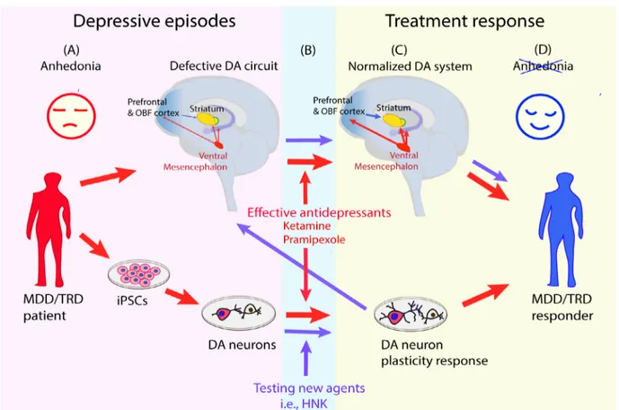
Figure 1 Schematic representation of a proposed translational model implementing human iPSC-derived dopaminergic neurons to assess neural plasticity induced by pharmacological agents potentially active on anhedonia.
(A) MDD/TRD subjects with low mood and prevalent anhedonia are profiled with (upper red arrow) neuroimaging for hypofunctional mesocor-ticolimbic dopaminergic system assessing prefrontal, orbitofrontal and striatal brain areas, as proposed by Pizzagalli et al. (2014) and Abdallah et al. (2017). iPSCs (lover red arrow) are generated from somatic cells donated by the same subjects and are differentiated to reproduce the neuron phenotype of the circuits involved, in this case DA neurons. (B) Red arrow: ketamine and pramipexole, effective anti-anhedonic treatments studied in clinical and neuroimaging trials, are tested in human iPSC-derived DA neurons at doses and exposure time compatible with the clinical use. Purple arrow: HNK is tested as new pharmacologic agent in human iPSC-derived DA neurons for neural plasticity. (C) Red arrow: treatments with ketamine and pramipexole result in neuroimaging normalization, clinical improvement and dendritic outgrowth in human iPSC-derived dopami-nergic neurons at exposures compatible with clinical use. Purple arrow: proposed neuroimaging study with HNK needed to confirm the transla-tional relevance of the positive human iPSC-derived dopaminergic in vitro test. DA: Dopaminergic; HNK: (2R,6R)-hydroxynorketamine; iPSCs: inducible pluripotent stem cells; MDD: major depressive disorder; OBF: orbitofrontal cortex; TRD: treatment-resistant depression.
1029 Collo G, Merlo Pich E (2020) A human translational model based on neuroplasticity for pharmacological agents potentially effective in
Treatment-Resistant Depression: focus on dopaminergic system. Neural Regen Res 15(6):1027-1029. doi:10.4103/1673-5374.270305 anti-anhedonic effects in animal models (Zanos et al., 2018).
In-triguingly, HNK does not bind to NMDAR but directly engages an AMPA-dependent mechanism (Shaffer et al., 2019). In human, HNK is slowing forming and reaches its peak at submicromolar concentrations (< 0.5 μM) 6–12 hours after administration, while the half-life of ketamine is about 2 hours (Zanos et al., 2018). Prediction from preclinical data indicates a pharmacological ef-fective concentration in human at 0.1 μM (Shaffer et al., 2019). Interestingly, the preclinical behavioral effects of HNK are blocked by the AMPAR antagonist NQBX, pointing to the critical role for AMPAR-dependent BDNF-TrkB signaling (Zanos et al., 2018). In human iPSC-derived dopaminergic neurons, 6-hour exposure to 0.5 μM HNK produced dendritic outgrowth when measured 3 days after dosing, effects similar to those obtained for 1-hour exposure to 1.0 μM ketamine (Collo et al., 2018b). These effects were blocked by pretreatment with the AMPA receptor antagonists NBQX and GYKI52466 and by the mTOR pathway blocker, rapamycin (Collo et al., 2018b).
It is tempting to contextualize these HNK data into the trans-lational working model described in Figure 1. In this cartoon, ketamine and pramipexole are considered as reference drugs: (1) they have published evidence of producing a treatment response in TRD/Mood Disorder patients by improving anhedonia; (2) they were shown to be capable of engagement and partial normalization of defective anhedonia neural circuits in TRD/Mood Disorder patients; (3) they produced in vitro structural neuroplasticity in iP-SC-derived human dopaminergic neurons; (4) they are active in vi-tro at concentrations compatible with those of clinical studies. No-tably, the full contextualization of HNK as novel agent with putative antianhedonic effects into this translational model is not complete. In fact, evidence of HNK as an independent therapeutic antidepres-sant for TRD is not available yet, clinical studies being in progress (https://clinicaltrials.gov/ct2/show/, NCT03977675). This holds true also for neuroimaging, whose evidence of HNK effects on an-hedonia neural circuits are indirectly associated with the effects of ketamine infusion measured at 24 hours after dosing (Abdallah et al., 2016), when the actions of approximately 12 hours exposure to pharmacological active HNK levels could have exerted its effects on neural circuits (Zanos et al., 2018). While further characterization in iPSC-derived dopaminergic neurons from MDD/TRD patients are needed, in Figure 1 we report an initial attempt for translating the available HNK in vitro data into clinical studies. Currently, HNK is in development at the NCAT/NIH (https://ncats.nih.gov/ chemtech/projects/active/ketamine).
In conclusion, we propose a translational approach driven by human biology in the attempt to conceptually linking molecular and cellular substrates with clinical and neuroimaging relevant information. The model is driven by human PKPD data from the pharmacological agent in consideration and focus on a neural cir-cuit-based hypothesis of psychiatric disorders. Interestingly, the possibility to study the molecular and cellular aspects of disorders and the mechanism of action of psychoactive drugs offered by iP-SC-derived neurons will be critical for driving the identification of novel targets.
This work is funded by Ministry of Education, University and Re-search (MIUR) ex-60% reRe-search fund University of Brescia, Italy. Emilio Merlo Pich is employee of Takeda Pharmaceutical Interna-tional AG.
Ginetta Collo*, Emilio Merlo Pich*
Department of Molecular and Translational Medicine, Section of Pharmacology, University of Brescia, Brescia, Italy; Department of Biomedicine, University of Basel, Basel, Switzerland (Collo G) Neuroscience Therapeutic Area Unit, Takeda Pharmaceuticals International, Zurich, Switzerland; Division of Brain Science, Imperial College, London, UK (Merlo Pich E)
*Correspondence to: Ginetta Collo, MD, [email protected];
Emilio Merlo Pich, MD, [email protected]. orcid: 0000-0003-1888-5049 (Ginetta Collo)
0000-0001-5478-235X (Emilio Merlo Pich)
Received: August 26, 2019
Peer review started: August 29, 2019 Accepted: October 12, 2019 Published online: December 10, 2019 doi: 10.4103/1673-5374.270305
Copyright license agreement: The Copyright License Agreement has been
signed by both authors before publication.
Plagiarism check: Checked twice by iThenticate. Peer review: Externally peer reviewed.
Open access statement: This is an open access journal, and articles are
dis-tributed under the terms of the Creative Commons Attribution-NonCommer-cial-ShareAlike 4.0 License, which allows others to remix, tweak, and build upon the work non-commercially, as long as appropriate credit is given and the new creations are licensed under the identical terms.
Open peer reviewers: Francisco Capani, Fundación Instituto Leloir,
Argenti-na; Suk Yu Yau, The Hong Kong Polytechnic University, Hong Kong, China.
References
Apel PJ, Ma J, CaAbdallah CG, Averill CL, Salas R, Averill LA, Baldwin PR, Krystal JH, Mathew SJ, Mathalon DH (2017) Prefrontal connectivity and glutamate transmission: Relevance to depression pathophysiology and ketamine treatment. Biol Psychiatry Cogn Neurosci Neuroimaging 2:566-574.
Cavalleri L, Merlo Pich E, Millan MJ, Chiamulera C, Kunath T, Spano PF, Collo G (2018) Ketamine enhances structural plasticity in mouse mes-encephalic and human iPSC-derived dopaminergic neurons via AM-PAR-driven BDNF and mTOR signaling. Mol Psychiatry 23:812-823. Collo G, Cavalleri L, Bono F, Mora C1, Fedele S, Invernizzi RW, Gennarelli
M, Piovani G, Kunath T, Millan MJ, Merlo Pich E, Spano P (2018a) Rop-inirole and pramipexole promote structural plasticity in human iPSC-de-rived dopaminergic neurons via BDNF and mTOR signaling. Neural Plast 2018:4196961.
Collo G, Cavalleri L, Chiamulera C, Merlo Pich E (2018b) (2R,6R)-Hy-droxynorketamine promotes dendrite outgrowth in human inducible pluripotent stem cell-derived neurons through AMPA receptor with tim-ing and exposure compatible with ketamine infusion pharmacokinetics in humans. Neuroreport 29:1425-1430.
Drysdale AT, Grosenick L, Downar J, Dunlop K, Mansouri F, Meng Y, Fetcho RN, Zebley B, Oathes DJ, Etkin A, Schatzberg AF, Sudheimer K, Keller J, Mayberg HS, Gunning FM, Alexopoulos GS, Fox MD, Pas-cual-Leone A, Voss HU, Casey BJ, et al. (2017) Resting-state connectivity biomarkers define neurophysiological subtypes of depression. Nat Med 23:28-38.
Duman RS, Aghajanian GK, Sanacora G, Krystal JH (2016) Synaptic plas-ticity and depression: new insights from stress and rapid-acting antide-pressants. Nat Med 22:238-249.
Dunlop BW, Nemeroff CB (2007) The role of dopamine in the pathophysi-ology of depression. Arch Gen Psychiatry 64:327-337.
Fawcett J, Rush AJ, Vukelich J, Diaz SH, Dunklee L, Romo P, Yarns BC, Escalona R (2016) Clinical experience with high-dosage pramipexole in patients with treatment-resistant depressive episodes in unipolar and bi-polar depression. Am J Psychiatry 173:107-111.
Mah L, Zarate CA Jr, Nugent AC, Singh JB, Manji HK, Drevets WC (2011) Neural mechanisms of antidepressant efficacy of the dopamine receptor agonist pramipexole in treatment of bipolar depression. Int J Neuropsy-chopharmacol 14:545-551.
Pizzagalli DA (2014) Depression, stress, and anhedonia: toward a synthesis and integrated model. Annu Rev Clin Psychol 10:393-423.
Shaffer CL, Dutra JK, Tseng WC, Weber ML, Bogart LJ, Hales K, Pang J, Volfson D, Am Ende CW, Green ME, Buhl DL (2019) Pharmacological evaluation of clinically relevant concentrations of (2R,6R)-hydroxynor-ketamine. Neuropharmacology 153:73-81.
Zanos P, Moaddel R, Morris PJ, Riggs LM, Highland JN, Georgiou P, Pereira EFR, Albuquerque EX, Thomas CJ, Zarate CA Jr, Gould TD (2018) Ket-amine and ketKet-amine metabolite pharmacology: insights into therapeutic mechanisms. Pharmacol Rev 70:621-660.
P-Reviewers: Capani F, Yau SY; C-Editors: Zhao M, Li JY; T-Editor: Jia Y [Downloaded free from http://www.nrronline.org on Monday, May 25, 2020, IP: 212.171.146.191]