POLYTECHNIC UNIVERSITY OF MARCHE
Ph.D. Course
LIFE AND ENVIRONMENTAL SCIENCES
curr. Biomolecular Sciences
Investigating the effectiveness of innovative
antimicrobial molecules against bacterial
species of clinical interest
Ph.D. student
Tutor
Dr. Laura Di Sante
Dr. Carla Vignaroli
INDEX
1. INTRODUCTION ... 1
1.1 Antimicrobial resistance: a long-standing issue ... 1
1.1.1 A multidrug resistant Gram-negative bacterium: Acinetobacter baumannii ... 4
1.1.2 A multidrug resistant Gram-positive bacterium: the methicillin resistant Staphylococcus aureus (MRSA) ... 8
1.2 Quinolones and fluoroquinolones ... 12
1.2.1 Bacterial topoisomerase type II and fluoroquinolones action mechanism ... 16
1.2.2 Fluoroquinolone-resistance mechanisms ... 19
1.3 The challenge of new antimicrobials: “the discovery void” ... 22
STATEMENT OF PURPOSE ... 28
2. MATERIALS E METHODS ... 30
2.1 Antimicrobial agents ... 30
2.2 Bacterial strains ... 30
2.3 Minimum inhibitory concentration (MIC) tests... 33
2.4 Minimal bactericidal concentration (MBC) tests... 34
2.5 Time-kill assay ... 34
2.6 Post antibiotic effect (PAE) ... 35
2.7 Selection of resistant S. aureus mutants ... 36
2.8 Characterization of acquired resistance ... 37
2.8.1 Total bacterial DNA extraction ... 37
2.8.3 Purification of PCR products ... 39
2.8.4 Sequences analysis ... 40
3. RESULTS ... 42
3.1 Susceptibility of control strains: MIC and MBC results ... 42
3.2 Time-kill assays ... 45
3.2.1 Time-kill performance of ciprofloxacin and levofloxacin .... 45
3.2.2 Time-kill performance of 73007 ... 48 3.2.3 Time-kill performance of 73009 ... 50 3.2.4 Time-kill performance of 73011 ... 52 3.2.5 Time-kill performance of 73012 ... 54 3.2.6 Time-kill performance of 73013 ... 56 3.2.7 Time-kill performance of 73015 ... 58 3.2.8 Time-kill performance of 73016 ... 60 3.3 PAE testing ... 62
3.4 In vitro selection of staphylococcal resistance ... 67
3.4.1 In vitro selection of S. aureus ATCC 29213 mutants ... 67
3.4.2 In vitro selection of S. aureus ATCC BAA 1720 mutants .... 72
3.4.3 Conserved domains analysis ... 74
3.5 Susceptibility of clinical strains to 73015 and 73016 NBTIs ... 76
4. DISCUSSION ... 78
Chapter 1 - INTRODUCTION
1
Chapter 1 – INTRODUCTION
1.1 Antimicrobial resistance: a long-standing issue
Since their clinical introduction in the 1930s, antibiotics have greatly influenced life on Earth (Rice, 2008). Ironically, antibiotics themselves have facilitated and contributed to the rapid dissemination of antimicrobial resistance (Pendelton et al., 2013). Actually, resistance to antimicrobials is a natural process that has been observed since the first antibiotics were discovered. The emergence of antibiotic resistance in bacterial pathogens is an inevitable consequence of antibiotic use and abuse and it has a significant negative impact on the outcome of therapies (French, 2005; Kollef, 2000). The propensity of antibiotic usage to favour the emergence of resistant pathogens is called antibiotic pressure revealing that their misuse promotes the emergence of resistance (Rolain et al., 2013). Antimicrobial resistance among the human pathogens arises when microorganisms, which cause infection, survive exposure to drug normally kill or inhibit the growth of bacteria. In absence of competition from other strains, resistant strains can spread, leading to the emergence of the so-called “superbugs”, bacteria untreatable with existing antimicrobials (O’Neill, 2016). Antibiotic resistance has increasingly become a serious threat to human health worldwide, with highly resistant pathogens of many difficult to treat species (Walker et al., 2009; Redgrave
et al., 2014). Moreover, during the last few years, there has been a
significantly increasing trend of bacterial multidrug resistance in many European countries. Data from European Antimicrobial Resistance Surveillance Network (EARS-net) have highlighted a wide variation in the occurrence of antimicrobial resistance associated with microorganism,
Chapter 1 - INTRODUCTION
2 drug and geographical region (EARS-net, 2013). One of the major concern about antimicrobial resistance, consists in the reduced chances of successfully treating such infections and in the resulting increase of morbidity and mortality by common bacterial diseases (Klevens et al., 2007, Akova, 2016a). The global spread of antibiotic resistance genes and their acquisition by clinically relevant microorganisms represents a serious problem for the health and welfare of both humans and animals (WHO, 2014).Multiple attempts have been made to quantify the local and global impact of antimicrobial resistance. Infections by resistant bacteria result in greater mortality, morbidity and costs of treatment compared to infections caused by their antimicrobial susceptible counterparts (Howard
et al., 2003; Tang et al., 2014). The mounting inability to prevent
infections with antimicrobial prophylaxis will massively and negatively affect the risk equation in performing more complex medical procedures (such as major surgery, cancer chemotherapy or organ/stem cell transplantation) (Tang et al., 2014). The growing numbers of antimicrobial-resistant pathogens, which are increasingly associated with nosocomial infections, place a significant burden on healthcare systems and have important global economic costs (Santajit & Indrawattana, 2016). It has been estimated that by 2050, 10 million lives per year and an economic output of 100 trillion of dollars are at risk due to the rise of infections caused by multidrug resistant bacteria. To date, 700,000 people die every year as consequence of bacterial infections (figure 1.1) (O’Neill, 2016). Political agendas, legislation, development of therapies and educational initiatives are essential to mitigate this increasing rate of antibiotic resistance (Frieri et al., 2016).
Chapter 1 - INTRODUCTION
3
Figure 1.1 Deaths attributable to antimicrobial resistance every year (O’Neill, 2016).
Although most antibiotic therapy is applied within the community, the greatest concentration of use per patient occurs in hospitals, hence nosocomial pathogens are likely to be the most resistant (French, 2010; O’Neill, 2016). However, the increasing prevalence of hospital and community-acquired infections caused by multidrug resistant bacterial pathogens is limiting the options for effective antibiotic therapy (Cassir et
al., 2014). Multidrug resistant Gram-positive and -negative bacterial
infections have resulted as difficult-to-treat or even untreatable with conventional antimicrobials (Frieri et al., 2016). Moreover, hospital infections are now microbiologically heterogeneous, being caused by many different species of multidrug resistant bacteria. Antibiotic usage selects, constantly, pathogens resistant also to last line antimicrobials (French, 2010). Data from the Centers for Disease Control and Prevention
Chapter 1 - INTRODUCTION
4 (CDC) have showed a rapidly increasing rates of infection due to methicillin-resistant Staphylococcus aureus (MRSA), vancomycin-resistant Enterococcus faecium (VRE), and fluoroquinolone-vancomycin-resistant
Pseudomonas aeruginosa strains (Boucher et al., 2009). Furthermore,
nosocomial infections caused by pan-drug resistant bacteria are now occurring. A lot of people die in hospitals for MRSA infection (Boucher & Corey, 2008) and in this setting several highly resistant Gram-negative pathogens such as Acinetobacter spp., multidrug resistant P. aeruginosa, carbapenem resistant Klebsiella spp. and Escherichia coli are emerging as significant pathogens worldwide (Falagas & Kasiakou, 2005; Falagas & Bliziotis, 2007; Urban et al., 2008). Recent report (Rice, 2008) basing on data from the Infectious Diseases Society of America has begun to refer to a clique of nosocomial pathogens, acronymically termed as “ESKAPE” pathogens. ESKAPE is an acronym for a group of bacteria, encompassing both Gram-positive and Gram-negative species: Enterococcus faecium,
Staphylococcus aureus, Klebsiella pneumoniae, Acinetobacter
baumannii, Pseudomonas aeruginosa and Enterobacter spp. These
microorganisms are capable of “escaping” the biocidal action of antibiotics and collectively represent new paradigms in pathogenesis, transmission and resistance (Rice, 2008). They are responsible of the bulk of antimicrobial resistant infections (Tang et al., 2014) amongst critically ill and immunocompromised individuals and are characterized by potential drug resistance mechanisms (Rice, 2010).
1.1.1 A multidrug resistant Gram-negative bacterium:
Acinetobacter baumannii
The incidence of infections caused by multidrug resistant Gram-negative bacteria has increased worldwide over the last decade. EARS-net (EARS-net, 2014) reported that the majority of Escherichia coli and Klebsiella
Chapter 1 - INTRODUCTION
5
pneumoniae isolates was resistant to at least one of the antimicrobial
tested, and many had combined resistance. To date, A. baumannii is also one of the most successful pathogens in the modern health care system (WHO, 2010). Multidrug resistant A. baumannii is a rapidly emerging pathogen in the hospital setting, where it causes severe infections (Jaward
et al., 1996; Fourier & Richet 2006).
The genus Acinetobacter consists of strictly aerobic, Gram-negative coccobacillary rods, oxidase negative, non-motile, usually nitrate negative and non-fermentative (Murray et al., 2003). Since 1986, the taxonomy of the genus Acinetobacter has been modified several times. Currently, at least 34 species can be distinguished within the genus, 23 of which have been assigned species name (Visca et al. 2011). Acinetobacter spp. are widely distributed in the environment and readily contaminate the hospital environment (Lin & Lan, 2014). They are the second non-fermenters most commonly isolated in human specimens. Acinetobacter spp. are able to survive on moist and dry surfaces, in foodstuff and on healthy human skin, especially in health care settings (Murray et al., 2003). Since they have a relatively long survival time on human hands, they can lead to high rates of cross contamination in nosocomial infections (Houang et al., 1998).
Acinetobacter spp. may also colonize or live in a patient without causing
infection or symptoms. Despite there are several species within genus, able to cause human disease, A. baumannii remains the most frequently isolated species and accounts for about 80% of reported infections (WHO, 2010). Moreover, hospital acquired infections, caused by A. baumannii, are most likely to involve the respiratory tract (related to endotracheal tubes or tracheostomies), urinary tract and wounds (including catheter sites) and many of them progress to septicaemia (Murray et al., 2003; Maragakis & Perl, 2008; Lin & Lan, 2014). These infections often occur
Chapter 1 - INTRODUCTION
6 in older patients, affected by chronic diseases and previously treated with antimicrobial therapies (Rodriguez-Baño et al., 2004; Lin et al., 2009). The mortality of patients with A. baumannii infections in hospitals and in the intensive care units has ranged from 7.8% to 23% and from 10% to 43%, respectively (Lin & Lan, 2014). A. baumannii rarely occur outside of health care settings; however, community-acquired A. baumannii has been reported in China and tropical Australia (WHO, 2010), and they have even affected patients with co-morbidities in the community (Falagas & Karveli, 2007). Previous studies on patients in the intensive care unit affected by bloodstream infections and burn infections due to carbapenem resistant Acinetobacter species, demonstrate an increased mortality rate (26% - 68%), as well as increased morbidity and length of stay in the intensive care unit (Maragakis & Perl, 2008). Antimicrobial resistance among Acinetobacter species has increased substantially in the past decade (Lockhart et al., 2007). Their extensive antimicrobial resistance may be due to the selective ability to prevent various molecules from penetrating their relatively impermeable outer membrane and to a large reservoir of resistance genes (EARS-net, 2014). Indeed, A. baumannii appears to be particularly effective at acquiring genetic material from other organisms and thus rapidly developing drug resistance (WHO, 2010). The antimicrobials still active against this pathogen include some fluoroquinolones (e.g. ciprofloxacin and levofloxacin), aminoglycosides (e.g. gentamicin, tobramycin and amikacin), carbapenems (imipenem, doripenem and meropenem), polymyxins (polymyxin B and colistin), sulbactam and tigecycline (net, 2014). However, data from EARS-net (2014) on 28 countries reporting antimicrobial susceptibility information revealed that in Europe the percentages of fluoroquinolones (ciprofloxacin or levofloxacin) resistant isolates ranged from 2.9 % to 95.3
Chapter 1 - INTRODUCTION
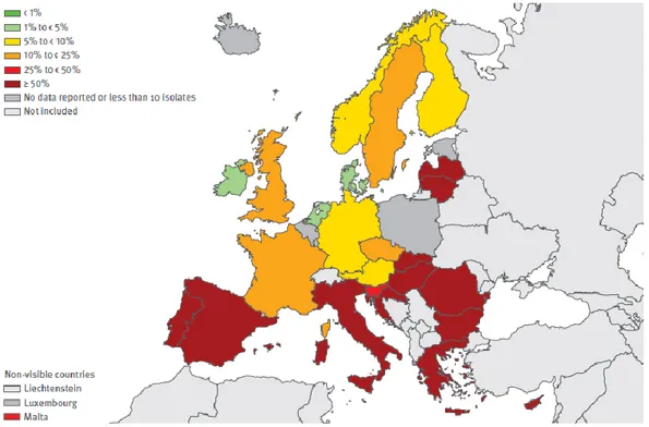
7 %. The highest percentages (≥ 50%) of resistant strains was encountered in many countries (e.g. Italy, Spain, Portugal, Greece, Cyprus, Hungary and Bulgaria) as showed in figure 1.2. The high levels of antimicrobial resistance in A. baumannii, reported from several countries in Europe, are of great concern, especially when resistance to the last-line treatment alternatives such as carbapenems occurs (EARS-net, 2014).
Moreover, the presence of multi drug resistant Acinetobacter spp. in the healthcare environment is of further concern since this pathogen can persist in the environment for long periods and is notoriously difficult to eradicate once established (Maragakis & Perl, 2008). This pathogen is emblematic of the mismatch between medical needs and the current antimicrobial research and development pipeline (Boucher et al., 2009).
Figure 1.2 Acinetobacter spp. Percentage (%) of invasive isolates with resistance to fluoroquinolones, by country (EARS-net, 2014).
Chapter 1 - INTRODUCTION
8
1.1.2 A multidrug resistant Gram-positive bacterium: the methicillin resistant Staphylococcus aureus (MRSA)
Staphylococci are Gram-positive coccal bacteria (0.5 to 1.5 µm in diameter), that occur singly and in pairs, tetrads, short chains (three or four cells), and irregular “grape-like” clusters. They are non-motile, non-spore-forming and catalase positive (Murray et al., 2003).
Staphylococci are widespread in nature and the role of S. aureus as one of the most important human pathogens is largely due to its virulence potential and ubiquitous occurrence as coloniser of humans, domestic animals, and livestock (Morgan, 2008). With non-fastidious growth requirements, staphylococci are mainly found living on the skin, skin glands, and mucous membranes of mammals and birds. They can be found in the mouth, blood, mammary glands, and intestinal, genitourinary and upper respiratory tracts of hosts (Murray et al., 2003; Gordon & Lowy, 2008). Staphylococcal infections represent a serious challenge for clinicians, particularly for serious infections such as bacteraemia, severe pneumonia and skin and soft-tissue infections. Staphylococci represent the second most frequent cause of hospital acquired infections (12.3%) in Europe (Campanile et al., 2015). S. aureus is both a commensal and pathogenic microorganism and the anterior nares are its main ecological niche (Wertheim et al., 2005) with approximately 20% of individuals permanently colonized, and 30% occasionally (Gordon & Lowy, 2008). Indeed, S. aureus is an opportunistic microorganism being responsible of severe infections. This “persistent pathogen” S. aureus, is even more challenging since has developed the resistance to methicillin and to other semi-synthetic penicillins, giving rise to the acronym “MRSA” (methicillin resistant S. aureus) (Sabath & Finland, 1962). A year after the methicillin introduction in clinical use (1959–1960), methicillin-resistant
Chapter 1 - INTRODUCTION
9 isolates were reported (Gordon & Lowy, 2008) and in the following ten years, it became widespread in Europe, Australia, and United States (Barber, 1961; Elek, 1965). This methicillin-resistant form has been the most important cause of antimicrobial resistant and healthcare-associated infections worldwide (EARS-net, 2014). Presently, and perhaps in a more virulent fashion, due to its association with Panton-Valentine leucocidin (PVL, an exotoxin active against neutrophils), MRSA has been also implicated in a surge of community-acquired infections that is reaching epidemic proportions (Vandenesch et al 2003; Labandeira-Rey et al 2007). Resistance to methicillin primarily derives from acquisition of the
mecA gene, not native in this species, which encodes the
penicillin-binding protein 2A (PBP2A) with a low penicillin-binding affinity for β-lactam antibiotics (Wu et al., 1996) thus preventing beta-lactams to inhibit cell wall synthesis. S. aureus is a versatile pathogen capable of causing a wide range of human diseases (Gordon & Lowy, 2008): the worldwide diffusion of MRSA increases morbidity and mortality rates in healthcare-associated infections, adding a further burden to S. aureus diseases (Köck
et al., 2010; Stefani et al., 2012). The occurrence of MRSA in Europe is
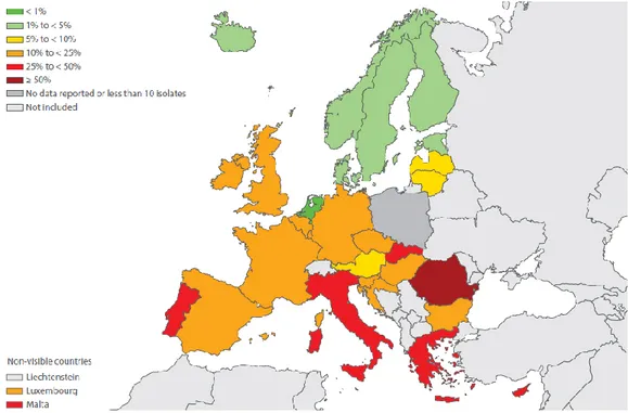
variable: MRSA percentages were generally lower in northern Europe and higher in the southern and south-eastern parts (figure 1.3).
Chapter 1 - INTRODUCTION
10
Figure 1.3 Staphylococcus aureus. Percentage (%) of invasive isolates with resistance to methicillin (MRSA), by country (EARS-net, 2014).
A recent epidemiological Italian survey (Campanile et al., 2015), aiming to determine the prevalence and antibiotic resistance of S. aureus in nosocomial infections, disclosed that S. aureus infections accounted for 11.6% and multidrug resistant MRSA accounted for approximately one-third of all isolates causing diseases. In this work, it has been found that, among clinical MRSA, percentages of isolates resistant to several antimicrobials (i.e. tetracycline, rifampicin, clindamycin and gentamicin) ranged from 12.6% to >39%. Higher percentages of resistant strains were found to erythromycin (65.4%) and fluoroquinolones (72.3 - 85.8%). Tackling the problem of MRSA infections is a top priority for public health systems worldwide. In most cases, glycopeptides (vancomycin and teicoplanin) are used as first-line antibiotics for treatment of specific MRSA infections. However, the selective pressure of these antibiotics has reduced susceptibility of some strains to glycopeptide with reports of
Chapter 1 - INTRODUCTION
11 clinical vancomycin-intermediate (VISA) and vancomycin-resistant (VRSA) S. aureus (Chambers & DeLeo, 2009).
Despite, a decreasing trend in MRSA infections has been reported, it remains a public health priority in Europe. Therefore, comprehensive strategies targeting all healthcare divisions (acute care, long-term care and ambulatory care) remain essential to continue reducing the spread of MRSA throughout the Europe (EARS-net, 2014).
Chapter 1 - INTRODUCTION
12
1.2 Quinolones and fluoroquinolones
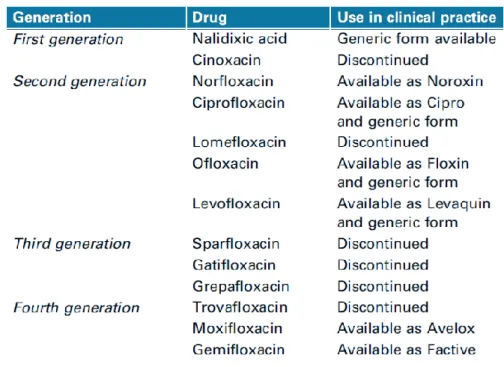
Unlike the first antibiotics discovered during the past century, the quinolone class of antimicrobial agents was not isolated from living organisms but, rather, was synthesized by chemists. Indeed, the quinolone class is structurally based on the fully synthetic naphthyridone (Andriole, 2005). Nalidixic acid, a related naphthyridone structure and the first member of the quinolone class, was accidentally discovered by George Lesher as synthesis by-product of the antimalarial compound chloroquine in 1962 (Lesher et al., 1962). This discovery led to the development of a series of quinolone compounds (Andriole, 1994) but its usage was strictly limited to the treatment of urinary tract infections caused by Gram-negative bacteria and resistance emerged quickly. As consequence of nalidixic acid discovery, other compounds were developed but only few were of signal importance: pharmaceutical chemists, modified the core of quinolone and the related chemical scaffolds (Domagala & Hagen 2003), generating compounds with greater potency, broader spectra of activity, improved pharmacokinetics and pharmacodynamics properties, and lower frequency of development of resistance (Andriole, 1999; Andriole, 2000). Therefore, changes have resulted in second-, third-, and fourth-generation fluoroquinolones, each defined by structural modifications and type of bacterial infection target including those by Gram-positive bacteria (Ball, 2000; King et al., 2000; Oliphant & Green, 2002).
Currently, there are four generations of quinolone/fluoroquinolone antibiotics as showed in table 1.1 (Redgrave et al., 2014).
Chapter 1 - INTRODUCTION
13
Table 1.1 Quinolones licensed for clinical use (Redgrave et al., 2014).
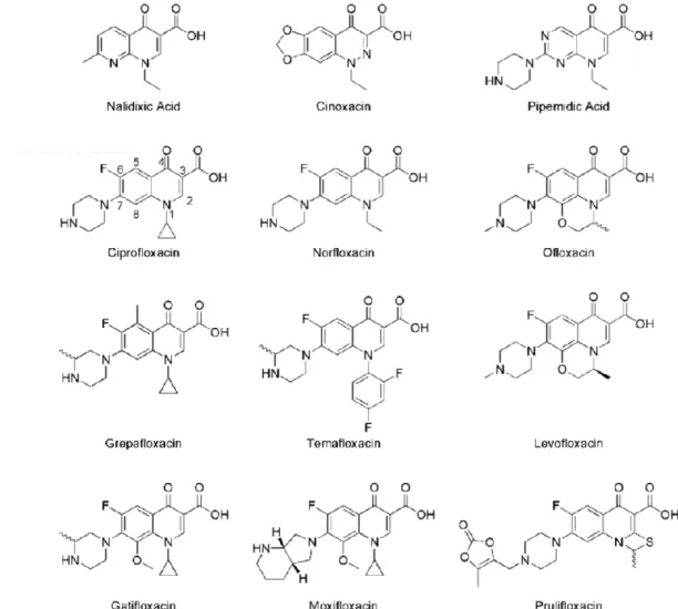
Fluoroquinolones (figure 1.4) differ from quinolones by the replacement of eight carbon atom of the backbone with a nitrogen atom and the addition of a fluorine substituent at position six (Ball, 2000). This addition became the common feature of the fluoroquinolone class with the introductions of norfloxacin in 1986 and ciprofloxacin in 1987 that showed substantially greater effectiveness against Gram-negative bacteria (Hooper & Jacoby, 2016). Actually, fluoroquinolone antibiotics are extensively used in a wide range of clinical applications because of their potency, spectrum of activity, oral bioavailability, and generally good safety profile (table 1.2; Owens & Ambrose 2000; Kim & Hooper 2014; Redgrave et al., 2014; Hooper & Jacoby, 2016). The most common fluoroquinolones prescribed are ciprofloxacin, levofloxacin, and moxifloxacin (Redgrave et al., 2014).
Chapter 1 - INTRODUCTION
14
Figure 1.4 Chemical structures of most relevant quinolones.
First generation quinolone antibiotics have been used for the treatment of uncomplicated urinary tract infections. The first generation agents were typically effective against a narrow range of Gram-negative bacteria, excluding Pseudomonas spp. (Oliphant & Green, 2002). Second generation quinolones were the first in class containing fluorine atom and they have expanded Gram-negative activity but limited efficacy against Gram-positive. Clinical infections treated using these second generation fluoroquinolones included urinary tract infections (both complicated and uncomplicated), pyelonephritis, sexually transmitted diseases, prostatitis, and localized skin and soft tissue infections (Oliphant & Green, 2002). Third generation fluoroquinolones were used also against respiratory
Chapter 1 - INTRODUCTION
15 infections. Broad-spectrum activity was further increased since they retained expanded negative efficacy and have improved Gram-positive coverage. Differences between third- and fourth generation fluoroquinolone were poorly distinguished. Fourth generation fluoroquinolones were used to treat similar infections as third generation drugs, but also have activity against anaerobic targets (Oliphant & Green, 2002).
Chapter 1 - INTRODUCTION
16
1.2.1 Bacterial topoisomerase type II and fluoroquinolones action mechanism
In both prokaryotes and eukaryotes, DNA exists as double strands forming a double-helix structure. Considering that each bacterium contains a chromosome composed of ~1300 µm long double-stranded DNA and bacteria are only ~2 µm in lenght and ~1 µm wide, they have to face a major topological problem (Andriole, 2005). Further twisting of this double-strand can occur throughout torsional force leading double helix to cross over on itself (Baranello et al., 2012). This process is called supercoiling and allows the chromosome to be highly condensed, hence can be placed into a bacterial cell. Supercoiling can be either positive or negative and this describes the direction in which twisting has occurred (Redgrave et al., 2014). The maintenance of supercoiling is essential in genetic processes that control gene expression and thereby determine the phenotype of a cell (Dorman et al., 1988; Dorman & Corcoran, 2009; Cameron et al., 2011; Webber et al., 2013). Changes in the global degree of supercoiling alter the expression of multiple genes including those involved in responses to stress and in pathogenesis (Peter, 2004; Baranello
et al., 2012; Webber et al., 2013). The degree of supercoiling of DNA is
not fixed and there is continuous remodelling of DNA topology in bacteria in response to environmental stress, growth stage, and cellular processes (Corbett et al., 2005; Barnarello et al., 2012; Arnoldi et al., 2013; Webber
et al., 2013). Topoisomerase I and topoisomerase II enzymes work in
opposition to control the level of twisting within DNA. Topoisomerase I reduces the number of negative supercoils (Barnarello et al., 2012). By contrast, topoisomerase II introduces negative supercoils, which unwind over-twisted DNA into a relaxed state and can further change the DNA topology (Barnarello et al., 2012). In bacteria, DNA supercoiling is
Chapter 1 - INTRODUCTION
17 controlled by type II topoisomerase enzymes, DNA gyrase and topoisomerase IV. Both are large heterodimeric enzymes composed of 2 pairs of subunits. The subunits of DNA gyrase are GyrA, and GyrB, 97-kDa and 90-97-kDa, respectively. The corresponding subunits of topoisomerase IV are ParC and ParE, 75 and 70 kDa, respectively (Jacoby, 2005). In S. aureus, topoisomerase IV subunits historically have also been referred as GrlA and GrlB (Hooper & Jacoby, 2016). The enzymes have homologous action but with subtle differences; although both DNA gyrase and topoisomerase IV relax positively supercoiled DNA, only DNA gyrase can introduce negative supercoils into relaxed DNA (Corbett et al., 2005). Topoisomerase IV has decatenating (unlinking) activity, allowing the segregation of catenated daughter chromosomes during cell division (figure 1.5; Corbett et al., 2005; Arnoldi et al., 2013; Drilica et al., 1999).
Figure 1.5 Action of type II topoisomerase on DNA (Redgrave et al., 2014).
The two enzymes work together in the replication, transcription, recombination and repair of bacterial DNA. The enzymes break both strands of double-stranded DNA, and, in an ATP-dependent reaction, pass
Chapter 1 - INTRODUCTION
18 a second DNA double helix through the break, which is then resealed (Kampranis et al., 1999).
Fluoroquinolones are potent inhibitors of bacterial type II topoisomerases: DNA gyrase and topoisomerase IV (Drlica, 1990; Drlica & Zhao, 1997; Hooper, 2001). They inhibit bacterial control of DNA supercoiling: at lower concentrations impaired DNA replication and at high concentrations determine cell death (Drlica, 1999; Drlica et al., 2009). Indeed, quinolones entrap DNA gyrase or topoisomerase IV in a drug-enzyme-DNA complex known as ternary complex (Drlica et al., 2008), with subsequent release of lethal, double-stranded DNA breaks (Hiasa & Shea, 2000). When a fluoroquinolone binds to the DNA-topoisomerase complex, the resulting ternary complex inhibits DNA replication and cell growth (Drlica & Zhao, 1997; Maxwell, 1997). In addition, this process stabilizes a catalytic intermediate covalent complex of enzyme and DNA. This can serve as a barrier to movement of the DNA replication fork (Wentzell & Maxwell 2000), to transcription complexes (Willmott et al. 1994) and finally to determine permanent double-strand DNA breaks (Drlica et al., 2008). Indeed, the binding of fluoroquinolones occurs within the enzyme at the target site of helix-4 in either GyrA or ParC, preventing the enzyme (DNA gyrase or topoisomerase IV) to re-ligate the DNA (Drlica et al., 2008). Quinolone interactions with DNA gyrase appear to result in more rapid inhibition of DNA replication than quinolone interactions with topoisomerase IV (Hooper & Jacoby, 2016). It could be possibly related to the overall proximity to the DNA replication complex of enzyme-binding sites on chromosomal DNA, with gyrase positioned ahead of the complex and topoisomerase IV behind it (Khodursky & Cozzarelli 1998). The targeting of either DNA gyrase or topoisomerase IV as the primary target by fluoroquinolones varies with
Chapter 1 - INTRODUCTION
19 bacterial species: quinolones can differ in their potency for the two enzymes. There is greater activity against DNA gyrase in Gram-negative bacteria and greater activity against topoisomerase IV in Gram-positive bacteria (Blanche et al. 1996; Pan & Fisher 1997; Strahilevitz & Hooper 2005). Therefore, in Gram-negative bacteria, DNA gyrase is more susceptible to inhibition by quinolones than topoisomerase IV, whereas, in Gram-positive bacteria, topoisomerase IV is usually the prime target, and DNA gyrase is intrinsically less susceptible (Drlica et al., 2008).
1.2.2 Fluoroquinolone-resistance mechanisms
Resistance to quinolones has been a problem ever since nalidixic acid was introduced into clinical practice more than 50 years ago (Jacoby, 2005). Despite prescribing guidelines now recommending reserving fluoroquinolone use, resistance continues to rise and constitute a relevant problem in clinical setting (Redgrave et al., 2014). Indeed, fluoroquinolone resistance has a significant clinical impact and can be mediated by multiple mechanisms (Oliphant & Green, 2002). Several mechanisms of resistance to fluoroquinolones (table 1.3) are currently recognized (Hooper, 2003).
Quinolone-resistance can be mediated by a transmissible route: genes encoding different resistance mechanisms and located on mobile genetic elements can decrease susceptibility to quinolone or fluoroquinolone antibiotics. This is known as plasmid-mediated quinolone resistance (PMQR) and the plasmid mediated resistance gene has been named qnr (Martinez et al, 1998). Data from a structural analysis of a Qnr protein suggest that fluoroquinolones resistance is achieved by the binding of the Qnr protein to the topoisomerase, physically preventing the intercalation of the antibiotic with the target enzyme (Tran & Jacoby, 2002; Xiong et
Chapter 1 - INTRODUCTION
20
al., 2011). The qnr genes generally confer to bacteria a modest protection
against fluoroquinolones (Martinez et al., 1998).
Table 1.3 Impact on susceptibility to ciprofloxacin of different resistance mechanisms (Redgrave et al., 2014)
a Based on data from E. coli b Based on data from S. aureus
To reach their targets, quinolones must cross the cell wall and cytoplasmic membrane of Gram-positive bacteria; in Gram-negative bacteria, quinolones must traverse an additional outer membrane barrier. Multidrug efflux pumps are capable of actively removing fluoroquinolones and other drugs from the bacterial cell. Both Gram-negative and Gram-positive bacteria have nonspecific, energy-dependent efflux systems, some of which are constitutively expressed and others are controlled by regulatory
Chapter 1 - INTRODUCTION
21 systems (Jacoby, 2005). These include transporters of various classes: in
S. aureus, for example, resistance to quinolones can be mediated by the
increased expression of norA, a gene that encodes a broad spectrum transporter for fluoroquinolones and other agents (Kaatz & Seo, 1995). Finally, the most common mechanism conferring high-level of fluoroquinolone resistance is the occurrence of point mutations in at least one of type II topoisomerases encoding genes, gyrA, gyrB, parC/grlA, and
parE/grlB. These mutations map within a DNA region defined as the
quinolone resistance-determining region (QRDR; Redgrave et al., 2014) corresponding to amino acid residues 67 to 106 of GyrA protein (Yoshida
et al., 1990) and to amino acid residues 63 to 102 of GrlA/ParC in E. coli
(Hooper & Jacoby, 2016). In S. aureus, the QRDR is represented by amino acid residues 68 to 107 of GyrA (Margerrison et al., 1992) and 64 to 103 of the GrlA (Ferrero et al., 1994). Mutations in the QRDR of topoisomerase type II encoding genes, result in amino acid substitutions that alter the target protein structure and subsequently the fluoroquinolone binding affinity of the enzyme (Piddock, 1999; Hooper, 2000). Although the primary target of fluoroquinolones differs in positive and Gram-negative, they will bind also to the secondary target and exert an antibacterial effect even if point mutations in the primary target have already occurred. Alteration of the primary target site can be followed by secondary mutations in lower affinity binding sites and highly resistant organisms will typically carry a combination of mutations within gyrA and
grlA (Redgrave et al., 2014). Indeed, once a first mutation arises in one of
the gene encoding DNA gyrase or topoisomerase IV, a reduction in the susceptibility of the target enzyme occur and additional mutations can further enhance resistance level (Ince & Hooper, 2003).
Chapter 1 - INTRODUCTION
22
1.3 The challenge of new antimicrobials: “the discovery void”
Antibiotics have been declared as one of the greatest medical advances in the modern age. The accelerated growth, diversity and burden of antimicrobial resistance became increasingly evident past the mid-1970s, although this was initially tackled by the development of new antimicrobial compounds that overcame the acquired resistance mechanisms (Tang et al., 2014). The revolutionary discovery of penicillin propelled the golden era of antibiotics, in which natural scaffolds and alternative versions of existing drugs were discovered. Currently the ever-increasing resistance (figure 1.6) and the management of bacterial infection could no longer be taken for granted (Brown & Wright, 2016).
Figure 1.6 Models of antibiotic drug discovery and development (Brown & Wright, 2016).
Chapter 1 - INTRODUCTION
23 Emphasis, in the resistance era, has fallen on target-based drug discovery to find broad-spectrum agents. In future a focus on innovative methods and unconventional targets should help to create narrow-spectrum agents and associated diagnostics (Brown & Wright, 2016).
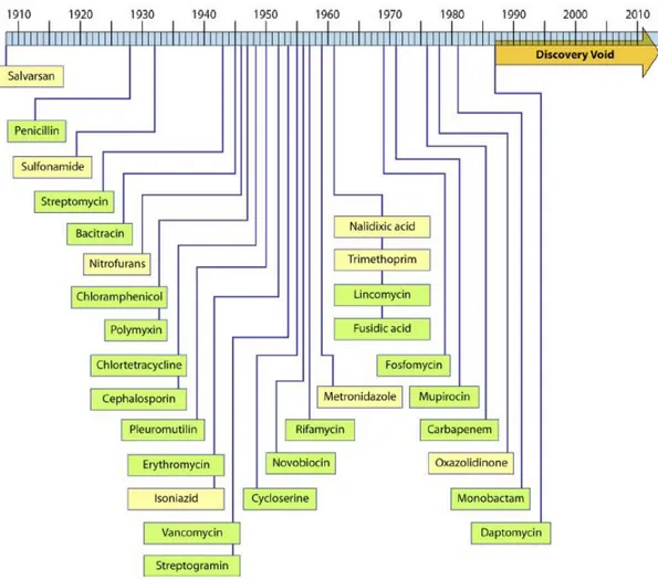
Over the past 25 years, the challenge of antibacterial discovery has retained the release of novel drug classes to extraordinarily low levels, even though discovery programs took place at large and small pharmaceutical companies as well as academic laboratories over this period (Silver, 2011). Since the 1990s there has been a “discovery void” (figure 1.7) of unknown extent in the research of new classes of antimicrobial compounds.
Figure 1.7 Timeline of antimicrobial drug discovery and illustration of the “discovery void.” Years indicated are those of reported initial discovery or patent (Silver, 2011).
Chapter 1 - INTRODUCTION
24 This problem was compounded by the persistent increase in new and modified mechanisms of antimicrobial resistance in disease-causing microbes (Talbot et al., 2006; Laxminarayan et al., 2013). Researchers have questioned if this “void” may be due to a lack of innovation or not: the simple definition of innovation is the act of introducing something new. However, it is noteworthy that almost all the drug discoveries (with few exceptions) have been unexpected, following to the screening of fermentation or chemical products for inhibition of bacterial growth (Silver, 2011).
Short-course dosing, impressive efficacy, limited side effects set the level whereby future antimicrobial compounds will be measured. However, this profile is unsustainable from both a practical and economic point of views. Most of large-pharma has severely reduced or eliminated infectious disease research and development, instead of pursuing more profitable therapeutics (Tomaras & Dunman, 2015) hence the route of exploiting novel antibacterial targets has met with significant failure over the last decade (Silver, 2011). It is alarming that although bacterial resistance continues to emerge, the rate at which antibiotics are being developed is decreasing (ECDC/EMEA, 2009). Although, there are a small number of novel compounds in the early clinical phase that might indicate the end of this “void”, in most cases their eventual developmental success is unclear. Indeed, new antibiotics discovery deals with several developing barriers:
i) scientific barrier, because it’s hard to find new classes of antibiotics and
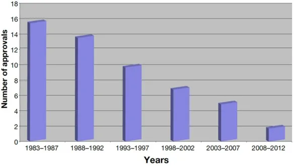
trials are not feasible; ii) regulatory barrier, because it’s difficult to get a licence from the Food and Drug Administration (FDA) and rules repeatedly change; iii) financial barrier, because antibiotics are not very profitable (Akova, 2016b). In 1983–1987 the FDA approved 16 new
Chapter 1 - INTRODUCTION
25 systemic antibiotics, but, since that time, antibiotic approvals have progressively been declined (see figure 1.8).
Figure 1.8 Antibiotic approvals by FDA over the last three decades in five-year increments (Cain, 2012).
Since 2008, only two systemic antibiotics have been approved. Moreover, just two new classes of broad-spectrum antibiotics have been validated in the last 40 years: the oxazolidinone (linezolid) and the lipopeptide (daptomycin) both effective against Gram-positive bacteria (Cain, 2012). Nowadays, agencies have provided incentives for large-pharma reinvestment in the antimicrobial research field but the damage has already been done due to the long-term consequences (Tomaras & Dunman, 2015). Moreover, reports of emerging multidrug-resistant Gram-negative bacteria, such as Acinetobacter spp., have stoked further concerns that there is not a robust pipeline addressing the issue posed by a highly diverse and variable spectrum of resistant pathogens (Cain, 2012). The resistance challenge with existing antibiotics needs of discovery and development of novel compounds with well differentiated modes of action to overcome the existing resistance mechanisms of
Chapter 1 - INTRODUCTION
26 bacteria spread in clinical settings (Perez et al., 2008; Karaiskos & Giamarellou, 2014).
Some efforts in the discovery of novel antibacterial agents have resulted in compounds with potent antimicrobial activity against multidrug resistant pathogens (Lahiri et al., 2015).
A wise strategy may be to identify novel scaffolds able to bind novel pockets on clinically well validated targets that are conserved among bacteria and distinct from human, with a different type of inhibition (Lahiri et al., 2015). Because of the attractive possibility of dual targeting, an area of active research has been the pursuit of inhibitors of type II topoisomerase: bacterial DNA type II topoisomerases are clinically validated as antibacterial drug targets by the development and use of fluoroquinolone (Silver, 2011). One of the promising class among these inhibitors are the Novel Bacterial Topoisomerase Inhibitors (NBTIs) reported by many groups (Bax et al., 2010; Miles et al., 2013; Dougherty
et al., 2014).
NBTIs inhibit both bacterial DNA gyrase and topoisomerase IV with minimal cross reactivity to human topoisomerase (Wiener et al., 2007; Black et al., 2008). Biochemical data (Bax et al., 2010), together with the co-crystal structure, have helped to understand the binding mode and the mechanism of inhibition of this class of inhibitors (Lahiri et al., 2015). Indeed, mechanistic and structural studies of NBTIs have revealed a binding site distinct from that of fluoroquinolones. Additionally, a novel mode of action of these inhibitors could be suggested by the absence of DNA-cleavage complex formation unlike the fluoroquinolones which stabilize a cleaved complex intermediate (Bax et al., 2010; Shapiro & Andrews, 2012). While the general mechanistic differences have been well established, the specific pathways of conformational changes remain
Chapter 1 - INTRODUCTION
27 poorly defined. Recently, structural advances in the field of bacterial topoisomerases (Bax et al., 2010; Vos et al., 2011; Laponogov et al., 2013) have significantly improved the understanding of this complex machinery, where association and dissociation of various subunits result in DNA cleavage/religation and DNA strand passage. This novel binding mode provides a structural basis explaining why NBTIs are able to overcome target-mediated fluoroquinolone resistance (Bax et al., 2010). Inhibitors of bacterial topoisomerases continue to expand beyond the quinolones but it remains to be verified that these agents can demonstrate clinical efficacy with minimal toxicity concerns.
These new topoisomerase inhibitors make advanced to the clinic and may be on track to be the first new class of clinically relevant DNA-targeted antibacterial agents (Bradbury & Pucci, 2008). Actually, novel compounds with dual target activity would have the advantage to reduce developmental frequency of antimicrobial resistance, due to the unlikely occurrence of two simultaneous mutations in both essential targets (Azam
et al., 2015). These agents could offer coverage against
quinolone-resistant strains, but further demonstrations of clinical usage are strictly required.
STATEMENT OF PURPOSE
28
STATEMENT OF PURPOSE
The increasing rate of bacterial resistance to clinical antimicrobial agents and its impact on treatment of infectious diseases has begun a unique problem throughout the world. Many antibacterial agents can induce resistance by different mechanisms hence many last-resort compounds are becoming increasingly ineffective. Treatment of bacterial infections caused by drug resistant strains relies on development of new agents that are able to overcome current resistance mechanisms. Therefore, there is a continuing need to develop newer and more potent antibiotics. Among the targets for the development of new antibacterial agents, bacterial topoisomerases remain a vibrant area of discovery. Novel Bacterial Topoisomerase Inhibitors (NBTIs) are a new class of compounds able to inhibit both bacterial DNA gyrase and topoisomerase IV.
A series of NBTIs have been developed and provided by the Italian pharmaceutical company Angelini S.p.A. (ACRAF). These molecules, still subject to confidentiality, should have a mechanism of action similar to that of fluoroquinolones, acting simultaneously on two microbial target.
This Ph.D. research is focused on the study of the effectiveness of these NBTIs against bacterial species of clinical interest.
To this aim molecular and microbiological methods have been used: to assess the susceptibility of Gram-positive and -negative reference
strains by MIC/MBC determination, time-kill assays and post antibiotic effect;
STATEMENT OF PURPOSE
29 to evaluate the occurrence in S. aureus of antimicrobial resistance to these NBTIs, investigating the ability of new compounds to select in vitro resistant mutants;
to assess their effectiveness against multidrug resistant A.
Chapter 2 – MATERIALS AND METHODS
30
Chapter 2 - MATERIALS AND METHODS
2.1 Antimicrobial agents
Ciprofloxacin and levofloxacin, were all purchased from Sigma Aldrich (Sigma-Aldrich Co., St. Louis, Mo, USA). Twelve NBTIs were supplied by the pharmaceutical company Angelini ACRAF as Dimethyl sulfoxide (DMSO)-soluble powder. The novel compounds tested in the present study are named as follow 73005, 73006, 73007, 73008, 73009, 73010, 73011, 73012, 73013, 73014, 73015, 73016.
2.2 Bacterial strains
Reference strains Escherichia coli ATCC 25922, Staphylococcus aureus ATCC 29213 and methicillin-resistant S. aureus ATCC BAA 1720 were used to test the activity of all compounds. Pseudomonas aeruginosa ATCC 27853 was used in susceptibility assays with 73009, 73015 and 73016 molecules.
A total of 38 Acinetobacter baumanii strains (Table 2.2.1) and 24 methicillin-resistant S. aureus (MRSA, Table 2.2.2) strains were evaluated for their susceptibility to molecules 73015, 73016 and ciprofloxacin. All strains were isolated and identified by Clinical Microbiology Laboratory, Ospedali Riuniti of Ancona. Both A. baumanii and MRSA strains were selected for their multidrug-resistance, including the fluoroquinolone resistance.
Chapter 2 – MATERIALS AND METHODS
31
Table 2.2.1: List of multidrug-resistant A. baumannii
Strains Clinical sample Isolation
period
A. baumannii 1 cerebrospinal fluid August 2015
A. baumannii 2 blood culture September 2015
A. baumannii 3 tracheal suctioning October 2015
A. baumannii 4 sputum October 2015
A. baumannii 5 bronchoaspirate (BAS) October 2015 A. baumannii 7 bronchoalveolar lavage (BAL) October 2015 A. baumannii 8 protected specimen brush (PSB) October 2015
A. baumannii 9 PSB October 2015
A. baumannii 10 blood culture October 2015
A. baumannii 11 BAS November 2015
A. baumannii 12 PSB November 2015
A. baumannii 13 PSB November 2015
A. baumannii 14 PSB November 2015
A. baumannii 15 BAL November 2015
A. baumannii 16 tracheal suctioning December 2015
A. baumannii 17 wound swabbing December 2015
A. baumannii 18 BAS December 2015
A. baumannii 20 PSB January 2016
A. baumannii 21 BAS January 2016
A. baumannii 22 PSB January 2016
A. baumannii 23 PSB January 2016
A. baumannii 24 tracheal suctioning January 2016
A. baumannii 25 BAS January 2016
A. baumannii 26 PSB January 2016
A. baumannii 27 wound swabbing January 2016
A. baumannii 28 PSB January 2016
A. baumannii 29 PSB February 2016
A. baumannii 30 tracheal suctioning February 2016
A. baumannii 31 sputum March 2016
A. baumannii 32 sputum March 2016
A. baumannii 33 wound swabbing March 2016
A. baumannii 34 wound swabbing March 2016
A. baumannii 35 urine March 2016
A. baumannii 36 urine March 2016
A. baumannii 37 wound swabbing March 2016
A. baumannii 39 rectal swab April 2016
A. baumannii 40 tracheal suctioning April 2016
Chapter 2 – MATERIALS AND METHODS
32
Table 2.2.2: List of multidrug-resistant MRSA
Strains Clinical sample Isolation
period
MRSA LU1 nasal swab April 2012
MRSA G auricular swab April 2012
MRSA 1 BAS September 2015
MRSA 2 sputum September 2015
MRSA 3 sputum September 2015
MRSA 5 BAS September 2015
MRSA 7 sputum September 2015
MRSA 15 blood culture September 2015 MRSA 16 blood culture October 2015
MRSA 17 BAS October 2015
MRSA 22 pharyngeal swab October 2015 MRSA 28 blood culture October 2015 MRSA 30 blood culture October 2015 MRSA 32 tracheal suctioning October 2015
MRSA 43 sputum November 2015
MRSA 49 BAL November 2015
MRSA 50 BAS November 2015
MRSA 52 sputum November 2015
MRSA 55 blood culture November 2015
MRSA 58 BAS November 2015
MRSA 59 blood culture January 2016 MRSA 60 blood culture January 2016
MRSA 61 blood culture March 2016
Chapter 2 – MATERIALS AND METHODS
33
2.3 Minimum inhibitory concentration (MIC) tests
MICs of ciprofloxacin and levofloxacin were determined by standard microdilution procedures as recommended by Clinical and Laboratory Standard Institute (M07- A9, CLSI 2012). MICs of all novel compounds supplied as DMSO-soluble powders, were determined following the scheme suggested by the CLSI for preparing dilutions of water-insoluble antimicrobial agents to be used in broth dilution susceptibility tests (Table 8B, M100-S25, CLSI, 2015).
The novel antibiotics were tested at final concentration ranging from 0.001 to 8 µg/ml, prepared from serial twofold dilutions in Cation-adjusted Mueller-Hinton broth (CAMHB, Sigma-Aldrich). Ciprofloxacin and levofloxacin were tested at final concentration ranging from 0.001 to 256 µg/ml. The overnight broth cultures, grown at 37°C in CAMHB (Sigma-Aldrich) were standardized to 0.1 optical density (ʎ=625 nm) and further diluted 1:100 in CAMHB in order to obtain 1×10⁶ CFU/ml as final
inoculum. Aliquots (50 µl) of standardized bacterial suspension were
distributed into the wells of a microtiter plate containing the different concentrations of the antibiotic to obtain a final bacterial concentration of 5×10⁵ CFU/ml in each well. The inoculated microtiter plates were
incubated at 37°C for 18-24 h.
S. aureus ATCC 29213 and E. coli ATCC 25922 were used as quality
control strains for MIC determination of clinical MRSA and A. baumannii strains, respectively.
Ciprofloxacin and levofloxacin MIC results for the reference strains S.
aureus ATCC 29213 and E. coli ATCC 25922 were assessed to be
consistent with the quality control ranges (M100-S25, CLSI, 2015). Ciprofloxacin MIC values for clinical strains were interpreted according
Chapter 2 – MATERIALS AND METHODS
34 to the susceptibility interpretive criteria reported in the appropriate CLSI Tables (M100-S25, CLSI, 2015). Since no breakpoints were available for novel compounds, their efficacy was established arbitrarily (MIC ≤2 µg/ml).
The MIC was defined as the lowest antibiotic concentration which yielded no visible growth.
2.4 Minimal bactericidal concentration (MBC) tests
Following performance of the MIC test by the broth microdilution method, Minimal bactericidal concentrations (MBCs) were determined to the evaluation of antibiotic bactericidal activity (Isenberg 1992, Clinical Microbiology Procedures Handbook, 1st Edition). After reading and
recording the MIC values, aliquots (10 µl) from all the wells showing no growth were spotted onto Mueller Hinton agar (MHA, Oxoid, Basingstoke, UK) plates. Viable counts were performed after plates incubation at 37°C for 24 h.
The MBC was defined as the lowest antibiotic concentration that reduces the viability of the initial bacterial inoculum by ≥99.9%.
2.5 Time-kill assay
After determination of MICs, these concentrations were used as starting point for in vitro time-kill studies. Ciprofloxacin, levofloxacin and the molecules proven to be effective (MIC ≤2 µg/ml) were investigated for their killing kinetics against the reference strains. Time-kill experiments were performed following the procedure reported by Isenberg (1992). Three doubling concentrations of each antibiotic (1, 2, and 4 times the MIC value) were tested. Drug free control tube were included in each
Chapter 2 – MATERIALS AND METHODS
35 experiment. Overnight bacterial cultures were standardized in order to obtain an initial inoculum ranged from 5×10⁵ to 5×10⁶ CFU/ml in
CAMHB (Sigma-Aldrich). Cultures were incubated at 37°C in a shaking bath. At 0-, 2-, 4-, 8-, and 24-h intervals (here and after T0, T2, T4, T8, T24
respectively), viable counts were performed in triplicate by spotting aliquots of 10 µl of the suitable dilutions onto MHA (Oxoid) plates. Plates were incubated at 37°C for 24 h. Compounds were considered bactericidal at the lowest drug concentration that reduced viable organism count by ≥3 Log10 CFU/ml. The bactericidal activity, was assessed for each molecule
as function of both time and concentration.
2.6 Post antibiotic effect (PAE)
Post antibiotic effect is the term used to describe the suppression of bacterial growth that persist after short exposure of organisms to antimicrobials (Lorian 4th edition, 1996).
PAE testing of ciprofloxacin, 73015 and 73016 was carried out as reported by Huband et. al (2015) with some modifications. Briefly, logarithmically growing bacterial cultures of E. coli ATCC 25922, S. aureus ATCC 29213, S. aureus ATCC BAA 1720, at a starting inoculum of 1×10⁶
CFU/ml, were exposed to different concentrations of drugs (i.e. 1×, 4× and 16×MIC) for 1h at 37°C. Drug free control tube was included in each experiment (unexposed tube). After 1h exposure, viable counts were carried out in triplicate by spotting aliquots (10 µl) of the suitable dilution onto MHA (Oxoid) plates; the drug removal were performed by bacterial cultures dilution (1:1000) in CAMHB (Sigma-Aldrich). Diluted cultures were incubated in shaking bath at 37°C. In order to monitor over time (for 5 hours) the ongoing suppression of bacterial growth, aliquots from each
Chapter 2 – MATERIALS AND METHODS
36 broth culture were periodically collected, diluted and plated to perform viable counts (in triplicate).
The following equation had been used for quantifying the PAE:
PAE = T – C
where T is the time required for the count of CFU in the test culture to increase 1 Log10 (10-fold) above the count observed immediately after
drug removal, and C is the time required for count of CFU in an untreated control culture to increase 1 Log10 above the count observed immediately
after completion of the same procedure used on the test culture for drug removal (Lorian 4th edition, 1996).
Indeed, PAE was defined as the difference between the times required for the cultures in the compound exposed and unexposed tubes to increase by 1 Log10 CFU/ml above the number present immediately after dilution
(1:1000) of the compound.
2.7 Selection of resistant S. aureus mutants
In vitro selection of S. aureus ATCC 29213 and S. aureus ATCC BAA
1720 mutants was carried out using single-step mutation method as reported by Drago et al. (2010) with some modifications. S. aureus ATCC 29213 mutants have been selected using the molecules 73009, 73011, 73012, 73013, 73015, 73016 and ciprofloxacin. S. aureus ATCC BAA 1720 mutants have been selected using the molecules 73015 and 73016. A bacterial culture in Mueller Hinton broth (MHB, Oxoid) of both S.
aureus strains were standardized to achieve a turbidity equivalent to a
suspension containing approximately 109 CFU/ml. Aliquots (100 μl) of
standardized bacterial cultures were spread onto MHA (Oxoid) plates containing antibiotic concentrations equal to 1, 2, 4, 8, 16, 32 times the
Chapter 2 – MATERIALS AND METHODS
37 MIC value. In addition, aliquots (100 μl in triplicate) of appropriate dilutions of the same standardized bacterial culture were also plated on antibiotic-free MHA (Oxoid) for the viable count of inoculum. After incubation at 37° C for 24-48 h, counts were performed for all plates. The frequency of mutation was calculated as the ratio between the number of colonies (CFU/100 µl) grown on antibiotic-containing plates and the number (CFU/100 µl) of the colonies of the initial inoculum. Putative resistant colonies were confirmed by 5 passages on MHA (Oxoid) containing the same concentration of antibiotic used for selection: this was followed by MIC tests. The isolates showing increments in MICs with respect to the MIC value of wild-type strain were considered mutants. All mutants were then plated on drug-free MHA for 5 more passages and the MICs were determined once more to confirm the stability of the developed resistance by mutants.
2.8 Characterization of acquired resistance
S. aureus resistant mutants, (one for each molecule tested), were analysed
for the presence of point mutations in genes encoding DNA gyrase (gyrA,
gyrB) and topoisomerase IV (grlA, grlB). 2.8.1 Total bacterial DNA extraction
Bacterial DNA of both mutants and wild-type strains (S. aureus ATCC 29213 and S. aureus ATCC BAA 1720), were obtained according to the method suggested by Hynes et al. (1992) in which crude cell lysates are used as target DNA. Bacterial overnight cultures (1 ml) in Brain Heart Infusion Broth (BHIB, Oxoid) were centrifuged (13000 rpm) at room temperature for 10 minutes in order to harvest bacterial cells. Pellets were resuspended in 1 ml of sodium chloride-EDTA buffer (10 mM
Tris-Chapter 2 – MATERIALS AND METHODS
38 HCl pH 8.0, 100 mM NaCl, 1 mM EDTA pH 8.0) supplemented with 20% sucrose (w/v, Sigma-Aldrich), 2,5 mg/ml lysozyme (Sigma-Aldrich) and 100 µg/ml lysostaphin (Sigma-Aldrich) and incubated at 37°C for 1h and 30 min. Samples were centrifuged (13000 rpm) at room temperature for 3 minutes and pellets were resuspended in 1ml of lysis buffer (50 mM KCl, 10 mM Tris-HCl pH 8.3, 0.1 mg/ml gelatin, 0.45% Nonidet P-40, 0.45% Tween 20) supplemented with proteinase K (Sigma-Aldrich) at final concentration of 5 µg/ml. After incubation at 60°C for 1h, the samples were heated at 95°C for 10 minutes in order to inactivate proteinase K and residual cellular proteases or nucleases and to denature the extracted DNA.
Bacterial DNA was analysed by agarose gel (0.8% in TAE buffer: 40 mM Tris-acetate; 1 mM EDTA pH 8.0) electrophoresis (80V for 1h). Green gel (at 0.7X final concentration, Fisher Molecular Biology), was used as intercalating agent and gel was observed using an ultraviolet transilluminator. Marker II (M-Medical srl, Milano, Italia) was used as standard molecular weight.
2.8.2 Polymerase Chain Reaction (PCR) assays
Bacterial DNA, obtained as previously described, was used in PCR assays to amplify gyrA, gyrB, grlA and grlB genes. The PCR reactions were performed in a 50 µl final reaction volume containing: 1X buffer, 200 µM deoxynucleoside triphosphate (Sigma-Aldrich), 0.6 mM of each primer pair (synthesised by Sigma-Aldrich), 1.5 U Taq DNA-Polymerase (DreamTaq DNA Polymerase, Thermo Fisher Scientific, Waltham, Massachusetts, USA) and 5 µl of bacterial DNA. The amplification program was as follows: 1 cycle of 4 min at 94°C, followed by 30 cycles of 30 sec at 94°C, 30 sec at 60°C, 3 min at 72°C; and a final extension step of 5 min at 72°C. After the PCR reaction, all amplicons were loaded on
Chapter 2 – MATERIALS AND METHODS
39 agarose gel (1 %) in TAE 1X, 80V. Green gel (at 0.7X final concentration, Fisher Molecular Biology), was used as intercalating agent and gel was observed using an ultraviolet transilluminator. GeneRulerTM 1 kb DNA
Ladder (Thermo Fisher Scientific) was used as standard molecular weight. Primer pairs used in PCR assays are listed in Table 2.8.2.
Table 2.8.2: Primer pairs used in this study
Target Primer Sequence (5’- 3’) Reference
gyrA gyrA-F1 AACTTGAAGATGCGATTGAAGCGGACC Huband et al.,
2014
gyrA-R1 CACCATCAAGACTTATCAATGAAATACC
gyrA2_F AGTGAATAAGGCTCGTATGA This study
gyrB gyrB-F1 TAGATGATTCGCGTCAAACG Huband et al.,
2014
gyrB-R1 AGTATACGACGATGTACTGG
grlA parC_grlA-F1 TGACAAAGTACAACCTAGACGTGAATGG Huband et al.,
2014
parC_grlA-R1 AATTTAACGATAAGTACTTGGTCAGC
grlA-parC2_F CGTGATGAAACTGATAGAACT This study
grlB parE_grlB-F1 AACGAACGTACGTTTGCAGG Huband et al.,
2014 parE_grlB-R1.1 TATTGAATTCACTAGATTTCCTCCTCATC
2.8.3 Purification of PCR products
The PCR products were purified using the Genelute PCR Clean-up kit (Sigma-Aldrich), that allows rapid purification of single-stranded or double-stranded PCR products (ranged from 100 bp to 10 kb) from other components, such as excess primers, nucleotides, DNA polymerase, oil and salts. The fluorometric quantitation of DNA (ng/µl) contained in the eluate, was determined using Quant-it™ dsDNA HS Assay Kit (Invitrogen kit, Milan, Italy). An aliquot (3 µl) of the purified DNA was added to a 200 µl final volume of the Quant-it™ dsDNA HS buffer
Chapter 2 – MATERIALS AND METHODS
40 containing the fluorophore (Quant-it™ reagent). The sample was incubated for 2 minutes at room temperature and then quantified.
2.8.4 Sequences analysis
Purified PCR products were subjected to sequencing by an external sequencing service, GATC Biotech (Cologne, Germany), that offers sequencing and bioinformatics solutions from single samples up to large scale projects (https://www.gatc-biotech.com/en/home.html). The method chosen to sequence amplicons was the Sanger sequencing approach: a simple and rapid method allowing to determine nucleotide sequences in single-stranded DNA. This technique, first commercialized by Applied Biosystems, is based on the incorporation of chain-terminating dideoxynucleotides by DNA polymerase during in vitro DNA replication (Sanger and Coulson, 1975).
The DNA was prepared according to the specification required by GATC Biotech: the DNA concentration of sample had to be ranged from 20 ng/µl to 80 ng/µl. Aliquot, 5 µl, of purified PCR product was mixed with 5 µl of each primer (5 pmol/µl) used in PCR assays. A total sample amount of 10 µl in 1,5 ml tube was sent to GATC Biotech to perform sequencing. The sequencing results were download from GATC web-site and the electropherograms were analysed using the Chromas software, available at http://www.technelysium.com.au/chromas.
The sequences of the four genes of mutant strains were compared with the sequences of the same genes of the wild-type strain to detect the occurrence of point mutations.
Analysis and comparison of the nucleotide sequences were carried out using the Basic Local Alignment Search Tool (BLAST) of the National Center for Biotechnology Information (NCBI) available at http://blast.ncbi.nlm.nih.gov/Blast.cgi. Functional domain of
Chapter 2 – MATERIALS AND METHODS
41 Topisomerase IV and DNA gyrase affected by point mutations, were identified using Conserved Domains search tool of NCBI available at https://www.ncbi.nlm.nih.gov/Structure/cdd/wrpsb.cgi.
Chapter 3 – RESULTS
42
Chapter 3 - RESULTS
3.1 Susceptibility of control strains: MIC and MBC results
The antibacterial activity of twelve novel NBTIs was first evaluated by MIC and MBC determination against three reference strains (Escherichia
coli ATCC 25922, Staphylococcus aureus ATCC 29213, the methicillin
resistant S. aureus ATCC BAA 1720) belonging to bacterial species of clinical interest. The susceptibility of the strains to the NBTIs compared to that to ciprofloxacin and levofloxacin is presented in table 3.1.1.
Table 3.1.1 MIC and MBC results Antimicrobial molecule Strain MIC µg/ml MBC µg/ml E. coli ATCC 25922 0.008 0.016
ciprofloxacin S. aureus ATCC 29213 0.5 1
S. aureus ATCC BAA 1720 128 256
E. coli ATCC 25922 0.016 0.03
levofloxacin S. aureus ATCC 29213 0.25 0.5
S. aureus ATCC BAA 1720 16 32
73005
E. coli ATCC 25922 8 ND*
S. aureus ATCC 29213 2 2
S. aureus ATCC BAA 1720 4 4
73006
E. coli ATCC 25922 >8 ND
S. aureus ATCC 29213 8 >8
S. aureus ATCC BAA 1720 8 >8
73007
E. coli ATCC 25922 1 2
S. aureus ATCC 29213 0.5 1
S. aureus ATCC BAA 1720 1 2
73008
E. coli ATCC 25922 8 >8
S. aureus ATCC 29213 0.5 0.5
S. aureus ATCC BAA 1720 0.5 0.5
73009
E. coli ATCC 25922 0.5 0.5
S. aureus ATCC 29213 0.5 0.5
S. aureus ATCC BAA 1720 0.5 >8
Chapter 3 – RESULTS
43
73010
E. coli ATCC 25922 8 >8
S. aureus ATCC 29213 8 8
S. aureus ATCC BAA 1720 8 >8
73011
E. coli ATCC 25922 2 2
S. aureus ATCC 29213 0.125 0.125
S. aureus ATCC BAA 1720 0.25 0.25
73012
E. coli ATCC 25922 8 >8
S. aureus ATCC 29213 0.125 0.25
S. aureus ATCC BAA 1720 0.125 0.25 73013
E. coli ATCC 25922 0.5 1
S. aureus ATCC 29213 0.125 0.125
S. aureus ATCC BAA 1720 0.25 0.25
73014
E. coli ATCC 25922 >8 ND
S. aureus ATCC 29213 4 8
S. aureus ATCC BAA 1720 4 8
73015
E. coli ATCC 25922 0.5 1
S. aureus ATCC 29213 0.25 4
S. aureus ATCC BAA 1720 0.5 1
P. aeruginosa ATCC 27853 2 4
73016
E. coli ATCC 25922 0.5 1
S. aureus ATCC 29213 0.03 0.5
S. aureus ATCC BAA 1720 0.016 2
P. aeruginosa ATCC 27853 >8 ND
*ND means not determined
Since no breakpoints were available for the novel NBTIs, efficacy for the compounds was arbitrarily established (MIC ≤ 2 µg/ml). Basing on MIC ranges, a fully effectiveness, intended as efficacy simultaneously against
E. coli and both S. aureus control strains, was observed only for molecules
73007 (MIC 0.5 to 1 µg/ml), 73009 (MIC 0.5 to 2 µg/ml), 73011 (MIC 0.25 to 2 µg/ml), 73013 (MIC 0.125 to 0.5 µg/ml), 73015 (MIC 0.25 to 2 µg/ml), 73016 (MIC 0.016 to 0.5 µg/ml). Molecule 73008 and 73012 were active only against staphylococci. P. aeruginosa ATCC 27853 was slightly susceptible to 73009 and 73015 compounds (MIC equal to 2 µg/ml) and resistant to 73016 molecule (MIC > 8 µg/ml).
Chapter 3 – RESULTS
44 The molecules 73005, 73006, 73010 and 73014 were ineffective (MIC equal to 4, 8 and >8 µg/ml) in bacterial growth inhibition of all control strains.
MBCs mostly corresponded to a concentration of one or two times MIC value. The MBC determination of compounds for which the MIC value exceeded the maximum concentration tested (MIC > 8 µg/ml) was not performed (ND in table 3.1.1). In few cases, MBC values were much greater than the related MIC: these concentrations, able to reduce the viability of the initial inoculum by ≥99.9%, correspond to 16× MIC (73009 against S. aureus ATCC BAA 1720, 73015 against S. aureus ATCC 29213 and 73016 against S. aureus ATCC 29213) and 125× MIC (73016 against S. aureus ATCC BAA 1720).