Dottorato di Ricerca in Biologia Applicata alla Salute dell’Uomo (BASU)
CICLO XXVII
Regulation of quorum sensing and virulence in
Pseudomonas aeruginosa
Regolazione del quorum sensing e della virulenza in
Pseudomonas aeruginosa
Dottoranda: Roslen Bondì
Docende guida: Prof.ssa Livia Leoni
“One sometimes finds what one is not looking for.”
“I have been trying to point out that in our lives
chance may have an astonishing influence and,
if I may offer advice to the young laboratory workers,
it would be this – never to neglect an extraordinary
appearance or happening.”
Alexander Fleming
“It is a capital mistake to theorize before one has data.
Insensibly one begins to twist facts to suit theories,
instead of theories to suit facts.”
“How often have I said to you that when you have eliminated the impossible,
whatever remains, however improbable, must be the truth?”
Sherlock Holmes
“Look deep into nature, and then you will understand everything better.”
“The true sign of intelligence is not knowledge but imagination.”
Albert Einstein
In loving memory of my mother, Stefania Moscato.
May she always rest in peace
1
CONTENTS pag. 1
ABSTRACT pag. 2
INTRODUCTION pag. 4
1. Phenotypic plasticity and the evolution of opportunistic pathogens pag. 4
2. Pseudomonas aeruginosa pag. 5
3. Quorum Sensing pag. 7
4. QS in Pseudomonas aeruginosa pag. 9
5. Regulation of QS in P. aeruginosa pag. 11
AIMS AND RATIONALE pag. 16
REFERENCES pag. 17
CHAPTER I: A new transcriptional repressor of the Pseudomonas aeruginosa
quorum sensing receptor gene lasR.
CHAPTER II: Affecting Pseudomonas aeruginosa phenotypic plasticity by
quorum sensing dysregulation hampers pathogenicity in murine chronic lung infection.
CHAPTER III: Characterization of the incoherent feedforward loop
governing quorum sensing in Pseudomonas aeruginosa.
CONCLUSIONS pag. 20
REFERENCES pag. 26
2
ABSTRACT
The asocial existence of the bacterial cells has been a major paradigm in microbiology for a long time. However, a huge amount of experimental evidences collected in the last 30 years has revealed that bacteria preferentially live in communities, in which the behavior of individual cells is coordinated by cell-cell communication systems to control phenotypic behaviors at the population level. Bacteria not only form well-organized communities, but they also exploit complex social cooperative and competitive interactions, mimicking multicellular organisms. Bacterial social life mainly relies on their ability to exchange information via chemical communication systems. In some cases, these systems allow a group of bacteria to trigger a unified and coordinated response to metabolic and environmental stimuli, so to accomplish tasks which would be difficult, if not impossible, to achieve for individual bacterial cells. One of the most studied bacterial cell-cell communication systems is quorum sensing (QS), by which a bacterial population coordinately reprograms gene expression in response to cell density. The QS-response is achieved when the concentration of a specific signal molecule, produced and secreted by single bacteria cells, reaches a threshold level, corresponding to a certain cell density, at which it is able to trigger a phenotypic and behavioral change in all the members of the bacterial population.
QS in different bacteria is involved in the regulation of a wide variety of physiological processes, including competence, bioluminescence, antibiotic biosynthesis, motility, plasmid conjugal transfer, biofilm formation, and production of bacterial virulence factors in plant, animal and human pathogens. Furthermore, evidence has been accumulated that some bacterial signal molecules are used not only for intra-species communication, but also to exchange information between bacteria of different species or genera occupying the same ecological niche, and also to interact with their eukaryotic hosts.
The QS system of the opportunistic human pathogen Pseudomonas aeruginosa is one of the best characterized bacterial communication systems, and it is now considered as a mayor model system for QS studies. In P. aeruginosa QS positively controls the expression of virulence related traits, such as virulence factor production and biofilm formation, and it is consequently involved in both acute and chronic infections. Moreover the QS circuit of P. aeruginosa is one of the most complex communication systems described up to date, since it is made up of four interconnected QS systems interwoven in an intricate regulatory network, the las, rhl, pqs and IQS systems. The four QS systems of P. aeruginosa are hierarchically organized and, in a wide range of cultural conditions, the las system is at the top of this hierarchy, being required for full activation of the other three QS circuits. Overall, the las QS system positively controls the complex regulative cascade involved in the expression of virulence-related phenotypes in P. aeruginosa, and for this reason it is considered one of the most promising target for the development of new anti-virulence drugs. Besides cell density, the las QS system is modulated by many regulators and signalling systems in response to metabolic and environmental cues. In particular, the timing and the extent of the QS response are
3 finely modulated at different levels by a plethora of transcriptional and post-transcriptional regulators. It is believed that this fine-tuning of the las QS system might play a major role during P. aeruginosa infections, even though this hypothesis has been poorly investigated in mammalian models of infection so far.
Despite in the last 20 years the huge efforts of the scientific community has lead to a refined knowledge of the molecular mechanisms controlling the expression of the las QS system in P. aeruginosa, our understanding of its regulation and actual role in the infection processes is far from complete. On these bases, the main aim of this PhD project has been to contribute to shed light on some unclear aspects of the las QS system regulation. In particular, this PhD work aimed at (i) identifying novel transcriptional regulators of the las QS system; (ii) evaluating the effect of a dysregulation in the timing and the extent of the las QS response on the ability of P. aeruginosa to establish both acute and chronic infections in a murine model system; (iii) investigating new emerging behavioral properties arising from the peculiar regulatory architecture of the las QS system.
Briefly, the search for novel direct regulators of LasR, the QS signal molecule receptor that activates the las QS system, lead to the identification of a new QS transcriptional regulator PA3699. This protein directly represses lasR transcription, and consequent expression of QS-controlled virulence phenotypes in P. aeruginosa.
The study of the effect of a dysregulation in the timing and extent of the QS response revealed that an anticipated activation of the las QS system does not significantly affect P. aeruginosa virulence in a mouse model of infection, while an increased activation of the las QS system beyond physiological levels impairs P. aeruginosa ability to establish chronic lung infections in mice.
The arrangement of the genes composing the las QS system resembles the architecture of a network motif, the type-1 incoherent feedforward loop (IFFL-1), which is known to confer peculiar regulatory properties to the expression of its output genes. In line with this resemblance, we demonstrated that the las IFFL-1 confers robustness to the expression of its output genes with respect to possible fluctuations in the levels of the LasR activator.
The main achievements of this PhD work have been published in two international peer-reviewed journals (Longo et al., 2013; Bondí et al., 2014), and have been collected in an additional manuscript almost ready to be submitted (Bondí et al., manuscript in preparation).
• Longo F, Rampioni G, Bondì R, Imperi F, Fimia GM, Visca P, Zennaro E, Leoni L (2013) A new transcriptional repressor of the Pseudomonas aeruginosa quorum sensing receptor gene lasR. PLoS One 8:e69554.
• Bondí R, Messina M, De Fino I, Bragonzi A, Rampioni G, Leoni L (2014) Affecting Pseudomonas aeruginosa phenotypic plasticity by quorum sensing dysregulation hampers pathogenicity in murine chronic lung onfection. PLoS
One. 9:e112105.
• Bondì R, Messina M, Longo F, Leoni L, Rampioni G. Characterization of the incoherent feedforward loop governing quorum sensing in Pseudomonas aeruginosa. Manuscript in preparation.
4
INTRODUCTION
1. Phenotypic plasticity and the evolution of opportunistic pathogens
Standard virulence evolution theory assumes that virulence factors are maintained in a pathogen microorganism to allow the optimal exploitation of the host, or a better transmission among the hosts (Anderson and May, 1982). An increasing number of studies demonstrate that many opportunistic pathogens (OPs) do not conform to these assumptions, with virulence factors being maintained to gain selective advantages in non-parasitic contexts (Woolhouse et al., 2001). The classical definition used in medical literature defines an OP as an organism that can become pathogenic following a perturbation to its host, while more recently OPs have been defined as pathogens that are non-obligate and/or non specialist of a focal host (Brown et al., 2012). The latter definition is simple and broader than the classical one and, applied to the human host, divides pathogen in four groups depending on the possible combination of the two conditions obligate/non-obligate and specialist/non-specialist (Table 1):
• Group 1: Specialist on humans and obligate parasite. This class includes pathogens specifically adapted to the human host (yellow box);
• Group 2: Specialist on humans and facultative parasite. This class includes commensal opportunistic pathogens (pink box);
• Group 3: Non-specialist on humans and obligate parasite. This class includes zoonotic opportunistic pathogens (green box);
• Group 4: Non-specialist and facultative parasite. This class includes environmental opportunistic pathogens (blue box).
Table 1. Classification of pathogens according to Brown definition of OPs.
Obligate parasite Facultative parasite Specialist on humans HIV, influenza virus (A,B,C),
Mycobacterium tubercolosis
Staphylococcus aureus, Streptococcus pneumoniae Non-specialist on humans Borrelia burgdorferi, Salmonella
spp.
Pseudomonas aeruginosa, Burkholderia cepacia
Modified from Brown et al., 2012.
According to this definition, only the Group 1 includes non-OP for which the standard virulence evolution theory can be still applied to investigate the mechanisms of virulence evolution, maintenance and expression, relating them with the survival and exploitation of the human host. The other groups of pathogens, conversely, all include OPs, and differ from each other for their
5 ecological niches besides the human host. Indeed, the Groups reported in Table 1 include: Group 2, commensal opportunists, adapted only to the human host but able to coexist peacefully with their hosts in the absence of perturbations; Group 3, zoonotic opportunists, that can colonize other animals besides the human host; Group 4, environmental opportunists, that normally live in the environment exploiting various ecological niches. For these three groups of OPs, the standard virulence evolution theory fails, and cannot explain the evolution, maintenance and regulation of virulence factors only in relation with the survival and colonization of the human host. Two key features seem to drive the evolution of the OPs, and are generalism and phenotypic plasticity. All human OPs are generalists, which means that they are able to grow in more than one environment, and many of them display remarkable phenotypic plasticity, defined as the ability to modify phenotypic expression in response to fluctuations in their environmental context. The study of the mechanisms involved in the plastic response that allows the adaptability of an OP to different environments provides information on why and when an OP expresses its virulence potential, possibly causing a disease. A deep understanding of the environmental conditions and the selective pressures that lead to the evolution, maintenance and expression of virulence traits in OPs, is required to develop new strategies to interfere with the plastic response that controls their pathogenic potential (Brown et al., 2012).
2. Pseudomonas aeruginosa
Pseudomonas aeruginosa is a highly adaptable OP that colonizes various environmental niches, including soil and marine habitats, plants, animals and humans. The ecological versatility of P. aeruginosa is reflected in its gene content, by its relatively large genome (6.3 Mbp) and genetic complexity (5570 open reading frames, ORFs), comparable to that of the simple eukaryote Saccharomyces cerevisiae (Stover et al., 2000). Compared to the majority of known sequenced bacterial genomes, the genome of P. aeruginosa possesses an overall larger number of genes coding for outer membrane proteins, efflux systems and multiple chemotaxis systems, which may contribute to its pathogenesis. Moreover, up to 10% of the assigned ORFs are classified as transcriptional regulators, reflecting the ability of P. aeruginosa to respond and adapt to environmental fluctuations (Stover et al., 2000; Goodman and Lory, 2004).
P. aeruginosa causes severe infections in hospitalised and immunocompromised patients; it is also the main cause of death in people affected by Cystic Fibrosis (CF) (Welsh et al., 2001). However, healthy people can also develop mild illnesses with P. aeruginosa, commonly after exposure to contaminated water. Ear infections, especially in children, and more generalized skin rashes may occur after exposure to inadequately chlorinated hot tubs or swimming pools. Eye
6 infections have occasionally been reported in persons using extended-wear contact lenses (Centers for Disease Control and Prevention).
The most common hospital-acquired P. aeruginosa infections are related to the use of medical devices, infections of wounds, burns and surgical sites. This infections are very frequent (11-13% of all nosocomial infections and 13.2-22.6% of infections in intensive care units), and associated with high morbidity and mortality rates when compared with infections caused by other bacterial pathogens (Osmon et al., 2004). This is mainly due to the fact that P. aeruginosa infections are hard to eradicate because this microorganism is intrinsically resistant to many antibacterials, including β-lactams, macrolides, tetracyclines, co-trimoxazole and most fluoroquinolones, and it is particularly prone to acquire new resistances in the hospital environment by horizontal gene transfer (Latifi et al., 1995). In CF patients P. aeruginosa plays a particularly important role. CF is a genetic disorder affecting approximately 1 in 2,500 newborns in the Caucasian population, and it is caused by a mutation in the gene coding for the transmenbrane conductance regulator, CFTR. The inability to regulate sodium and chloride transport due to aberrant CFTR increases airway secretion viscosity, and within the resulting thick mucus in the lung P. aeruginosa finds a favorable niche (Lyczak et al., 2000). From 80% to 95% of CF patients die because of respiratory failure due to chronic P. aeruginosa infection and concomitant airway inflammation. Up to 97% of CF patients are infected with P. aeruginosa by the age of 3 years (Lyczac et al., 2002; Murray et al., 2007).
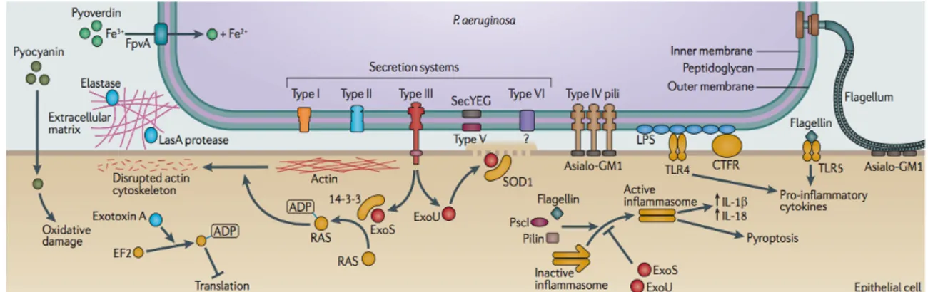
The capacity of P. aeruginosa to produce such diverse infections, is due to a large number of virulence factors, such LasA and LasB elastases, exotoxin A, phospholipase C, protease IV, PrpL protease, pyocyanin, siderophores, hydrogen cyanide, and rhamnolipids, and also to the production of biofilm, that allows the colonization of host tissues and the protection of bacterial cells from the immune system and antibiotics therapies. These factors are collectively capable of causing extensive tissue damage, bloodstream invasion and dissemination in humans and other mammals (Fig. 1; Smith and Iglewski, 2003).
Fig. 1. Overview of P. aeruginosa virulence determinants. P. aeruginosa has both cell-associated (flagellum, pili,
lipopolysaccharides, alginate) and extracellular virulence factors. The type III secretion apparatus is used to inject exotoxins S, T, Y, and U in the host cell. Modified from Hauser and Ozer, 2011.
7 P. aeruginosa can adopt two different lifestyles that reflect two different strategies of infection, the acute and the chronic infection. The acute infection is rapid, systemic, carried out by a planktonic bacterial community expressing high levels of virulence factors and typically has a severe outcome; in patients with damaged airways from mechanical ventilation, trauma, or antecedent viral infection, P. aeruginosa colonization of the respiratory tract is often followed by acute pneumonia, sepsis, and death (Sadikot et al., 2005). Conversely, during chronic infections bacterial proliferation is limited to a specific host tissue (e.g., in the CF lung or in association with medical devices), and bacteria can persist in the host for extended periods of time, adopting a slow-growing sessile lifestyle and forming biofilm. In the biofilm mode of growth bacteria are more resistant to the host immune system and prolonged antibiotic therapies, and they produce limited amount of virulence factors (Furukawa et al., 2006; Coggan et al., 2012). In P. aeruginosa both the acute and the chronic infections are positively controlled by quorum sensing, a communication system that regulates gene expression in response to cell density (Rutherford and Bassler, 2012).
3. Quorum Sensing
Quorum sensing (QS) is a communication system by which a bacterial population coordinately reprograms gene expression in response to cell density, and it is based on the production, secretion and perception of signal molecules. In different bacteria QS is involved in the regulation of a wide variety of physiological processes, including genetic competence, bioluminescence, antibiotic biosynthesis, motility, plasmid conjugal transfer, biofilm formation, and the production of bacterial virulence factors in plant, animal and human pathogens (Miller and Bassler, 2001; Williams and Cámara, 2009). QS communication systems rely on the production of different classes of signal molecules and on different mechanisms of signal response in Gram-positive and Gram-negative bacteria.
The most intensively investigated QS systems in Gram-negative bacteria employ N-acylhomoserine lactones (AHLs) as signal molecules. AHL biosynthesis is typically catalysed by LuxI-family synthases, which transfer an acyl group from an acylated acyl carrier protein (acyl-ACP) to the methionyl amine of S-adenosyl-L-methionine (SAM), after which cyclization of the methionyl moiety to homoserine lactone occurs. The length of the acyl side chain (usually from 4 to 18 carbons), saturation and oxidation state at position 3, determine the resulting AHL structure, and thus signal-specificity. Short-chain AHLs generally freely diffuse across membranes, while there is some evidence for active efflux of AHLs with longer acyl side-chains. AHLs generally function by binding to a cognate intracellular receptor protein belonging to the LuxR-family. In most cases, the
8 LuxR receptor-AHL complex binds to target promoters, activating gene expression (Miller and Bassler, 2001; Atkinson and Williams, 2009).
In Gram-positive bacteria QS systems generally rely on genetically encoded peptides as signal molecules, often termed ‘autoinducing peptides’ (AIPs). AIPs are expressed as inactive pro-peptides via canonical ribosomal synthesis, and later processed and modified to generate the active QS signal. AIPs are not freely diffusible across membranes. AIP perception by the receiver cell is usually mediated by sensor kinases, which transduce the signal from the membrane to cognate response regulators inside the cell via a phosphorylation cascade (Miller and Bassler, 2001; Atkinson and Williams, 2009).
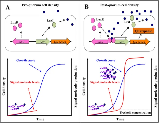
Fig. 2. Quorum sensing. Schematic representation of a general LuxR/LuxI-like QS system. (A) At the pre-quorum cell
density an acyl-homoserine lactone (AHL) signal molecule is produced by a LuxI-like protein at a basal level (red curve), and diffuses outside the producing cells accumulating in the extracellular environment. (B) At the post-quorum cell density the signal molecule reaches a threshold concentration and binds to an intracellular LuxR-like cognate receptor; the LuxR/AHL complex activates QS-regulated genes and triggers the expression of the luxI-like gene, generating a positive feedback loop which lead to a rapid increase in signal molecule production (red curve). The blue curve is representative of a general growth curve; solid black arrows indicate positive control; dashed black arrows indicate information flow; dark blue stars represent the QS signal molecules.
9 The first QS system was described in the early 1970s in the marine bacterium Vibrio fischeri, in which it controls bioluminescence emission. This QS system was based on the production of the signal molecule N-3-(oxohexanoyl)homoserine lactone (3OC6-HSL). In V. fischeri the gene luxI
encodes the synthase LuxI that, at low cell density, synthesizes 3OC6-HSL at a basal level; 3OC6
-HSL accumulates in the extracellular environment proportionally to cell density of the bacterial culture, and when it reaches a certain threshold concentration, corresponding to the “quorum” cell density, it binds to and activates its cognate intracellular receptor LuxR, encoded by the gene luxR (Fig. 2A). The LuxR/3OC6-HSL complex triggers the transcription of genes involved in
bioluminescence production, and also of the gene luxI gene, generating a positive feedback loop which lead to a rapid increase in the concentration 3OC6-HSL (Fig. 2B; Nealson et al., 1970; Fuqua
et al., 1994).
4. QS in Pseudomonas aeruginosa
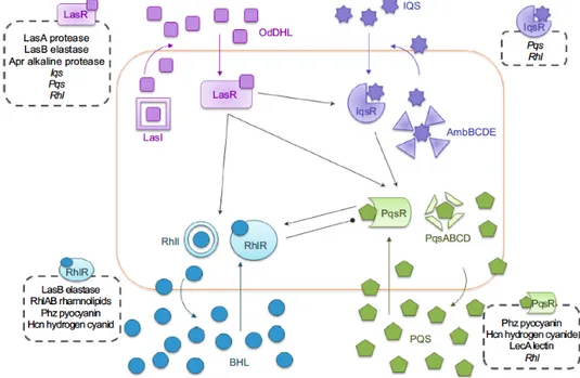
As previously described, in P. aeruginosa both acute and chronic infections are positively controlled by QS (Rutherford and Bassler, 2012). P. aeruginosa has four QS systems that are interconnected and hierarchically arranged: in rich medium, the las QS system is at the top of this hierarchy, because it is required for full activation of the other three QS systems, the rhl, the pqs, and the recently characterized IQS systems (Latifi et al., 1996; Pesci et al., 1997; Gallagher et al., 2002 Kiratisin et al., 2002; Deziel et al., 2004; Xiao et al., 2006 Lee et al., 2013) (Fig. 3).
Fig. 3. Schematic representation of the four QS signaling networks in P. aeruginosa. Black arrows indicate positive
10 The las system consists of the LuxR-like transcriptional regulator LasR (encoded by the lasR gene), and of the LuxI-like AHL synthase LasI (encoded by the lasI gene), that directs the synthesis of the QS signal molecule N-(3-oxododecanoyl)homoserine lactone (3OC12-HSL) (Fig. 4; Schuster
and Greenberg, 2006). Similarly to the V. fischeri lux QS system previously described, at low-cell density the 3OC12-HSL molecule is synthesized by LasI at a basal level, and is secreted into the
surrounding medium; as a consequence, no QS-response occurs (Pearson et al., 1999). With increasing cell density, the signal molecule accumulates until its concentration reaches the threshold level; at this critical concentration, 3OC12-HSL binds its cognate receptor, the QS-activator LasR
(Fuqua et al., 1996). The LasR/3OC12-HSL complex triggers lasI transcription, generating a
positive feedback loop that leads to the amplification of 3OC12-HSL production; as a consequence,
the QS-system becomes active (Seed et al., 1995). Indeed, the LasR/3OC12-HSL complex also
activates the rhl, pqs and IQS QS systems, and acts as a global transcriptional regulator, drastically reprogramming P. aeruginosa transcriptome (Fig. 4; Schuster et al., 2003). As a whole, the QS circuit regulates about 7% of all the P. aeruginosa genes, and has a key role in the infection processes being required for the production of many virulence factors and for biofilm formation (Kirisits and Parsek, 2006; Schuster and Greenberg, 2006).
The involvement of QS in P. aeruginosa pathogenicity is highlighted by the observation that QS-deficient strains are less virulent than the wild type counterparts in all the animal and plant infection models tested up do date: mice, Drosophila melanogaster, Caenorhabditis elegans, Dictyostelium discoideum, and Arabidopsis thaliana (Smith and Iglewski, 2003, Juhas et al., 2005; Diggle et al., 2006; Rampioni et al., 2010). Moreover, 3OC12-HSL directly stimulates interferon-γ
and interleukin-8 production, inhibits interleukin-12 and tumor necrosis factor α, promotes immunoglobulin-E production, and causes apoptosis in macrophages and neutrophils. In this respect, 3OC12-HSL can be considered as virulence factors itself (Wagner et al., 2006).
Also the rhl QS system relies on the production of an acyl-homoserine lactone as signal molecule, the N-butanoylhomoserine lactone (C4-HSL). C4-HSL is synthesised by the LuxI-like
enzyme RhlI, and it is released into the extracellular environment; as the bacterial population grows, C4-HSL binds to its LuxR-like cognate receptor RhlR, and the RhlR/C4-HSL complex
modulates the expression of target genes. The pqs system uses the alkyl-quinolone signals 2-heptyl-3-hydroxy-4(1H)-quinolone (PQS) and 2-heptyl-4-hydroxyquinoline (HHQ) as signal molecules. Both PQS and HHQ can activate the LysR-like protein PqsR that, once activate upon signal molecule(s)-binding, acts as a transcriptional regulator. The IQS system has been characterized very recently, and it is based on the production of the signal molecule 2-(2-hydroxyphenyl)-thiazole-4-carbaldehyde (IQS); the genes involved in IQS synthesis are a non-ribosomal peptide synthase
11 genes of the ambBCDE operon, and the transcriptional regulator IqsR is the IQS receptor. When bacteria are grown in complex media, such as LB, the four QS systems are interconnected and hierarchically organized, with the las QS system placed at the top of the QS cascade, since it is required for full activation of the rhl, pqs and IQS systems. Therefore the regulative cascade leading to the expression of virulence phenotypes in P. aeruginosa mainly relies on the las QS system (Lee and Zhang, 2015).
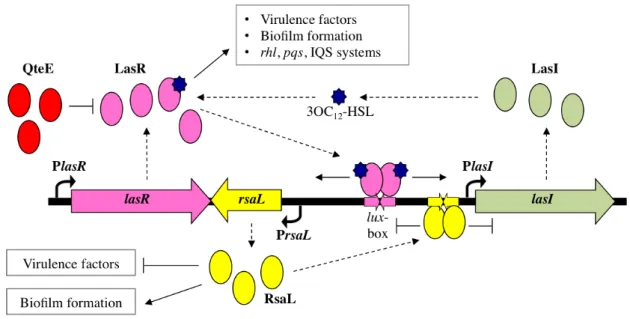
Fig. 4. Schematic representation of P. aeruginosa las QS system. The signal molecule 3OC12-HSL is produced by the synthase LasI at a basal level until the “quorum” cell density is reached; at the “quorum” cell density 3OC12-HSL binds to and activates its cognate receptor LasR, that in turn promotes the expression of (i) genes involved in virulence factors production and in biofilm formation, (ii) the rhl, pqs and IQS systems, (iii) the lasI gene, generating a positive feedback loop which leads to a rapid increase in 3OC12-HSL production, (iv) the rsaL gene, that counteracts the positive feedback loop by repressing lasI transcription. The premature activation of the las system at low cell density is prevented by the pre-quorum repression exerted by QteE on LasR. Solid arrows indicate positive control; T-lines indicate negative control; dashed arrows indicate information flow; blue stars represent the 3OC12-HSL signal molecules.
5. Regulation of QS in P. aeruginosa
The las QS system is more complex than the minimal lux-like QS system exemplified by V. fischeri and described in Fig. 2, because it undergoes a fine modulation of the timing and the extent of 3OC12-HSL production. As an example, in V. fischeri exogenous provision of 3OC6-HSL to a
low cell-density culture activates the lux QS system (Fuqua et al., 1994). Conversely, the exogenous provision of 3OC12-HSL to a P. aeruginosa low cell-density culture is not sufficient to
activate the las QS system (Witheley et al., 1999). This difference is due to a strong pre-quorum repression exerted on the P. aeruginosa las system by multiple repressors, such as the LasR post-translational repressors QscR, QslA and QteE (Chugani et al., 2001; Ledgham et al., 2003; Sienhel
12 et al., 2010; Seet et al., 2011). QscR has been characterized by Chugani et al. in 2001 for its ability to repress some QS regulated genes, and a mutant defective in qscR shows an anticipation of 3OC12-HSL production (Fig. 5). Its mechanism of action was investigated by Ledgham et al. in
2013, and revealed that QscR, in the absence of 3OC12-HSL, forms heterodimers with LasR unable
to bind DNA; however, since QscR has a functional DNA binding site, the reason why the heterodimers with LasR are impaired in DNA binding is not clear. Exogenous provision of high levels of 3OC12-HSL disrupt QscR inhibition of LasR (Chugani et al., 2001; Ledgham et al., 2003;
Coggan et al., 2012). Similarly to QscR, also QslA represses LasR via protein-protein interaction; for QslA however, Seet et al. in 2011 demonstrated that its interaction with LasR prevents LasR binding to the promoter of the lasI gene (PlasI); QslA is also able to disrupt a pre-formed LasR-PlasI complex, and the exogenous provision of 3OC12-HSL does not affected QslA repression on
LasR, suggesting that QslA may control the overall QS threshold (Seet et al., 2011; Coggan et al., 2012). QteE was first characterized by Sienhel et al. in 2010 for its role in modulating the QS threshold level; it has been supposed that QteE affects LasR stability at low cell density. Further characterization of QteE is discussed in the Chapter II of this thesis; the work of Bondì et al. of 2014 demonstrates that a P. aeruginosa mutant strain defective in qteE shows an anticipation of 3OC12-HSL production and of QS-related virulence phenotypes expression with respect to the wild
type strain (Sienhel et al., 2010; Coggan et al., 2012; Bondì et al., 2014) (Fig. 5).
The timing of the QS response seems to be very important in P. aeruginosa considering that its regulation is committed to three distinct regulators; it is possible, however, that they are not simply redundant, but respond to different external cues to control the exact timing of QS activation in different environmental conditions (Coggan et al., 2012).
Besides the pre-quorum repression, another difference between the traditional lux-like QS systems and the P. aeruginosa las system is the post-quorum repression exerted by the repressor of lasI, RsaL. As previously described, the traditional lux-like QS systems consist of the luxI-like gene, encoding the synthase of the signal molecule, and of the luxR-like gene, encoding the receptor of the signal molecule; conversely, in the las system the gene rsaL is part of the las genetic locus together with lasI and lasR, and its transcription is activated by the LasR/3OC12-HSL complex (Fig.
4). RsaL is the only transcriptional regulator, besides LasR, known to bind the lasI promoter region (Rampioni et al., 2006). RsaL represses lasI transcription in a post-quorum phase of growth, and therefore it counteracts the positive feedback loop generated by the LasR/3OC12-HSL complex
(Rampioni et al., 2007). In the traditional QS systems, when the threshold concentration of the signal molecule is reached, the positive feedback loop leads to the accumulation of the signal molecule during the all growth curve; conversely, in the las system, 3OC12-HSL production reaches
13 a stationary level before the end of exponential phase of growth due to the RsaL-dependent homeostatic regulation of lasI transcription (Fig. 4 and Fig. 5; Chugani et al., 2001; Ward et al., 2004; Rampioni et al., 2007). RsaL is also a global regulator that in P. aeruginosa regulates more than 300 genes, including genes involved in virulence factors production (pyocyanin and hydrogen cyanide), in the efflux of antibiotic outside the cell, and in biofilm formation (Rampioni et al., 2007; Rampioni et al., 2009).
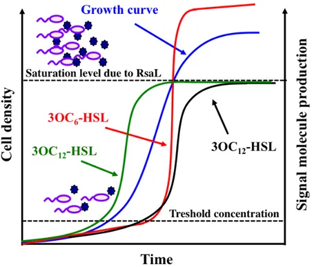
Fig. 5. Schematic representation of 3OC6-HSL production in V. fischeri and of 3OC12-HSL production in P.
aeruginosa. In V. fischeri, the positive feedback loop generated by the LuxR/3OC6-HSL complex leads to the accumulation of the signal molecule during the all growth curve (red curve); in P. aeruginosa, the levels of 3OC12-HSL reach a saturation level before the end of exponential phase of growth because the positive feedback loop generated by the LasR/3OC12-HSL complex is counteracted by the RsaL repressive effect on lasI transcription (black curve). The mutation of the qscR or qteE genes anticipates 3OC12-HSL production (green curve). Dark blue stars represent the QS signal molecules.
P. aeruginosa QS circuit is integrated into a wide regulatory network, and is modulated by environmental and metabolic signals beside cell density (Soberón-Chávez et al., 2005; Dunn and Stabb, 2006; Duan and Surette, 2007; Coggan et al., 2012). If considering only the regulators acting on the las QS system, a number of transcriptional and post-transcriptional regulators have been described that exert a direct or an indirect regulation on its activity. However, in most cases, their physiological role and molecular mechanism of action remain unclear (Coggan et al., 2012). Some
14 of these regulators and their effect on the las system are summarized in Fig. 6, and the best characterized ones are also described below.
Besides QscR, QslA and QteE, that have been previously described, MvaT and AlgQ negatively regulate the expression of LasR, while Vfr and GacA positively regulate its expression. MvaT belongs to family of the histone-like nucleoid structuring (H-NS), that are involved in compacting chromosomal DNA (Dorman, 2004). In P. aeruginosa MvaT regulates the expression of genes involved in virulence, in arginine metabolism, in antibiotic resistance and in biofilm formation, through direct binding to the promoters or through indirect regulation (Diggle et al., 2002; Vallet et al., 2004; Westfall et al., 2004 and 2006; Li et al., 2009). In an mvaT mutant the production of 3OC12-HSL is anticipated with respect to the wild type strain, suggesting its role as repressor of the
las system (Diggle et al., 2002).
AlgQ (also named AlgR2) was originally identified as a regulatory protein involved in alginate production. It also regulates the synthesis of secreted virulence factors, up-regulating neuraminidase and siderophore synthesis and down-regulating rhamnolipids and extracellular proteases synthesis. AlgQ negatively modulates the expression of lasR by the directly binding to its promoter region (Ledgham et al., 2003).
Vfr was first identified in P. aeruginosa as a virulence factor regulator, due to its positive effect on the production of several virulence factors, such as proteases and exotoxin A. It is a member of the cAMP receptor protein (CRP) family, and links the QS circuit and the cyclic adenosine monophosphate (cAMP) signaling system by directly activating lasR transcription (Albus et al., 1997). Differently from Escherichia coli, cAMP levels in P. aeruginosa are not related to glucose uptake and utilization (West et al., 1994), and the role played by cAMP in P. aeruginosa physiology, as well as the environmental stimuli to which Vfr responds, are still unknown (Lazdunkski et al., 2007).
GacA is the response protein of the two-component system GacS/GacA, and it activates the transcription of LasR and also of its repressor QscR. Moreover, GacA is essential for the expression of the two non-coding regulatory RNAs RsmZ and RsmY, that work in tandem with the regulator RsmA. RsmA is a pleiotropic post-transcriptional regulator that together with RsmZ and RsmY controls the production of secondary metabolites both directly and indirectly. RsmA represses lasI transcription and exerts a negative effect on 3OC12-HSL production. In a rsmA mutant strain the
expression of lasI is prematurely induced and reaches higher levels with respect to the wild type strain (Lazdunkski et al., 2007).
15 VqsR is a LuxR-like protein with no signal molecule-binding domain. Its synthesis is dependent on the LasR/3OC12-HSL complex, and in turn VqsR represses lasR transcription. A mutant
defective in vqsR is impaired in 3OC12-HSL production (Lazdunkski et al., 2007).
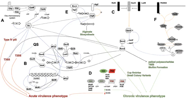
Fig. 6. Schematic representation of the regulatory pathways that modulate P. aeruginosa lifestyle. Lines depict
direct regulatory mechanisms within given signaling pathways, as well as cross-talk between regulatory systems. Arrows represent positive regulation; T-lines indicate negative regulation; blue lines represent transcriptional regulation while purple lines represent enzymatic reactions; orange lines depict post-translational modification events; black lines illustrate post-translational regulation events; dashed lines indicate unknown mechanisms of regulation. (A) cAMP/Vfr signaling. (B) Quorum Sensing. (C) Gac/Rsm pathway. (D) c-di-GMP signaling. (E) MucA signaling. (F) HptB signaling. Modified by Coggan et al., 2012.
16
AIMS AND RATIONALE
P. aeruginosa resistance to antibiotics demand the discovery of new therapeutic approaches. Targeting bacterial virulence instead of bacterial growth is an alternative approach to antimicrobial therapy that offers promising opportunities to inhibit pathogenesis and its consequences without placing immediate life-or-death pressure on the target bacterium (Cegelski et al., 2008). However, as described above, virulence in P. aeruginosa is due to the production of an arsenal of virulence factors: for this reason the appropriate anti-virulence drug should target the regulatory networks controlling virulence instead of a single virulence factor (Rasko and Sperandio, 2010). In this context, QS is a good target for anti-virulence drugs because it controls genes involved in multiple virulence factors production and in biofilm formation, playing a positive role in the instauration of both the acute and the chronic infection in humans (Rutherford and Bassler, 2012). Moreover, QS is interwoven in an intricate regulatory network (Fig. 6; Venturi, 2006; Coggan et al., 2012) and it has been suggested that also the regulators of QS play an important role during the infections; therefore, QS regulators could be good candidates for the development of anti-virulence drugs.
The general aim of this PhD project has been the study of the regulation of the P. aeruginosa las QS system, an. This project is structured in three main objectives: (i) the identification of novel transcriptional regulators controlling LasR expression (Longo et al., 2013; Chapter I); (ii) the study of the effect of a dysregulation in timing and magnitude of the las QS response on P. aeruginosa ability to establish infections; (Bondì et al., 2014; Chapter II); (iii) the investigation of new possible regulative properties arising from the peculiar architecture of the las QS system (Bondì et al., manuscript in preparation; Chapter III).
17
REFERENCES
• Albus AM, Pesci EC, Runyen-Janecky LJ, West SE, Iglewski BH (1997) Vfr controls quorum sensing in
Pseudomonas aeruginosa. J Bacteriol 179:3928-3935.
• Anderson R, May R (1982) Coevolution of hosts and parasites. Parasitology 85:411-426.
• Atkinson S, Williams P (2009) Quorum sensing and social networking in the microbial world. J R Soc Interface 6:959-978.
• Bondí R, Messina M, De Fino I, Bragonzi A, Rampioni G, Leoni L (2014) Affecting Pseudomonas aeruginosa phenotypic plasticity by quorum sensing dysregulation hampers pathogenicity in murine chronic lung infection. PLoS
One 9:e112105.
• Brown SP, Cornforth DM, Mideo N (2012) Evolution of virulence in opportunistic pathogens: generalism, plasticity, and control. Trends Microbiol 20:336-342.
• Cámara M, Williams P, Hardman A (2002) Controlling infection by tuning in and turning down the volume of bacterial small-talk. Lancet Infect Dis 2:667-676.
• Cegelski L, Marshall GR, Eldridge GR, Hultgren SJ (2008) The biology and future prospects of antivirulence therapies. Nat Rev Microbiol 6:17-27. Erratum in: Nat Rev Microbiol (2009) 7:836.
• Chugani SA, Whiteley M, Lee KM, D’Argenio D, Manoil C, Greenberg EP (2001) QscR, a modulator of quorum-sensing signal synthesis and virulence in Pseudomonas aeruginosa. Proc Natl Acad Sci USA 98:2752-2757.
• Coggan KA, Wolfgang MC (2012) Global regulatory pathways and cross-talk control Pseudomonas aeruginosa environmental lifestyle and virulence phenotype. Curr Issues Mol Biol 14:47-70.
• Diggle SP, Cornelis P, Williams P, Cámara M (2006) 4-quinolone signalling in Pseudomonas aeruginosa: old molecules, new perspectives. Int J Med Microbiol 296:83-91.
• Dorman CJ (2004) H-NS: a universal regulator for a dynamic genome. Nat Rev Microbiol 2:391-400.
• Duan K, Surette MG (2007) Environmental regulation of Pseudomonas aeruginosa PAO1 Las and Rhl quorum-sensing systems. J Bacteriol 189:4827-4836.
• Dunn AK, Stabb EV (2006) Beyond quorum sensing: the complexities of prokaryotic parliamentary procedures. Anal
Bioanal Chem 387:391-398.
• Fuqua C, Winans SC, Greenberg EP (1996) Census and consensus in bacterial ecosystems: the LuxR-LuxI family of quorum-sensing transcriptional regulators. Annu Rev Microbiol 50:727-751.
• Fuqua WC, Winans SC, Greenberg EP (1994) Quorum sensing in bacteria: the LuxR/LuxI family of cell density-responsive transcriptional regulators. J Bacteriol 176:269-275.
• Furukawa S, Kuchma SL, O’Toole GA (2006) Keeping their options open: acute versus persistent infections. J
Bacteriol 188:1211-1217.
• Goodman AL, Lory S (2004) Analysis of regulatory networks in Pseudomonas aeruginosa by genomwide transcriptional profiling. Curr Opin Microbiol 7:39-44.
• Gupta R, Schuster M (2013) Negative regulation of bacterial quorum sensing tunes public goods cooperation. ISME J 7:2159-2168.
• Hauser A, Ozer EA (2011) Pseudomonas aeruginosa. Nat Rev Microbiol Vol. 9 no.3. Poster.
• Juhas M, Wiehlmann L, Salunkhe P, Lauber J, Buer J, Tümmler B (2005) GeneChip expression analysis of the VqsR regulon of Pseudomonas aeruginosa TB. FEMS Microbiol Lett 242:287-295.
18
• Kirisits MJ, Parsek MR (2006) Does Pseudomonas aeruginosa use intercellular signalling to build biofilm communities? Cell Microbiol 8:1841-1849.
• Latifi A, Winson MK, Foglino M, Bycroft BW, Stewart GS, Lazdunski A, Williams P (1995) Multiple homologues of LuxR and LuxI control expression of virulence determinants and secondary metabolites through quorum sensing in
Pseudomonas aeruginosa PAO1. Mol Microbiol 17:333-343.
• Lazdunski AM, Ventre I, Bleves S (2007) Cell-cell communication: Quorum Sensing and regulatory circuits in
Pseudomonas aeruginosa. In: Ramos JL, Filloux A (ed), Pseudomonas, A model system in Biology vol. 5; 279-310.
• Ledgham F, Soscia C, Chakrabarty A, Lazdunski A, Foglino M (2003) Global regulation in Pseudomonas
aeruginosa: the regulatory protein AlgR2 (AlgQ) acts as a modulator of quorum sensing. Res Microbiol 154:207-213.
• Liang H, Duan J, Sibley CD, Surette MG, Duan K (2011) Identification of mutants with altered phenazine production in Pseudomonas aeruginosa. J Med Microbiol 60:22-34.
• Longo F, Rampioni G, Bondí R, Imperi F, Fimia GM, Visca P, Zennaro E, Leoni L (2013) A new transcriptional repressor of the Pseudomonas aeruginosa quorum sensing receptor gene lasR. PLoS One 8:e69554.
• Lyczac JB, Cannon CL, Pier GB (2000) Establishment of Pseudomonas aeruginosa infection: lessons from a versatile opportunist. Microbes Infect 2:1051-1060.
• Lyczak JB, Cannon CL, Pier GB (2002) Lung infections associated with cystic fibrosis. Clin Microbiol Rev 15:194-222.
• Miller MB, Bassler BL (2001) Quorum sensing in bacteria. Annu Rev Microbiol 55:165-199.
• Murray TS, Egan M, Kazmierczak BI (2007) Pseudomonas aeruginosa chronic colonization in cystic fibrosis patients. Curr Opin Pediatr 19:83-88.
• Nealson KH, Platt T, Hastings JW (1970) Cellular control of the synthesis and activity of the bacterial luminescent system. J Bacteriol 104:313-322.
• Osmon S, Ward S, Fraser VJ, Kollef MH (2004) Hospital mortality for patients with bacteremia due to
Staphylococcus aureus or Pseudomonas aeruginosa. Chest 125:607-616.
• Pearson JP, van Delden C, Iglewski BH (1999) Active efflux and diffusion are involved in transport of Pseudomonas
aeruginosa cell-to-cell signals. J Bacteriol 181:1203-1210.
• Rampioni G, Bertani I, Zennaro E, Polticelli F, Venturi V, Leoni L (2006) The quorum-sensing negative regulator RsaL of Pseudomonas aeruginosa binds to the lasI promoter. J Bacteriol 188:815-819.
• Rampioni G, Pustelny C, Fletcher MP, Wright VJ, Bruce M, Rumbaugh KP, Heeb S, Cámara M, Williams P (2010) Transcriptomic analysis reveals a global alkyl-quinolone-independent regulatory role for PqsE in facilitating the environmental adaptation of Pseudomonas aeruginosa to plant and animal hosts. Environ Microbiol 12:1659-1673. • Rampioni G, Schuster M, Greenberg EP, Bertani I, Grasso M, Venturi V, Zennaro E, Leoni L (2007) RsaL provides
quorum sensing homeostasis and functions as a global regulator of gene expression in Pseudomonas aeruginosa. Mol
Microbiol 66:1557-1565.
• Rampioni G, Schuster M, Greenberg EP, Zennaro E, Leoni L (2009) Contribution of the RsaL global regulator to
Pseudomonas aeruginosa virulence and biofilm formation. FEMS Microbiol Lett 301:210-217.
• Rasko DA, Sperandio V (2010) Anti-virulence strategies to combat bacteria-mediated disease. Nat Rev Drug Discov 9:117-128.
• Rutherford ST, Bassler BL (2012) Bacterial quorum sensing: its role in virulence and possibilities for its control. Cold
19
• Sadikot RT, Blackwell TS, Christman JW, Prince AS (2005) Pathogen-host interactions in Pseudomonas aeruginosa pneumonia. Am J Respir Crit Care Med 171:1209-1223.
• Schuster M, Greenberg EP (2006) A network of networks: quorum-sensing gene regulation in Pseudomonas
aeruginosa. Int J Med Microbiol 296:73-81.
• Schuster M, Lostroh CP, Ogi T, Greenberg EP (2003) Identification, timing, and signal specificity of Pseudomonas
aeruginosa quorum-controlled genes: a transcriptome analysis. J Bacteriol 185:2066-2079.
• Seed PC, Passador L, Iglewski BH (1995) Activation of the Pseudomonas aeruginosa lasI gene by LasR and the
Pseudomonas autoinducer PAI: an autoinduction regulatory hierarchy. J Bacteriol 177:654-659.
• Seet Q, Zhang LH (2011) Anti-activator QslA defines the quorum sensing threshold and response in Pseudomonas
aeruginosa. Mol Microbiol 80:951-965.
• Siehnel R, Traxler B, An DD, Parsek MR, Schaefer AL, Singh PK (2010) A unique regulator controls the activation threshold of quorum-regulated genes in Pseudomonas aeruginosa. Proc Natl Acad Sci USA 107:7916-7921.
• Smith RS, Iglewski BH (2003) Pseudomonas aeruginosa quorum-sensing systems and virulence. Curr Opin
Microbiol 6:56-60.
• Soberón-Chávez G, Aguirre-Ramírez M, Ordóñez L (2005) Is Pseudomonas aeruginosa only "sensing quorum"? Crit
Rev Microbiol 31:171-182.
• Stover CK, Pham XQ, Erwin AL, Mizoguchi SD, Warrener P, Hickey MJ, Brinkman FS, Hufnagle WO, Kowalik DJ, Lagrou M, Garber RL, Goltry L, Tolentino E, Westbrock-Wadman S, Yuan Y, Brody LL, Coulter SN, Folger KR, Kas A, Larbig K, Lim R, Smith K, Spencer D, Wong GK, Wu Z, Paulsen IT, Reizer J, Saier MH, Hancock RE, Lory S, Olson MV (2000) Complete genome sequence of Pseudomonas aeruginosa PAO1, an opportunistic pathogen.
Nature 406:959-964.
• Vallet I, Diggle SP, Stacey RE, Cámara M, Ventre I, Lory S, Lazdunski A, Williams P, Filloux A (2004) Biofilm formation in Pseudomonas aeruginosa: fimbrial cup gene clusters are controlled by the transcriptional regulator MvaT. J Bacteriol 186:2880-2890.
• Van Delden (2004) Virulence factors in Pseudomonas aeruginosa. In Ramos JL (ed), Pseudomonas, Vol. II, pp. 3-45, Kluwer Academic / Plenum Publishers, NY.
• Venturi V (2006) Regulation of quorum sensing in Pseudomonas. FEMS Microbiol Rev 30:274-291.
• Wagner VE, Frelinger JG, Barth RK, Iglewski BH (2006) Quorum sensing: dynamic response of Pseudomonas
aeruginosa to external signals. Trends Microbiol 14:55-58.
• Ward JP, King JR, Koerber AJ, Croft JM, Sockett RE, Williams P (2004) Cell-signalling repression in bacterial quorum sensing. Math Med Biol 21:169-204.
• Welsh MJ, Ramsey BW, Accurso F, Cutting G (2001) The metabolic and molecular basis of inherited diseases. New
York: McGraw-Hill. 8:5121-5188.
• West SE, Sample AK, Runyen-Janecky LJ (1994) The vfr gene product, required for Pseudomonas aeruginosa exotoxin A and protease production, belongs to the cyclic AMP receptor protein family. J Bacteriol 176:7532-7542. • Whiteley M, Lee KM, Greenberg EP (1999) Identification of genes controlled by quorum sensing in Pseudomonas
aeruginosa. Proc Natl Acad Sci USA 96:13904-13909.
• Williams P, Cámara M (2009) Quorum sensing and environmental adaptation in Pseudomonas aeruginosa: a tale of regulatory networks and multifunctional signal molecules. Curr Opin Microbiol 12:182-191.
Chapter I
A new transcriptional repressor of the Pseudomonas
aeruginosa Quorum Sensing receptor gene lasR
Francesca Longo
1, Giordano Rampioni
1, Roslen Bondì
1, Francesco
Imperi
2, Gian Maria Fimia
3, Paolo Visca
1,
Elisabetta Zennaro
1and Livia Leoni
11
Department of Sciences, University Roma Tre, Rome, Italy,
2
Department of Biology and Biotechnology “Charles Darwin”, Sapienza University,
Rome, Italy,
3
A New Transcriptional Repressor of the Pseudomonas
aeruginosa Quorum Sensing Receptor Gene lasR
Francesca Longo1, Giordano Rampioni1, Roslen Bondì1, Francesco Imperi2, Gian Maria Fimia3, Paolo
Visca1, Elisabetta Zennaro1, Livia Leoni1*
1 Department of Sciences, University Roma Tre, Rome, Italy, 2 Department of Biology and Biotechnology “Charles Darwin”, Sapienza University, Rome, Italy, 3 National Institute for Infectious Disease “Lazzaro Spallanzani”, Rome, Italy
Abstract
Pseudomonas aeruginosa pathogenic potential is controlled via multiple regulatory pathways, including three quorum sensing (QS) systems. LasR is a key QS signal receptor since it acts as a global transcriptional regulator required for optimal expression of main virulence factors. P. aeruginosa modulates the QS response by integrating this cell density-dependent circuit to environmental and metabolic cues. Hence, QS also controls the adaptation to challenging environmental niches, such as infection sites. However, little is known about the molecular mechanisms connecting QS and other signalling pathways. In this work, DNA-affinity chromatography was used to identify new lasR transcriptional regulators. This approach led to the identification and functional characterization of the TetR-like transcriptional repressor PA3699. This protein was purified and shown to directly bind to the lasR promoter region in vitro. The induction of PA3699 expression in P. aeruginosa PAO1 cultures repressed lasR promoter activity and the production of LasR-dependent virulence factors, such as elastase, pyocyanin, and proteases. These findings suggest a role for PA3699 in P. aeruginosa pathogenicity. P. aeruginosa genome encodes at least 38 TetR-family proteins, and PA3699 is the eighth member of this group functionally characterized so far and the first one shown to bind the lasR promoter in vitro.
Citation: Longo F, Rampioni G, Bondì R, Imperi F, Fimia GM, et al. (2013) A New Transcriptional Repressor of the Pseudomonas aeruginosa Quorum Sensing Receptor Gene lasR. PLoS ONE 8(7): e69554. doi:10.1371/journal.pone.0069554
Editor: Tom Coenye, Ghent University, Belgium
Received May 03, 2013; Accepted June 10, 2013; Published July 5, 2013
Copyright: © 2013 Longo et al. This is an open-access article distributed under the terms of the Creative Commons Attribution License, which permits unrestricted use, distribution, and reproduction in any medium, provided the original author and source are credited.
Funding: This work was supported by grants from the Ministry of University and Research of Italy (Futuro in Ricerca 2010 - RBFR10LHD1; www.miur.it), University Roma Tre (Internationalization Project; www.uniroma3.it), and the Italian Cystic Fibrosis Research Foundation (FFC 14/2010 and FFC 13/2011; www.fibrosicisticaricerca.it). The funders had no role in study design, data collection and analysis, decision to publish, or preparation of the manuscript. Competing interests: The authors have declared that no competing interests exist.
* E-mail: [email protected]
Introduction
The definition of quorum sensing (QS) was coined about 20 years ago to describe a communication system based on the production, detection, and response to signal molecules, allowing bacterial populations to trigger a coordinated response at a threshold cell density [1]. Today it is well known that QS regulation plays a key role in a number of relevant bacterial processes, including colonization of plants and animal tissues, and production of antibiotics [2,3].
In many bacteria, the QS regulatory device is interwoven with other global regulatory networks responsive to different environmental cues (e.g., temperature, pH, osmolarity, oxidative stress, nutrient starvation). These networks cross-talk in the bacterial cell in order to determine the optimal survival strategy [4]. The advantage of integrating QS with other regulatory pathways becomes a compelling necessity when the QS response controls multiple functions and its activation commits bacteria to a strong reorganization of the whole cellular metabolism [5].
One of the most studied model organisms in QS research is
Pseudomonas aeruginosa. This versatile bacterium is able to
thrive in a wide range of environmental niches, including the human body. The high adaptability of P. aeruginosa is reflected by its behaviour as a pathogen. In humans, P. aeruginosa causes community- and hospital-acquired infections, colonizing different body districts like lungs, eyes, ears, urinary tract, injured skin (burns and wounds). Such infections are often difficult to eradicate as a consequence of antibiotic resistance and biofilm formation [6]. In particular, P. aeruginosa chronic lung infection is the major cause of death in cystic fibrosis patients, a genetic disease affecting about 1/3,000 newborns in the Caucasian population [6].
P. aeruginosa pathogenicity strongly depends on the fine
and coordinated regulation of a wide array of virulence factors [7,8]. This is achieved thanks to a complex network of regulatory and signalling pathways controlling virulence-related phenotypes, in response to environmental cues and bacterial population structure [9]. Among these pathways, a preeminent role in P. aeruginosa virulence is played by three
interconnected QS systems, based on the production of different signal molecules. The QS signal molecule N-3-oxo-dodecanoyl-homoserine lactone (3OC12-HSL) is synthesized by
LasI, encoded by the lasI gene, and its cognate receptor is the cytoplasmic transcriptional regulator LasR, encoded by the
lasR gene. 3OC12-HSL progressively accumulates in the bacterial culture and at a threshold concentration it binds LasR. The LasR/3OC12-HSL complex, in turn, activates transcription
of hundreds of genes, including the genes coding for the receptors of the other two QS systems, based on N-butanoylhomoserine lactone (C4-HSL) and
2-heptyl-3-hydroxy-4-quinolone (PQS) signal molecules. As a whole, the
P. aeruginosa QS circuit has a key role in pathogenesis,
regulating the production of virulence factors, the formation of biofilm and the expression of antibiotic efflux pumps [10].
The three QS systems of P. aeruginosa are interwoven and connected to other regulatory pathways [10–12]. Indeed the expression of genes involved in the synthesis and perception of QS signal molecules is finely regulated at the transcriptional and post-transcriptional level in response to various metabolic and environmental stimuli [12–15]. However, the proteins involved in this regulation and their mechanisms of action are largely unknown [11,12]. To date only few transcriptional regulators have been shown to directly bind the promoters of genes involved in P. aeruginosa QS signal synthesis and perception. In particular, RsaL directly represses lasI transcription [16,17], while Vfr directly activates the lasR promoter region [18]. Moreover, AlgR2 (AlgQ), besides acting as an anti-sigma factor for the vegetative sigma RpoD [19], can also bind to and downregulate the lasR promoter [20]. However, the stimuli controlling the transcriptional activity of Vfr, AlgR2 (AlgQ) and RsaL are still unknown.
The objective of this work has been the identification of novel transcriptional regulators of LasR expression. To this aim, P.
aeruginosa cytoplasmic proteins able to bind the lasR promoter
region have been picked-up by DNA-affinity chromatography and identified by mass spectrometry. This led to the functional characterization of the TetR-like protein PA3699, which acts as a novel repressor of lasR transcription and of P. aeruginosa virulence factors production.
Results and Discussion
Identification of P. aeruginosa proteins interacting with the lasR promoter
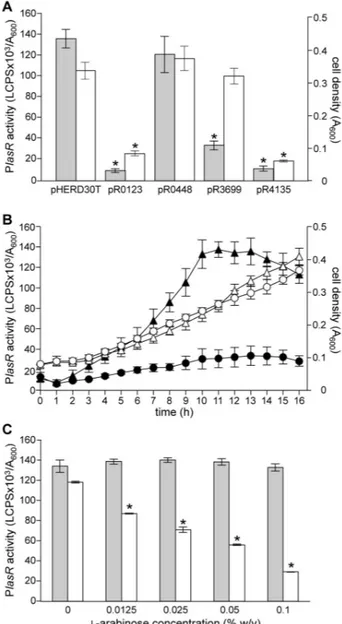
To increase the probability of fishing proteins interacting with the lasR promoter (PlasR), the activity of PlasR was preliminarily monitored along the P. aeruginosa PAO1 growth curve by means of a PlasR::lacZ transcriptional fusion (Figure 1A Table S2). PlasR activity increased steadily during the exponential phase and reached a plateau at the onset of the stationary phase. The maximal promoter activity was maintained for about four hours, and then declined (Figure 1A), suggesting that at the end of the exponential phase and during the stationary phase of growth, one or more transcriptional regulator(s) bind to PlasR and affect its promoter activity. Hence, crude protein extracts were prepared from P.
aeruginosa PAO1 cultures grown to A600 = 2.0 and A600= 5.0,
corresponding to late exponential and stationary phase of growth, respectively (Figure 1A).
To prepare the DNA-affinity chromatography matrix, a biotinylated double-stranded DNA fragment, corresponding to the lasR promoter region cloned in the PlasR::lacZ transcriptional fusion, was immobilized on a streptavidin-conjugated chromatography resin. Protein crude extracts were independently incubated with the matrix and, after extensive washing, proteins specifically bound to PlasR were eluted, separated by SDS-PAGE, and identified by MALDI-TOF mass spectrometry (Table 1). The experiment was performed in duplicate for each crude extract. Examples of SDS-PAGE gels are shown in Figure 1B. Overall, twelve protein bands were reproducibly detected on the SDS-PAGE gels (Table 1), while Figure 1. PlasR activity and PlasR-affinity chromatography. (A) Growth curve of the P. aeruginosa PAO1 wild type strain carrying the pMPlasR::lacZ plasmid (dashed line) and corresponding PlasR promoter activity (solid line). The points of the growth curve at which the protein crude extracts for DNA-affinity chromatography were prepared are indicated by arrows. (B) SDS-PAGE analysis of proteins bound to the PlasR promoter region. Protein crude extracts were prepared from P. aeruginosa PAO1 cultures grown in LB broth to the indicated cell densities (A600). Bands analysed by
MALDI-TOF mass spectrometry are indicated by arrows, and the corresponding protein name is reported.
doi: 10.1371/journal.pone.0069554.g001
A New Repressor of P. aeruginosa QS and Virulence
no protein bands were visible in control experiments with beads uncoupled to DNA (data not shown). Among these, RpoA, Ssb and TufB are involved in general DNA processing, while the PhaF phasin is involved in polyhydroxyalkanoate segregation during cell division [21]. Moreover three proteins not involved in DNA processing (i.e., Lon, PepA and GroEL) were also retrieved (Table 1).
Of the two transcription factors previously known to bind PlasR (i.e., Vfr and AlgR2) [18,20], only Vfr was picked-up (Table 1), indicating that the DNA-affinity chromatography approach here described allows identification of some, but not all, the transcription factors specific for PlasR. Therefore, besides AlgR2, other regulators of this promoter may have escaped our analysis.
Last, but of primary importance with respect to our aims, among the picked-up proteins PA0123, PA0448, PA3699 and PA4135 were annotated in the P. aeruginosa genome as putative transcription factors with unknown function (Table 1). On the basis of their sequence, PA0123 and PA0448 are annotated as putative LysR-like family transcriprional regulators [22]. Interestingly, the three-dimensional structure of PA3699 and PA4135 has been recently solved (Protein Data Bank accession numbers, 3KKD and 2FBI, respectively). On the basis of primary sequence and structural features, PA3699 and PA4135 can be assigned to the TetR-like and MarR-like family of transcriptional regulators, respectively [22]. In this view, the above results provide the first experimental evidence
that PA0123, PA0448, PA3699 and PA4135 are expressed in
P. aeruginosa cultures and able to bind DNA in vitro. On the
basis of the above considerations, these four factors were selected for further analysis as novel putative lasR regulators (Table 1).
In vivo characterization of putative lasR transcriptional regulators
In order to study the in vivo effect of PA0123, PA0448, PA3699 and PA4135 on PlasR activity, a set of P. aeruginosa PAO1 in frame deletion mutants in the corresponding genes was generated. As a control, also a mutation in the vfr gene was introduced in P. aeruginosa PAO1. A genetic cassette carrying the PlasR::lux transcriptional fusion was introduced in a chromosomal neutral site of the five mutant strains and of P.
aeruginosa wild type to conveniently measure PlasR activity as
a function of light emission along the growth curve. The DNA region cloned in this genetic cassette was identical to that used as a bait in the DNA-affinity chromatography experiment.
In accordance with literature data, PlasR activity was strongly decreased as a consequence of vfr mutation (Figure S1) [18], while no significant differences with respect to the wild type activity were observed in the PA0123, PA0448, PA3699 and PA4135 mutant strains (Figure S1). Similar results were obtained by using the PlasR::lacZ transcriptional fusion as reporter system (data not shown).
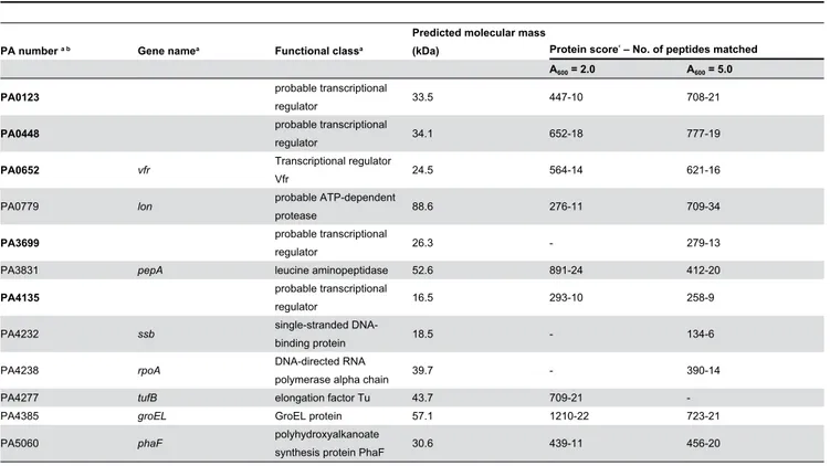
Table 1. List of PlasR-binding proteins identified by MALDI-TOF analysis from the SDS-PAGE gels shown in Figure 1B.
PA number a b Gene namea Functional classa
Predicted molecular mass
(kDa) Protein scorec – No. of peptides matched
A600 = 2.0 A600 = 5.0
PA0123 probable transcriptional
regulator 33.5 447-10 708-21
PA0448 probable transcriptional
regulator 34.1 652-18 777-19
PA0652 vfr Transcriptional regulator
Vfr 24.5 564-14 621-16
PA0779 lon probable ATP-dependent
protease 88.6 276-11 709-34
PA3699 probable transcriptional
regulator 26.3 - 279-13
PA3831 pepA leucine aminopeptidase 52.6 891-24 412-20
PA4135 probable transcriptional
regulator 16.5 293-10 258-9
PA4232 ssb single-stranded
DNA-binding protein 18.5 - 134-6
PA4238 rpoA DNA-directed RNA
polymerase alpha chain 39.7 - 390-14
PA4277 tufB elongation factor Tu 43.7 709-21
-PA4385 groEL GroEL protein 57.1 1210-22 723-21
PA5060 phaF polyhydroxyalkanoate
synthesis protein PhaF 30.6 439-11 456-20
a. PA number, gene name and functional class refer to the Pseudomonas Genome Database annotation [22].
b Proteins identified only when using PlasR as DNA bait are in bold characters.
c. The protein score is according to the Mascot programme (scores ≥ 76 correspond to an error probability p < 0.05 in our data set) [37].
A New Repressor of P. aeruginosa QS and Virulence