UNIVERSITÀ DEGLI STUDI DI PISA
PhD Course in Basic and Developmental Neuroscience Head: Prof. Giovanni Cioni
Early intervention centred on infant massage
in preterm infants:
effects on brain development
Tutor
Prof. Giovanni Cioni
Candidate
M. Giulia D’Acunto
CYCLE XXIV (2009-2011)
SSD MED39
A mio padre Antonio, che mi ha trasmesso la passione per il lavoro e la
curiosita’ per la conoscenza e mi ha insegnato il rispetto per la vita.
Contents
ACKNOWLEDGEMENTS . . . 1 INTRODUCTION AND GENERAL OUTLINE OF THE THESIS . . .
1.1 Preterm birth and developmental risk . . . 1.2 Early brain plasticity as a potential window of opportunity . . . 1.3 From early plasticity to early intervention . . . 1.4 Infant massage and its influence on brain function . . . 1.5 The PREMM project . . . 1.6 Outline of the thesis . . . 2 EARLY BRAIN PLASTICITY . . .
2.1 General principles of early brain plasticity . . . 2.2 How early is early? . . . 2.3 Cerebral plasticity and the different systems . . . 2.3.1 Language . . . . 2.3.2 The sensori-motor system . . . 2.3.3 The visual system . . . 2.4 Cerebral plasticity and environmental enrichment . . .
2.4.1 The animal model . . . 2.4.2 Infant massage as a form of environmental enrichment in the human being . . . . 2.5 Conclusions . . . . 3 EARLY INTERVENTION . . .
3.1 General principles of early intervention . . . 3.2 Family-centred care . . . 3.3 Developmental care interventions . . .
3.3.1 Newborn Individualized Developmental Care and Assessment Program (NIDCAP) . . . 3.3.2 Massage therapy . . . 3.3.3 Kangaroo care . . . 9 13 14 15 16 16 17 18 21 22 23 25 25 26 29 31 31 33 34 37 38 39 40 40 43 44
3.3.4 Other programmes . . . 3.4 Conclusions . . . . 4 EFFECTS OF PRETERM INFANT MASSAGE ON BRAIN ELECTRICAL ACTIVITY 4.1 Background . . . . 4.2 Methods . . . .
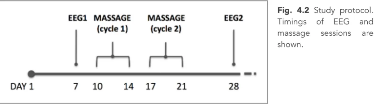
4.2.1 Participants . . . 4.2.2 Massage therapy . . . 4.2.3 Pre-massage (T0) and post-massage (T1) EEG . . . . 4.2.4 Statistical analysis . . . 4.3 Results . . . . 4.3.1 Within-group analysis . . . 4.3.2 Between-group analysis . . . 4.4 Discussion . . . . 4.5 Conclusions . . . . 5 THE PRETERM EARLY MATERNAL MASSAGE (PREMM) PROJECT: THE
PROTOCOL . . . . 5.1 General aim of the PREMM project . . . 5.2 Background . . . . 5.3 Objectives . . . 5.4 Methods . . . . 5.4.1 Recruitment . . . 5.4.2 Intervention . . . 5.4.3 Outcome measures . . . 5.5 Significance and expected results . . . 6 THE PREMM PROJECT: PRELIMINARY FINDINGS ON NEURODEVELOPMENT, MOTHER’S MENTAL HEALTH MOTHER-INFANT RELATIONSHIP . . . 6.1 Introduction . . . . 6.2 Results of baseline (T0) and term (T1) assessments . . .
6.2.1 Differences between groups in the neurological exam . . . 6.2.2 Differences between groups in vision assessment . . . 6.2.3 Differences between groups in mothers’ questionnaires . . . 6.3 Results of the 12-month assessments (T12) . . . 6.3.1 Differences between groups in mothers’ questionnaires . . . 6.4 The Infant Observation . . .
44 45 47 48 50 50 51 52 53 53 53 55 55 57 59 60 60 63 64 64 64 66 69 71 72 72 73 73 74 76 76 77
6.4.1 Summary of the qualitative findings of the Infant Observation . . . 6.5 Discussion . . . . 7 THE PREMM PROJECT: PRELIMINARY FINDINGS ON EARLY BRAIN
STRUCTURAL AND FUNCTIONAL DEVELOPMENT . . . 7.1 Introduction . . . . 7.2 Methods . . . .
7.2.1 Participants . . . 7.2.2 Clinical assessments . . . 7.2.3 Neonatal neuroimaging . . . 7.2.4 Neonatal High-density EEG . . . 7.3 Results . . . .
7.3.1 Whole-group correlations . . . 7.3.2 Independent groups (massage Vs standard care) correlations . . . 7.4 Discussion . . . 8 GENERAL CONCLUSONS AND FUTURE PERSPECTIVES . . . 8.1 General conclusions . . . 8.2 Limitations of the PREMM study . . . 8.3 Future perspectives . . . REFERENCES . . . APPENDIX I – Massage Booklet . . . APPENDIX II – Publications . . . 79 81 85 86 86 86 86 87 88 90 90 92 93 95 96 97 98 101 113 119
Acknowledgements
This PhD has represented for me a unique opportunity for professional and personal growth and, as it sometimes serendipitously happens, some of the themes I have encountered and explored in my research have nurtured and have been nurtured by my personal experience, in a phase of profound change, as is transition towards parenthood. I consider myself extremely fortunate for the opportunity to count on the mentorship, constant support and friendship of many people who have enriched, inspired and filled with sense my experience. I feel towards them deeply grateful.First of all, I would like to thank Prof. Giovanni Cioni for his mentorship over almost a decade and for having always encouraged and trusted me. I feel truthfully grateful to him for his guidance and for having always respected my choices. He also gave me the opportunity to develop a large part of my PhD in Australia, an experience of priceless value. He inspired this project together with the group of Prof. Lamberto Maffei of the CNR in Pisa, and in particular with Nicoletta Berardi and Alessandro Sale.
My deep gratefulness also goes to Professor Paul Colditz and Professor Roslyn Boyd who welcomed me and my husband Andrea in their research groups, believed in our ideas and supported the development of the Preterm Early Maternal Massage at the University of Queensland. Paul, thank you so much for your mentorship, for having generously shared your knowledge and having always helped me when I needed it. You taught me how to be proactive and to overcome the sense of insecurity I felt when trying to put together our project team. Thank you and Rhonda for welcoming us in your family and for making us feel at home even on the other side of the world. Ros, I am deeply grateful to you for having been an excellent mentor and a sincere friend, for all the practical and moral support you generously gave Andrea and me, for being a source of inspiration, for having taught me so much about research methodology and for always sustaining me in pursuing my own ideas.
A very special thanks goes to Penny Love and her family for being such precious, supportive and caring friends. As a research partner, thank you so much for your enriching and always original contribution to meetings and discussions and for always helping us to keep mother-infant relationship in the centre of our thinking. The PREMM project would not have been possible without the invaluable support and professionalism of Naoni Ngenda and Sonia Sam, who still carry on the
recruitment and intervention in Brisbane. Naoni and Sonia, thanks for your constant support, for all your help in the development of the intervention protocol and in every single phase of the project, for sharing with me all the achievements and all the troubles encountered along the way.
I am very grateful to Janine Oostenbroek, who took up the challenging role to coordinate the project after my departure, carrying out the heavy job in an utmost elegant and accurate way, and to Koa Whittingham for the essential contribution to the development of the study protocol, and in particular for the selection of questionnaires assessing maternal mental health and mother-infant relationship. Thanks to Prof. Stephen Rose, Ms. Kerstin Pannek, Dr. Simon Finnigan and Ms. Preethi Mathew for their priceless support in the most advanced and challenging aspects of my project, the neuroimaging and electrophysiological analyses. I learned many things from them, and among those I learned the importance of multidisciplinary work in research.
I could never forget all the other Australian friends and their families: Yoon, Marjorie, Bronwin and her adorable and brave kids, Margit, Megan, Susan, Kerry, Lisa, Lucia e Vincenzo, Simon and Nicky, for their friendship, generosity and support, that made our experience down-under warm and unforgettable. Thanks to the supervisor Margot Lynch and all the other participants to the Infant Observation work group I took part in while in Brisbane; and thanks to Carolyn and Thomas, who weekly welcomed me in the intimate space of their house for more than one year and gave me the unique opportunity to learn about the development of mother-infant relationship and infant emotional development in an ecological setting.
Many people helped me since my return to Italy for the last part of my PhD. First of all, Ada Bancale who has been involved from the beginning in the studies on massage and contributed to the development of new ways of thinking to this project. She has been essential in these last months for setting-up the recruitment in the Neonatal Unit of the Pisa University Hospital. And with her, the staff of the Unit and in particular Prof. Antonio Boldrini and Dr. Laura Bartalena. My gratitude also goes to Cristina Tessore, who trained me as a teacher of infant massage and made me profoundly understand the importance of the early mother-baby dialogue mediated by loving touch and to Benedetta Costa, President of the Italian Association of Infant Massage, for always encouraging and sustaining me in the involvement of mothers in the intervention.
I am especially grateful to Dr. Gabriele Masi who gave me plenty of flexibility regarding my work in the clinical department in order to be able to complete my
PhD work and write my thesis and to my colleague and friend Francesca, for her invaluable support, encouragement and positiveness.
And a huge thanks to Vittorio, for having shared with me 15 (!) years of study and exams, laughter and tears and the deepest friendship.
Last but not least, I’d like to thank all the babies and their families who participated to our studies, both in Australia and Italy. They made me appreciate how our everyday work as physicians relies on the generosity of the families who accepted in the years to give their contribution to research.
Writing a PhD thesis while being a working mum with a less-than-two child has proven to be a sometimes very difficult and overwhelming experience: my profound gratefulness goes to my family and especially my mother, for supporting and sustaining me with her presence and loving care to Marco and myself.
But the biggest acknowledgment of all goes to my husband Andrea, who had the crazy thought to share with me over the last 4 years, a professional path, besides life. Mixing professional and personal life has not always been easy, and at times we had to cope with never-ending discussions and diverging ideas. But, at the end of the day (as Australians would say), these have been the true engines of our research and a unique chance for growth as a couple. I thank you Andrea for your patience, your tireless and irreplaceable help for this PhD thesis, your constant encouragement and your love.
I need finally to thank from the deep of my heart my son Marco, who was conceived and lived a substantial part of his “intra-uterine life” in Australia, in a moment of big personal transformation, for bringing day by day infinite joy and energy to us and for blessing us with a sense of completeness to our lives.
Chapter 1
Introduction and general
1.1 Preterm birth and developmental risk
Prematurity affects more than 2% of live births and can be associated with long-term neurodevelopmental abnormalities, including motor disability, reduced cognitive performance and behavioural problems with very high costs for the
health systems [1]. Preterm-born infants show an increased risk for
neuro-developmental outcome that is related to a number of factors. Firstly, the set of events that led to a premature birth (genetic, malformative, infectious, hypoxic disorders etc) may themselves be a causative factor of cerebral damage. Also, the immaturity of the nervous system associated with that of the cardio-vascular and respiratory system, expose the brain to a higher risk of direct and indirect damage, especially of vascular origin [2, 3]. Such risk is also increased by the condition of being exposed to an environment and to stimulations that, even though trying to simulate intrauterine life, cannot be considered as fully physiological. A last very important aspect, which especially emerged in the last few years, is represented by the role played by infections and inflammations in the genesis of the CNS damage in preterm infants. It was demonstrated, indeed, that the presence of maternal chorioamnionitis, apart from increasing the risk of preterm birth, is positively associated with the damage to cerebral white matter and the evolution towards cerebral palsy [4].
MR imaging studies in term-born infants and preterm infants at term-equivalent age have shown differences between the normal appearing preterm brain at term-equivalent age and the brain of term-born infants. Premature exposure to the ex-utero environment alters brain development in the white matter, cortex, deep grey matter and vascular pattern [5-12]. The abnormalities particularly involve cerebral neuronal regions including both cortex and deep nuclear structures. The pattern of cerebral alterations is related most significantly to the degree of immaturity at birth and to concomitant WM injury [7].
While most studies have focused on medically ill preterm infants, comparatively fewer studies have focused on neurodevelopmental outcomes in seemingly healthy, asymptomatic preterm infants, that is, infants who do not suffer from significant medical complications in the postnatal period or require extensive medical intervention or support. It is of great interest that the studies that have investigated neurodevelopmental outcomes in low-risk preterm-born infants have
demonstrated that they are still at higher risk of learning, behavioural, motor and visual problems in later life [13, 14].
1.2 Early brain plasticity as a potential window of opportunity
Preterm brain is not only more vulnerable. It is also more plastic and thus able, under specific circumstances, to adapt and compensate for lesion-induced functional impairments. In broad terms, mechanisms of cerebral plasticity are thought to be more powerful during development. For example, children are more fast-paced than adults in learning a new language or in achieving complex skills such as playing a musical instrument [15]. Similarly, children lacking proper environmental inputs early in life are more prone to have abnormal development of the functions related to those inputs (the concept of critical periods) [16]. The presence of more powerful mechanisms of neuronal plasticity during early phases of development should imply that recovery from brain damage is more effective for early lesions compared to similar lesions occurring later in life. The picture is however far more complex, as many other aspects influence outcome beyond the timing of the insult, including the location and extent of injury (e.g., focal vs. diffuse), the clinical correlates (e.g., presence of seizures), or the individual genetic susceptibility [17].
The time boundaries of early lesion-related plasticity have never been clearly defined. Changes in cerebral plasticity, which influence the effects of brain damage, are gradual during development, and the sensitive periods are now known to be different for the various functional subsystems [16]. Also, the types of brain insult are extremely variable during development and affect the nervous system in ways that are directly dependent on the level of maturation at the moment they occur. For these and other reasons, the boundaries between early and late lesions are necessarily blurry. As in the present project we are interested in exploring neuroplasticity in very young brains (the preterm brain), we got interested in the mechanisms of adaptive plasticity secondary to preterm brain damage. A review of the mechanisms of early lesion-related brain plasticity will be the topic of chapter 2.
1.3 From early plasticity to early intervention
As outlined in the previous sections, it is today well recognised that preterm infants are at higher risk for developmental and cognitive delays, as well as difficulties in mother-infant relationship across infancy [18, 19]. Follow-up studies of preterm individuals into the school years, consistently found reduced cognitive performance and increased behavioural problems in these children. To minimize the consequences of preterm birth it is of utmost importance to take the highest advantage of enhanced early brain plasticity by devising evidence-based intervention programs centred on the optimization of infant’s environment.
Increasing evidence suggests that the course of mental development is more open to inputs from the caregiving environment, and the parental style is more central in shaping the child’s cognitive development, as compared with motor and growth. It was shown that the neurodevelopmental and cognitive skills of premature infants are influenced by a variety of maternal factors, including the provision of skin-to-skin contact, the frequency of maternal touch, or the degree of maternal postpartum depressive symptoms [18]. Interactions with parents may be compromised for premature infants for several reasons: the emotional response of parents to preterm birth, an altered parental role as a non-caregiver in the nursery, the infant’s characteristics, the NICU environment and the prolonged parent–infant separation [18].
Early experience can modify the anatomy of the rapidly developing brain, which implies that early intervention may alter developmental paths and improve health, educational and social outcomes. This has prompted the use of early interventions for preterms, aimed at different targets in the complex interplay of biology and environment influencing development. Historically, initial interventions were purely sensory based and focused on providing external stimuli; while in contrast, later interventions aimed to minimize the stress of the NICU environment (developmental care). Later still, the recognition of the importance of the caregiving environment was broadened to target interventions including parents. A review of the principles and main known paradigms of early intervention will be the topic of chapter 3.
1.4 Infant massage and its influence on brain function
While during intrauterine life the fetus is protected by noxius external stimuli and benefits from positive physiological stimulations such as the continuous and
gentle tactile stimulation deriving from the movements inside the womb, preterm birth exposes the fetus to a highly stressful, non-physiological environment with unfiltered stimuli and deprives it from the positive stimulations it would normally experience in the womb, potentially resulting in detrimental effects on the development of the immature brain.
On these grounds, early intervention programmes based on the manipulation of the extra-uterine environment have been used in preterm infants with the aim of optimizing the infant’s sensory experience and thus potentially improving development and functional outcome [20]. Among these, infant massage and interventions based on the provision of skin-to skin contact have widely demonstrated positive effects mainly on somatic indexes (such as weight gain acceleration), the reduction of stress and related biomarkers even during invasive procedures as well as the reduction of the overall stay in the NICU [21].
Effects on brain development of these interventions have been dramatically less explored in the scientific literature, probably also due to the challenges correlated with the implementation of early instrumental evaluations on such vulnerable populations. We therefore tested the hypothesis that infant massage can affect the maturation of brain function by performing, in a RCT, an EEG spectral analysis, a quantitative measure of brain electrical activity that in preterm infants is highly sensitive to brain maturation. The study is reported in chapter 4.
1.5 The PREMM Project
Despite the recent advances in the understanding of the mechanisms of early intervention, and in particular of infant massage on neurodevelopment and brain maturation, there are at least two key aspects that have received little attention in the scientific literature so far.
A first aspect that has been poorly investigated is the contribution of a more intensive parental involvement to the efficacy of the intervention. Growing evidence suggests that mother-infant early relationship and interaction has a powerful positive influence on the modulation of the effects of early developmental care. A recent meta-analysis reported clinically meaningful effects of neurodevelopmental interventions that involve parents up to an age of 36 months [21]. Despite this growing evidence, the full involvement of parents in early intervention programs and developmental care is still very limited.
A second aspect that is still poorly understood concerns the neurobiological underpinnings of the therapy-related functional improvements in preterm infants undergoing early intervention. We recently showed that an early intervention in preterm newborns based on infant massage can have significant effects on quantitative components of the electroencephalographic activity of preterm babies assessed at term-equivalent age, resulting in a pattern more similar to that observed in term-born infants [38] (see chapter 4). However, no studies have explored the effect of infant massage on finer structural and functional aspects of brain development such as volumetric measures, structural connectivity or functional networking.
As part of the PhD project, we set up a RCT aiming at addressing these specific issues, called Preterm Early Maternal Massage (PREMM). The main purpose of this part of the project has been to explore the short and medium term effects of infant massage on brain development in low-risk preterm infants, by means of advanced neurophysiological and neuroimaging techniques applicable to the preterm infant, many of which have been developed specifically for these studies thank to the expertise of the neurophysiologists and physicists at the University Of Queensland Centre of Clinical Research (Brisbane, AUS). Other objectives were to evaluate the effects of the direct involvement of the mother in providing the intervention, making it more flexible and tailored on the characteristics of the mother-infant dyad. Effects on mothers’ mental health and mother-infant relationship are hence also explored. The full protocol and the preliminary results on the neuroimaging, electrophysiological and clinical outcome measures will be exposed in chapters 5,6 and 7.
1.6 Outline of the thesis
The following aspects will be treated in the thesis:
Chapter 2: an overview about the studies exploring early brain plasticity in the different CNS systems after brain damage or as an effect of environmental stimulations;
Chapter 3: the rationale for early intervention in infants at neuro-developmental risk and the relevance of a family-centred care;
Chapter 4: the effects of preterm infant massage on the maturation of quantitative components of brain electrical activity, such as spectral power;
Chapter 5: the presentation of the full protocol of the PREMM project (Preterm Early Maternal Massage), a multicentre international RCT on early intervention in preterm infants;
Chapter 6: the preliminary findings about the short-term effects of maternal infant massage on neurodevelopment, mothers’ mental health and mother-infant relationship;
Chapter 7: the preliminary findings on the short-term effects of maternal infant massage on brain development as measured by advanced neuroimaging and neurophysiological techniques;
Chapter 2
Early Brain Plasticity
From (see appendix II):
1 - Cioni G, D'Acunto MG, Guzzetta A. Perinatal brain damage in children. Neuroplasticity, early
intervention, and molecular mechanisms of recovery. Prog Brain Res. 2011; 189:139-54
2 - Cioni G, D'Acunto MG, Guzzetta A. Plasticity in the developing brain: functional reorganization
after early brain damage. In: Cerebral Plasticity: New Perspectives. Chalupa LM, Berardi N, Caleo M, Galli-Resta L and Pizzorusso T Editors. MIT Press. 2011
3 - Guzzetta A, D'Acunto MG, Rose S, Tinelli F, Boyd R, Cioni G. Plasticity of the visual system after
early brain damage. Dev Med Child Neurol. 2010;52(10):891-900
4 – Cioni G, Guzzetta A, D’Acunto G. Cerebral Plasticity and Functional Reorganization in Children with Congenital Brain Lesions. In: Neonatology. Practical approach to neonatal management. Buonocore G, Bracci R, Weideling M Editors. Springer Italia, 2011
2.1 General principles of early brain plasticity
Cerebral plasticity plays an important role during brain development. For example, children learn a new language or achieve complex skills, such as playing a musical instrument, faster than adults. In a classic experiment on string players, the extent of the cortical representation of the left digits was found to be inversely correlated with the age at which the person had begun to play, indicating a larger amount of cerebral plasticity in subjects with earlier exposure to training [15]. Similarly, children lacking proper environmental inputs early in life are more likely to have abnormal development of the functions related to those inputs (the concept of critical periods) [16].
The presence of mechanisms for neuronal plasticity during early phases of development should mean that recovery from brain damage is better for early lesions compared to similar lesions acquired later in life. This principle was first suggested by Paul Broca in 1865 [22] and then systematically explored by Margaret Kennard in the late 1930s [23]. Since then, studies carried out in different species have partially refuted this general principle. The picture appears to be more complex. Account needs to be taken of factors other than the timing of the insult; these include the location and extent of injury (e.g., focal or diffuse), the clinical correlates (e.g., presence of seizures), and the genetic susceptibility of the subject [17].
One of the most important predictors of the efficacy of functional reorganization seems to be the distribution of the damage, i.e., whether diffuse or focal [24, 25]. Strikingly, children with early unilateral left hemisphere damage may develop normal language, while lesions at a similar site and of similar extent in the adult brain result in aphasia [26]. Even if an entire hemisphere is removed at an early stage of development (for instance for the treatment of severe epilepsy), children can develop normal language and cognitive function [27]. Also, children with unilateral ischemic stroke are able to develop normal cognitive functions, which they maintain over time [28].
By contrast, children who sustain an early generalized cerebral insult (e.g., global hypoxia or traumatic brain injury) recover slower with a poorer outcome, compared to adults with similar lesions [29]. The mechanism most often invoked for this greater vulnerability to early damage is that, at an early stage, cognitive development is highly dependent on the integrity of diffuse neural networks; thus the transient disruption of developing attention, memory, and learning functions
undermines the effective acquisition of new abilities [24]. Conversely, at an older age, cognitive abilities, which have already been acquired, may be spared and impairment is limited to functions directly related to the final area of damage. The aim of this chapter is to outline recent discoveries relating to mechanisms of functional reorganization in the young brain, as a basis for the identification of possible non-pharmacological early interventions.
2.2 How early is early?
The time boundaries of early brain damage have never been clearly defined. This is due to the complexity of the task rather than to lack of effort. Cerebral plasticity, which influences the effects of brain damage, has a gradual effect during development and vulnerable periods are now known to be different for the various functional sub-systems [16]. The types of brain insults vary during development and affect the nervous system in ways that depend on the level of maturity at the time they occur. The boundaries between early and late lesions are therefore necessarily fuzzy. This chapter explores the differences of the young brain’s response to damage and focuses on lesions occurring before or around birth (also called congenital lesions), which are more frequent and have been more extensively studied than later ones.
A relevant aspect of the pathophysiology of congenital brain injury is the stage of cerebral development at the time of the insult, which may be either prenatal or perinatal [30]. Because of the complexity and speed of maturation during pregnancy, the response to a harmful event varies with gestational age with different neuropathological and clinical outcomes. Our understanding has grown thanks to non-invasive neuroimaging techniques, first ultrasonography, and, more recently, computerised tomography (CT) and magnetic resonance imaging (MRI). These new methods, increasingly applied to children, allow in vivo investigation of cerebral lesions by monitoring their evolution and providing further insight about the relationship between lesion and its effect on function.
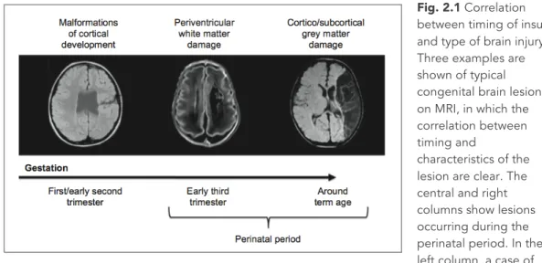
In broad terms, types of congenital brain damage can be grouped according to timing (Fig. 2.1). Lesions occurring during the first half of gestation (and in particular the first trimester) give rise to cortical malformations. These lesions can vary in size, location and distribution, resulting in different clinical pictures. The underlying mechanisms of cerebral plasticity are likely to be similarly variable.
Fig. 2.1 Correlation between timing of insult and type of brain injury. Three examples are shown of typical congenital brain lesions on MRI, in which the correlation between timing and
characteristics of the lesion are clear. The central and right columns show lesions occurring during the perinatal period. In the left column, a case of schizencephalia, a malformation of cortical development secondary to an insult occurred during the early phases of brain development. In the central column, a case of periventricular white matter damage secondary to an intraventricular hemorrhagic insult occurred at the beginning of the third trimester of gestation. In the right column, a case of ischemic infarction of the territory of the middle cerebral artery occurred around birth in a term born infant.
The second group of lesions are those occurring around the early third trimester of gestation (approx. 25−34 weeks of gestation). The most typical of this group are periventricular leukomalacia and intra-ventricular haemorrhage. The first is a diffuse lesion due to ischemia or infection-inflammation, while the second is a haemorrhagic lesion, usually limited to within the ventricles, but sometimes developing into a periventricular parenchymal infarction, usually unilateral. The third group includes lesions around term (typically lesions affecting the term infant at around birth). The most relevant are hypoxic-ischemic encephalopathy (HIE) and focal cerebral stroke. HIE is a bilateral and diffuse ischemic lesion, while a stroke is a focal lesion of arterial origin, with similar neuropathology to the stroke observed in adults.
The distribution of the lesion, i.e., focal unilateral compared with diffuse bilateral, appears to be the single most important factor that influences the effectiveness of the plastic reorganization of brain function. Congenital lesions therefore provide an interesting model for studying cerebral reorganization because they include different combinations of distribution (focal, diffuse) and timing (early gestation, late gestation and term).
2.3 Cerebral plasticity and the different systems
The effects of a cerebral lesion are related to the site of the lesion. This implies different involvement of the various systems in different subjects, with complex and heterogeneous functional correlates. Although subjects with cerebral damage that is acquired early show a clinical phenotype consisting of impairment of language, sensori-motor and visual systems, we will consider each system independently, focusing on cerebral plasticity and differentiating the effects of early and late lesions.
2.3.1 Language
Language processing generally (i.e., in 95% and 98% of adults) occurs in the left hemisphere of the brain. How this special ability develops, its nature and what happens when the left hemisphere is damaged by a cerebral injury during early development are still matters of debate. More than 30 years ago, invasive techniques such as the Wada test demonstrated that language developed in the right hemisphere of patients with early left hemispheric brain lesions [31]. How this happens and the consequences for brain function are starting to be clarified by the application of advanced functional neuroimaging techniques, such as positron emission tomography (PET) and functional magnetic resonance imaging (fMRI).
An important aspect relates to where (and indirectly how) in the right hemisphere language function is organized. In 2002, Martin Staudt and coworkers [32] used fMRI and a language task to explore the topography of right-hemispheric language organization after early left brain injury by unilateral periventricular lesions. They showed a remarkable similarity between activation in the left hemisphere in normal controls and activation in the right hemisphere in brain injured patients, with identical distribution of the known cortical areas of the language circuit. These findings indicated that reorganization of language in the right hemisphere occurs in areas that are homotopic (i.e., in the same location) to those normally involved, strongly suggesting a near-equipotentiality of the two hemispheres at birth as far as the ability to develop language control is concerned.
Similar findings were found in patients with malformations of cortical development [33, 34]. In those subjects, however, epileptic seizures, which are known to alter cerebral reorganization, were almost invariably present. This made
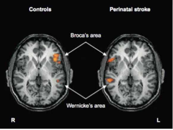
it hard to explore whether shifting language processing could also hap- pen in the absence of seizures and/or with later damage (early third trimester). More recently, Guzzetta et al. demonstrated a similar pattern of contralateral homotopic reorganization in patients with arterial stroke at term (Fig. 2.2) [35]. The study also showed that a shift of language function after a stroke at term is more common than for earlier lesions, suggesting a direct influence of timing on the pattern of reorganization. If this pattern is confirmed, it might suggest that, as for other systems (see below), the hemispheric specialization of language develops because of competition between the two hemispheres. If a left (non-epileptogenic) lesion occurs during the early third trimester when cerebral plasticity is highly active, the affected hemisphere is more likely to maintain its genetic advantage over the contralateral hemisphere and eventually develop control over language. When a left lesion occurs at term or during early development when the potential for plasticity is reduced, the non-affected hemisphere might take over language development. This possibility becomes progressively less available during later development, with later lesions invariably resulting in an intra-hemispheric reorganization of function and different degrees of language disturbance.
Fig. 2.2 Language representation in patients with left perinatal stroke and right-hemispheric reorganization of language. fMRI shows the activation of regions of the right hemisphere which are contralateral and homotopic to the regions of the language circuit activated in normal controls (group analysis performed on eight patients and 10 normal controls.
2.3.2 The sensori-motor system
When a cerebral cortical or subcortical lesion involves the motor system, neuroplastic mechanisms should be able to drive recovery of voluntary movements, restoring an adequate cortical impulse to the spinal motor neurons and inter-neurons.
In the case of a cerebral lesion, two major mechanisms are available for restoring an efficient re-connection of the motor cortex with the spinal cord. The first
involves reorganisation of the ipsilateral cortex within the primary motor cortex or in non-primary motor areas. The second mechanism is specific for lesions during early development. It is based on the existence of bilateral motor projections originating in the primary motor areas during the first weeks of life post-term; these connect each hemisphere with both sides of the body. These fibres withdraw during later development, but may persist in the case of cerebral damage, giving rise to a contralesional reorganization of motor function, but this mechanism is limited to early brain damage (Fig. 2.3).
Fig. 2.3 Schematic representation of the main types of reorganization of sensorimotor function following early brain damage. (a) Ipsilesional reorganization of motor and sensory function. Both functions are reorganized in the affected hemisphere, in regions around the lesion. In this case,
functional impairment is mainly related to the extent of the damage of the sensorimotor system. (b) Contralesional reorganization of motor function and ipsilesional reorganization of sensory function. Motor and sensory function of the affected limb are processed by different hemispheres. In this case, functional impairment is related not only to the extent of the damage of the sensorimotor system but also to the presence of the functional dissociation.
fMRI is able to provide relevant information on the type of reorganization occurring in each patient. Integration with other techniques to provide temporal resolution, such as Transcranial Magnetic Stimulation (TMS), demonstrates the existence of cortical-spinal monosynaptic connections. TMS has shown that in subjects with early lesions of the motor cortex, there is significant bilateral corticospinal innervation of spinal motoneuron pools and that these persist in the healthy hemi- sphere. In these subjects, activation of the intact motor cortex elicits large responses in both ipsi- and contra- lateral muscles, with similar latencies and thresholds.
But what are the consequences of having found this specific type of motor reorganization after early damage? It appears that this pattern of SM reorganization (contralesional reorganization) is determined during the first year of life, and possibly even within the first few months [36]. This is not only a consequence of the size and site of the lesion, but is strongly influenced by what happens after damage (action dependent reorganization): there is a complex
interaction between the residual motor output from the affected hemisphere and a somato-sensory feedback from the affected limb – the hypothesis has been called “amblyopia of the cortico-spinal system”[36]. Confirmation of this hypothesis would emphasise the importance of an early time window (the first months of life) for therapeutic intervention. This is especially true when considering that children with contra-lesional reorganization, i.e., when the unaffected hemisphere directly controlling both hands, achieve less good hand-motor performance, making this pattern of reorganization potentially maladaptive [37, 38].
Cerebral lesions affecting the motor system often involve the sensory system as well, and may lead to a functional deficit. These functions can be studied in vivo with techniques like somato-sensory evoked potentials, magnetoencephalog- raphy and fMRI with sensory stimulation. These approaches have demonstrated that, by contrast with the motor system, the intra-hemispheric (ipsilesional) reorganization of primary sensory function is the principal, if not the only, compensatory mechanism for brain damage of the sensory system, even when this occurs very early during development [37].
The mechanisms underlying this phenomenon are not fully understood. However, two elements seem to be of special relevance. The first is the lack of an anatomical substrate for contralesional reorganization, even during the early stages of development, in contrast to what happens to the motor sys- tem. The second is the possibility that at least for some types of early lesions, thalamo-cortical fibres are still developing when the insult occurs, thus allowing a bypass of the lesion and reconnection with the sensory cortex [39].
It is of considerable interest that the different reorganizational potentials of the sensory and the motor systems often result in an inter-hemispheric dissociation of these functions, with the sensory system being reorganised in the affected hemisphere and the motor system being shifted contralaterally (Fig. 2.3). There is some evidence to support the hypothesis that such a dissociation could lead to functional deficits in tasks requiring good sensory-motor integration (such as stereognosia). In light of these findings, a specific target of early therapeutic intervention might be activation of the sensori-motor cortex of the affected hemisphere to enhance the competitive ability of a damaged corticospinal system during development and, by so doing, to mitigate the consequences of injury on motor function.
2.3.3 The visual system
There have been few investigations of reorganization of the visual system after early lesions in humans. However, visual function has been more studied in animals, especially the cat, than any other system. The sparse scientific evidence from humans on the specificity of reorganizational mechanisms after early damage is summarized below (Fig. 2.4).
The correlation between damage to the optic radiations or the occipital cortex and the corresponding visual field deficit is far less strong in the case of an early lesion than for a lesion occurring later in life. This might be a direct expression of greater cerebral plasticity in the young child. It may, at least in part, have a similar neurophysiological basis to that observed for the somato-sensory system; in particular there is a possibility that thalamo-cortical fibres develop after the injury and so bypass it. The precise characteristics and limits for plasticity involving thalamo-cortical connections are not fully understood. Some data suggest that up to term, structural modifications of the geniculo-striate pathway enable functional reorganization of the visual system. A combination of fMRI and diffusion tensor tractography was used in a recent longitudinal study of an infant with a perinatal left arterial stroke [40, 41]. The stroke spared the primary visual cortex but involved the optic radiations. At 3 months, cortical activation could only be observed in the unaffected side; diffusion tensor imaging (DTI) was unable to show the presence of optical radiations in the affected hemisphere [40]. At 20 months, the infant was re-tested using the same protocol and surprisingly showed definite fMRI activation, an indirect sign of functional reorganization. This was further supported by clear structural modifications on diffusion tractography [41]. Unfortunately, assessment of visual fields could not be performed because of the subject’s young age. However, regardless of the possible presence of functional impairment, the imaging data seem to support the existence of a process of reorganization at the level of the thalamocortical pathway, and an ability to restore at least partially a functional connection between the lateral geniculate body and the occipital cortex.
Even when there is a visual field deficit, patients with early damage seem to have fewer difficulties in environmental navigation and exploration. These data are in line with findings from animal models, which showed clearly that ablation of the whole primary visual cortex in the newborn animal did not affect visual orientation, which, by contrast, was massively impaired after a similar lesion in adult animals. Studies on cats showed that this phenomenon is linked to re-
organization of the pathways connecting subcortical visual structures (the lateral geniculate nucleus, superior colliculus and pulvinar) directly to the extra-striatal ipsi- and contra- lateral visual centers. This could also apply to humans in some certain degree, as shown, for example, by increased activation on fMRI of extra-striatal structures after stimulation of the affected hemifield [42].
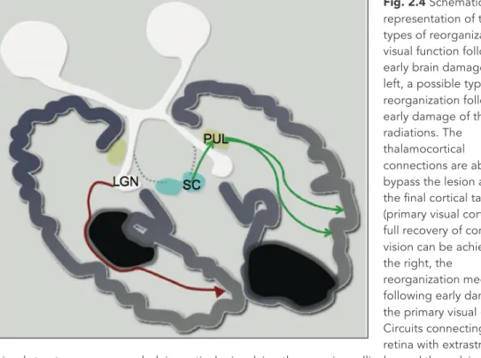
Fig. 2.4 Schematic representation of the main types of reorganization of visual function following early brain damage. On the left, a possible type of reorganization following early damage of the optic radiations. The
thalamocortical connections are able to bypass the lesion and reach the final cortical target (primary visual cortex). A full recovery of conscious vision can be achieved. On the right, the
reorganization mechanisms following early damage to the primary visual cortex. Circuits connecting the retina with extrastriatal visual structures are expanded, in particular involving the superior colliculus and the pulvinar. A full recovery of conscious vision cannot be achieved, but a high degree of functional compensation is obtained, consisting of near normal exploratory visual behavior and navigation. LGN, lateral geniculate nucleus; SC, superior colliculus; Pul, pulvinar.
Reorganization of visual function appears to be more effective after early brain damage. This may be due either to reconnection with targeted structures or to the use of compensating circuits. Even if such circuits are not able to restore conscious visual perception on the contralesional hemifield, they seem to allow for good compensation in spatial orientation and localisation. These conclusions are being confirmed by a study in progress at the Stella Maris Scientific Institute; this is aimed at demonstrating how, in a visual search task, congenital hemianopic subjects perform normally, whereas patients with acquired hemianopia are significantly compromised [43].
2.4 Cerebral plasticity and environmental enrichment
2.4.1 The animal modelResearch on development and plasticity of the central nervous system (CNS) increasingly uses the model of environmental enrichment. The latter has revealed unexpected effects of the environment on cognitive functions, behaviour and aging processes, providing information on a cellular and molecular basis [44, 45]. Although the vast majority of studies based on environmental enrichment protocols have been carried out in animals, some evidence on their application in humans is now available [21].
The first observations about the effects of enrichment were made almost serendipitously at the end of the forties by the Canadian neuropsychologist Donald Hebb [46]. From time to time he used to take one or two rats from their laboratory cages and bring them home for some weeks as pets for his children. He noticed that these rats would gradually become more curious, less frightened, and more prone to explorative behaviour. In particular, he observed that once these rats were brought back to the laboratory, their performance in several behavioural tests was better than those of rats that had never left their usual cages. These pioneering observations inspired various studies on the rat, done principally at Berkeley University, where a group of neuroscientists, coordinated by Mark Rosenzweig, demonstrated how the experience of what they called an ‘enriched environment’ led to a significant and consistent improvement in tasks involving cognitive functions, especially learning and memory [47].
The exposure to a complex environment is obtained by raising rats in numerous groups, in larger than standard cages, equipped with stairs, tunnels, various coloured objects, frequently changed nesting materials which enhance exploratory behaviour, and running wheels for spontaneous exercise. It was shown that one of the most significant effects of environmental enrichment involve hippocampal-dependent performance, such as spatial memory (evaluated with a Morris water maze), both through direct performance improvement (active effect) and through a reduction in progressive cognitive decline normally associated with the aging process (protective effect) [48, 49]. At the same time, several studies showed the influence of environmental enrichment on emotional reactions and stress, supporting its potential anxiolytic effect, mediated, for instance, by preventing enhancement of cortisol levels in response to induced stress (a low intensity electric shock) [50].
Further experimental evidence showed that the improvement in behavioural performance in environmentally enriched animals is accompanied by anatomical modifications of the cerebral cortex – for example, an increase in cortical weight and thickness, increased neuronal cellular bodies and cell body dimensions, and dendritic structural modifications (enhanced arborisation in the pyramidal cells of layers II, IV, and V in the occipital cortex, increased dendritic length and arborization field, and an increase in synaptic spine length by up to 10 per cent in basal dendrites) [51].
Another structure highly sensitive to environmental enrichment is the hippocampus, where modifications similar to those reported for the cortex were found, involving pyramidal cells of CA1 and CA3 areas and the dentate gyrus [48, 52].
Despite the large amount of data collected for the adult animal model, the possibility that the complex stimulation provided by environmental enrichment had effects on the early stages of CNS development has been explored only relatively recently. Taking visual system development as a paradigm of nervous system development, it was shown that exposure to environmental enrichment from birth in the rat prevented the effects of dark rearing on cortical visual development [49]. This suggested that there are factors contributing to the development of the visual cortex through which environmental enrichment establishes its effects that are not under the direct control of visual experience. Other studies in the rat showed that rearing the animal in environmentally enriched conditions leads to a significant acceleration of visual system development, revealed at behavioural, neurophysiological and molecular level [53]. In particular, these animals, compared to controls reared in standard conditions, exhibit earlier eye opening and faster visual acuity development. Some of the principal changes observed in enriched rats were first evident at very early ages (7 to 15 days from birth), when pups still spend almost all the time in the nest. The precocity of these events makes a direct effect of environmental enrichment on the pups unlikely; it has thus been hypothesized that environmental enrichment encourages a higher level of maternal care toward pups (in terms of physical contact, licking and grooming behaviour, and so on) that would act as an indirect mediator of the enrichment effects on visual system development. It was soon demonstrated that the increased licking behaviour and physical contact experienced by environmentally enriched pups is accompanied in the first week of life by higher levels of brain-derived neurotrophic factor
(BDNF), a decisive neurotrophin for visual cortex plasticity in the early stages of development during a specific critical period [53, 54].
A recent study underlines the key role of insulin-like growth factor 1 (IGF-1) in mediating the effects of environmental enrichment on visual system development. In particular, it provokes an increase in IGF-1-positive neurones in the visual cortex. Increasing the IGF-1 levels in the visual cortex of non-enriched rats by means of osmotic minipumps leads to an acceleration of visual acuity development, while blocking the action of IGF-1 on the visual cortex in enriched animals with IGF-1 receptor antagonists blocks the action of IGF-1 on the development of visual acuity [55]. The effect of IGF-1 on the enhancement of neuronal activity had been demonstrated previously [56], as had its role in several pre- and postnatal events that guide central nervous system development, such as cell proliferation control, glycogenesis, neurogenesis, neuronal survival, differentiation, synaptogenesis, and myelination [57, 58].
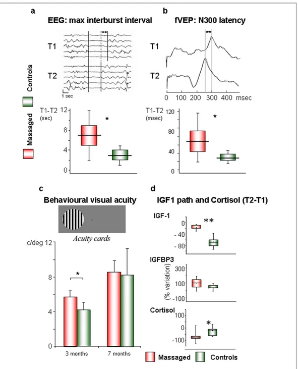
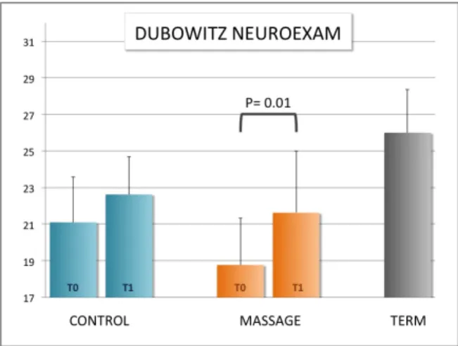
2.4.2 Infant massage as a form of environmental enrichment in the human being Infant massage, described in the previous sections, could be a valid form of environmental enrichment for two main reasons: it represents a well known and widely used intervention in Neonatal Intensive Care Units (NICU) and, as already mentioned above, data in rodents show that pups reared in an enriched environment receive in their first days of life a greater amount of tactile stimulation, through maternal licking, grooming and physical contact, thus suggesting that tactile stimulation represents a crucial component in early environmental enrichment. Smooth massage performed on preterm (<37 weeks) or low birthweight infants (<2500 g) has some positive effects on development and behaviour [21]. Massage has an effect in both countering the stress-inducing stimuli of the NICU (bright light, constant noise, and so on) and providing an additional amount of tactile stimulation, thus constituting an instrument capable of assisting growth and development in these selected newborns. However, as reported above, clinical evidence of the effect of the massage in preterm development is still weak. Preliminary data resulting from a recent study in preterm infants (See chapter 4, Fig. 4.1) show that massage increases IGF-1 blood levels and leads to an acceleration of visual system maturation and to a modification of electroencephalographic activity [59].
It is reasonable that IGF-1 could play a role as a mediator of the effects of therapeutic massage on visual development in infants, as in rats. This could occur through an acceleration of the maturation of the intracortical inhibitory circuits that shape the receptive fields of the visual cortex [60, 61]. The presence of lower plasma levels of IGF-1 and IGF-1 binding protein in premature subjects has been correlated with an increased incidence of retinopathy of prematurity (ROP) [62, 63]. Therapeutic massage, causing an increase in plasma IGF-1 and to a lesser extent IGF binding protein-3, could have a clinical application in preterm infants, especially between 30 and 35 weeks of postmenstrual age when typically ROP is induced [63].
The effect of massage is not limited to the visual system, as shown by a significant difference in EEG power between massaged neonates and controls [64]. There is evidence that such changes in the EEG in preterm infants approaching term are a positive phenomenon, probably related to an increase in synaptic density and connectivity [65, 66]. Recently, a direct link was shown between environmental enrichment, synaptic plasticity, and the spectral power of slow wave activity, sustaining the concept that the sleep EEG is strongly influenced not just by the length of the previous waking period, but also in general by its quality [67, 68]. This study maintains the idea that therapeutic massage favours the maturation of bioelectrical cerebral activity through a process similar to that occurring in utero in term neonates, probably by attenuating the discrepancy between the intrauterine and the extrauterine environment.
2.5 Conclusions
How can we summarize present knowledge on cerebral plasticity and reorganization following early brain damage? The emerging concept, based on studies on both human and non-human subjects, is that functional reorganization in children is similar to that in adults, but there are differences. An understanding of the specific mechanisms of cerebral plasticity in infancy is far from complete. Such knowledge will, however, be essential for the definition and development of therapies based on sound neurobiological and neurophysiological principles. Information is needed about not only the type of treatment, but also its timing, dosage, and means of administration.
Preliminary data support the view that infant massage represents a model of environmental enrichment in the human that shares significant characteristics with
animal models. Effects of infant massage are observed at the electrophysiological, behavioural, and molecular level. The visual system seems particularly sensitive to environmental enrichment effects, even when the enrichment is not directly focused on increasing visual stimulation.
Brain mapping techniques, and in particular fMRI, have provided answers to many questions. Implementations of other technologies, for example advanced brain structural and functional connectivity, will provide further insights on the mechanisms of lesion- and environment-related plasticity.
Chapter 3
Early Intervention
From (see appendix II):
5 - Cioni G, D'Acunto MG, Paolicelli PB. Early Intervention. In: Neurology of the infant. Mariani
3.1 General principles of early intervention
The concept of early intervention was introduced into clinical management some decades ago but its importance has increased considerably over the last 20 to 30 years, thanks to new methods of intervention and greater systematic use in clinical practice. A greater push for intervention programmes has been created by the increasing number of surviving preterm infants with brain lesions and motor, cognitive and behavioural disabilities [69, 70]. In fact, the survival rate for extremely low birthweight preterm babies has been increasing while the disability rate has remained constant. Up to 50 per cent of these infants later show developmental disabilities and disorders (5–15 per cent cerebral palsy) [71, 72]. In the current literature three main at-risk populations have been identified for early intervention: i) high-risk children as a result of low socioeconomic status and limited home environmental stimulation; ii) children with disorders causing developmental delay (for example, Down syndrome, sensory impairment); iii) children at biological risk because of conditions that could lead to developmental disorders (for example, preterm birth, low birthweight, asphyxia).
According to Hadders-Algra [73], early intervention consists of ‘multidisciplinary services provided to children from birth to 5 years of age to promote child health and well-being, enhance emerging competencies, minimize developmental delay, remediate existing or emerging disabilities, prevent functional deterioration, and promote adaptive parenting and overall family functioning’. According to this definition, ‘early intervention’ includes both prevention and rehabilitation, and may be interpreted as two different phases of the same process for those children who, at a later age, show a specific neurodevelopmental dysfunction that requires particular therapeutic programmes (physical, linguistic, cognitive, educational, behavioural).
Depending on when they are applied, early intervention programmes may be categorized as neonatal intervention programmes, focused on the environment, infant and parents, and mainly designed to minimize the stress on infants in neonatal intensive care (NICU), and postnatal intervention programmes, which begin soon after discharge or in the first year of life, with or without an in-patient hospital component, aimed at enhancing infant development.
Family-focused programmes involve family participation at centres or through home visits. These interventions are directed at improving parenting skills and relationships. By optimizing caregiving behaviour, parent-child interactions are
facilitated and subsequent child development is enhanced. Parents are effective agents for maximizing the developmental performance of their children. They play an essential role as active participants in early intervention and are involved in identifying goals and specific needs [74-76].
A recent Cochrane review [77] suggests that interventions with components that focus on the parent-infant relationship have a greater impact on cognitive skills for infant and preschool subjects than interventions focused only on infant development or parental support. Although interventions focused solely on infant development had the greatest impact on motor development, the effect was not significant and the studies were of low quality.
3.2 Family-centred care
Recent reports suggest there have been important modifications in the approach to health care, changing its main objective. This is now shifting from providing treatment to supporting and improving the individual as a whole, thus ensuring a better all-round quality of life. The family is considered to be the ecological system of child development in which parents and family, as primary caregivers, play a vital role in ensuring the health and well-being of children. The child grows up and performs in the family context and here learns and selects social behaviours; the family has a key role in promoting the child’s developmental potential [75].
Family-centred care is an approach to the planning, delivery, and evaluation of health care based on a partnership between health professionals and patient families [78]. It is a current and very widespread model of early intervention, rehabilitation and other health problems, not only in childhood but also for adults. As reported in the definitions proposed by the Canadian Center for Childhood Disability Research and by the Institute for Family-Centred Care, the main concepts of this health care model for both children and adults are the central role of the parents as experts in their child’s needs, and the importance of partnership between parents and providers of services.
Great efforts are now being devoted to demonstrating the efficacy and utility of this new model of health care by means of valid and reliable measures [79, 80]. However, the development of measures that quantify human interactions is especially difficult as they must take into account the presence of many subjective factors [80, 81]. The importance of a family-centred care is confirmed by a recent
meta-analysis reported significant clinical effects of early interventions involving parents up until 36 months [19] and a RCT recently published demonstrating that a preventive care program for very preterm infants and their families improved behavioral outcomes for infants and reduced anxiety and depression for primary caregivers [82].
3.3 Developmental care interventions
A family-centred approach is a fundamental part of early intervention for preterm infants in the NICU. The environment of the NICU, so different from the intrauterine environment, may have a negative impact on neurodevelopmental outcome in the preterm infant. The infant’s sensory experiences in the NICU – including exposure to bright lights, loud sounds, and frequent disturbing interventions – has been assumed to have negative effects on the immature brain, with alterations in subsequent development [83-85]. Taking into account the importance of experience on animal brain plasticity, particularly during critical periods, Als proposed an intervention called Developmental Care, aimed at minimizing the negative impact and stress of environmental exposure on brain development in NICU at such a critical time [86]. Developmental care is a broad category of interventions mainly based on positioning, clustering of nursery activities, modification of external stimuli and individualized developmental care interventions.
The Newborn Individualized Developmental Care and Assessment Program (NIDCAP) is without any doubt the best known early intervention programme in the NICU. It is particularly common and widespread in the USA and Sweden. 3.3.1 Newborn Individualized Developmental Care and Assessment Program (NIDCAP)
’Developmental care’, introduced in the mid 1980’s [87] is an approach that was designed to modify the NICU environment so as to minimize the stress experienced by the preterm infant, whose rapidly developing brain is particularly vulnerable to a stressful environment. The detrimental effects of this stress could have short and long term implications for neurobehavioural development. A negative impact of the NICU environment can be shown by the preterm infant in different ways, such as altering physiological parameters like heart rate and
oxygen saturation or affecting infant growth due to increased energy expenditure occurring during routine nursery care.
A number of elements are included within the developmental care concept such as control of external stimuli (vestibular, auditory, visual, tactile), clustering of nursery care activities, and positioning or swaddling of the preterm infant so as to provide a sense of containment similar to the intrauterine experience. Programs such as the NIDCAP utilize a combination of these strategies depending on the individual needs of the infant, thus including a pre-assessment using an instrument designed for this purpose that was developed from the assessment protocol of the Brazelton Neonatal Behavioral Assessment Scale [87]. This involves the evaluation of the respiratory status, colour, visceral responses (e.g. gagging, hiccoughing), motor state (e.g. tone, posture), facial expressions (e.g. grimace, smile), and attention, as an individualized assessment used to appraise infant’s tolerance to the environment and caregiving activities.
Als articulated a theory of subsystem differentiation (Synactive Theory) and integration within the newborn in interaction with the environment, identifying three main subsystems: (1) the autonomic subsystem (color fluctuations, breathing patterns, and visceral stability); (2) the motor subsystem (body tone, posture, and movement); and (3) the state subsystem (range of available states, state robustness and modulation, and transition from one state to another). Within the infant’s state subsystem, the infant’s alertness and attentional and interactive ability is assessed as well [88].
Two categories of behaviors derive from each of these 3 subsystems, approach/self-regulatory behaviors and stress behaviors. The infant has strategies or behaviors available to move toward and take in stimuli (approach/self-regulatory behaviors) if the input is appropriate in timing, complexity, and intensity in relation to the infant’s thresholds of functioning. Conversely, the infant has strategies to move away from or avoid inputs that are too complex or intense or are inappropriately timed. Such behaviors are thought of as stress behaviors. These behaviors may include a color change from a pinkish hue to pale (autonomic system), stretching out of his or her legs and feet away from his or her body (motor subsystem), and/or facial grimacing (state subsystem). The meaning