Clinical approach to anterior adhesive restorations using resin composite veneers.
22
0
0
Testo completo
(2) MANGANI ETopAL yrig. n. Abstract. ot. Q ui. by N ht. No C t fo rP ub lica tio n te ss e n c e. fo r. Scientific progress in adhesive dentistry. neers depends mainly on the tooth prepa-. has led to more conservative techniques,. ration, which should be confined to enamel,. both direct and indirect, to solve esthetic. involve proximal contact areas, maintain. problems in anterior teeth. This article will. the cervical enamel margin, and incorpo-. discuss only indirect techniques, which are. rate the incisal edge to increase veneer. clearly superior in complex cases in which. resistance and enable correct placement.. it will be difficult to recreate harmonious. Although no clinical follow-up similar to that. tooth shape and color. After reviewing the. of ceramic materials is available, the latest-. literature and highlighting the properties of. generation resin composites offer interesting. this technique, the indications and benefits. features. They can withstand mechanical. compared to the direct technique will be. stress, have excellent esthetic properties,. assessed. This is followed by a step-by-. and, most importantly, can be repaired. step description of operative procedures,. intraorally without impairing their physico-. from treatment planning to relining and. chemical and mechanical properties.. polishing. of. the. cemented. adhesive. restoration. The long-term success of ve-. (Eur J Esthet Dent 2007;2:188–209.). 189 THE EUROPEAN JOURNAL OF ESTHETIC DENTISTRY VOLUME 2 • NUMBER 2 • SUMMER 2007.
(3) CLINICAL APPLICATION. creased. In these cases, clinicians often. treatment planning to relining and polish-. prefer the use of complete crowns to re-. ing of the cemented adhesive restoration.. store esthetics.1. Finally, two clinical cases are presented to. fo r. Remarkable progress in adhesion re-. n. In the past few years, patient requests for. ot. Q ui. by N ht. esthetic treatment of anterior teeth have in-. pyrig No Co t fo rP ub lica tio sessed. This is followed by at step-by-stepn ess c e e n from description of operative procedures,. illustrate the concepts discussed.. search has led to the introduction of more reliable adhesive systems and highly filled hybrid resin composites with microparti-. History. cles, which offer better chemicophysical properties and adequate mechanical prop2. erties.. The use of ceramic veneers is not a recent development; in fact, in 1938 Pincus6 de-. Today, this evolution is clinically repre-. scribed a technique used during Holly-. sented by the choice of direct and indirect. wood filmmaking. A common adhesive for. techniques. Direct techniques are one-ses-. total prostheses was used to retain veneers. sion procedures performed chairside by. to the tooth surface. The introduction of ad-. directly applying resin composite to the. hesive systems and their continuous im-. dental surface. They are used for simple. provement has significantly helped the. restorations using an anatomic layering. evolution and increasing clinical success. procedure, which aids the clinician in cor-. of these techniques.. rectly defining the color and shape of the. The materials of choice for veneers are. tooth, using the residual dental structure as. porcelain and resin composite. Baked. reference.3. dental ceramics are made up of two com-. Indirect techniques demand at least two. ponents: a glass matrix and crystalline in-. sessions and the collaboration of a dental. clusions.7,8 In particular, those used in. technician,4 who will manufacture a veneer. restorative dentistry are reinforced hetero-. to be luted to the prepared dental surface.. geneous porcelains that contain a greater. Indirect techniques are preferred in more. percentage. complex cases, in which restoring harmo-. porcelain fused to metal.9. of. crystalline. phase. than. nious tooth shape and color is highly de-. Ceramic has always been the material. pendent on variables such as the clini-. of choice for indirect anterior restorations. cian’s skill and the technique and material. because of its effectiveness in reproducing. used. Further, it should be considered. the structure and translucency of the natu-. whether the patient will be more or less. ral tooth. Long-term follow-up evaluations. compliant about a prolonged session.5. of ceramic veneers show excellent bio-. In this article, only indirect techniques are discussed, and the most commonly. compatibility and very good chemical stability.10,11. used materials will be considered: dental. The use of resin composite to build an-. porcelain and the more recent resin com-. terior indirect restorations is more recent;. posites. After reviewing the literature and. only in the last few years has research. highlighting the properties of each tech-. identified materials that offer good polisha-. nique, the indications and benefits com-. bility, hardness, and wear resistance.12,13. pared to the direct technique will be as-. Such materials belong to a class of highly. 190 THE EUROPEAN JOURNAL OF ESTHETIC DENTISTRY VOLUME 2 • NUMBER 2 • SUMMER 2007.
(4) MANGANI ETopAL yrig. Q ui. by N ht. No C t fo rP ub lica tio Metal-ceramic crown repair t ess c e n en Discoloration caused by. ■. fo r. are made up of glass filler (70% to 85% in weight) and particles varying from 0.04 to. pulpal necrosis or inadequate. 3 μm in size.. root canal therapy. ot. ■. n. filled microparticle hybrid composites and. In the authors’ opinion, however, this. Indications. final item should actually be considered a contraindication, since the result is often. Thanks to improved oral hygiene habits. affected by dischromic relapses of the. and the greater reliability of recent esthetic. treated tooth, which may have a negative. materials, the indications for anterior indi-. effect on esthetics over time.. rect adhesive restorations have enlarged to cover most patients.4 However, many authors argue that adhesive restorations should not be used in patients with parafunctions. Magne et al14 reported that the. Choice of technique and material. success rate of ceramic veneers is reduced. The advantages offered by indirect tech-. to 60% in patients with bruxism. Further,. niques compared to direct techniques are. they showed a decreased wear resistance. as follows16:. in resin composite restorations and a high incidence of fractures in ceramic restora-. ■. Superior esthetic result. tions for these patients. Universally, the fol-. ■. Adequate abrasion resistance. lowing clinical situations are considered in-. ■. Biocompatibility with soft tissues. dications for anterior indirect restorations:. ■. Dimensional and chromatic stability. 15. over time ■. Enamel hypoplasia, waves, stains,. ■. Strong bond between the two adhesive. grooves, etc. interfaces (luting agent/etched enamel. ■. Enamel abrasions. and luting agent/etched porcelain or. ■. Congenital imperfect amelogenesis. postpolymerized resin composite). caused by hormones or tetracyclines ■. ■. ■. ■. ■. Chromatic or distrophic alterations. Obviously,. these. advantages. are. a. caused by fluorosis. result of both the intrinsic properties of the. Numerous esthetically unsatisfactory. material (ceramics) and the superior qual-. superficial restorations. ity obtained with extraoral work (resin. Coronal fractures located primarily. composites). The disadvantages com-. on the palatal side. pared to direct techniques are now being. Retention of deciduous teeth. reconsidered, as resin composites can. with a reabsorbed root. actually give excellent results in anterior. Lateral incisor agenesia, when the. indirect restorations when used as an. canine has transposed to that position. alternative to porcelain.. ■. Volume anomalies (microdens). ■. Diastemas. the material to be used will now be pre-. ■. Alignment defects. sented.. Several useful parameters for selecting. 191 THE EUROPEAN JOURNAL OF ESTHETIC DENTISTRY VOLUME 2 • NUMBER 2 • SUMMER 2007.
(5) Q ui. by N ht. pyrig No Co t fo rP ub lica tio n Position of the margins and te e s s c e n preparation shape. ■. It has been shown that filler hardness and. ■. n. Wear resistance. ot. CLINICAL APPLICATION. fo r. Type of ceramic. dimension influence resin composite wear when opposed by a natural tooth. The filler. Resin composites are more elastic. hardness should be equal or lower than. and less affected by microfractures; thus,. that of hydroxiapatite.17. their use is particularly recommended in. It has also been demonstrated that when. patients with parafunctions.. resin composites are subject to a final curing phase by means of both light and heat inside a special oven, they show a remark-. Laboratory procedures and cost. able increase in mechanical properties (wear resistance, microhardness, etc) and. Resin composite veneers require easier. physical properties (solubility, thermal ex-. laboratory. pansion coefficient, elasticity module, brit-. veneers, even though veneers built with the. tleness) compared to resin composites. refractory cast technique take less time than. cured with light alone.18–22 Further, microin-. those fabricated with other methods. Gen-. filtration, as measured with dye penetration,. erally, indirect restorations require a greater. decreases dramatically.22. amount of time and result in more technical. These procedures may affect resin composite brightness, so the postpolymeriza-. procedures. than. ceramic. difficulties, which explains the higher overall cost of ceramic restorations.26,27. tion temperature should be controlled. In 1991, Rueggeberg et al23 suggested that a final cycle at 100°C for 5 minutes is appro-. Repair potential. priate. Although ceramics show greater wear resistance than resin composites,. If necessary as a result of secondary caries,. they also cause greater wear on the enam-. margin subsiding, or other complications,28. el of the antagonist teeth.. resin composite veneers are easily repairable intraorally. Marginal inaccuracies during luting can be corrected by curing a. Elasticity module and brittleness. small amount of material in place; however, this procedure may jeopardize the per-. According to many clinicians, fractures are. formance of the definitive restoration.29. more likely in adhesive restorations made. The intraoral repair of ceramic veneers. with ceramic than with resin composite,. is also possible, and the clinical success. because ceramics are stiffer and transmit. depends on many factors. The type of ce-. higher functional stresses to the adhesive. ramic used affects the choice of adhesive. 24,25. interface.. To reduce this risk, the following parameters should be considered:. material and clinical repair procedure. Glass-based ceramics can be etched with hydrofluoric acid for 2 minutes, followed by application of silane and bonding. ■. Relationship between resin composite cement and porcelain thickness. 192 THE EUROPEAN JOURNAL OF ESTHETIC DENTISTRY VOLUME 2 • NUMBER 2 • SUMMER 2007. resin.30,31.
(6) MANGANI ETopAL yrig. uses silica acid–modified aluminum oxide particles to sandblast the surface to be bonded. The sand bonds tribochemically. fo r. lain is the CoJet System (3M ESPE), which. n. Another method for bonding resin to porce-. ot. Q ui. by N ht. No C t fo rP ub lica tio n Treatment planning tes se nc e Diagnostic waxup and color registration. (ie, creates a chemical bond using me-. The esthetic result of an anterior restoration. chanical energy) to the substrate. The sur-. depends on the shape and color, which are. face should then be silanized and bonding. the key parameters of dental esthetics.3,5,39. resin should be applied. The CoJet system. For. can be applied to aluminum oxide ceram-. should be used to evaluate the shape of. ics and zirconium oxide ceramics.30,32,33. the final restoration. Prior to tooth prepara-. this. purpose,. diagnostic. waxups. It can be difficult to select the proper pro-. tion, the clinician will plan the final shape of. cedure for a long-lasting repair and optimal. the veneer, type of preparation, and posi-. esthetic result when resin composites are. tion of the finishing margins. Using a color. used to seal and repair ceramic veneers,. scale and a 5,000-K lighting system—which. especially for small or medium defects.. should eliminate every source of reflected. Large defects or fractures at the incisal. light and make it easier to discern the var-. margin of ceramic veneers can be re-. ious shades of gray—it will be possible to. paired, when possible, by adhesively luting. detect all five dimensions of tooth color. As. a lab-made partial veneer to the fractured. reported by Vanini et al,3,39 the traditional. 34. way to detect tooth color (hue, chrome, val-. veneer.. ue) can be redefined to include five parameters: basic chromaticity, value, intensives, opalescents, and characterizations.. Polishability Surface finishing and final polishing are simpler for resin composite restorations. Tooth preparation. than ceramic restorations.26 It has been suggested that an aggressive finishing of. The guidelines regarding tooth prepara-. ceramic veneers may remove the surface. tion for a veneer have changed drastically. glaze, thus increasing plaque retention and. in the last few years. Initially, no preparation. authors. was recommended.40–42 As a result, there. assert that in these cases an accurate. was a remarkable increase in tooth thick-. intraoral polishing (specific rubber cups,. ness and, consequently, proximal and gin-. diamond paste, etc) can restore the sur-. gival overcontour. Shani et al43 reported a. face to its initial condition.36–38. 90% failure rate for restorations of unpre-. gingival. inflammation.. 35. Other. pared teeth. Today, most authors16,40,44–47 agree on the importance of a conservative tooth preparation, which offers the following advantages:. 193 THE EUROPEAN JOURNAL OF ESTHETIC DENTISTRY VOLUME 2 • NUMBER 2 • SUMMER 2007.
(7) CLINICAL APPLICATION. Q ui. by N ht. pyrig No Co t fo rP ub lica tio Mild preparation for dischromic teeth n te ss e n c e Overpreparation for morphologic. between tooth surface and. ■. resin composite cement. modifications: conoids, diastemas. More stable veneer fitting due. with black triangles (accentuate. to precise reference marks. proximal and mild buccal preparation),. Reduced possibility of proximal or. and changes to incisor length or. horizontal overcontouring. prominence (up to 2 mm can be. (with consequent emergence profile. added without inducing stresses). alteration) ■. fo r. ■. ■. ot. ■. More effective adhesive bond. n. ■. ■. More extensive preparation for fractured elements or teeth with. Preserved marginal tissue health. acquired morphologic modifications Concerning. Enamel reduction. Vanini et al. resin 5. composite. veneers,. emphasized this material’s. Ideally, the preparation should be confined. ability to limit the entity of reduction due to. to enamel,1,16,48 though Pippin et al1 con-. the low elasticity module and resulting high. firmed the need to remove the aprismatic. capacity to absorb functional stresses. In. enamel isles located mainly in cervical ar-. fact, it is not mandatory to have a minimum. eas. These zones should be analogous to. thickness of 0.5 mm for resin composite;. 49. in third mo-. Perdigao and Lopes56 suggested a 0.2- to. lars at a distance between 103 to 756 μm. 0.4-mm reduction at the cervical third, a. from the cementoenamel junction, and by. 0.3- to 0.6-mm reduction at the middle. other authors in anterior teeth at a distance. third, and a 1.5-mm reduction at the incisal. of 0.4 mm from the cementoenamel junc-. third.. those identified by Gasperic. tion. This is the same area where many cli-. Regarding interproximal surfaces, these. nicians commonly position the finishing. same authors argue that the preparation. line, which is why it is often difficult to ob-. must extend to the contact area without in-. 50–52. volving it; conversely, Christensen57 and. tain a solid bond in this area. According. to. Caleffi. and. Berardi,. 16. enamel removal should not exceed 0.3 to. Caleffi and Berardi16 suggest including half the contact area in the preparation.. 0.6 mm, depending on tooth dimension,. A study by Magne and Douglas,58 in. shape, and pathosis. According to Ferrari. agreement with Douglas,59 reported that. 53. the enamel thickness and exten-. interproximal enamel reduction does not. sion in the cervical area of anterior teeth. decrease tooth resistance, while buccal. do not allow a 0.5-mm reduction without. reduction does. The buccal surface of the. et al,. 54. showed. tooth-veneer complex seems to be more. that enamel removal should not be less. subject to compressive stresses, even. than 0.5 mm or greater than 2 mm to. though the use of porcelain instead of. avoid decreasing the resistance of ceram-. enamel seems to recover the mechanical. ic veneers.. properties of the restoration.. dentin exposure. Chalifoux et al. Magne et al55 listed the types of preparation based on the various indications:. It is generally agreed that the position of the cervical margin is a key factor in soft tissue reaction. Pippin et al1 underlined that. 194 THE EUROPEAN JOURNAL OF ESTHETIC DENTISTRY VOLUME 2 • NUMBER 2 • SUMMER 2007.
(8) MANGANI ETopAL yrig. oral hygiene are key factors to guarantee. which preparation affords good resistance. the periodontal health of restored teeth.. to ceramic fracture analyze samples using. n. fo r. the presence of a cervical enamel edge,. ot. Q ui. by N ht. polishing of restoration margins, and good. No C t fo rP ub lica tio low correct insertion. It shouldt be noted ess c e n en that most studies aiming to determine loading systems that are directed from the incisal margin and parallel to the long axis of the tooth; however, functional and. Preparation type. parafunctional loads (the latter having 6 Identifying which type of tooth preparation. times the intensity)68 acting on the palatal. is least likely to lead to veneer fracture is. surface of maxillary anterior teeth are di-. still controversial.60 This is especially true. rected horizontally.69. for ceramic veneers, which theoretically. Porcelain is exposed to fracture when. should be exposed to minimal occlusal. subjected to tensile forces. The maxillary. load.41 Toh et al61 suggested that ceramic. central incisors are mostly affected by this. veneers should be used only to solve es-. kind of stress, because the compressive. thetic (and not functional) problems, while. forces applied on maxillary incisal mar-. Friedman62 reported that ceramic veneers. gins by mandibular teeth are less than in. can restore the anterior guide if there is an. other teeth.63 According to the manufactur-. appropriate incisal length. In a 15-year fol-. er, the maximum tensile resistance of. low-up study, fracture, detachment, and. feldspar. microleakage were the most common. Jeneric/Pentron).49. ceramic. is. 25 MPa. (Avantè,. causes for ceramic veneer failure.63 Frac-. Some authors64,70 report that a frame. ture alone represented 67% of the failures.. preparation (ie, with no incisal reduction). According to this study, the fractures were. seems to be one of the most resistant.. primarily cohesive and located at the in-. Greater fracture values were found in oth-. cisal margin, where stresses are higher.. er types of preparation, especially when a. Several types of preparation have been 64. described in the literature. Hui et al. palatal chamfer was present. Additionally,. and. no significant differences were observed. later Clyde and Gilmour65 described three. when increasing the height of ceramics. different designs: (1) a “window” or “frame”. unsustained by dental tissues from 1 to. preparation, where a 1-mm incisal margin. 4 mm.64 Therefore, the amount of unsus-. is preserved; (2) a preparation with a 0.5-. tained ceramic is not a critical factor for. to 1-mm incisal bevel; and (3) and a. fractures. Conversely, the palatal chamfer,. preparation with lingual extension.. which is common to both preparation. To increase veneer translucency, Wein66. types, appears to be the true weak point.. suggested a 1-mm incisal reduction. Fractures at the chamfer level extending to. with rounded angles and edges, while. the buccal side of the veneer were ob-. berg. 67. Sheet and Taniguchi. described a prepa-. served. Thus, it may be preferable to elim-. ration with a pronounced palatal chamfer. inate the palatal chamfer and reduce at. and rounded margin for adequate ceram-. least 2 mm at the incisal level, while leav-. ic thickness. Castelnuovo et al60 advised a. ing a butt-joint palatal margin.. preparation incorporating the incisal mar-. An experimental study by Magne et al,14. gin to increase veneer resistance and al-. carried out by thermocycling the restora-. 195 THE EUROPEAN JOURNAL OF ESTHETIC DENTISTRY VOLUME 2 • NUMBER 2 • SUMMER 2007.
(9) CLINICAL APPLICATION. sible to prevent porcelain fractures. These. Troedson and Derand48 examined the. authors further suggested that the ceram-. stress distribution in ceramic veneers. ic thickness should be at least 3 times that. made with three different cervical designs:. demon-. strated the need for a good ceramic thickness and minimum resin composite thickness to reduce thermal contraction and polymerization stresses. Thus, preparation becomes a critical factor in long-term veneer. predictability.. The. same. study. showed that by providing an adequate resin composite/ceramic ratio, only a buttjoint configuration reduced the risk of ceramic fracture after veneer insertion, as caused by resin composite contraction and thermal changes in the oral cavity. Further, the butt-joint configuration with no palatal extension allows enamel preservation at the restoration margin, which is essential to obtain an effective bond between the veneer and dental surface. The authors concluded that frame preparations with a butt-joint margin and a 2-mm incisal reduction are the most reliable because they guarantee adequate mechanical resistance to the restoration and allow better characterization of the incisal third. Moreover, compared to the palatal chamfer configuration, the butt-joint with no palatal extension is simpler for the clinician, has a better definition when reproduced with a cast, and is more easily read by the dental technician. Further, a flat surface allows better veneer support and, as opposed to the palatal chamfer and extension configuration, has only one insertion axis. This considerably reduces the risk of fracture in the unsustained ceramic portion.60. 196 THE EUROPEAN JOURNAL OF ESTHETIC DENTISTRY VOLUME 2 • NUMBER 2 • SUMMER 2007. fo r. of the cement. Another study. 71. n. tion, reported that by increasing the porce-. ot. Q ui. by N ht. lain/resin composite cement ratio it is pos-. pyrig No Co t fo rP ub lica tio n Regarding the configuration of te the cervical e s s c e n third of the preparation, a recent study by. (1) a “feather-edge” configuration (modified razor-edge configuration), (2) chamfer configuration, and (3) shoulder configuration. The results showed that in the presence of moderate stress, the cervical margin design does not influence veneer success. Further, when occlusal loads have various directions reflecting the forces applied on the tooth during mastication, a shoulder configuration is preferable. This study also demonstrated that veneer adhesion is the most important factor to reduce compression and traction forces. The importance of pre-restoration (buildup) in cases where the tooth is very compromised must be discussed. Magne and Douglas58 evaluated the stress distribution in teeth with coronal fractures. These teeth were divided into two groups: (1) teeth restored with ceramic veneers after a resin composite buildup, and (2) teeth restored with ceramic veneers only. The results showed that pre-restoring compromised teeth is a valuable method for guaranteeing better flexibility. A separate study72 showed that fracture resistance of porcelain is influenced by the elasticity module of the material used for the pre-restoration. Such procedures also provide an adequate geometry to the preparation and guarantee the restoration material will have constant thickness in every portion, which is an essential requirement for obtaining a thin layer of resin composite cement and greater control of material contraction.73.
(10) MANGANI ETopAL yrig. Q ui. by N ht. No C t fo rP ub lica tio n te Incisal reduction and butt-joint ss e n c e with no palatal extension. ot. n. fo r. Operative phases The operative phases and preparation types are identical for ceramic or resin. The first phases are identical to the frame. composite veneers. The preparation pro-. preparation. A 2-mm incisal reduction. cedures and necessary tools that, accord-. should follow, providing clean incisal and. ing to evidence-based literature, are able to. palatal margins.. guarantee a long-term success will now be briefly examined. The use of a silicone template based on a diagnostic waxup is advised during preparation to control tooth. Preparation with palatal extension. reduction. The preparation procedures will now be summarized.. After carrying out the first two phases of the frame preparation and performing the incisal reduction, two interproximal (mesial. Frame preparation. and distal) steps should be taken, which should join at the palatal side. The palatal. For the cervical third preparation, a guide-. margin, as explained later, may be located. groove should be executed along all cer-. at the incisal third (chamfer) or inside the. vical contours by means of a round bur. It. palatal cavity (shoulder), depending on the. is very important to preserve a sufficient. veneer extension. The chosen palatal fin-. enamel rim above the cementoenamel. ishing line configuration should be a con-. junction.. sequence of the bur design itself and be. For the mesial, distal, and incisal prepa-. clearly visible in cross-sectional images.. rations, the contact points should not be entirely involved, in order to avoid creating undercuts that will prevent proper veneer. Provisional restoration. insertion. A sufficient enamel rim should be preserved at the incisal level.. Since most of the preparation is contained. For the buccal preparation, a longitudi-. to enamel, patients seldom complain about. nal guide-groove should be executed to. intolerable postpreparation sensitivity. Most. gain better control over reduction depth.. authors prefer to avoid the application of. These grooves should be connected by. provisional restorations for several reasons.. means of a chamfer bur, respecting the dif-. First, any provisional restoration represents. ferent reduction angles of the whole buc-. a chemicophysical-irritating factor that may. cal side.. cause tissue inflammation.27 Furthermore,. For preparation finishing at cervical and. provisional restorations are unstable and. buccal level, fine-grit burs (red and yellow. unesthetic. In addition, according to se-. rings) or medium-abrasive rubber burs. veral authors,16,74–77 the use of temporary. should be used.. cements or adhesive resins would compromise the effectiveness of adhesive procedures; however, Tjan and Nementz78 argued that the total etch technique can. 197 THE EUROPEAN JOURNAL OF ESTHETIC DENTISTRY VOLUME 2 • NUMBER 2 • SUMMER 2007.
(11) by N ht. n. 87. Q ui. remove any residue left on the tooth. pyrig No Co t fo rP ub lica tio material. To restore the mechanical prop-n te s e nc e erties of the restoration, furtherspostpoly-. fo r. surface. With no provisional restorations, oral hy-. ot. CLINICAL APPLICATION. merization is necessary.. giene procedures will be easier for the patient, thus guaranteeing ideal health of the periodontal tissues with a remarkable im-. Luting procedures. provement of the tooth-periodontium complex once the restoration is luted. In this. Luting procedures are a crucial step in the. case, it is extremely important that the. overall treatment. Indirect adhesive restora-. restoration be completed within a maxi-. tions are retained by means of microme-. mum time frame of 3 to 5 days.. chanical adhesion between the resin com-. If significant tooth sensitivity develops,. posite cement and both the tooth surface. the application of fluoride-based gels us-. and restorative material. Marginal quality. ing an individual tray can be used. Paul. and precision of luted restorations is strict-. and Sharer79 demonstrated that applying a. ly dependant on the following factors.. bonding resin after cavity preparation (dual bonding technique) reduces postoperative sensitivity, which is generally con-. Adhesive potential of substrate. sidered a symptom of bacterial contamination and hydrodynamic phenomena.80,81. As mentioned previously, the best internal. If necessary, such as with very large. adaptation and marginal seal are obtained. preparations and/or after patient request, it. when the preparation is entirely surround-. is possible to execute chairside direct pro-. ed by enamel.4 It has been demonstrated. visional self-curing acrylic resin veneers,. that exceedingly aggressive etching, ex-. using a silicone template based on an es-. cessive dryness of the etched surface, or. thetic diagnostic waxup. These provisional. high compression of the collagen fiber net-. veneers should be temporarily fixed by po-. work during impression taking or luting. sitioning a small amount of resin compos-. procedures23,80 may cause denaturation. 82–85. ite without any adhesive procedures.. and/or collapse of the collagen fibers. A hybrid layer only partially impregnated with resin would allow enzyme and ion infiltra-. Veneer clinical check A veneer should be tried on to evaluate the marginal adaptation, stability, shape, and color. No corrections by adding material can be performed on porcelain veneers, and all occlusal checks should be performed after luting to avoid accidental fracture.86 If resin composite veneers are used, it is possible to modify them before luting by roughening their surface, applying bonding resin, and adding a small amount of. 198 THE EUROPEAN JOURNAL OF ESTHETIC DENTISTRY VOLUME 2 • NUMBER 2 • SUMMER 2007. tion. This phenomenon, called nanoleakage, can cause adhesive bond failure and restoration detachment. The dual bonding technique (ie, the application of a bonding resin at the end of the preparation) can stabilize adhesion because it does not allow contamination caused by impression materials and temporary cement, but still preserves the adhesive interface from the abovementioned procedures.77.
(12) MANGANI ETopAL yrig. Q ui. by N ht. No C t fo rP ub lica tio n concentration, veneer execution te type, e s s c e n and ceramic type, influence morphology 89,90. ot. n. Adhesive potential of ceramic. fo r. Adhesion between resin composite and. and thus the retentive capacity of etched. porcelain is based on a combination of mi-. ceramics.96,99 It has been reported that in-. cromechanical and physical bonds, which. creased etching time increases the initial. has been shown in vitro to be more effec-. microfractures of ceramic surfaces, thus. tive than either type of bond by itself.74,88,89. reducing the flexural resistance of the ve-. By etching the veneer surface with hydro-. neer.43,100 To obtain a significant increase in. fluoric acid and then applying silane, the. surface energy, etching with 7.5% to 10%. bond between resin composite cement. hydrofluoric acid for 2 to 10 minutes is. and porcelain is stronger than that be-. necessary.48. tween cement and etched enamel.47,89–91. Ultrasonic devices are useful to remove. Scanning electronic microscope (SEM). acid residues on ceramic surfaces.101 An. imaging of etched ceramics has shown a. SEM analysis of treated feldspar ceramics. structure filled with micropores,47,89,92 which. surfaces showed more retentive surface in. increases the available surface for adhe-. terms of penetrability.94,102. sion and allows the micromechanical bond of resin composites. Many authors report that the adhesive bond between the cement and treated surface. depends 39,93–95. used.. on. the. type. of. Adhesive potential of resin composite. silane. Treating the silanized surface. For inlays, roughening the internal surface. with heat (about 100°C) has been proven. of resin composite veneers is effective to. to create an adhesive bond that is twice as. create microretentions and obtain a bond. strong.96 Pretreated ceramic bonds are. between the resin layered on the restora-. negatively influenced by external factors. tion and the free radicals. This should. such as water absorption, changes in tem-. preferably be performed via sandblasting. perature,. latex. or a coarse-grit diamond bur.103–105 Once. In these. the veneer has been roughened, it should. cases, retreating the contaminated surface. be cleaned with alcohol and silanized with. with 37% phosphoric acid followed by. thermal treatment.. and. contamination. by 97. gloves, saliva, and the fit checker.. silanization may restore the original adhesive potential. However, Sheth et al97 argued that the original bond strength can-. Resin composite cement. not be restored after contamination. Stacey47 observed that 43% of ceramic. Cement type. veneer adhesive failures were found at the enamel-cement-composite interface. Par-. The use of light-curing cement is preferred. ticularly, the micromechanical bond was. to a dual-curing cement101 because it allows. lower at the cervical level. Arakawa et al98. a longer working time for luting procedures.. confirmed this result by observing minimal. It also makes it easier to remove any ex-. size interdigitations in this area. Many fac-. cess before light activation. Finally, the. tors, such as etching time, etching agent. chemical stability of light-curing cements. 199 THE EUROPEAN JOURNAL OF ESTHETIC DENTISTRY VOLUME 2 • NUMBER 2 • SUMMER 2007.
(13) CLINICAL APPLICATION. 40% to 50% of the light emitted; therefore,. minimal contraction73 and leave a smaller. its thickness will be the main factor influenc-. gap between the tooth and restoration.111. fo r. 92,106. n. helps maintain esthetics over time. It should. ot. Q ui. by N ht. be noted, however, that ceramic absorbs. pyrig No Co t fo rP ub lica tio rect veneer placement and marginal pre-n te ss e nhave ce a cision; also, a thin cement layer will. Color. Die spacer application provides a layer. and opacity, on the other hand, have only. of about 25 μm or more depending on the. a minimal influence.92,107,108. number of coats.112 Kramer et al113 reported. ing the amount of light absorbed.. Linden107 demonstrated that ceramic. that recent luting cements have excellent. opacity becomes an important factor when. flow characteristics with mean film thick-. ceramic thickness is greater than 0.7 mm.. nesses ranging from 8 to 21 μm. The. In these cases, O’Keefe et al92 suggested. flowability of viscous luting composites or. doubling the veneer exposition time. Con-. conventional restorative composites can. versely, Linden et al107 argued that the only. be increased by using an ultrasonic tip. solution is to use dual-curing cement,. and/or by preheating the resin composite. because thick ceramic seems to prevent. itself.. the cement from reaching sufficient hard-. A finite element model study by Magne. ness. It is also better to use a highly filled. et al,71 which aimed to highlight the effects. resin composite cement (at least 65% in. of thermal changes and resin composite. volume) with a medium-high viscosity, be-. cement shrinkage on microfracture forma-. cause of its low thermal expansion coeffi-. tion in ceramic veneers, reported some in-. cient and lower polymerization shrink-. teresting results. It was demonstrated that. 44,78,109. This is essential for ceramic. the amount of stress generated by resin. veneer luting, because it allows the cre-. composite shrinkage on the surface and at. ation of an elasticity module gradient. the restoration interface is strictly due to the. through the dental surface, resin compos-. ratio of porcelain to cement thickness. As. age.. 110. ite cement, and porcelain surface.. The. mentioned before, the ceramic thickness. viscosity of this kind of cement is very high,. should be at least 3 times that of cement;. so resin composite preheating is required. if this ratio decreases, a preparation con-. to decrease the viscosity when it is applied. figuration with an incisal reduction and. as a thin layer on the veneer surface. Next,. palatal butt-joint is needed to reduce the. the veneer should be positioned gently on. palatal fracture risk caused by restoration. the tooth preparation.. insertion or resin composite shrinkage and thermal changes in the oral environment. This problem is relevant mostly at the. Cement thickness. restoration margins, where ceramic thickness is remarkably reduced compared to. A meticulous application of the operative. that of resin composite cement.. protocol requires a highly precise impres-. As has already been discussed, fracture. sion of the preparation. Once the stone cast. risk is significantly less likely in resin com-. is made, die spacer should be applied on-. posite veneers. Resin composite is not as. to the cast to provide space for the luting. fragile as ceramic, and may absorb func-. agent. The resin composite cement layer. tional stress through the adhesive interface. should be as thin as possible to allow cor-. to a greater extent.25 Thus, it is not impera-. 200 THE EUROPEAN JOURNAL OF ESTHETIC DENTISTRY VOLUME 2 • NUMBER 2 • SUMMER 2007.
(14) MANGANI ETopAL yrig. ramic restorations.. A number of materials are now available. Q ui. by N ht. greater than adhesive cement, as for ce-. No C t fo rP ub lica tio n te Finishing and polishing ss e n c e. ot. n. fo r. tive to obtain a material thickness 3 times. on the market. These procedures are performed to correct any superficial defects,. Application and polymerization. smooth irregular surfaces, and obtain perfect. Once the provisional restoration has been. marginal. continuity. between. the. 4. restoration and tooth surface.. removed using hand instruments, and af-. It has been shown that polishing ce-. ter field isolation using rubber dam and. ramic veneers may remove the superficial. floss binding, the teeth should be cleaned. glaze, which could be a cause of plaque. with pumice and ultrafine brushes. During. retention and gingival irritation.120 Accord-. this phase, some authors prefer the use of. ing to Magne et al,37 ceramic should be. a bicarbonate spray to easily remove. polished intraorally to avoid this problem. residual pigmentation.74,76,77,79 The tooth sur-. and obtain a surface with analogous prop-. face and internal side of the restoration. erties to one finished in the laboratory.. surface should be treated to prepare them 2. The best finishing of a ceramic veneer. for adhesion, and cement should then be. is that obtained on a laboratory bench;. applied onto both surfaces with a spatula. therefore, if possible, the authors absolute-. and dragged to the margins with a small. ly advise not to touch ceramic veneers with. brush.114 Since there may be a slight loss. abrasive tools during luting procedures.. in value after restoration luting, a white. Regarding resin composite veneer finish-. mass should be used during this phase.. ing, the operative sequence comprises the. For resin composite veneer luting, it is rec-. following: fine-grit diamond burs, silicone. ommended to use the same material from. points, brushes impregnated with dia-. which the veneer itself is made.. mond paste, and aluminum oxide. These. The veneer should be positioned and. will guarantee a brilliant restoration.2,110. any excess cement removed using brush-. Sealing the restoration margin using a. es, probes, and floss. The restoration. flowable resin composite is an effective. should then be light cured (at least 1. way to fill eventual marginal microdefects.. minute per side) to guarantee an optimal degree of conversion of the material.115–117 It is well known that oxygen hinders com-. Restoration maintenance. plete monomer conversion.118,119 so a glycerine-based gel should be applied to the. As with esthetic inlays/onlays, a recall vis-. interface during final light curing to avoid. it should be performed every year to check. the formation of an inhibited layer (air. the functional stability, closure margins,. block).. and health of the surrounding gingival tissues, as well as to evaluate any esthetic modifications required. After veneer surface polishing with aluminum oxide, margin etching and sealing with flowable resin composite should be performed.. 201 THE EUROPEAN JOURNAL OF ESTHETIC DENTISTRY VOLUME 2 • NUMBER 2 • SUMMER 2007.
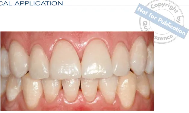
(15) by N ht. n. 129. Q ui. fo r. Evaluation parameters. pyrig No Co t fo rP ub lica tio the gingival contour. All authors agreen te ss ehygiene, nc e that in the presence of good oral. ot. CLINICAL APPLICATION. margins that are precise, well-finished, and. Esthetic result. placed above the gingival level will guar-. Long-term studies of the esthetic behavior. antee good periodontal health.35,65,129–132. of resin composite veneers are very prom-. To demonstrate these concepts, two. ising, thanks to the excellent esthetic prop-. clinical cases demonstrating the most. erties of recent materials. Several stud-. prevalent indications for ceramic or resin. ies28,121,122 have confirmed the color stability. composite veneers will now be presented.. of porcelain veneers over time. Indirect resin composite restorations are useful for masking severe discoloration or. Clinical Case Reports. achieving a certain esthetic effect. Because the veneer should be luted with the. Case 1. same material used for its fabrication, the clinician has a greater choice of color. This case involved remaking incongruous. shades, making it easier to produce slight. Class 4 restorations on the maxillary cen-. modifications of the esthetics.. tral incisors, restoring shape/color defects on the maxillary lateral incisors and canines using ceramic veneers, and correcting enamel hypoplasia on the mandibular. Periodontal response. incisors (Figs 1 and 2). Only a few long-term studies confirming. A clinical situation such as this is one of. the good integration of resin composite. the clearest indications for esthetic veneer. veneers with the surrounding periodontal. treatment, especially for patients who have. tissues have been published. Indirect tech-. repeatedly sought a perfect esthetic inter-. niques allow a level of marginal precision. vention and instead found their smile ruined. that is absolutely comparable to that of ce-. by unsatisfactory restorations. Today, it is. ramics. In addition, the latest-generation. possible to achieve excellence even with di-. resin composites show enhanced superfi-. rect techniques; however, in the authors’. cial polishing and smoothness character-. opinion, in cases like this it is simpler and. istics. Thus, it is possible to achieve prom-. more predictable to use indirect techniques.. ising. results. in. terms. of. periodontal. integrity.. Figures 3 and 4 show the preparations of the maxillary and mandibular teeth. The. A number of long-term studies demon-. veneers received from the dental techni-. strated the biocompatibility and low plaque. cian are shown in Figs 5 and 6. After repli-. retention of ceramic.35,123–130 Peumans et al120. cating the cast, the ceramic (Avanté,. reported mild plaque presence at the mar-. Jeneric/Pentron) was layered and baked. ginal level 5 years after luting as a result of. in the oven. The veneers were tried in, ad-. glaze removal during finishing.. hesively luted (with the use of rubber dam),. Margin positioning plays a fundamental. and polished. The cement used was Cali-. role in periodontal tissues response, which. bra (Dentsply). The final result is shown in. suffers when the margin is placed far from. Figs 7 to 9.. 202 THE EUROPEAN JOURNAL OF ESTHETIC DENTISTRY VOLUME 2 • NUMBER 2 • SUMMER 2007.
(16) MANGANI ETopAL yrig. n. fo r. Figs 1 and 2. Initial situation.. Figs 3 and 4. Preparations of the maxillary and mandibular teeth.. Figs 5 and 6. Veneers for the maxillary and mandibular teeth.. Figs 7 and 8. Final result.. ot. Q ui. by N ht. No C t fo rP ub lica tio n te ss e n c e. 203 THE EUROPEAN JOURNAL OF ESTHETIC DENTISTRY VOLUME 2 • NUMBER 2 • SUMMER 2007.
(17) CLINICAL APPLICATION. n. fo r. Fig 9. ot. Q ui. by N ht. pyrig No Co t fo rP ub lica tio n te ss e n c e. Final result.. Case 2. Conclusions. This case involved replacing two direct restorations on the maxillary right lateral. The success of anterior indirect restora-. incisor and left central incisor, and correct-. tions depends on proper treatment plan-. ing enamel anomalies on the right central. ning and application of the operative pro-. incisor and left lateral incisor (Fig 10).. tocol. Medium- and long-term follow-up. Resin composite veneers (Enamel Plus. studies35,65,129–134 of ceramic veneers have. HFO, Micerium) were used and luted with. shown satisfactory results regarding es-. the same resin composite material. The. thetic result and periodontal response. Re-. dentin bonding agent used was EnaBond. specting every step of the treatment proto-. (Micerium).. col will reduce the percentage of failure in. In this case, a conservative preparation. cases with a high risk of veneer fracture.. confined to the enamel layer was used. Despite the small number of studies in the. (Fig 11). The manufactured resin compos-. literature, it should be underlined that resin. ite veneers are shown in Fig 12. The labo-. composite veneers have undeniable ad-. ratory procedures for this material are. vantages and may represent the future of. much simpler than for ceramic veneers; the. this technique for the following reasons:. material is layered on the cast and each increment is light cured. A final postpolymer-. ■. Luting procedures are less complicat-. ization treatment inside a light-heat oven is. ed and risky thanks to the resin com-. necessary, but no baking is required. Use. posite’s greater ability to absorb the. of rubber dam not only provides operating. polymerization stress of the adhesive cement.. field isolation, but also allows the clinician to carry out each operative step with ab-. ■. Chairside finishing and polishing ma-. solute calm and concentration (Fig 13). The. neuvers are simpler and less likely to. final result is shown in Fig 14.. increase fracture risk, as in ceramic.. 204 THE EUROPEAN JOURNAL OF ESTHETIC DENTISTRY VOLUME 2 • NUMBER 2 • SUMMER 2007.
(18) MANGANI ETopAL yrig. Q ui. by N ht. No C t fo rP ub lica tio n te ss e n c e. ot. n. fo r. Fig 10. Initial situation.. Fig 11. A conservative preparation was carried out.. Fig 12. Resin composite veneers.. Fig 13. Rubber dam was used for field isolation.. Fig 14. ■. Final result.. Resin composite veneers can be. References. modified before luting without compromising either their mechanical proper-. 1.. ties or adhesive potential. ■. Cost is lower due to simpler laboratory procedures, and the range of applica-. 2.. tion is wider than with ceramic. With these considerations in mind, it is the. 3.. authors’ opinion that in the near future the use of ceramic in anterior indirect adhe-. 4.. sive restorations will decrease, while the use of resin composite will increase. 5.. Pippin DJ, Mixson JM, Soldan-Els AP. Clinical evaluation of restored maxillary incisors: Veneers vs PFM crowns. J Am Dent Assoc 1995;126:1523–1529. Mangani F, Sigalot C, Vanini L. Intarsi in resina composita nel restauro estetico dei settori lateroposteriori. Dent Mod 2001;2:25–64. Vanini L, Mangani F. The five dimensions of the color of the teeth in esthetic dentistry. Pract Periodontics Aesthet Dent 2001;1:10–16. Dietschi D, Spreafico R. Restauri Adesivi Nonmetallici: Attuali Concetti per il Trattamento Estetico dei Denti Posteriori. Milano: Scienza Tecnica Dentistica Ed Int, 1997. Vanini L, De Simone F, Tammaro S. Indirect composite restorations in the anterior region: A predictable technique for complex cases. Pract Periodontics Aesthet Dent 1196;9:795–802.. 205 THE EUROPEAN JOURNAL OF ESTHETIC DENTISTRY VOLUME 2 • NUMBER 2 • SUMMER 2007.
(19) Q ui. by N ht. pyrig No Co t fo rP ub lica tio n te ss e n c e n. fo r. 6.. 7.. 8.. 9.. 10.. 11.. 12.. 13.. 14.. 15.. 16.. Pincus CR. Building mouth personality. J South Cal Dent Assoc 1938;14:125–129. McLean JW. The future of dental porcelain. In: McLean JW (ed). Dental Ceramics: Proceedings of the International Symposium of Ceramics. Chicago: Quintessence, 1983:13–40. Sadoun M, Degrange M. Les céramiques dentaires 2ème partie: Les nouvelles ceramiques. J Biomatériaux Dentaire 1987;3:61–69. Schmid M, Fischer J, Salk M, Strub J. Mikrogefuge Leucit verstarter Glaskeramiken. Schweis Monatsschr Zahnmed 1992;102:1046–1053. Anusavice KJ. Degradability of dental ceramics. Adv Dent Res 1992;6:82–89. Shafer R, Kaper HK. Die chemische Loslichkeit von Dentalkeramiken. Dtsch Zahnartzl Z 1993;48:625–628. Mangani F, Ciogli M, Botti F, Cocchia D. Intarsi estetici in vetropolimero. Dent Mod 1999;3:49–62. Martin-Paoletti V. Un nuovo sistema ceramico per intarsi ceramici preformati. Dent Mod 1998;5:55–67. Magne P, Kwon KR, Belser UC, Hodges JS, Douglas WH. Crack propensity of porcelain laminate veneers: A simulate operatory evaluation. J Prosthet Dent 1999;81:327–334. Magne P, Dietschi D, Holz J. Esthetic restoration for posterior teeth: Practical and clinical consideration. Int J Periodontics Restorative Dent 1996;16:105–116. Caleffi A, Berardi D. Veneers in Porcellana Mordenzata: Esperienze Cliniche e Nuove Metodiche. Dalle Porcellane Tradizionali alle Ceramiche Integrali a Pressione. Verona: Resch Ed, 1994.. 17.. 18.. 19.. 20.. 21.. 22.. 23.. 24.. 25.. 26.. 27.. Burgoyne AR, Nicholls JI, Brudvik JS. In vitro two-body wear of inlay-onlay composite resin restoratives. J Prosthet Dent 1991;65:206–214. Bauch JR, de Lange C, Davidson CL. The influence of temperature on some physical properties of dental composites. J Rehabil 1981; 8:309–17. De Gee AJ, Pallav P, Werner A, Davidson CL. Annealing as a mechanism of increasing wear resistance of composites. Dent Mater 1990;6: 266–270. Shinkai K, Susuki S, Leinfelder KF, Katoh Y. How heat treatment and thermal cycling affect wear of composite resin inlays. J Am Dent Assoc 1994;125:1467–1472. Wendt SL Jr, Leinfelder KF. Clinical evaluation of heattreated resin composite inlay: 3-years results. Am J Dent 1993;5:258–262. Wendt SL Jr, Leinfelder KF. The clinical evaluation of heat-treated composite resin inlays. J Am Dent Assoc 1990;120:177–181. Rueggeberg FA, Harvey DK, Evans AL. Color changes in post-cure heat-treated resin composite. Am J Dent 1991; 4:171–176. Dietschi D, Maeder M, Holz J. In vitro evaluation of marginal fit ad morphology of fired ceramic inlays. Quintessence Int 1992;23:271–278. Krejci I. Standortbestimmung in der konservierenden zahnmedizin. Schweiz Monatschr Zahnmed 1993;103:614–619. Burke FJT, Qualtrough AJE. Aesthetic inlays: Composite or ceramic. Br J Dent 1994; 22:53–60. Christensen GJ. Alternative for the restoration of posterior teeth. Int Dent J 1999;39: 155–161.. 206 THE EUROPEAN JOURNAL OF ESTHETIC DENTISTRY VOLUME 2 • NUMBER 2 • SUMMER 2007. ot. CLINICAL APPLICATION. 28. Davies BR, Millar BJ, Wood DJ, Bubb NL. Strength of secondary cured resin composite inlays repairs. Quintessence Int 1997;28:415–418. 29. Mitsaki-Matsou H, KaranikaKouma A, Papadoyiannis Y, Theodoridou-Pahine S. An in vitro study of the tensile strength of composite resin repaired with the same or another composite resin. Quintessence Int 1991;22: 475–481. 30. Frankenberger R, Kramer N, Sindel J. Repair strength of etched vs silica-coated metal-ceramic and all-ceramic restorations. Oper Dent 2000;25:209–215. 31. Guler AU, Yilmaz F, Yenisey M, Guler E, Ural C. Effect of acid etching time and a selfetching adhesive on the shear bond strength of composite resin to porcelain. J Adhes Dent 2006;8:21–25. 32. Atsu SS, Kilicarslan MA, Kucukesmen HC, Aka PS. Effect of zirconium-oxide ceramic surface treatments on the bond strength to adhesive resin. J Prosthet Dent 2006;95:430–436. 33. Kim BK, Bae HE, Shim JS, Lee KW. The influence of ceramic surface treatments on the tensile bond strength of composite resin to all-ceramic coping materials. J Prosthet Dent 2005;94:357–362. 34. Moghadam B. Intraoral repair of fractured porcelain using porcelain laminate veneer. Pract Periodontics Aesthet Dent 1994;6:65–68. 35. Kihn PW, Barnes DM. The clinical evaluation of porcelain veneers: A 48-month clinical evaluation. J Am Dent Assoc 1998;29:211–221. 36. Goldstein GR, Barnhard BR, Penugonda B. Profilometer SEM and visual assessment of porcelain polishing methods. J Prosthet Dent 1991; 65:627–634..
(20) MANGANI ETopAL yrig. n. fo r. 37.. Magne P, Pintado MR, DeLong R. Wear of enamel and veneering ceramics laboratory and chairside finishing procedure. J Prosthet Dent 1999;82:669–679. 38. Ward MT, Tate WH, Powers JM. Surface roughness of opalescence porcelains after polishing. Oper Dent 1995; 20:106–110. 39. Vanini L. Light and color in anterior composite restorations. Pract Periodontics Aesthet Dent 1996;8:673–682. 40. Calamia JR. Etched porcelain facial veneers: A new treatment modality based on scientific and clinical evidence. N Y J Dent 1983;53:255–259. 41. Christensen GJ. Veneering of teeth. State of art. Dent Clin North Am 1985;29:373–391. 42. Horn RH. Porcelain laminate veneers bonded to etched enamel. Dent Clin North Am 1983;27:671–684. 43. Shani FJ, Shortall ACC, Marquis PM. Clinical performance of porcelain laminate veneers. A retrospective evaluation over a period of 6.5 years. J Oral Rehabil 1997; 24:553–559. 44. Friedman MJ. Augmenting restorative dentistry with porcelain veneers. J Am Dent Assoc 1991;122:29–34. 45. Garber DA, Goldstein RE, Feinman RA. Porcelain Laminate Veneers. Chicago: Quintessence, 1987. 46. Shneider PM, Messer LB, Douglas WH. The effect of enamel surface reduction in vitro on the bonding of composite resin to permanent human enamel. J Dent Res 1981;60:895–900. 47. Stacey GD. A shear stress analysis of the bonding of porcelain veneers to enamel. J Prosthet Dent 1993;70: 395–402.. 48.. 49.. 50.. 51.. 52.. 53.. 54.. 55.. 56.. 57.. 58.. Troedson M, Derand T. Effect of margin design, cement polymerization, angle of loading on stress in porcelain veneers. J Prosthet Dent 1999;82:518–524. Gasperic D. Micromorphometric analysis of cervical enamel structure of human upper third molars. Arch Oral Biol 1995;40:453–457. Peumans M, Van Meerbeek B, Yoshida Y. Porcelain veneer bonded to tooth structure: An ultra-morphological FE-SEM examination of the adhesive interface. Dent Mater 1999;15:105–119. Tjan AH, Dun JR, Sanderson IR. Microleakage patterns porcelain and castable ceramic laminate veneers. J Prosthet Dent 1989;61:276–282. Zaimoglu A, Karaaglacliooglu L, Utacli C. Influence of porcelain materials and composite luting resin on microleakage of porcelain laminate veneers. J Rehabil 1992;19: 319–327. Ferrari M, Patroni S, Balleri P. Measurement of enamel thickness in relation to reduction of etched laminate veneers. Int J Periodontics Restorative Dent 1992;23: 407–413. Chalifoux PR. Porcelain veneers. Curr Opin Cosmet Dent 1994:58–66. Magne P, Magne M, Belser U. The esthetic width in mixed prosthodontics. J Prosthodont 1999;82:106–118. Perdigao J, Lopes M. Dentin bonding: Questions for the millennium. J Adhesive Dent 1991;(3):191–209. Christensen G. Porcelain veneer update 1993. Clinical Research Associates Newsletter 1993;17(6). Magne P, Douglas WH. Porcelain veneers: Dentin bonding optimization and biometric recovery of the crown. Int J Prosthodont 1999;12:111–121.. 59.. 60.. 61.. 62.. 63.. 64.. 65.. 66.. 67.. 68.. 69.. ot. Q ui. by N ht. No C t fo rP ub lica tio n te ss e n c e. Douglas WH. The esthetic motif in research and clinical practice. Quintessence Int 1989;20:739–745. Castelnuovo J, Tjan AHL, Philips K, Nicholls JI, Kois JC. Fracture load and mode of failure of ceramic veneers with different preparations. J Prosthet Dent 2000;83: 171–180. Toh GC, Setcos JC, Weinstwin AR. Indirect dental laminate veneers: An overview. J Dent 1987;15:117–124. Friedman M. Multiple potential of etched porcelain lamiante veneers. J Am Dent Assoc 1987;114(special issue):83E–87E. Friedman MJ. A 15-year review of porcelain veneer failure—A clinician’s observations. Compend Contin Educ Dent 1998;19:625–636. Hui KK, Williams B, Davis EH, Holt RD. A comparative assessment of the strengths of porcelain veneers for incisor teeth dependent on their design characteristic. Br Dent J 1991;171:51–55. Clyde JS, Gilmour A. Porcelain veneers: A preliminary review. Br Dent J 1998;164: 9–14. Weinberg LA. Tooth preparations for porcelain veneers. NY State Dent J 1989;55: 25–28. Sheet CG, Taniguchi T. Advantages and limitations in the use of porcelain veneer restorations. J Prosthet Dent 1990;64:406–411. Gibbs CH, Mahan PE, Mauderli A, Lundeen HC, Walsh EK. Limits of human bite strength. J Prosthet Dent 1986;56:226–229. Nicholls JI. Tensile bond of resin cements to porcelain veneers. J Prosthet Dent 1988;60:443–447.. 207 THE EUROPEAN JOURNAL OF ESTHETIC DENTISTRY VOLUME 2 • NUMBER 2 • SUMMER 2007.
(21) Q ui. by N ht. pyrig No Co t fo rP ub lica ti Lu R, Harcourt teJK, Tyas MJ, on Alexander B. An ssinvestigation e nc e. 72.. 73.. 74.. 75.. 76.. 77.. 78.. 79.. 80.. 81.. 82.. 83.. 84.. 85.. 86.. 87.. 88.. 89.. 90.. Paul SJ, Sharer P. The dual bonding technique: A modified method to improve adhesive luting procedures. Int J Periodontics Restorative Dent 1997;17:537–545. Bergenholtz G. Effect of bacterial products on inflammatory reactions in the dental pulp. Scand J Dent Res 1997;85:122–129. Eidenbenz S. Kopierschleifen Keramischer Formkorper [thesis]. Zürich: University of Zürich, 1992. Elledge DA, Hart JK, Schorr BL. A provisional restoration technique for laminate veneer preparations. J Prostet Dent l989;62:139–142. Hayakawa T, Horie K, Aida N, Kanaya H, Kobayashi T, Murata Y. The influence of surface conditions and silane agents on the bond of composite to dental porcelain. Dent Mater 1992;8:238–240. Meijering AC, Creugers NH, Mulder J, Roeters FJ. Treatment times for three different types of veneer restorations. J Dent 1995;23:21–26. Nuixon RL. Masking severely tetracycline-stained teeth with cement laminate veneers. Pract Periodontics Aesthet Dent 1996;8:227–235. Banks R. Conservative posterior ceramics: A literature review. J Prosthet Dent 1990; 63:619–626. Burtscher P. Stability of radicals in cured composite material. Dent Mater 1993;9: 218–221. Fett HP, Mormann WH, Krejci I, Lutz F. The effect of short bevel and silanization on marginal adaptation of computer machined mesio-occlusodistal inlays. Quintessence Int 1991;22:823–829. Stagel I, Nathanson D, Hsu CS. Shear strength of the composite bond to etched porcelain. J Dent Res 1987; 66:1460–1465. Calamia JR, Simonsen RJ. Effect of coupling agents on bond strength of etched porcelain. J Dent Res 1984; 63:179.. 208 THE EUROPEAN JOURNAL OF ESTHETIC DENTISTRY VOLUME 2 • NUMBER 2 • SUMMER 2007. 91.. fo r. 71.. Andreasen FM, Flugge E, Daugaard-Jensen J, Munksgaard EC. Treatment of crown fractured incisors with laminate veneers restoration. An experimental study. Endod Dent Traumatol 1992; 8:30–35. Magne P, Verslius A, Douglas WH. Effect of luting composite shrinkage and thermal loads on the stress distributions in porcelain veneers. J Prosthet Dent 1999;82: 335–344. Schwartz R, Davis R, Mayhew R. Effect of ZOE temporary cement on the bond strength of a resin luting cement. Am J Dent 1990;3:28–30. Dietschi D, DeSiebenthal G, Neveu-Rosenstand L, Holz J. Influence of the restorative technique and new adhesives on the dentin marginal seal and adaptation of resin composite class II restorations: An in vitro evaluation. Quintessence Int 1995;26: 717–726. Lacy AM, Fowell I, Watanabe LG. Resin dentin bond strength following treatment with temporary cements. J Dent Res 1991;70:397. Millstein PL, Hazan E, Nathanson D. Effect of aging on temporary cementation in vitro. J Prosthet Dent 1991;65:768–771. Terata R. Characterization of enamel and dentin surfaces after removal of temporary cement: Study on removal of temporary cement. Dent Mater 1993;12:18–28. Woody TL, Davis RD. The effect of eugenol-containing and eugenol-free temporary cements on microleakage in resin bonded restorations. Oper Dent 1992;17:175–80. Tjan AH, Nementz H. Effect of eugenol-containing endodondtic sealer on retention of prefabricated posts luted with adhesive composite resin cement. Quintessence Int 1992;23:839–844.. n. 70.. 92.. 93.. 94.. 95.. 96.. 97.. 98.. 99.. 100.. ot. CLINICAL APPLICATION. of the composite resin/porcelain interface. Aust Dent J 1992;37:12–19. O’Keefe KL, Pease PL, Herrin HK. Variables affecting the spectral transmittance of porcelain through porcelain veneer samples. J Prosthet Dent 1991;66:434–438. Calamia JR, Vaidyanathan J, Vaidyanathan T, Hirsh S. Shear bond strength of etched porcelains [abstract 1098]. J Dent Res 1985;64: 828. Jones JE, Boksman L. Effect of etching technique on the clinical performance of porcelain veneers. Quintessence Dent Technol 1986;10: 635–657. Lacy AM, LaLuz J, Watanabe LG, Dellinges M. Effect of porcelain surface treatment on the bond to composite. J Prosthet Dent 1988;67: 288–291. Roulet JF, Soderholm KJ, Longmate J. Effect of treatment and storage conditions on ceramic/composite bond strength. J Dent Res 1995;74: 381–387. Sheth J, Jensen M, Tolliver D. Effect of surface treatment on etched porcelain bond strength to enamel. Dent Mater 1988;4:328–337. Arakawa Y, Takahashi Y, Sebata M. The effect of acid etching on the cervical region of the buccal surface of the human premolar, with special reference to direct bonding techniques. Am J Orthod 1979;76:201–208. Carstensen W. The effect of different phosphoric acid concentration on surface enamel. Angle Orthod 1992; 62:51–58. Hussain MA, Bradford EW, Charlton G. Effect of etching on the strength of aluminious jacket crowns. Br Dent J 1979;147:89–90..
(22) MANGANI ETopAL yrig. by N ht. fo r. 126.. 127.. 128.. 129.. 130.. 131.. 132.. 133.. 134.. ot. n. 113. Kramer N, Lohbauer U, Frankenberger R. Adhesive luting of indirect restorations. Am J Dent 2000;13:60D–76D. 114. Noack MJ, Roulet JF, Bergmann PA. New method to lute tooth-colored inlays highly filled composite resins [abstract 1528]. J Dent Res 1991;70:475. 115. Breeding LC, Dixon DL, Caughman WF. The curing potential of light-activated composite resin luting agents. J Prosthet Dent 1991; 65:512–518. 116. Darr AH, Jacobson PH. Conversion of dual cure luting cements. J Oral Rehabil 1995;22:43–47. 117. Hasegawa EA, Boyer DB, Chan DC. Hardening of dualcured cements composite resin inlays. J Prosthet Dent 1991;66:187–192. 118. Barnes CE. Mechanism of vinyl polymerisation. I. Role of oxygen. J Am Chem Soc 1945;67:217–220. 119. Bovey FA, Kalthoff IM. Inibition and retardation of vinyl polymerisation. Chem Rev 1948;42:491–525. 120. Peumans M, Van Meerbeek B, Lambrechts P, VuylstekeWauters M, Vanherle G. Fiveyear clinical performance of porcelain veneers. Quintessence Int 1998;29:211–221. 121. Davis BK, Aquilino SA, Lund PS, Diaz-Arnold AM, Denehy GE. Colorimetric evaluation of the effect of porcelain opacity on the resultant color of porcelain veneers. Int J Prosthodont 1992;5:160–166. 122. Davis BK, Johnston WM, Saba RF. Kubelka-Munk reflectance theory applied to porcelain veneer system using a colorimeter. Int J Prosthodont 1994;7:227–233. 123. Calamia JR. Clinical evaluation of etched porcelain veneers. Am J Dent 1989;2: 9–15. 124. Chan C, Weber H. Plaque retention on restored teeth with full-ceramic crown: A comparative study. J Prosthet Dent 1986;56:666–671.. Q ui. 101. Alberts HF. Bonded Tooth Colored Restoratives: Indirect Bonded Restorations. Santa Rosa: Alto Books, 1989. 102. Peumans M, Van Meerbeek B, Lambrechts P, Vanherle G. Porcelain veneers: A review of the literature. J Dent 2000; 28:163–177. 103. Boyer DB, Chan KC, Reinhardt JW. Build-up and repair of light cured composites: Bond strength. J Dent Res 1984;63:1241–1244. 104. Latta MA, Barkmeier WW. Bond strength of resin cement to a cured composite inlays material. J Prosthet Dent 1994;72:189–93. 105. Swift EJ Jr, Brodeur C, Cvitko E, Pires JA. Treatment of composite surfaces for indirect bonding. Dent Mater 1992;8:193–196. 106. Brodbelt RHW, O’Brien WJ, Fan PL. Translucency of dental porcelain. J Dent 1980;59: 70–75. 107. Linden JJ, Swift EJ, Boyer DB. Photo-activation of resin cements through porcelain veneers. J Dent Res 1991;70: 154–157. 108. Strang R, McCrosson J, Muirhead GM, et al. The setting of visible light-cured resin beneath etched porcelain veneers. J Br Dent 1987;163: 141–149. 109. Nakabayashi N, Kojima K, Masuhara E. The promotion of adhesion by infiltration of monomers into tooth substrates. J Biomed Mater Res 1982;16:265–273. 110. Toffenetti F. Compositi sugli anteriori: Un restauro definitivo. Riv Odont 1990,1:43–62. 111. van Dijken JW, Horstedt P. Marginal breakdown of 5-year-old direct composite inlays. J Dent 1996;24:389–394. 112. Campbell SD. Comparison of conventional paint-on die spacers and those used with the all-ceramic restorations. J Prosthet Dent 1990;63: 151–155.. No C t fo rP ub lica tio n 125. Hahn R, Weiger tR, Netuschil e L, Bruch M. Microbial accuss e ce n mulation and vitality on different restorative materials. Dent Mater 1993;9:312–316. Janeko C, Smales RJ. Anterior crown and gingival health. Aust Dent J 1979;24:225–30. Kourkouta S, Walsh TF, Davis LG. The effect of porcelain laminate veneers on gingival health and bacterial plaque characteristics. J Clin Periodontol 1994;22:1–14. Newcombe GM. The relationship between the location of subgingival crown margin and gingival inflammation. J Periodontol 1974;45:151–154. Peumans M. The Clinical Performance of Veneer Restorations and Their Influence on the Periodontium [thesis]. Leuven, Belgium: Katholieke Universiteit Leuven, 1997. Walls AWG. The use of adhesively retained allporcelain veneers during the management of fractured and worn anterior teeth. Part II: Clinical result after 5-years follow-up. Br J Dent 1995; 178:337–339. Jager K, Stern M, Wirz J. Laminates-reif fur die Praxis? Quintessenz 1995;46: 121–130. Rucker ML, Richter W, MacEntee N, Richardson A. Porcelain and resin veneer clinically evaluated: 2 year result. J Am Dent Assoc 1990;121:595–596. Christensen GJ, Christensen RP. Clinical observation of porcelain veneers: A three year report. J Esthet Dent 1991;3:174–179. Meijering AC, Creughers NH, Roeters J, Mulder J. Survival of three types of veneer restorations in a clinical trial: A 2.5-year interim evaluation. J Dent 1998;26:563–568.. 209 THE EUROPEAN JOURNAL OF ESTHETIC DENTISTRY VOLUME 2 • NUMBER 2 • SUMMER 2007.
(23)
Figura


Documenti correlati
We have proved analytically the actual service properties of a large family of fair-queueing sched- ulers, ranging from WF 2 Q+ to its fast, approx- imated variants, and of
It is very important to be sure during preparation of the tooth for coronary restoration that the margin of preparation is not coincident with the contact points of
The fact that GPS geodetic slip rates astride the Centennial Shear Zone are 10x faster than the paleoseismic slip rates for individual faults, like the Lima Reservoir fault, may
In order to reveal the role of Ece2 in the developing cerebral cortex in vivo, acute KD of this gene’s expression was performed by in utero electroporation (IUE) of microRNA (and
I motivi di necessità e ur- genza sono ben illustrati dal Presidente del TAR nel provvedimento; essi sono l’«aggravamento del rischio sanitario anche in ambito
The present quantum chemical results provides a detailed atom- istic description of the reaction channels for H additions to CO on water ices. However, these results are limited
This family was identified two decades ago in ruminants, but its role(s) remained largely unknown. Investigating cellular functions and mechanism of action of BCNT proteins
Abstract—This work presents a novel approach to distributed training of deep neural networks (DNNs) that aims to overcome the issues related to mainstream approaches to data
