Research Article
Motor Cortex Function in Fibromyalgia: A Study by Functional
Near-Infrared Spectroscopy
Eleonora Gentile
,
1Katia Ricci
,
1Marianna Delussi,
1Filippo Brighina
,
2and Marina de Tommaso
11Applied Neurophysiology and Pain Unit, SMBNOS Department, Bari Aldo Moro University, Polyclinic General Hospital,
Via Amendola 207 A, 70123 Bari, Italy
2Department of Experimental Biomedicine and Clinical Neurosciences (BioNeC), University of Palermo, Via del Vespro 143,
90127 Palermo, Italy
Correspondence should be addressed to Marina de Tommaso; [email protected] Received 25 July 2018; Accepted 31 December 2018; Published 16 January 2019
Academic Editor: Hartmut G¨obel
Copyright © 2019 Eleonora Gentile et al. This is an open access article distributed under the Creative Commons Attribution License, which permits unrestricted use, distribution, and reproduction in any medium, provided the original work is properly cited. Previous studies indicated changes of motor cortex excitability in fibromyalgia (FM) patients and the positive results of transcranial stimulation techniques. The present study aimed to explore the metabolism of motor cortex in FM patients, in resting state and during slow and fast finger tapping, using functional Near-Infrared Spectroscopy (f NIRS), an optical method which detects in real time the metabolism changes in the cortical tissue. We studied 24 FM patients and 24 healthy subjects. We found a significant slowness of motor speed in FM patients compared to controls. During resting state and slow movement conditions, the metabolism of the motor areas was similar between groups. The oxyhemoglobin concentrations were significantly lower in patients than in control group during the fast movement task. This abnormality was independent from FM severity and duration. The activation of motor cortex areas is dysfunctional in FM patients, thus supporting the rationale for the therapeutic role of motor cortex modulation in this disabling disorder.
1. Background
Fibromyalgia (FM) is a disabling disease with widespread muscle pain, associated with fatigue, sleep disorders, cog-nitive impairment, and a number of other physic and psy-chopathological symptoms [1]. It is a complex and often poorly curable syndrome, with unclear pathophysiological mechanisms. Most experimental evidence suggests a cen-tral failure in pain modulation with abnormal response to nociceptive stimuli [2–4]. Increasing evidence is in favor of the pivotal role of the motor function in contributing to pain syndrome outcomes [5]. As a matter of fact, patients with chronic pain have deficits in motor performances, with compromised coordination and control force [6, 7]. How-ever, definite data about the functional condition of motor cortex in FM patients is still lacking. Cortical excitability characteristics are not constant in time, but they depend upon clinical status. In addition, different neurophysiological methods could give different results [8]. Meta-analyses
provided evidence of increased M1 long-interval intracortical inhibition in chronic pain [5]. The possible basal condition of motor cortex inhibition and the benefit of its recovery in pain control may explain the often-reported success of excitatory neuromodulation of M1 in patients with fibromyalgia [8– 10].
Brain hemodynamic activity is considered a good mea-sure of cortical activation during the performance of a motor task. In neuroimaging studies the finger tapping task is widely used to investigate the function of the motor cortex [11–13].
Functional Near-Infrared Spectroscopy (f NIRS) is a non-invasive technique that allows a real time detection of blood flow and metabolism changes in the cerebral cortical tissue. To measure functional brain activity, f NIRS employs emit-ters, named sources, that release optical radiation within head tissue detected by scalp detectors. Thanks to its low cost, good temporal resolution, portability, and movement tolerance, the f NIRS is a useful tool in experimental neuroimaging studies on motor functions [14].
Volume 2019, Article ID 2623161, 7 pages https://doi.org/10.1155/2019/2623161
task
(3) To detect f NIRS changes during finger tapping speed test and correlate it to motor performance
(4) To correlate f NIRS and clinical data in FM patients
2. Materials and Method
The study included consecutive outpatients who came for the first time to our Unit in the period between January and June 2017. The selection criterion was the diagnosis of fibromyalgia according to the ACR criteria [1]. Controls were selected among hospital staff. Exclusion criteria were less than 8 years of education; any peripheral or central nervous system (CNS) disease, including spinal cord diseases and radiculopathies; psychiatric disease; diabetes; active and/or positive history for thyroid insufficiency; renal failure; autoimmune diseases; inflammatory arthritis; systemic connective tissue disease; present or previous history of cancer; and use of drugs acting on the CNS or chronic opioid therapy. All the participants were right handed, as confirmed by Edinburgh Handedness Inventory [15]. The patients group included 20 females and
4 males patients (Mage= 42,17 SD= 10,05; age range from 23
to 59 years), the control group included 19 female and 5 male
control subjects (Mage= 40,00 SD= 14,64; age range from 22 to
60 years). The experimental research received the approval of the Ethics Committee of the Bari Policlinic General Hospital. All the participants signed a written informed consent. 2.1. Clinical Examination. The neurologist submitted all patients to the standard neurological examination, including thorough bedside sensory testing. In accordance with recent studies [16, 17], the FM patients filled out the fibromyalgia-linked invalidity questionnaire-FIQ [18] and the Multidimen-sional Assessment of Fatigue-MAF [19]. Patients received indications on the modalities of the responses by a psychol-ogist. In view of the correlation between f NIRS and clinical data, we considered the Wide Pain Index (WPI) according to the recent ACR diagnostic criteria (2010) [1].
2.2. Experimental Study Design. The participants sat down on a comfortable chair in a quiet room. The researcher trained the participants how to perform the finger tapping task and the resting state. The motor task consisted of pressing a toggle button with the thumb of the right hand. We evaluated the motor cortex activation during the finger tapping tasks and the speed test.
pretask baseline was 1 minute in duration.
2.3. FNIRS Procedure. The hemodynamic activity was recorded by f NIRS compatible cap. We used the f NIRSport instrument (Wearable f NIRS Imaging System) to detect the f NIRS signal. To acquire oxy-hemoglobin concentration changes, we employed f NIRStar 14.2 Acquisition Software (Version 14, Revision 2, Release Build, 2016-04-15 (c) NIRx Medizintechnik GmbH, Berlin, Germany; https://www.nirx .net.
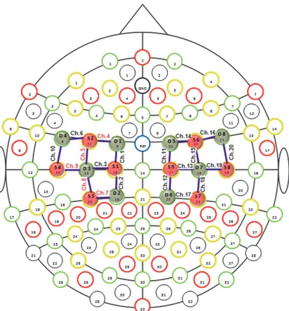
We positioned the f NIRS optodes (sources and detectors) on the cortical regions of interest, which was on the motor cortex, according to the predefined montages tab by f NIRStar 14.2 (Figure 1).
The f NIRS recording channel consists of a source that emits the near-infrared light, which spreads through the bio-logical tissue and is received by the detectors. The distances between sources and detectors were 30 mm. The sampling rate of f NIRS signal was 7.81 Hz. The variations in oxygenated hemoglobin concentration were measured in mmol/L. For the present experiment we used 20 recording channels, 10 for each side of motor cortex, consisting of 8 sources and 8 detectors communicating with each other. Before placing the optodes on the scalp, the technician freed the surface of the skin from the hair, in order to advance the passage of the optical radiation through the brain tissue. We also positioned a covering black cap, to avoid the interference of environmental light. The technician carried out a signal quality check by quick automated calibration using the signal acquisition software. Before proceeding with the recording, the experimenter ascertained the good quality of the f NIRS signal.
2.4. FNIRS Analysis. We used the NIRSlab Software (v2014.12; NIRx Medical Technologies LLC) to process raw data. The raw f NIRS signal was digitally bandpass-filtered offline at 0.01-0.3 Hz. Moreover, the optical density data were converted into relative oxyhemoglobin concentration variations, according to modified Beer-Lambert Law [20]. To evaluate the cerebral regions of interest, we considered the oxyhemoglobin concentration variation, in accordance with several experimental studies, which indicate this parameter as the most sensitive for the assessment of the cortical metabolism during finger tapping task execution [21, 22]. We thus considered the difference between the mean values of oxyhemoglobin concentrations in the resting state and finger
Figure 1: Channels and optodes configuration. In this figure the lines in purple color are the recording channels. Red circles are the sources and green circles are the detectors.
tapping tasks, divided for the standard deviation of mean baseline values [23]. For each subject and each recording channel, the effect size (Cohen’s d) was calculated.
The speed during fast finger tapping task was calculated through the number of clicks per second by manual compu-tation.
2.5. Statistical Analysis. The IBM SPSS Statistics software, version 21, was employed. The values of oxyhemoglobin concentration in resting state, slow and fast movement finger tapping tasks were compared among groups, considering the channels as variable, with a MANOVA test, complete factorial type III model. We merged the mean oxyhemoglobin levels of the significant channels into a Region of Interest (ROI). The finger tapping speed was compared between groups by one way ANOVA. The ROI values were correlated with the finger tapping speed using the linear regression test in FM and control groups. They were also correlated with age and clinical features by Spearman correlation test. We considered
for each statistical analysis p< 0.05 as significant.
3. Results
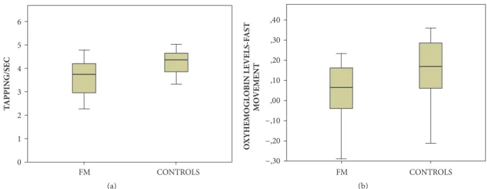
3.1. Finger Tapping Speed. Movement was significantly slower in FM patients, compared to healthy controls (Figure 2(a)).
3.2. Oxyhemoglobin Levels
3.2.1. Resting State. In resting state the oxyhemoglobin con-centration was similar between FM patients and controls (MANOVA F –Roy Square 0.45, Degree of Freedom Hypoth-esis (H-DF) 20; error DF 27, p 0.96).
3.2.2. Slow Finger Tapping Task. Oxyhemoglobin changes occurring during slow finger tapping task were similar between patients and controls (MANOVA: F -Roy Square-1.2, hypothesis DF 20, error DF 27 p 0.31).
3.2.3. Fast Finger Tapping Task. The oxyhemoglobin levels were lower in FM groups during the fast finger tapping task, at least on Ch-4,5,7,8,9 corresponding to the left side (MANOVA: F –Roy Square – 1.08; hypothesis DF 20, error DF 27 p 0.41, single p values; Ch-4 0.010; Ch-5 0.043; Ch-7 0.027; Ch-8 0.030; Ch-9 0.034). We thus averaged channels values into a unique ROI value (Figures 2(b) and 3).
3.2.4. Correlations. The linear regression analysis between motor speed and oxyhemoglobin changes during fast finger tapping task, detected on the ROI, was not significant either in patients or in controls (FM patients: beta 1.12 p 0.55; control: beta 0.17 p 0.41).
FIN 0 FM CONTROLS (a) FM CONTROLS O −,30 (b)
Figure 2: (a) Mean values and standard deviation of speed values in fibromyalgia patients (FM) and controls. One way ANOVA F 10 DF 1 p 0.003. (b) Mean values and standard deviation of oxyhemoglobin levels change on the ROI –CH4,5,7,8,9 during fast movement in fibromyalgia patients (FM) and controls. One way ANOVA F 6.44 DF 1 p 0.015.
No correlation was present between oxygenated hemoglobin and illness duration (Spearman test -0.178 n.s.), disability linked to fibromyalgia (-0.102 n.s.), WPI score (-0.180 n.s.), and fatigue ( -0.192 n.s.). No correlation emerged between finger tapping speed and the same clinical variables, excluding fatigue, which was more severe in slower patients (Spearman test -0.466 p 0.011).
4. Discussion
In the present study we evaluated motor cortex function using functional Near-Infrared Spectroscopy in FM patients compared to control subjects.
Main results consisted of normal oxyhemoglobin levels in resting state and during slow finger tapping task in FM patients, while patients showed lower metabolic activation on the left hemisphere during fast finger tapping task. This was not linearly correlated with the motor slowness patients exhibited in comparison to controls nor with clinical features. In the next paragraph, these results will be discussed in detail. Recent studies have shown that the baseline characteris-tics of M1 are altered in patients with fibromyalgia [5–7] and that activity patterns in response to experimentally induced pain are abnormally enhanced [4, 24]. Studies using TMS methods and assessing cortical silent period and intracortical inhibitions found a reduction of inhibitory activity and increase of network excitability in cortical motor regions [5, 25]. The fMRI studies showed that during acute painful stimulation, FM patients showed increased M1 activation, a sign of a dysfunctional behavior aiming at exerting a full inhibitory control over the pain-related neural circuits [24].
Our f NIRS results were not in agreement with a basal dysfunction of motor cortex in FM patients, as the oxy-hemoglobin levels were quite similar to controls. However, the oxygenated hemoglobin concentration, used to detect changes in cerebral hemodynamics, has no absolute values, so
it could be quite variable across subjects. [26]. The abnormal function of motor cortex could thus appear under specific procedures of activation. During slow finger tapping task, we did not observe a different level of oxyhemoglobin change in patients compared to controls, while this emerged during the speed task.
The regions we identified as critical for the metabolic changes induced by the right-hand movement corresponded to the left prefrontal regions. The primary and probably sup-plementary motor cortex related to the moving hand had a reduced activation in patients. This result may be in apparent disagreement with the hyper excitability of motor networks emerging in FM patients in TMS experiments [24]. However, this paradoxical hyper excitability may be a compensatory phenomenon to a basal dysfunctional performance of motor networks [8]. Changes of cortical excitability may result in complex changes of cerebral blood flow and oxygenated hemoglobin [27]. The reduced motor cortex metabolism may thus be a final result of the prolonged activation of pain-related circuits, with a chronic inhibition of motor network and paradoxical increase of excitatory transmission [25]. The speed test revealed a reduced motor performance in FM patients. Fibromyalgia patients are affected by slowness in motor initiation [28, 29]. A study on f NIRS-related finger tapping changes in FM patients confirmed reduced motor performances and reduced activation of several brain areas that showed enhanced oxygenated hemoglobin levels in controls [30]. Our study cannot resolve the question of whether the motor impairment could be a consequence or a cause of cortical motor network dysfunction. Motor speed in the finger tapping task and oxyhemoglobin changes of motor cortex were not linearly correlated, either in patients or in controls. In line with the results of previous studies [31] we did not find a significant relationship between the finger tapping speed and the levels of cortical activation. In any case, in FM patients motor cortex dysfunction seemed to emerge during energy-demanding tasks, while
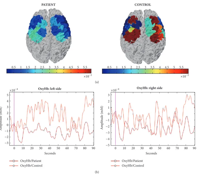
PATIENT 0.5 1 1.5 2 2.5 3 3.5 4 4.5 5 5.5 0.5 1 1.5 2 2.5 3 3.5 4 4.5 5 5.5 ×10−5 ×10−5 CONTROL (a) 5 4 3 2 1 0 −1 −2 −3 Seconds 0 10 20 30 40 50 60 70 80 90 Seconds 0 10 20 30 40 50 60 70 80 90
OxyHb: left side
Am pl it ude (mM) Am pl it ude (mM) 3 2 1 0 −1 −2 −3 −4 −5 ×10−4
×10−4 OxyHb: right side
OxyHb/Patient OxyHb/Control OxyHb/Patient
OxyHb/Control
(b)
Figure 3: (a) Hemodynamic changes in a FM patient (female, 46 years old) and a control subject (female, 47 years old) during fast movement condition according to statistical parametric mapping methods. (b) Raw oxygenated hemoglobin levels during fast movement in the same cases. The Grand Average data of right and left channels are shown.
oxygenated hemoglobin was normal during the slow move-ment and in the resting condition. The metabolic deficit emerging during fast finger tapping task seems a basal feature of FM, not correlated with disease duration and severity.
This means that it could not be a consequence of the prolonged inhibition exerted by the pain circuits activation, but a basal condition able to facilitate pain maintenance. Following this line of thinking, cortical motor dysfunction and movement impairment could characterize FM at its onset [32]. In the present FM sample, fatigue, which is a fundamental symptom of the disease, [1] was correlated with motor slowness, but not with oxygenated hemoglobin during fast finger tapping task. This could further confirm that motor failure is not in direct dependence with the motor circuits functional status, but other factors could cooperate as psychopathologic traits [33], which consideration was beyond the aim of the present study.
5. Conclusion
Overall, the activation of motor cortex areas during fast movement is dysfunctional in FM patients [34, 35]. These data could further support the role of movement [36], M1 transcranial modulation (particularly with new stimulation approaches as focal tDCS and tRNS) [37, 38] and even combination of both approaches [39] in the treatment of this disabling disease.
Data Availability
The f NIRS data used to support the findings of this study are restricted by the Ethics Committee of the Bari Policlinic General Hospital in order to protect patient privacy.
Conflicts of Interest
The study was supported by Bari Aldo Moro University Research funds.
References
[1] F. Wolfe, D. J. Clauw, M. Fitzcharles et al., “The American College of Rheumatology preliminary diagnostic criteria for fibromyalgia and measurement of symptom severity,” Arthritis
Care & Research, vol. 62, no. 5, pp. 600–610, 2010.
[2] A. Rodr´ıguez, J. Tembl, P. Mesa-Gresa, M. ´A. Mu˜noz, P. Montoya, and B. Rey, “Altered cerebral blood flow velocity features in fibromyalgia patients in resting-state conditions,”
PLoS ONE, pp. 1–21, 2017.
[3] G. A. Reyes del Paso, C. I. Montoro, and S. Duschek, “Reaction time, cerebral blood flow, and heart rate responses in fibromyal-gia: Evidence of alterations in attentional control,” Journal of
Clinical and Experimental Neuropsychology, vol. 37, no. 4, pp.
414–428, 2015.
[4] A. T. O’Brien, A. Deitos, Y. Tri˜nanes Pego, F. Fregni, and M. T. Carrillo-de-la-Pe˜na, “Defective Endogenous Pain Modulation in Fibromyalgia: A Meta-Analysis of Temporal Summation and Conditioned Pain Modulation Paradigms,” The Journal of Pain, vol. 18, pp. 30073–30077, 2018.
[5] W.-J. Chang, N. E. O’Connell, P. R. Beckenkamp, G. Alhassani, M. B. Liston, and S. M. Schabrun, “Altered Primary Motor Cortex Structure, Organization, and Function in Chronic Pain: A Systematic Review and Meta-Analysis,” The Journal of Pain, vol. 19, no. 4, pp. 341–359, 2018.
[6] C. J. C. Lamoth, O. G. Meijer, A. Daffertshofer, P. I. J. M. Wuisman, and P. J. Beek, “Effects of chronic low back pain on trunk coordination and back muscle activity during walking: changes in motor control,” European Spine Journal, vol. 15, no. 1, pp. 23–40, 2006.
[7] P. J. M. Bank, C. E. Peper, J. Marinus, J. J. Van Hilten, and P. J. Beek, “Intended and unintended (sensory-) motor coupling between the affected and unaffected upper limb in complex regional pain syndrome,” European Journal of Pain, vol. 19, no. 7, pp. 1021–1034, 2015.
[8] J. P. Lefaucheur, “The use of repetitive transcranial magnetic stimulation (rTMS) in chronic neuropathic pain,”
Neurophys-iologie Clinique, vol. 36, pp. 117–124, 2006.
[9] F. Fregni, S. Freedman, and A. Pascual-Leone, “Recent advances in the treatment of chronic pain with non-invasive brain stimulation techniques,” The Lancet Neurology, vol. 6, no. 2, pp. 188–191, 2007.
[10] A. Passard, N. Attal, R. Benadhira et al., “Effects of unilateral repetitive transcranial magnetic stimulation of the motor cortex on chronic widespread pain in fibromyalgia,” Brain, vol. 130, no. 10, pp. 2661–2670, 2007.
tific Reports, vol. 8, p. 3341, 2018.
[15] R. C. Oldfield, “The assessment and analysis of handedness: the Edinburgh inventory,” Neuropsychologia, vol. 9, no. 1, pp. 97–113, 1971.
[16] M. de Tommaso, A. Federici, R. Santostasi et al., “Laser-evoked potentials habituation in fibromyalgia,” The Journal of Pain, vol. 12, no. 1, pp. 116–124, 2011.
[17] M. de Tommaso, M. Nolano, F. Iannone et al., “Update on laser-evoked potential findings in fibromyalgia patients in light of clinical and skin biopsy features,” Journal of Neurology, vol. 261, no. 3, pp. 461–472, 2014.
[18] C. S. Burckhardt, S. R. Clark, and R. M. Bennett, “The Fibromyalgia Impact Questionnaire (FIQ): development and validation,” The Journal of Rheumatology, vol. 18, pp. 728–733, 1991.
[19] B. Tack, Dimensions and correlates of fatigue in older adults
with rheumatoid arthritis, [Unpublished doctoral dissertation],
School of Nursing, University of California, San Francisco, Calif, USA, 1991.
[20] M. Cope, D. T. Delpy, E. O. Reynolds, S. Wray, J. Wyatt, and P. van der Zee, “Methods of quantitating cerebral near infrared spectroscopy data,” Advances in Experimental Medicine and
Biology, vol. 222, pp. 183–189, 1988.
[21] G. Strangman, J. P. Culver, J. H. Thompson, and D. A. Boas, “A quantitative comparison of simultaneous BOLD fMRI and NIRS recordings during functional brain activation,”
NeuroIm-age, vol. 17, no. 2, pp. 719–731, 2002.
[22] T. Zama and S. Shimada, “Simultaneous measurement of electroencephalography and nearinfrared spectroscopy during voluntary motor preparation,” Scientific Reports, vol. 5, Article ID 16438, 2015.
[23] M. Balconi and M. E. Vanutelli, “Brains in Competition: Improved Cognitive Performance and Inter-Brain Coupling by Hyperscanning Paradigm with Functional Near-Infrared Spectroscopy,” Frontiers in Behavioral Neuroscience, vol. 11, p. 163, 2017.
[24] L. Castillo Saavedra, M. Mendonca, and F. Fregni, “Role of the primary motor cortex in the maintenance and treatment of pain in fibromyalgia,” Medical Hypotheses, vol. 83, no. 3, pp. 332–336, 2014.
[25] A. Thibaut, D. Zeng, W. Caumo, J. Liu, and F. Fregni, “Cor-ticospinal excitability as a biomarker of myofascial pain syn-drome,” PAIN Reports, vol. 2, Article ID e594, 2017.
[26] H. Obrig, “NIRS in clinical neurology—a ‘promising’ tool?”
NeuroImage, vol. 1, pp. 535–546, 2014.
[27] H. Takai, A. Tsubaki, K. Sugawara et al., “Effect of transcranial direct current stimulation over the primary motor cortex on cerebral blood flow: A time course study using near-infrared
spectroscopy,” Advances in Experimental Medicine and Biology, vol. 876, pp. 335–341, 2016.
[28] O. Rasouli, E. A. Fors, P. C. Borchgrevink, F. ¨Ohberg, and A.-K. Stensdotter, “Gross and fine motor function in fibromyalgia and chronic fatigue syndrome,” Journal of Pain Research, vol. 10, pp. 303–309, 2017.
[29] I. D. Costa, A. Gamund´ı, J. G. Miranda, L. G. Franc¸a, C. N. De Santana, and P. Montoya, “Altered Functional Performance in Patients with Fibromyalgia,” Frontiers in Human Neuroscience, vol. 26, pp. 11–14, 2017.
[30] A. Eken, D. G¨okc¸ay, C. Yılmaz, B. Baskak, A. Baltacı, and M. Kara, “Association of Fine Motor Loss and Allodynia in Fibromyalgia: An f NIRS Study,” Frontiers in Human
Neuro-science, vol. 26, pp. 11–14, 2017.
[31] J. H. Jenkins, R. E. Passingham, and D. J. Brooks, “The effect of movement frequency on cerebral activation: a positotron emis-sion tomography study,” Journal of the Neurological Sciences, vol. 151, pp. 195–205, 1997.
[32] L. D. Ellingson, M. R. Shields, A. J. Stegner, and D. B. Cook, “Physical activity, sustained sedentary behavior, and pain mod-ulation in women with fibromyalgia,” The Journal of Pain, vol. 13, no. 2, pp. 195–206, 2012.
[33] I. Trofimova and W. Sulis, “There is more to mental illness than negative affect: comprehensive temperament profiles in depression and generalized anxiety,” BMC Psychiatry, vol. 18, p. 125, 2018.
[34] R. Peyron, B. Laurent, and L. Garc´ıa-Larrea, “Functional imag-ing of brain responses to pain. A review and meta-analysis,”
Neurophysiologie Clinique, vol. 30, pp. 263–288, 2000.
[35] A. Salerno, E. Thomas, P. Olive, F. Blotman, M. C. Picot, and M. Georgesco, “Motor cortical dysfunction disclosed by single and double magnetic stimulation in patients with fibromyalgia,”
Clinical Neurophysiology, vol. 111, no. 6, pp. 994–1001, 2000.
[36] M. K. H. Bement and K. A. Sluka, “Exercise-induced analgesia: an evidence-based review,” in Mechanisms and Management of
Pain for the Physical Therapist, K. A. Sluk, Ed., vol. 10, Chapter
10, pp. 177–201, 2nd edition, 2016.
[37] L. Castillo-Saavedra, N. Gebodh, M. Bikson et al., “Clinically Effective Treatment of Fibromyalgia Pain with High-Definition Transcranial Direct Current Stimulation: Phase II Open-Label Dose Optimization,” The Journal of Pain, vol. 17, no. 1, pp. 14–26, 2016.
[38] M. Curatolo, G. La Bianca, G. Cosentino et al., “Motor cortex tRNS improves pain, affective and cognitive impairment in patients with fibromyalgia: preliminary results of a randomised sham-controlled trial,” Clinical and Experimental
Rheumatol-ogy, vol. 105, pp. 100–105, 2017.
[39] M. E. Mendonca, M. Simis, L. C. Grecco, L. R. Battistella, A. F. Baptista, and F. Fregni, “Transcranial direct current stim-ulation combined with aerobic exercise to optimize analgesic responses in fibromyalgia: A randomized placebo-controlled clinical trial,” Frontiers in Human Neuroscience, vol. 10, p. 68, 2016.
Stem Cells
International
Hindawi
www.hindawi.com Volume 2018
Endocrinology
International Journal ofHindawi www.hindawi.com Volume 2018 Hindawi www.hindawi.com Volume 2018 BioMed Research International
Oncology
Journal of Hindawi www.hindawi.com Volume 2013 Hindawi www.hindawi.com Volume 2018Oxidative Medicine and Cellular Longevity Hindawi www.hindawi.com Volume 2018
PPAR Research
Immunology Research Hindawi www.hindawi.com Volume 2018 Journal ofObesity
Journal of Hindawi www.hindawi.com Volume 2018 Hindawi www.hindawi.com Volume 2018 Computational and Mathematical Methods in Medicine Hindawi www.hindawi.com Volume 2018Behavioural
Neurology
Ophthalmology
Journal of Hindawi www.hindawi.com Volume 2018 Hindawi www.hindawi.com Volume 2018Research and Treatment