Presence of cadmium residues
in muscle, liver and kidney
of Bubalus bubalis and
histological evidence
Roberta Barrasso,1Edmondo Ceci,1 Laura Stinga,2Giuseppina Tantillo,1 Giancarlo Bozzo1
1Department of Veterinary Medicine, University of Bari “Aldo Moro”; 2Specialization degree in Food
Inspection of Animal Origin, Veterinary Medicine, University of Bari “Aldo Moro”, Bari, Italy
Abstract
Cadmium (Cd) concentrations were evaluated in the samples of kidney, liver and muscle of sixty-six buffaloes regularly slaughtered. Forty were raised in Campania, in the territory between the province of Naples and Caserta and twenty-six were bred in Apulia, in the province of Bari. Two aliquots were prepared for the renal and hepatic samples: one intended for the chem-ical analysis and the other one intended for histological investigations. Muscle samples were the subject of purely chemical investi-gation. In the group of forty animals raised in the Campania region, the limits imposed by EC Reg. 1881/2006 and EC Reg. 488/2014 were exceeded in three renal sam-ples, which showed values of 1.53, 1.22 and 1.1 mg/kg respectively; in three hepatic samples, which presented values of 0.72, 0.64 and 0.61 mg/kg, and in five muscle samples, with values of 0.16, 0.16, 0.09, 0.08 and 0.07, respectively. On the other hand, in the group of animals raised and slaughtered in the province of Bari, none of the twenty-six samples examined exceeded the limits imposed by the European regula-tions. The histological analysis showed typ-ical, but not pathognomonic lesions in the renal samples from the animals raised in the provinces of Naples and Caserta. The levels of Cd contamination found in the samples examined suggest that it would be correct to exclude from the human consumption, as it happens for the equines, the kidney and the liver, especially from the animals raised in some geographical areas with a high rate of pollution.
Introduction
Elements can be classified as “essen-tial” and “non-essen“essen-tial” following their
functions in biota. Non-essential elements are very resistant to natural degradation and keep their toxicity unchanged for a long time; for this reason, their environmental concentration should be monitored and maintained at very low levels (Lara et al., 2012; Paßlack et al., 2014; Binkowski et al., 2016).
There is a broad range of toxic effects caused by metals, including carcinogenici-ty, impaired reproduction, teratogenicicarcinogenici-ty, immunosuppression, cardiovascular and pulmonary diseases, nephrotoxicity and neurotoxicity (Roychoudhury et al., 2010). Aiming to protect human health and ensure the quality of food, an important component of any food safety programme is the control and monitoring of residues and contami-nants in living organisms (EFSA, 2004a).
Environmental pollution, the main sources of which constitute increasing industrial production, transportation and plant protection chemicals used in agricul-ture, results in serious health hazards to humans as well as animals (Demirel et al., 2008). Among the farm animals, horses are the species that most easily accumulates the metal in kidney, liver and muscle, but also cattle, sheep and goats can contain high concentrations of Cd, if fed with highly contaminated feed (Groten et al., 1994; Plumlee et al., 1996; Giofrè et al., 2000).
The compounds of some non-essential elements, most cadmium compounds, are relatively easily soluble in water, have high mobility and tend to accumulate in living organisms (Nagajyothi et al., 2008). Cd concentrations in the food chain are of cur-rent interest for risk assessment. The Scientific Report of EFSA (2012) regarding the cadmium dietary exposure in the European population, has indicated that the ingestion of liver and kidneys of horses, ruminants and wildlife can considerably contribute to human overall Cd exposure. According to the World Health Organization (WHO) and the United Nations Food and Agriculture Organization (FAO) in 2003, the amount of Cd that can be ingested weekly by an adult man is 500 μg, i.e. 7 μg/kg of weight body correspond-ing to 1 μg/kg per day and a dose of 100 μg/dL is considered fatal. The maximum admissible limit in drinking water is 3 μg/L; in foods the average concentration should not exceed 0.04 - 0.05 mg/kg (D.M. of 12/29/2003). The International Agency for Research on Cancer (IARC) and the US National Toxicology Program have classi-fied the Cd as a “category 1 carcinogen”.
Cd contamination is of particular importance when it derives from plant prod-ucts, as they represent the raw material for the production of feed for zootechnical
pur-poses. The latter, destined for livestock ani-mals, are therefore the weak link in the entire production chain (Mantovani et al., 2002). Cd levels in air, water and soils have been increasing during recent years, partic-ularly because of human activity. It is wide-ly introduced into the food chain and nor-mally found in variable concentrations in foods of plant or animal origin (Antoniou et al., 1989).
The most serious problem about cadmi-um is that this metal is characterized by a remarkable capacity for penetration into the cell, by a high bio-persistence and by a par-ticular difficulty of elimination (Nordberg et al., 1994). Winds, as well as rainwater, play a fundamental role in the spread of metal even in areas very far from the main pollution sites. The Cd of the atmosphere contributes about 23% to the pollution of the water as it is transported by atmospheric currents even at great distances from the most urbanized centres (Mantovani et al., 2002). The accumulation of Cd in the soil is continuous and systematic, so that even the vegetables, through the root system, absorb Correspondence: Roberta Barrasso, Department of Veterinary Medicine, University of Bari “Aldo Moro”
Strada Provinciale per Casamassima km 3, 70010 Valenzano (BA), Italy.
Tel: +39.080.5443851 - Fax: +39.080.5443855. E-mail: [email protected]
Key words: Cadmium, Bubalus bubalis, Muscle, Liver, Kidney.
Acknowledgments: The authors thank Stefano Sportelli for providing invaluable technical assistance in the laboratory.
Contributions: RB, EC, GB and GT designed the experiment; GB, EC and LS performed the experimental procedures; RB conducted the statistical analysis. All authors contributed equally writing the paper.
Conflict of interest: the authors declare no potential conflict of interest.
Funding: none.
Received for publication: 10 July 2018. Revision received: 27 September 2018. Accepted for publication: 12 October 2018. This work is licensed under a Creative Commons Attribution-NonCommercial 4.0 International License (CC BY-NC 4.0).
©Copyright R. Barrasso et al., 2018 Licensee PAGEPress, Italy
Italian Journal of Food Safety 2018; 7:7684 doi:10.4081/ijfs.2018.7684
Non-commercial
it in a constant way, involving the contami-nation of the herbivores (Mantovani et al., 2002).
The elective deposit sites, in all animal species, are represented by the cortical por-tion of the kidney, especially in the first part of the proximal contorted tubule, and by the liver. Renal injury induced by Cd is charac-terized first of all by proximal tubular dys-function, which is believed to be irre-versible at advanced stages (Mitsumori et al., 1998). Other typical effects of Cd expo-sure are: (i) disturbances of calcium metabolism, (ii) hypercalciuria and (iii) for-mation of stones in the kidney. Finally, high exposure can lead to lung and prostate can-cer.
In the kidneys and liver can be found values equal to 50-60% of all the Cd present in the organism. To a lesser extent, the metal is deposited at the level of the muscle masses and in smaller quantities in the pan-creas, testes, spleen, breast tissue, ovaries, uterus, vascular walls and arterioles (Pozzali et al., 1995).
In general, the level of contamination of organs and muscle tissue is directly propor-tional to the geographical area in which these animals are bred and to their age (Al-naemi HS, 2011). The Buffalo breeding rep-resented a new reality. Until recently the buffalo farming, bred mainly in Campania, in the lower Lazio, Apulia and Calabria, was finalized almost exclusively to the use of milk and its derivatives and the produc-tion of the buffalo meat was totally rejected. Today, it is believed that even the males, about 50% of the births, represent an impor-tant economic resource thanks to the dietet-ic-nutritional and organoleptic characteris-tics of its meat and the wide commercial possibilities of this product (Mehmood et al., 2014). The buffalo meat is a food suit-able for the needs of modern life, character-ized by its typical bright red colour for its contents in stearic and linoleic acids, also has a cholesterol content significantly lower than that of beef meat. It lends itself well to the various meat preparations, it is more tender than the beef meat also because gen-erally these animals are mostly bred in the wild, free to graze (Mehmood et al., 2014). This aspect implies that if the breeding area and/or the feed used are very contaminated, they affect the meat. To date, this species is not yet included in the EC Regulation 1881/2006, nor in the most recent EC Regulation 488/2014. Also the National Residual Plan (NRP) assimilates, with a note from the Ministry of Health No. 0021474-P-01/12/2009, the buffalo species to the bovine ones.
According to the EC Regulation 1881/2006 latest amended by EC
Regulation 488/2014, the maximum level for Cd in liver and kidney of bovine ani-mals, sheep, pig, poultry and horse is 0.50 mg/kg and 1.0 mg/kg fresh weight respec-tively; while in meat (excluding offal) of bovine animals, sheep, pig and poultry is 0.050 mg/kg fresh weight.
In order to acquire data on the Cd con-centrations in the buffalo species (Bubalus bubalis), a less investigated specie of zootechnical interest and mistakenly assimilated to the bovine, the aim of the present study was to provide preliminary indications about the incidence of this non-essential element in samples of liver, kidney and muscle (longissimus dorsi) of Bubalus bubalis. We compared Cadmium concentra-tion in different farms in Apulia and Campania regions, considered at risk of environmental contamination and we tried to assess a correlation between Cd levels in tissues and organs and the presence of his-tological lesions.
Materials and Methods
Sampling
The study was conducted in the period between March and May 2018 on a total of sixty-six buffaloes regularly slaughtered, aged between 4 and 82 months and bred in free paddocks outdoors. Forty were raised in Campania, in the territory between the province of Naples and Caserta and twenty-six were bred in Apulia, in the province of Bari. The animals were divided into three experimental groups, according to their dif-ferent age: a group under 6 months of age (group A), a group aged between 7 months and 3 years (group B) and a group of ani-mals aged more than 3 years (group C).
After post-mortem examination, per-formed by the Official Veterinarian and before the entry of the carcasses in the pre-cooling and pre-cooling tunnel, samples of kid-ney, liver and muscle (longissimus dorsi) were collected. Then the samples were transported, under refrigeration, to the labo-ratories of the Food Safety Section of the Department of Veterinary Medicine at Bari University, where two aliquots were pre-pared for the renal and hepatic samples: one intended for the chemical analysis (stored frozen) and the other one intended for histo-logical investigations (fixed in 4% buffered formalin). Muscle samples were the subject of purely chemical investigation.
Samples for chemical testing: atomic
absorption spectrometry
For the qualitative and quantitative detection of cadmium, all the samples were
subjected to digestion at 120°C for 240 minutes in a DK6 Heating Digester (VELP Scientifica). For each animal, aliquots of 2,5 g of muscle and 0,5 g of liver and kid-ney were placed into a 25 mL glass diges-tion vessel and 8 mL of concentrated nitric acid (HNO3) and 3 mL of hydrogen
perox-ide (HClO4) were added to the vessel.
After cooling, the final volume of the solution was made up to 25 mL with dis-tilled water. High-quality water, obtained using a Milli-Q system (Millipore), was used exclusively. Cd standard solutions (1000 mg/mL-1) were obtained from
Panreac (Spain) and diluted as necessary to obtain working standards. Concentrated nitric acid (65% w/v Merck), hydrogen per-oxide (30% w/v Fluka), and ammonium dihydrogenphosphate (Fluka) were also used. A Solar M Series-Unicam (Cambridge UK) 939QZ atomic absorption spectrome-ter equipped with a GF90 electrothermal atomizer (ET-AAS). Pyro lytic platforms were obtained from ATI-Unicam. This instrument is equipped with both a deuteri-um-arc background corrector and a Zeeman correction device, which facilitates compar-ison between both correction modes. Argon was used as the inert gas. Background-cor-rected integrated absorbance was used in all cases as the analytical signal. Each sample was subjected, in a graphite furnace, to the thermal cycle divided into: drying, pyroly-sis, atomization and final cleaning. Measurements were performed at 228.8 nm for Cd and hollow cathode lamps were operated at 7 mA. The graphite furnace temperature program for the determination of Cd in meat samples by ET-AAS using the digestion technique was as follows: (i) Drying 1: °C; ramp/°C s-1135; hold/s: 70; 10; 20. (ii) Drying 2: °C; ramp/°C s-1136; hold/s: 100; 5; 60. (iii) Pyrolysis: °C; ramp/°C s-1137; hold/s: 900; 100; 35. (iv) Atomization: °C; ramp/°C s-1138; hold/s: 2000; Full Power; 4. (v) Cleaning: °C; ramp/°C s-1139; hold/s: 2400; 1000; 4.
Purge gas (argon) flow rate: 2 L min-1
140 in all steps, except in atomization, when the gas flow was interrupted. To decrease the risk of Cd contamination, the use of glassware was reduced to a minimum and plastic (polypropylene) vessels of the type commonly used to collect clinical sam-ples were used to prepare and store the solu-tions or suspensions. Pipette tips were also of polypropylene. All the glassware and plastic ware was nitric acid-washed and rinsed with ultrapure water. An external cal-ibration curve was constructed to determine Cd. The working standards were prepared by serial dilution of stock solutions with the addition of 0.014 mole/L-1148 nitric acid.
The limit of detection (LOD) is expressed
Non-commercial
as three times the standard deviation (SD) of the mean result using a large number of blanks (n≥20). The limit of quantification (LOQ) is expressed as two times the LOD (6 SD). The limits of quantification (LOQ) for the element were estimated as 0.005 mg/kg-1Cd 152 dry weight. The precision
(i.e. internal reproducibility; measured as residual standard deviation RSDr in %) was calculated to be 6-10% for Cd in the present study (n=30). The analytical procedure was validated using certified reference material (BCR 668). Each sample was analysed in duplicate and the error did not exceed 7%.
Statistical analysis
A statistical descriptive analysis based on central tendency and concentration indexes was carried out for the two groups of animals according to their geographical origin, each one divided in three subgroups of different age. We realized box plots in which the trends of the Cd concentration in the three organs involved in the study (kid-ney, liver and muscle) were highlighted to provide a framework of the changes in its amount according to the geographical provenience, the age and the samples. The results were expressed as average, median, Standard Error (SE), Standard Deviation (SD), kurtosis, skewness, minimum and maximum. Then, we carried out the para-metric analysis (r Pearson correlation) of the Cd concentrations in the various tissues to verify a possible correlation among the three samples studied.
Samples for histological testing
The samples of kidney and liver collect-ed for histological examination were pro-cessed as follows: (i) fixed in 10% neutral buffered formalin for 48 hours; (ii) embed-ded in paraffin wax; (iii) sectioned at 4 μm; (iv) stained with Haematoxylin Eosin (H.E.) and finally (v) observed by optical microscopy (Eclipse 50i - Nikon instru-ment).
Results
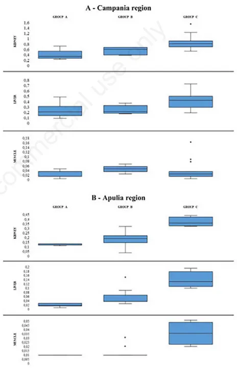
The results of the statistical descriptive analysis based on central tendency and con-centration indexes of the Cd concon-centration are shown in Table 1 and Figure 1. In the group of forty animals raised in the Campania region, the limits imposed by EC Reg. 1881/2006 and EC Reg. 488/2014 (1 mg/kg for the kidney, 0.50 mg/kg for the liver and 0.050 mg/kg for muscle) were exceeded in three renal samples, which showed values of 1.53, 1.22 and 1.1 mg/kg respectively; in three hepatic samples, which presented values of 0.72, 0.64 and
0.61 mg/kg, and in five muscle samples, with values of 0.16, 0.16, 0.09, 0.08 and 0.07, respectively. On the other hand, in the group of animals raised and slaughtered in the province of Bari, the level of Cd con-tamination was much lower. Indeed, none of the 26 samples examined exceeded the limits imposed by the European Regulations.
The strongest relationship in Cd con-centrations occurred between liver and kid-ney tissue (r = 0,8318 in the samples of the Campania region and r = 0,8421 in the sam-ples of the Apulia region), even if this rela-tionship was present also between liver
tis-sue and muscle (r = 0,5516 in the samples of the Campania region and r = 0,7218 in the samples of the Apulia region, respec-tively) and between kidney tissue and mus-cle (r = 0,5458 in the samples of the Campania region and r = 0,6142 in the sam-ples of the Apulia region, respectively) (Table 2).
The histological analysis showed typi-cal, but not pathognomonic lesions in the renal samples from the animals raised in the provinces of Naples and Caserta. Moreover, these lesions were visible only in the sam-ples with a concentration of the metal of 1.53, 1.22 and 1.1 mg/kg. These samples
Figure 1. Distribution of Cd concentrations.
Non-commercial
showed extended histological alterations of the whole renal morpho-functional unit; in fact, a very compromised kidney picture was observed: (i) the proximal convoluted tubules appeared dilated, filled with pro-tein-like amorphous substance and with localized necrotic areas and (ii) the renal glomerulus showed an alteration of epithe-lial cells and the capillaries were clearly visible (Figure 2). On the other hand, in the renal samples in which the concentration of Cd was between 0.75 and 0.95 mg/kg, the alterations were found only in the proximal convoluted tubules. Histological examina-tion conducted on the hepatic parenchyma, showed only different degrees of steatosis, both in the samples from the animals raised in the province of Bari, and in the group of animals raised in the provinces of Naples and Caserta.
Discussion
Cadmium was present in all examined samples, both in the offal and in the muscu-lar tissue, but the most amuscu-larming values were showed in the animals raised in the Campania region, in the provinces of Naples and Caserta. Indeed, only in the kid-neys, livers and muscles of the animals from Campania region were found Cd lev-els exceeded the European limits for human
consumption. The eleven samples examined that exceeded the limits imposed by the European Regulation concerned only six animals: (i) two presented the exceeding of the limits only in the liver and muscle respectively; (ii) three showed the exceed-ing of the limits in two organs simultane-ously (two both in the kidney and in the muscle and one both in the liver and in the muscle); (iii) finally, one animal presented the exceeding of the limits in all three sam-ples.
Moreover, in agreement with other studies (Al-naemi HS, 2011; Binkowski et al., 2016), the Cd concentration depended on the animal age, showing lower concen-tration in the younger buffaloes (<6 months) than in the older ones (groups B and C). This correlation was confirmed by the classes of homogeneity constructed that, although different in number, were optimal since the dispersion around the average was low within the groups (Table 1). On the other hand, no differences were observed
depending on the sex of the animals, which was in accordance with further studies (Paßlack at al., 2014).
Among the relationships studied, the strongest occurred in Cd concentrations between liver tissue and kidney tissue (r = 0,8318 in the samples of the Campania region and r = 0,8421 in the samples of the Apulia region) (Table 2). This relationship was generally known and was described in many species (Binkowski et al., 2016; Binkowski and Sawicka-Kapusta, 2015a; Nordberg et al., 2007). In fact, the liver and kidneys are especially prone to xenobiotic induced injury due to their central role in xenobiotic metabolism (Massanyi et al., 2014).
Conversely, the histological picture resulted independent of the animal age; in fact, it was observed both in subjects who have reached slaughter in the first months of their life (group A), and in those at the end of their careers (groups B and C). Table 1. Descriptive statistics.
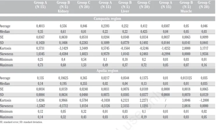
Group A Group B Group C Group A Group B Group C Group A Group B Group C (N 15) (N 5) (N 20) (N 15) (N 5) (N 20) (N 15) (N 5) (N 20)
Kidney Liver Muscle Campania region Average 0,4013 0,556 0,846 0,2393 0,252 0,412 0,0307 0,05 0,046 Median 0,35 0,61 0,81 0,22 0,22 0,425 0,04 0,05 0,03 SE 0,0367 0,0630 0,0510 0,0284 0,0348 0,0334 0,0037 0,0063 0,0099 SD 0,1420 0,1408 0,2283 0,1099 0,0779 0,1492 0,0144 0,0141 0,0441 Kurtosis 0,3731 -3,1429 3,3449 0,5745 -0,1564 -0,5246 -1,4252 2,0000 3,1717 Skewness 1,0345 -0,4384 1,6023 0,9579 1,0143 0,2483 -0,2994 0,0000 1,9556 Minimum 0,25 0,4 0,54 0,1 0,18 0,2 0,01 0,03 0,01 Maximum 0,73 0,68 1,53 0,49 0,37 0,72 0,05 0,07 0,16 Apulia region Average 0,135 0,18625 0,365 0,0217 0,0544 0,1375 0,01 0,013125 0,035 Median 0,14 0,195 0,355 0,02 0,04 0,13 0,01 0,01 0,035 SE 0,0034 0,0159 0,0240 0,0031 0,0076 0,0189 0,0000 0,0018 0,0065 SD 0,0084 0,0634 0,0480 0,0075 0,0305 0,0377 0,0000 0,0070 0,0129 Kurtosis 1,4286 0,9866 0,5784 -0,1038 6,2121 2,2271 / 3,0046 -1,2000 Skewness -1,5367 -0,1712 1,0154 -0,3126 2,3155 1,1293 / 2,0816 0,0000 Minimum 0,12 0,05 0,32 0,01 0,03 0,1 0,01 0,01 0,02 Maximum 0,14 0,32 0,43 0,03 0,15 0,19 0,01 0,03 0,05
SE: standard error; SD: standard deviation.
Table 2. Correlations (r Pearson) of metal concentrations in various tissues.
Correlation Campania region Apulia region
Kidneys vs Liver 0,8318 0,8421 Kidneys vs Muscle 0,5458 0,6142 Liver vs Muscle 0,5516 0,7218
Non-commercial
Conclusions
The buffaloes of our investigation were all suitable for slaughter following the ante-mortem visit and therefore suitable for human consumption following the post-mortem inspection. The most worrying data emerged after the survey conducted on the samples from Campania region was the exceeding of the limits allowed in the mus-cle (0.05 mg/kg).
As demonstrated in recent studies (Oladipo et al., 2016; Dumkova et al., 2016), histological lesions had a diagnostic role only in the renal samples in which Cd concentrations were higher than 0.75 mg/kg. Therefore, at concentrations of Cd ranging from 0.75 to 0.95 mg/kg, the lesions were limited to proximal contorted tubules; while the lesions involved the whole morpho-functional unit of the kidney as the metal concentration increased.
The levels of Cd contamination found in the samples examined suggest that it would be correct to exclude from the human consumption, as it happens for the equines, the kidney and the liver, especially from the animals raised in some geographical areas with a high rate of pollution. This may be possible by mapping the risk areas and by a continuous and constructive collaboration
between the ASL and the people responsible for the environmental control.
As showed by other studies (Bozzetta et al., 2011; Bozzo et al., 2011), which have experimentally used histological methods for the detection of chemical compounds or to reveal food fraud, the survey showed that, to improve the effectiveness of tradi-tional control protocols (ante and post mortem visit), screening methods that eval-uate indirect and objective parameters are necessary. Therefore, it is crucial to consid-er the histological examination as a diag-nostic tool to be integrated in the traditional chemical investigation, favouring the possi-bility of animal experimentation in this field to verify experimentally when lesions occur. Indeed, steatosis or other forms of hepatic degeneration were not pathog-nomonic and not referable with certainty to Cd intoxication, a correlation that could be confirmed only by using animal experimen-tation.
Only chemical analysis, combined with histological examination, is able to provide a complete, clear picture of the animal’s condition, as this makes it possible to corre-late the renal damage with the actual metal concentration. These actions should be combined with testing of the environmental pollution to which the animals are exposed
since the quality of the animal products des-tined for human consumption is affected by several factors acting throughout the pro-duction chain. Whereas a recent study (Bozzo et al., 2018) reported that the per-ception of animal welfare has increased among European consumers, the welfare of livestock must be preserved and a good environment is a fundamental pre-requisite to improve livestock production quality.
The present data underline the rele-vance of buffalo organs in the food chain for human Cd exposure and confirm the sci-entific opinion of the EFSA Panel on Contaminants in the Food Chain (CON-TAM 2009; 2011), according to which the human Cd exposure should be reduced. Therefore, it is important to keep studying the cadmium levels tolerated by human and denounce what might be underestimated chemical hazards in meat. Considering that the buffalo breeding is in continuous growth thanks to the dietetic-nutritional and organoleptic characteristics of its meat, it should be considered the possibility of inserting this animal species in the commu-nity Regulations (EC Reg. 1881/2006; EC Reg. 488/2014) as a separate species and not in common with the bovine one.
References
Al-naemi HS, 2011. Estimation of lead and cadmium levels in muscles, livers and kidneys of slaughtered cattle in Mosul city. Vet. Public Hlth. Dept., College of Vet. Med., Mosul Univ., Iraq. Vol. 39 n. 3.
Antoniou V, Tsoukali-Papadopoulou H, Epivatianos P, Nathanael B, 1989. Cadmium concentrations in beef con-sumable tissues in relation to age of ani-mals and area of their breeding. Bull Environ Contam and Toxicol 43:915-919.
Binkowski ŁJ, Merta D, Przystupinska A, Sołtysiak Z, Pacon J, Stawarz R, 2016. Levels of metals in kidney, liver and muscle tissue and their relation to the occurrence of parasites in the red fox in the Lower Silesian Forest in Europe. Chemosphere 149:161-167.
Binkowski ŁJ, Sawicka-Kapusta K, 2015a. Cadmium concentrations and their implications in Mallard and Coot from fish pond areas. Chemosphere 119:620-625.
Bozzetta E, Pezzolato M, Maurella C, Varrello K, Richelmi GB, Draisci R, Ferranti C, D’Angelo A, Caramelli M, 2011. Development of an enhanced histopathological approach to detect low-dose dexamethasone illicit treat-Figure 2. Kidney. (i) H.E. (Haematoxylin Eosin) 40x: alteration of epithelial cells and
glomerular capillaries (G); dilatation and necrotic areas of proximal convoluted tubules (T). (ii) H.E. 20x: protein material (P); small areas of oedema (E).
Non-commercial
ment in veal calves. Food Addit Contam 28:9, pp. 1187-1192.
Bozzo G, Bonerba E, Ceci E, Colao V, Tantillo G, 2011. Determination of ochratoxin A in eggs and target tissues of experimentally drugged hens using HPLC-FLD. Food Chem 126:278-1282.
Bozzo G, Barrasso R, Marchetti P, Roma R, Samoilis G, Tantillo G, Ceci E, 2018. Analysis of stress indicators for evalua-tion of animal welfare and meat quality in traditional and Jewish slaughtering. Animals 8:43.
Demirel S, Tuzen M, Saracoglu S, Soylak M, 2008. Evaluation of various diges-tion procedures for trace element con-tents of some food materials. J. Hazard. Mater 152:1020-1026.
Dumkova J, Vrlikova L, Vecera Z, Putnova B, Docekal B, Mikuska P, Fictum P, Hampl A, Buchtova M, 2016. Inhaled Cadmium Oxide Nanoparticles: Their in Vivo Fate and Effect on Target Organs. Int J Mol Sci 17(6):874. European Commission, 2006. Regulation of
the European Parliament and of the Council of 19 December 2006 setting maximum levels for certain contami-nants in foodstuffs, 1881/2006/EC. In: Official Journal, L 364/5, 20/12/2006. European Commission, 2014. Regulation of
the European Parliament and of the Council of 12 May 2014 amending Regulation (EC) No 1881/2006 as regards maximum levels of cadmium in foodstuffs, 488/2014/EU. In: Official Journal, L 138/75, 13/5/2014.
European Food Safety Authority, 2012. Cadmium dietary exposure in the European population. The EFSA Journal 10(1):2551.
European Food Safety Authority, 2009. Scientific Opinion of the Panel on Contaminants in the Food Chain (Question No EFSA-Q-2007-138). Adopted on 30 January 2009. The EFSA Journal 980:1-139.
FAO/WHO (Food and Agriculture
Organization/World Health
Organization), 1993. Evaluation of cer-tain food additives and contaminants (Forty-first report of the Joint FAO/WHO Expert Committee on Food Additives). WHO Technical Report Series, No. 837. [TRS 837-JECFA 41].
Giofrè F, Saladino A, Caparello G, Marino D, Naccari F, 2000. Livelli di cadmio in campioni di muscolo e fegato di cavalli allevati nella regione Calabria. Large Anim Rev 6(2):5-9.
Groten JP, Van Bladeren PJ, 1994. Cadmium bioavailability and health risk in food. Trends Food Sci Technol 5:50-55.
IARC, 1972. Monographs on the Evaluation of the Carcinogenic Risk of Chemicals to Humans. Geneva: World Health Organization, International Agency for Research on Cancer. Available at: http://monographs. iarc.fr/ENG/Classification/index.php. Italian Ministry of Health, 2003. Attuazione
della direttiva n. 2003/40/CE della Commissione nella parte relativa ai cri-teri di valutazione delle caratcri-teristiche delle acque minerali naturali di cui al decreto ministeriale 12 novembre 1992, n. 542, e successive modificazioni, non-ché alle condizioni di utilizzazione dei trattamenti delle acque minerali naturali e delle acque di sorgente, Ministerial Decree 29/12/2003.
Italian Ministry of Health, 2009. Direzione generale della sicurezza degli alimenti e della nutrizione. National Residual Plan, 2009. Available at: http://www.salute.gov.it/imgs/C_17_ pubblicazioni_1296_allegato.pdf. Lara PCP, Fabrino HJF, Germano A, & Da
Silva JBB, 2012. Development and val-idation of a method for Cd, Pb and As analysis in bovine, equine and poultry liver by inductively coupled plasma mass spectrometry. Food Additives & Contaminants: Part A 29:4, 609-616. Mantovani P, Piccinini S, 2002. Le fonti di
apporto di metalli pesanti ai terreni. L’informatore agrario vol. 20, pp 29-33. Massanyi P, Stawarz R, Halo M, Formicki G, Lukac N, Cupka P, Schwarcz P, Kovacik A, Tusimova E, Kovacik J, 2014. Blood concentration of copper, cadmium, zinc and lead in horses and its relation to haematological and bio-chemical parameters. J Environ Sci Health, Part A 49:973-979.
Mehmood A, Sarfraz RA, Qudoos A, Akbar F, 2014. Appraisal of Some Heavy Metals in Organ Meat from Non-Industrialized Areas of Faisalabad, Pakistan. Global Vet 12 (1):98-103.
Mitsumori K, Shibutani M, Sato S, Onodera H, Nakagawa J, Hayashi Y, Ando M, 1998. Relationship between the devel-opment of hepato-renal toxicity and cadmium accumulation in rats given minimum to large amounts of cadmium chloride in the long term: preliminary study. Arch Toxicol 72:545-552. Nagajyothi PC, Dinakar N, Prasad TNVKV,
Suresh C, Damodharam T, 2008. Heavy metal toxicity: Industrial effluent on groundnut (Arachis hypogaea L.) seedlings. J. Appl. Sci. Res. 4:110-121. Nordberg GF, Fowler BA, Nordberg M, Friberg LT, 2007. Handbook on the Toxicology of Metals. Elsevier, London.
Nordberg GF, Jin T, Nordberg M, 1994. Subcellular targets of cadmium nephro-toxicity: cadmium binding to renal membrane proteins in animals with or without protective metallothionein syn-thesis. Environ. Health Perspect 102 Suppl. 3:191-194.
Oladipo OO, Ayo JO, Ambali SF, Bisalla M, 2016. Evaluation of hepato-renal impairments in Wistar rats co-exposed to low-dose lead, cadmium and man-ganese: insights into oxidative stress mechanism. Toxicol Mech Methods 15:1-32.
Paßlack N, Mainzer B, Lahrssen-Wiederholt M, Schafft H, Palavinskas R, Breithaupt A, Neumann K, Zentek J, 2014. Concentrations of strontium, bar-ium, cadmbar-ium, copper, zinc, man-ganese, chromium, antimony, selenium and lead in the equine liver and kidneys. SpringerPlus 3:343.
Plumlee KH, Johnson B, Gardner IA, 1996. Heavy metal concentrations in injured Racehorses. Vet Hum Toxicol 38(3):204-206.
Pozzali U, Rattizzi C, Tessore F, 1995. Residui involontari nelle carni di equi-no: il cadmio. Il Progresso Veterinario 2:53-55.
Roychoudhury S, Massanyi P, Bulla J, Choudhury MD, Lukac N, Filipejova T, Trandzik J, Toman R, Almasiova V, 2010. Cadmium toxicity at low concen-tration on rabbit spermatozoa motility, morphology and membrane integrity in vitro. J. Environ. Sci. Health. Part A 45:1374-1383.