ROLES FOR ORC1 AND CDC6 IN THE REGULATION
OF HUMAN DNA REPLICATION
Roberta Paolinelli
Ph.D. Thesis
in
Molecular Biology
Supervisors: Prof. Mauro Giacca Dr. Ramiro Mendoza-Maldonado
Scuola Normale Superiore
“...Non vorrei che pensaste che mi sto montando la testa o, errore anche peggiore, che la mia è proprio una grossa scoperta. Né una cosa, n’è l’altra. Non mi monto affatto la testa e so perfettamente che quanto ho trovato non è che un piccolissimo spiraglio (…). Se gli sviluppi saranno limitati, come è probabile, poco importa. Ciò non toglie che io abbia passato delle ore di inverosimile gioia…”.
I don't want you to think I'm full of myself or, even worse, that mine is really a great discovery. Not one nor the other. I'm not full of myself and I know perfectly well that what I found is just a small grain (...). And even if what will develop next will be small, it doesn't matter. Nonetheless, I spent some hours of true happiness..."
to Massimo,
for his (un)patient waiting to share our lives
and
to Ramiro,
for his extraordinary genius.
CONTENTS
INTRODUCTION ... 11
1 DNA REPLICATION INITIATION... 11
1.1 THE PRE-REPLICATION COMPLEX (PRE-RC)... 13
1.1.1 Origin Recognition Complex (ORC) ...14
1.1.2 Cell division cycle 6 (Cdc6)...17
1.1.3 Cdc10-dependent transcript 1 (CDT-1)...18
1.1.4 Mini-Chromosome Maintenance proteins (MCMs) ...19
1.2 ASSEMBLY OF THE PRE-RC ... 20
1.3 ORIGIN ACTIVITY, CELL-CYCLE PROGRESSION AND CHECKPOINTS... 23
1.4 CDK-DEPENDENT REGULATION OF HUMAN CDC6 PROTEIN... 25
1.5 ORC AND GENE SILENCING... 31
2 THE E2F/RB COMPLEX... 40
2.1 THE E2F/RB COMPLEX AND THE CELL CYCLE... 41
2.2 THE E2F/RB COMPLEX AND DNA REPLICATION... 43
3 PROTEIN ACETYLATION ... 47
3.1 CHROMATIN-MODIFYING ENZYMES AND HISTONE ACETYLATION... 47
3.2 HISTONE ACETYL-TRANSFERASE FAMILIES... 49
3.3 ACETYLASES IN COMPLEXES... 56
3.4 ACETYLATION AND PROTEIN FUNCTION... 57
3.5 GCN5 ACETYLTRANSFERASE... 58
3.5.1 GCN5 , P/CAF and cell cycle progression ...63
3.6 PROTEIN ACETYLATION AND DNA REPLICATION... 67
4 MULTISITE PROTEIN MODIFICATION... 71
4.1 HISTONE CODE... 73
4.2 SWITCH- AND GAUGE-LIKE EFFECTS OF MULTISITE MODIFICATION... 74
4.3 PROTEIN ACETYLATION AND PHOSPHORYLATION... 74
4.3.1 Examples of coordinated post-translational protein modification by phosphorylation and acetylation ...76
SUBNUCLEAR DISTRIBUTION OF THE LARGEST SUBUNIT OF THE HUMAN ORIGIN COMPLEX DURING THE CELL CYCLE... 87
1 SUMMARY ... 87
2 RESULTS ... 88
The subnuclear distribution of Orc1p changes during the cell cycle... 88
Protein determinants involved in Orc1p* focalization ... 96
In vitro interaction between human Orc1p and HP1 ... 100
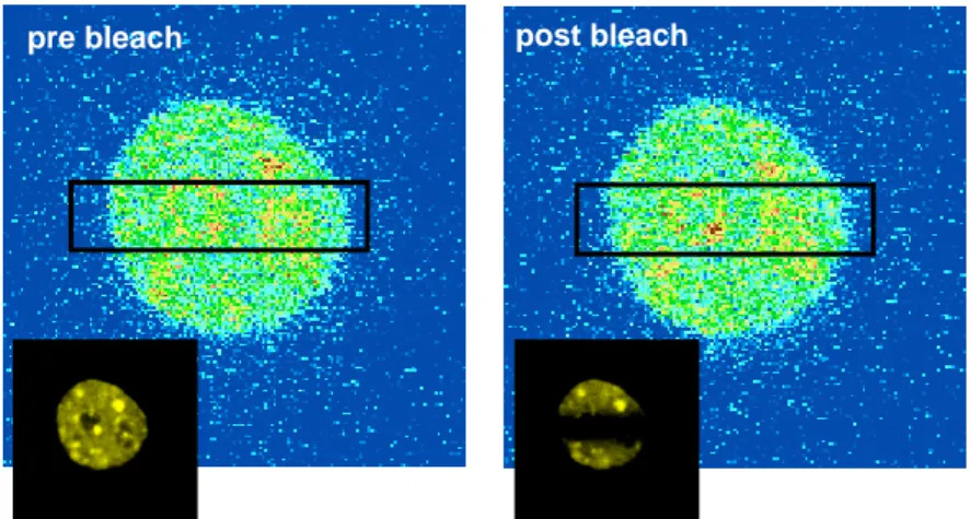
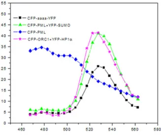
Visualization of direct Orc1p-HP1α interaction in human cells by Fluorescence Energy Transfer (FRET) ... 102
Orc1p focalization survives TSA and RNase A treatments... 105
Contents
4 ADDENDUM: FLUORESCENCE RESONANCE ENERGY TRANSFER (FRET) MICROSCOPY IMAGING OF LIVE CELL PROTEIN LOCALIZATIONS 110
4.1 FRET APPLICATIONS IN CELL BIOLOGY... 112
4.2 FLUORESCENCE RESONANCE ENERGY TRANSFER FROM CYAN TO YELLOW... 113
4.3 HP1Α/ORC1 IN VIVO INTERACTION BY ACCEPTOR PHOTOBLEACHING... 115
SPECIFIC INTERACTION OF THE RETINOBLASTOMA PROTEIN WITH ORC1 AND ITS RECRUITMENT TO HUMAN ORIGINS OF DNA REPLICATION .... 123
1 SUMMARY ... 123
2 RESULTS ... 123
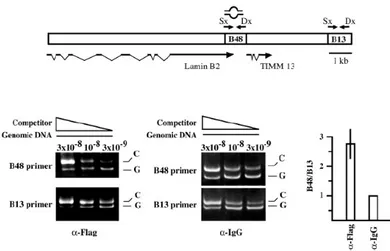
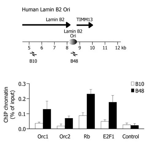
Rb and E2F1 proteins are recruited to human origins of DNA replication.... 123
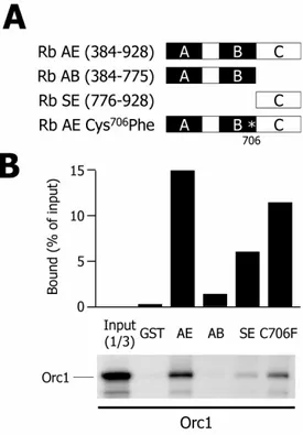
Orc1 specifically interacts with Rb in vitro ... 126
E2F1 competes with Orc1 for Rb-binding ... 132
Endogenous Orc1 forms a stable complex with hypo-phosphorylated Rb in human cells ... 134
Visualization of Orc1-Rb interaction inside the cells by Fluorescence Resonance Energy Transfer (FRET) ... 137
Orc1 and E2F1 are recruited to the lamin B2 origin at different temporal windows of G1 phase ... 141
Downregulation of Orc1 blocks cells in G1 and increase binding of ... 146
E2F-1 to origin DNA ... 146
3 DISCUSSION... 151
ACETYLATION OF HUMAN CDC6 BY GCN5 ACETYLTRANSFERASE REGULATES SITE-SPECIFIC, CDK-MEDIATED PROTEIN PHOSPHORYLATION IN THE S PHASE OF THE CELL CYCLE. ... 155
1 SUMMARY ... 155
2 RESULTS ... 156
Cdc6 associates with a nuclear HAT and is acetylated in vivo... 156
GCN5 acetyltransferase binds and acetylates Cdc6 in vitro and in vivo... 159
GCN5-mediated acetylation of Cdc6 affects Ser106 phosphorylation ... 171
GCN5 acetylates Cdc6 in early S-phase... 176
GCN5 and Cdc6 complex with Cyclin A/CKD2 in early S phase... 181
Acetylation and S106-phosphorylation of Cdc6 regulate its subcellular localization... 188
K3R mutations block cell cycle progression and stabilize Cdc6... 201
3 DISCUSSION... 205
INTRODUCTION
1 DNA replication initiation
The exact duplication of a genome once per cell division is required for every proliferating cell. To achieve this goal, eukaryotes adopt a strategy that limits every replication origin to a single initiation event within a narrow window of the cell cycle by temporally separating the assembly of the pre-replication complex (pre-RC) from the initiation of DNA synthesis (Lei and Tye, 2001).
Eukaryotic genomes are very large and the process of DNA replication is restricted to the S phase of the cell cycle: as a consequence replication must start at thousands of different chromosomal locations that are specifically selected; these sites are referred to as origins of DNA replication. The initiation of DNA replication must be strictly controlled to ensure that DNA is replicated once and only once per cell cycle. Therefore, origins of DNA replication are the key points to understand the cell cycle controls that are imposed on the process of DNA replication.
The first essential event in the initiation of DNA synthesis is the local opening of the duplex to provide access to the template strands. Origins of replication serve to increase the efficiency of the replication process by providing loci for the assembly of multi-protein complexes that mediate DNA synthesis.
In the original replicon model proposed over 40 years ago, Jacob, Brenner and Cuzin postulated the existence of two important elements required for replication initiation: the replicator and the initiator (Jacob and Brenner, 1963). The replicator is where replication starts, namely the cis-acting sequence within the genome, whereas the initiator is what binds the replicator, the positive trans-acting factor able to recognize a specific sequence of the genome that overlaps with the replicator. In response to the appropriate cellular signals, the initiator directs the local unwinding of chromatin and recruits additional factors to initiate the process of DNA replication.
Introduction
Compared to the simpler prokaryotic genomes, eukaryotic DNA replication is much more complex as genomes are larger and cell growth and differentiation have to be coordinated within a complex, developing, multi-cellular organism (Huberman, 1995). The main difference between prokaryotic and eukaryotic replication origins consists in the way in which chromosomal DNA is synthesized. In the latter case, in fact, genomes are larger and the duplication of chromosomal DNA relies on the activity of many different origins, the activation of which has to be strongly coordinated in space and time (DePamphilis, 1993; Kornberg, 1991). The advantage of this mechanism, besides reducing the overall time required to duplicate the entire genome, is that the generation of single-stranded DNA is much more localized and transient, helping preserving the genome integrity (DePamphilis, 1993). The initiation of DNA replication is mediated by a complex protein machinery that is assembled at each replication fork and that acts in concert to unwind the parental strands and carry out simultaneous synthesis of the two progeny strands (Bell and Dutta, 2002; Diffley, 1992; Diffley et al., 1995). The overall situation concerning the regulation of DNA replication in eukaryotic genomes appears to be far more complex than that in bacteria or DNA viruses, where replication occurs starting from a single origin. The initiation of DNA replication in lower eukaryotes is similar to that observed in bacteria in that it occurs at well-defined, site-specific origins of DNA replication that are recognized by specific initiator proteins (DePamphilis, 1993). However, although in the unicellular yeast S. cerevisiae the genome is duplicated from 250 to 400 replication origins, defined site-specifically, higher eukaryotic systems are expected to harbor a number of origins that is at least 100 times higher, and, at present, no sequence specific replicators have been found (DePamphilis, 1999; Gilbert, 2001; Todorovic et al., 1999). Nonetheless, in both simple and complex eukaryotes, replication origins are activated at each cell cycle, driving the replication of a limited region of the
Introduction
replication function that does not depend on the origin sequence itself (Gerbi and Bielinsky, 2002; Gilbert, 2004; McNairn and Gilbert, 2003; Mechali, 2001; Pasero and Gasser, 2002).
1.1 The pre-Replication Complex (pre-RC)
Initiator proteins have been identified and extensively characterized during the past 15 years, since the Origin Recognition Complex (ORC) was first isolated in yeast cells (Bell and Stillman, 1992). Subsequently, the Cdc6 protein and the minichromosome maintenance (MCM) protein complexes were isolated (Koonin, 1993; Zhou and Jong, 1993) and altogether these factors have been shown to be evolutionary conserved in metazoans (Cocker et al., 1996; Fujita et al., 1999; Gavin et al., 1995; Kearsey and Labib, 1998; Saha et al., 1998; Tugal et al., 1998). More recently, a new member of the pre-RC has been isolated, CDT-1, and also found in all the different eukaryotes analyzed (Devault et al., 2002; Maiorano et al., 2000; Nishitani et al., 2000; Tada et al., 2001; Wohlschlegel et al., 2000). The conservation of all these factors in metazoans corroborates the notion that their function is required for origin activity in all the different organisms. A current model for the process of initiation of DNA replication is the following. Starting from late mitosis, the ORC, Cdc6, CDT-1 and other proteins cooperate to load the MCM proteins onto chromatin to form licensed pre-replication complexes (pre-RCs) at sites that have the potential to become origins of DNA replication (Bell and Dutta, 2002; Lei and Tye, 2001; Takisawa et al., 2000). At the beginning of the S phase, cyclin-dependent kinases (CDKs) and the Cdc7 kinase (also named Dbf4-dependent kinase, DDK) cooperate to signal initiation of DNA replication at a subset of the pre-RCs, mediating the unwinding of the double helix at the origin and the recruitment of additional essential factors responsible for the synthesis process (Blow and Hodgson, 2002; Diffley, 2001; Diffley, 2004). The ordered recruitment or the activation of these proteins is believed to be responsible for controlling the process of initiation of DNA replication in terms of both space and time, as well as their subsequent inactivation or removal is believed to be necessary to prevent re-replication
Introduction
during a single S phase. For this reason, initiator proteins are crucial in regulating origin activity.
1.1.1 Origin Recognition Complex (ORC)
The Origin Recognition Complex (ORC) is a six-subunit complex (Orc 1-6) that acts as the initiator, likely selecting the sites for subsequent initiation of replication at eukaryotic origins of replication. First identified in S. cerevisiae as the ARS ACS binding factor (Bell and Stillman, 1992), it was subsequently found to be a very conserved element of chromosomal replication in all eukaryotes. Studies in Xenopus egg extracts demonstrated that the Xenopus analogue XlORC is required for initiation of replication in this organism (Carpenter et al., 1996). Similarly, recessive mutations in multiple Drosophila ORC subunits were shown to give rise to lethal phenotypes (Austin et al., 1999). In humans, although isolated a few years ago (Gavin et al., 1995; Tugal et al., 1998) and shown to be required for replication activity from the OriP of Epstein-Barr virus transfected in human cells (Chaudhuri et al., 2001; Dhar et al., 2001; Schepers and Diffley, 2001), hsORC has been only recently shown to be directly involved in the initiation of DNA replication (Abdurashidova et al., 2003; Ladenburger et al., 2002; Mendez et al., 2002; Todorovic et al., 2005). ORC drives the formation of the pre-RCs at replication origins, and one of its best-characterized features is its ability to bind DNA. In the yeast model, the ORC complex marks the origin throughout the cell cycle, binding to specific sites that map within the A and B1 elements of the yeast origin, spanning a region of ∼30 bp (Bell et al., 1993; Micklem et al., 1993; Rao and Stillman, 1995). Conversely, in higher eukaryotes the ORC-DNA interaction is still unclear. Both in vivo and in vitro studies indicate that ORC is present at origins of replication (Kreitz et al., 2001; Mendez et al., 2002; Natale et al., 2000; Okuno et al., 2001). In Drosophila, ORC binds both
Introduction
the fly Sciara coprophila (Bielinsky et al., 2001). Chromatin immunoprecipitation (ChIP) studies demonstrated the association of hsORC with the OriP of the Epstein-Barr virus (Chaudhuri et al., 2001; Dhar et al., 2001; Schepers and Diffley, 2001). More recently, some authors have shown its interaction with human replication origins (Abdurashidova et al., 2003; Ladenburger et al., 2002; Todorovic et al., 2005). In contrast to the yeast model, some of the members of the ORC complex are believed to be displaced from the origin site after initiation of DNA replication (Kreitz et al., 2001) suggesting a more dynamic interaction between mammalian ORC and origin DNA. Both Xenopus and Drosophila ORC cannot be extracted as a stable complex (Natale et al., 2000; Thome et al., 2000). Biochemical studies have demonstrated that human ORC is formed by a core sub-complex of 4 subunits, Orc2-5 (Dhar et al., 2001; Vashee et al., 2001). Recent studies in mammalian cells suggest that not all ORC subunits remain tightly associated as part of the complex throughout the cell cycle (Bell and Dutta, 2002). Unlike ORC from budding yeast, Drosophila, and Xenopus, the subunits of the SpORC and mammalian ORC are difficult to extract as a stable complex (Moon et al., 1999; Natale et al., 2000; Thome et al., 2000). For example, SpOrc4p is retained on chromatin under conditions that elute the remainder of SpORC (Moon et al., 1999). Similarly, whereas mammalian Orc2p is found constitutively on the chromatin, mammalian Orc1p is removed from the chromatin at the end of S phase and rebinds only as cells re-enter G1 (Kreitz et al., 2001; Natale et al., 2000; Tatsumi et al., 2000). Studies in Homo sapiens
suggest that Orc1 may be proteolyzed during S phase as a mechanism to prevent re-replication (Kreitz et al., 2001; Mendez et al., 2002); however, other studies have found HsORC1p to be stable throughout the cell cycle (Okuno et al., 2001; Saha et al., 1998) (T. Kelly, personal communication in (Bell and Dutta, 2002)). Yet another study has observed that Hamster Orc1p is stable through the cell cycle but is regulated in its association with chromatin by cell cycle regulated ubiquitination (Li and DePamphilis, 2002). These substantial differences are unlikely to be due to simple technical differences but instead might indicate variations in the regulation of this key factor in different cell lines. In S. cerevisiae, ORC binding to DNA requires the Orc1-5 subunits, four of which
Introduction
(1,2,4 and 5) contact DNA directly. The Orc6 subunit does not seem to be required for DNA binding, but it is essential for replication (Lee and Bell, 1997). In S. pombe, ORC-origin binding is mediated uniquely by Orc4, which is able to recognize and bind specifically AT-rich sequences through its AT-hook DNA binding motif (Kong and DePamphilis, 2001; Kong and DePamphilis, 2002). The specificity of mammalian ORC binding to DNA is very low, due to its limited ability to distinguish specific sequences, as more recently reported (Remus et al., 2004; Schaarschmidt et al., 2004; Vashee et al., 2003). Moreover, the difficulties in identifying well-defined ORC binding sites in species other than yeast raise the possibility that other DNA binding factors may contribute and facilitate ORC localization and origin selection. In support to this hypothesis are some results obtained in Drosophila, where ORC has been shown to interact with the transcription factor E2F-1 and the disruption of such interaction reduced chorion amplification (Asano and Wharton, 1999; Royzman et al., 1999). ORC binding to DNA requires ATP. However, studies in both S. cerevisiae and
Drosophila indicate that only ATP binding and not its hydrolysis is required for DNA binding by ORC. ATP binding is mediated by the Orc1 subunit in both yeast and fly, an observation that suggests conservation of function through evolution (Austin et al., 1999; Chesnokov et al., 2001; Klemm et al., 1997). Recent work has shown that ssDNA stimulates ATP hydrolysis, suggesting that, once bound to the origin, ORC is retained in an ATP-bound state and that DNA unwinding stimulates its hydrolysis (Lee et al., 2000). Additional data indicate that ATP binding might be needed for Cdc6 interaction (Klemm and Bell, 2001).
The so-named "ORC cycle" is therefore the premier step in preventing rereplication of DNA during a single cell division cycle: the ORC not only selects the sites where prereplication complexes are assembled and DNA replication begins, it is the first in a series of multiple coherent pathways that determines when prereplication complexes are assembled. Data from yeast, frogs, flies and
Introduction
is not restored until mitosis is complete and a nuclear membrane is present. In yeast, frogs and mammals, the same cyclin-dependent protein kinase [Cdk1(Cdc2)] that initiates mitosis also inhibits assembly of functional ORC/chromatin sites. In yeast, ORC remains bound to chromatin throughout cell division, but in the metazoa either ORC or the Orc1 subunit appears to cycle on and off the chromatin (DePamphilis, 2005).
ORC functions go beyond DNA replication: recent findings show that the complex is able to promote the formation of transcriptionally silent, late-replicating, chromosomal domains (see paragraph 1.5 for details).
1.1.2 Cell division cycle 6 (Cdc6)
Identified in a screen for proteins involved in controlling cell cycle progression, Cdc6 is a member of the AAA+ ATPases protein family, strictly related to Orc1 and, to a limited extent, to Orc4, Orc5 and to the MCM2-7 proteins (Lee et al., 2000). Cdc6 is essential in the formation of pre-RC at origins of DNA replication (Cocker et al., 1996; Liang et al., 1995) and requires ORC to associate with DNA (Blow and Tada, 2000; Romanowski et al., 2000) and is in turn required for the association of the MCMs (Cook et al., 2002; Kearsey et al., 2000; Mendez and Stillman, 2000; Yanow et al., 2001). Periodic transcription of the yeast Cdc6 gene in rapidly proliferating cells starts late in mitosis and this correlates with the appearance of the Cdc6 protein (Hateboer et al., 1998). It is believed that Cdc6 is synthesized at this stage of the cell cycle because it contributes to the inactivation of CDKs at the end of mitosis to inhibit cyclin B-CDK complexes (Calzada et al., 2001; Cook et al., 2002; Tanaka et al., 1997; Weinreich et al., 2001).
The ATPase domains of human Cdc6 and Orc1 are critical for DNA replication: recent work has established that an artificial recruitment Cdc6 or Orc1 to a DNA sequence can create a functional origin of replication (Takeda et al., 2005). Moreover, Randell and colleagues have recently found that Cdc6 is an ORC- and origin DNA-dependent ATPase that functions at a step preceding ATP hydrolysis by ORC: inhibiting Cdc6 ATP hydrolysis stabilizes CDT-1 on origin DNA and
Introduction
prevents Mcm2-7 loading and, in contrast, the initial association of Mcm2-7 with the other pre-RC components does not require ATP hydrolysis by Cdc6 (Randell et al., 2006). Importantly, these coordinated yet distinct functions of ORC and Cdc6 ensure the correct temporal and spatial regulation of pre-RC formation. The initiation protein Cdc6, like other initiator factors, is post-translationally modified in a cell-cycle dependent manner thereby preventing re-replication events to occur (see 1.4 paragraph for details).
1.1.3 Cdc10-dependent transcript 1 (CDT-1)
Originally identified in S. pombe (Hofmann and Beach, 1994), CDT-1 has been recently shown to be a key element in the formation of the pre-RC and, moreover, in the regulation of the “once per cell cycle” replication feature. It is periodically expressed under the control of the transcription factor Cdc10, which also controls the expression of Cdc6 in different species (Hofmann and Beach, 1994). In S. pombe, CDT-1 was shown to be an essential factor for origin licensing, similar to Cdc6, as its over-expression alone or together with Cdc6 induces high levels of re-replication (Gopalakrishnan et al., 2001; Nishitani et al., 2000; Tada et al., 2001; Yanow et al., 2001). At the same time, Xenopus CDT-1 was shown to be required for origin licensing in terms of MCM protein loading (Gillespie et al., 2001; Maiorano et al., 2000). In both cases, CDT-1 associated to DNA in an ORC-dependent manner. CDT-1 can be found in all organisms (Blow and Tada, 2000; Rialland et al., 2002; Wohlschlegel et al., 2000). Its identification in S. cerevisiae is relatively recent (Hodgson et al., 2002; Tanaka and Diffley, 2002); in this organism, it is required for proper MCMs loading, therefore to form a complete, functional pre-RC (Takahashi et al., 2003). Despite the apparent redundancy in their roles, CDT-1 expression differs from that of Cdc6. In fact, the factor peaks during the second half of G1, being its expression somewhat delayed with respect to Cdc6 expression (Ballabeni et al.,
Introduction
second mechanism used by cells to control origin firing and prevent re-replication (Wohlschlegel et al., 2000). Geminin is a known inhibitor of DNA replication that acts by preventing MCM loading onto origins and displays its activity from S to M phase, thus preventing unwanted additional firing events (McGarry and Kirschner, 1998). Geminin was shown to interact with CDT-1 during the S phase, targeting it for degradation thereby preventing MCM loading until the following G1 and hence re-replication (Tada et al., 2001; Thomer et al., 2004; Wohlschlegel et al., 2000). CDT1 degradation, following ubiquitination, was shown to also occur in response to UV irradiation (Hu et al., 2004). In
Xenopus egg extracts, CDT-1 is the key feature preventing re-replication of DNA (Li and Blow, 2005). XCDT-1 is downregulated late in the cell cycle by two different mechanisms: proteolysis, which occurs in part due to the activity of the anaphase-promoting complex (APC/C), and inhibition by geminin.
Recently, some authors have reported that replication-dependent proteolysis of CDT-1 requires its interaction with proliferating cell nuclear antigen (PCNA), a homotrimeric processivity factor for DNA polymerases. Moreover, mutation of the PCNA-interaction motif yields a stabilized Cdt1 protein that induces re-replication. DDB1, a component of the Cul4 E3 ubiquitin ligase that mediates human CDT-1 proteolysis in response to DNA damage, is also required for replication-dependent CDT-1 destruction. Thus, PCNA functions as a platform for CDT-1 destruction, ensuring efficient and temporally restricted inactivation of a key cell-cycle regulator (Arias and Walter, 2006).
1.1.4 Mini-Chromosome Maintenance proteins (MCMs)
MCM proteins have also been found in all eukaryotic cells and represent the functional analogs of bacterial dnaC helicases (Tye, 1999). Discovered as important factors for the maintenance of plasmids in cells (Sinha et al., 1986), they play a key role in the cell cycle control of chromosome replication as they distinguish replication competent (licensed) chromatin during the G1 phase from replication-incompetent chromatin during the G2 phase and mitosis (Labib et al., 2001; Labib et al., 2000). Moreover, MCMs have been shown to be part of the
Introduction
active pre-RC, being loaded just before origin firing (Chong et al., 1995; Madine et al., 1995).
All eukaryotes appear to have exactly six MCM protein analogs, each one falling into one of the existing classes (MCM2-7). This observation argues, that each MCM protein has a unique and important function (Kelly and Brown, 2000). MCMs require the coordinate function of ORC, Cdc6 and CDT-1 to be loaded onto chromatin (Aparicio et al., 1997; Tanaka et al., 1997). Interestingly, once the MCMs have been loaded, the ORC and Cdc6 proteins can be displaced from chromatin without preventing replication initiation, suggesting that the primary role of these proteins is to load MCMs (Chong et al., 1995; Hua and Newport, 1998; Rowles et al., 1999). Moreover, in higher eukaryotes, in contrast to ORCs and Cdc6, MCMs do not appear to be displaced from origins as firing occurs and, by ChIP experiments, MCMs have been recently shown to localize with the replication machinery (Labib et al., 2001; Labib et al., 2000; Lee and Hurwitz, 2001; Zhou and Elledge, 2000). This implies that they play an active role as replicative helicases in both the initiation and the elongation processes. In S. cerevisiae, each MCM appears to be required for replication (Labib et al., 2000) whereas in higher eukaryotes only a subset of them, MCM4, MCM6 and MCM7, were shown to be required for DNA helicase activity (Ishimi and Komamura-Kohno, 2001; Ishimi et al., 2000; Schwacha and Bell, 2001; You et al., 2003). These same subunits were recently shown to display a preference for AT rich sequences, suggesting a possible role of these proteins in the recognition of the origin site (You et al., 2003). Recent work shows that the accumulation on chromatin of an additional member of the MCM protein family, human MCM8 (hMCM8), occurs during the early G(1) phase, before the hMCM2-hMCM7 complex binds. hMCM8 interacts in vivo with hCdc6 and hOrc2 resulting a crucial component for pre-RC assembly (Volkening and Hoffmann, 2005).
Introduction
early G1, involves the ordered assembly of the pre-replicative complex (pre-RC) at potential replication origins. The assembly of this multiprotein complex is initiated by the association of the Origin Recognition Complex (ORC1-6), which is required to recruit both Cdc6 and CDT-1 proteins that are then loaded on ORC-bound chromatin independently of one another (Blow and Tada, 2000). ORC, Cdc6 and CDT-1 are together required for the loading of the Minichromosome Maintenance (MCM2-7) proteins on the origins during G1. Extensive evidence supports a nucleotide-binding role of many of these factors. MCM2-7, Orc1/4 and 5 and Cdc6 have consensus motifs for nucleotide binding and mutations in these motifs result in nonfunctional proteins (Schepers and Diffley, 2001). Moreover, in vitro studies indicate that there are at least two ATP-requiring steps in pre-RC formation: ORC association with origins and the subsequent recruitment of Cdc6 and MCMs (Harvey and Newport, 2003; Klemm and Bell, 2001; Schwacha and Bell, 2001; Seki and Diffley, 2000). The loading of all these factors results in the origins becoming “licensed” for DNA replication in the subsequent S-phase.
The second phase (firing step) involves the activity of numerous other proteins or protein complexes that associate with some of the pre-RC marked origins prior to successful initiation of DNA synthesis. These proteins include regulatory factors as well as components of the DNA replication fork, such as Cdc7-Dbf4, Cdc28, MCM10 and cyclin-dependent kinases (CDKs), that modulate the activity of the chosen origin by loading the Cdc45 protein and inducing the initiation of a pair of replication forks (Bell and Dutta, 2002). Being the factors involved in the assembly of the pre-RC and in the replication process very conserved among different organisms, all models of metazoan replication are largely based on the yeast paradigm just described. Recent advances in DNA microarray technology have enabled eukaryotic replication to be studied at whole-chromosome and genome-wide levels . These studies, in both S. cerevisiae and higher eukaryotes, have provided new insights into the mechanisms that influence origin selection and the temporally co-ordinated activation of replication initiation from these sites (MacAlpine et al., 2004). The comprehensive nature of the microarray-based studies has revealed clear connections between chromosome organization
Introduction
and the pattern of replication. For example, in yeast, the centromeric proximal sequences are consistently early replicating and telomeric regions are consistently late replicating. The metazoan studies reveal a recurring theme of gene-dense transcriptionally active regions of the genome replicating before gene-sparse regions.
Figure 1. Assembly of replication-competent chromatin and the post-replicative state of origins. ORC, Cdc6, CDT-1 and MCM proteins bind to chromatin sequentially during G1 phase, licensing the DNA for replication. At the G1–S phase transition, pre-replicative complexes (pre-RCs) are activated and disassembled. ORC binds to DNA throughout the cell cycle in budding and fission yeasts, but in mammalian cells only some of the ORC subunits are tightly associated with chromatin at all stages. Cdc6 is phosphorylated and either
Introduction
However, the dynamic association of ORC and other replication proteins with origin DNA to form the pre-RC might be different in higher eukaryotes, especially given the lack of a target consensus sequence responsible for ORC-origin interaction (Gilbert, 2001; Gilbert, 2004; Mechali, 2001). As a matter of fact, the way in which these factors recognize or are recruited to the origin site is still to be uncovered (Blow and Tada, 2000; Cimbora and Groudine, 2001; Quintana and Dutta, 1999). Therefore, it can be concluded that the same function is performed by the same set of conserved factors in all eukaryotes while the mechanisms underlying these processes can vary depending on the organism.
1.3 Origin activity, cell-cycle progression and checkpoints
To ensure that each replication origins fires efficiently and only once per cell cycle, eukaryotic cells have evolved a remarkable molecular switch which, when turned on, promotes just a single initiation event from each origin per S-phase. The temporal separation of pre-RC assembly and origin activation steps is a key feature of the replication checkpoint that ensures that new pre-RC cannot assemble on origins which have already been fired (Diffley, 2001; Diffley, 2004). The heart of this mechanism is the tightly regulated assembly of the pre-RC complex; the activity of several cell-cycle regulated kinases is central to this regulation (Bell and Dutta, 2002). CDKs and DDKs (Dbf4-dependent kinases) are essential for triggering the initiation of DNA replication from origins that contain preassembled pre-RC. While DDKs seem to act on MCMs (Lei and Tye, 2001), CDKs appear to play a direct role in preventing the assembly of new pre-RCs. Because CDK activity remains high from S phase onset to the end of the following mitosis, re-licensing cannot occur until the beginning of the next cell cycle (Ballabeni et al., 2004; Diffley, 2004; Noton and Diffley, 2000; Tanaka and Diffley, 2002). At least three of the components of the pre-RC (ORC, Cdc6 and MCMs) are phosphorylated by CDKs to prevent re-replication and pre-RC assembly (Furstenthal et al., 2001; Lei and Tye, 2001). Moreover, CDKs have also been implicated in controlling the time of replication initiation at specific origins (Zou and Stillman, 1998). Cell cycle progression, as well as the control of
Introduction
genomic integrity, is under continuously surveillance by cell cycle checkpoints (Nyberg et al., 2002; Zhou and Elledge, 2000). It is thus conceivable that one of the steps at which these checkpoint act by blocking cell cycle progression is DNA replication origin activation. Given the complexity and the importance of the S phase for the maintenance of genome integrity, many different checkpoint pathways are clearly active within this window of time, as demonstrated by studies in the yeast model (Bartek et al., 2004). In addition, and most importantly, the fact that the induction of genotoxic stresses during S phase causes a delay but not an arrest implies that replication origins are differentially regulated depending on the time of their firing (Merrick et al., 2004). Experiments carried out in the Xenopus cell-free system indicated that one of the main consequences of the induction of a replication checkpoint is the regulation of the recruitment of key members of the pre-RC to the origin site. Double-strand breaks allowed the assembly of complete pre-RCs, but prevented Cdc45 interaction with pre-RC in an ATM- and/or ATR- dependent, but Mre11-independent fashion (Costanzo and Gautier, 2003; Costanzo et al., 2001; Costanzo et al., 2000). In budding yeast, HU treatment blocked forks progression from early-origins and prevented the firing of late-origins, and this mechanism was shown to depend on Rad53 and Mec1, homologs of human ATM and Chk2 (Santocanale and Diffley, 1998). The same conclusion was obtained also following induction of double-strand breaks, and the protein involved in this regulation was shown to be yeast Orc2 (Shirahige et al., 1998). Recently, the
Drosophila CDT-1 protein was reported to be phosphorylated and degraded in a Cyc E-Cdk2 dependent fashion, thereby preventing rereplication (Thomer et al., 2004). Moreover, also Cdc6 was recently reported to be directly involved in the control of rereplication (Mimura et al., 2004). Altogether, these data suggest the same factors involved in the formation of the pre-RC are also involved in the regulation of the replication process at different stages during the cell cycle,
Introduction
1.4 CDK-dependent regulation of human Cdc6 protein
As introduced in the previous paragraph, prior to cell division, the eukaryotic genome is duplicated during S phase of the cell-cycle. To ensure that only one single round of DNA replication occurs per cell cycle, a strict regulation is imposed. In G1/S phase, preRCs are assembled at origins of replication. During G1 the protein levels of both Cdc6 and CDT-1 increase, and they bind to the ORC complex. Since Cdc6 and CDT-1 are essential for the subsequent loading of the helicase MCM complex, these factors are thought to license the cell for replication. It has been established that not all origins are activated at the same time in S-phase. Some origins appear to be activated in early S phase, which has been shown to correlate with active transcription, whereas other origins are activated late in S-phase. Therefore, in the existing model preRCs are individually activated and fired (Duursma and Agami, 2005b). This firing is thought to be executed through phosphorylation of the preRC components by cyclin dependent kinases (CDKs) and the Cdc7-Dbf4 kinase (Lei and Tye, 2001). Apart from its role in activating DNA replication, CDK phosphorylation inhibits the formation of new replication complexes and inactivates components of the fired preRCs. Only at the end of mitosis CDK activity decreases due to degradation of mitotic cyclins, which allows new preRC formation in the G1 phase of the next cell cycle.
How exactly CDK activity both activates the initiation of replication at origins and inhibits the firing of origins that have already been activated is at present not clear. It has been proposed that the level of CDK activity is the primary determinant of this “replication switch” (Jallepalli and Kelly, 1997). Low kinases levels would be sufficient to trigger origin activation, whereas high CDK activity would result in disassembly of preRCs and inhibition of new preRC formation. However, this model would have the risk of aberrant regulation at intermediate levels of CDK activity.
Another possibility would be that the specific phosphorylation of replication proteins determines whether the origin is activated or inhibited. Whether these proteins are phosphorylated or not depends on their availability or their
Introduction
accessibility. In this model, E3-ubiquitin ligase proteins like the anaphase promoting complex (APC) and the Skp1-cullin-F box SCF complex could play a central role in determining the availability of the initiation proteins by regulating their cell cycle dependent destruction.
At present several proteins of the mammalian preRC have been described to be phosphorylated by CDKs and as a result degraded or inactivated. First, human Orc1 was shown to be phosphorylated by cyclin A/CDK2 and degraded in a SCFSkp2-dependent manner (Mendez et al., 2002). Second, it was demonstrated
that cyclin A-dependent CDT1 phosphorylation in S-phase induces its SCFSkp2
-dependent degradation (Li et al., 2003; Liu et al., 2004; Sugimoto et al., 2004). Notably, CDT1 activity remained regulated by geminin in G2/M and G1-phase of the cell cycle. Third, CDK phosphorylation decreases the helicase activity of the MCM-complex. However, phosphorylation of an initiation protein by CDK2 that is essential for activation of DNA replication has not been revealed.
Intriguingly, in addition to Orc1, CDT1 and the MCM proteins also the licensing protein Cdc6 was previously recognized as a CDK target. Cdc6 phosphorylation by Cyclin-A/CDK2 was described to occur in S-phase and to result in its translocation from the nucleus to the cytosol and subsequent degradation (Delmolino et al., 2001; Herbig et al., 2000; Jiang et al., 1999; Petersen et al., 1999; Saha et al., 1998). However, these studies were all performed with ectopically expressed and tagged wild-type Cdc6 or Cdc6 that had been mutated in several phosphorylation sites. Later on, this model has been challenged by the finding that only ectopically expressed Cdc6 or the soluble endogenous form are translocated to the cytosol, whereas the chromatin-bound form persist through S and G2 phases (Coverley et al., 2000; Mendez and Stillman, 2000). Notably, Cdc6 phosphorylated on serine 54 was also shown to remain chromatin bound in S-phase (Alexandrow and Hamlin, 2004).
Introduction
APCCdh1 mediated destruction. Moreover, in line with these results, Mailand and
colleagues showed that phosphorylation of Cdc6 prevents its Cdh1-dependent ubiquitination (Mailand and Diffley, 2005). The fact that Cdc6 stability is controlled by CDK2 implies regulation through the p53 pathway in stress responses. DNA damage induces stabilization and activation of the p53 transcription factor, which results in increased synthesis of the CDK inhibitory protein p21cip1 (Fei and El-Deiry, 2003). Morevoer, enhanced Cdc6 destruction
was observed following DNA damage in a p53 and p21cip1-dependent manner;
Cdc6 is regulated in a p53-dependent manner in non stressed cells (Duursma and Agami, 2005a).
Both Cdc6 and CDT1 are the licensing factors of DNA replication. Therefore, this key step in initiation of DNA replication appears to be regulated by several independent pathways. Whereas the abundance of Cdc6 protein in G1/S phase is positively regulated by cyclin E/CDK2 phosphorylation through protecting it from APCCdh1-dependent degradation, CDT1 is negatively regulated during
S-phase by Cyclin A phosphorylation in a SCFSkp2-dependent manner (Li et al.,
2003; Liu et al., 2004; Sugimoto et al., 2004).
However, this model is more complex since CDT1 activity is also inhibited by geminin (Sugimoto et al., 2004). As both Cdc6 and geminin (McGarry and Kirschner, 1998) are regulated by the APC a paradox emerged. How is the APC-dependent destruction of both an activator (Cdc6) and an inhibitor (geminin) of DNA replication coordinated to ensure that Cdc6 and CDT1 are present in the same time-frame to allow efficient preRC assembly? Based on the last results, a model for the assembly of the preRCs has been proposed (Fig. 2). Geminin is degraded in an APC-dependent manner at the end of G2/M and in early G1 phase of the cell cycle. Due to the degradation of geminin the levels of Cdt1 will rise during G1. Cdc6 transcription is regulated by E2F transcription factors (Hateboer et al., 1998; Yan et al., 1998), hence Cdc6 transcription increases in G1 phase. Yet, Cdc6 protein levels will only be stabilized in the course of G1 as the activity of CDK2/Cyclin E increases.
Introduction
Figure 2. A schematic model of the regulation of DNA replication licensing in G1 phase of the cell cycle. Licensing of replication origins occurs in a time-frame where geminin is degraded by the APC, resulting in active CDT-1, and Cdc6 is protected from APC-dependent degradation by CDK2/cyclin E phosphorylation of serine 54 (Duursma and Agami, 2005b).
Phosphorylation of Cdc6 at serine 54 will protect Cdc6 from APCCdh1-dependent
destruction and the protein is allowed to accumulate. This provides the cells with a period of time in which both licensing factors CDT-1 and Cdc6 are present. Binding of these proteins to the ORC complex at origins results in recruitment of the MCM-complex and formation of a preRC. Thus, phosphorylation of Cdc6 serine 54 could be the primary determinant of the timing of preRC formation at G1/S transition. Another question that arises is whether the CDK2-dependent phosphorylation of Cdc6 not only stabilizes the protein but also plays a role in recruitment of the MCM-complex. Interestingly, defective MCM loading was observed in mouse embryonic fibroblasts (MEFs) lacking both cyclin E1 and E2. Cells that reentered the cell cycle from quiescence showed chromatin bound
Introduction
de depicted whereby phosphorylation of Cdc6 plays a crucial role in MCM recruitment to the chromatin.
The regulation of DNA replication initiation in mammalian cells is based on the finding that Cdc6 is stabilized by CDK phosphorylation. As shown in the model (Fig. 3), Cdc6 phosphorylation was sufficient to load MCM6 onto the chromatin. Cdc6 is a key CDK2/cyclin E target in the replication complex for activation of origins in G1-phase. Stabilization of Cdc6 by phosphorylation together with the destruction of geminin results in binding of both Cdc6 and CDT-1 to the ORC complex at origins of replication leading to the loading of the MCM-complex. Next, independent of further CDK activity, the MCM complex recruits Cdc45, which in turn recruits DNA polymerase. Thus, opposing the hypothesis that CDK-dependent phosphorylation of MCM proteins results in origin firing, some authors have proposed that CDK activity is crucial for activating replication origins by phosphorylating Cdc6 (Duursma and Agami, 2005b).
Figure 3. A proposed model for the regulation of initiation of DNA replication. Geminin and Cdc6 are both degraded by the APC, until Cdc6 is stabilized by CDK2/ Cyclin E activity in course of G 1 phase. This is the step in origin firing that requires CDK activity. Once Cdc6 and CDT-1 license the chromatin, the MCM-complex will be recruited. The subsequent loading of Cdc45 and DNA-polymerase a occur in a CDK independent manner, but what is required for this loading remains to be determined (Duursma and Agami, 2005b).
Introduction
This model is consistent with the fact that all CDK-phosphorylated MCM proteins identified until now are negatively regulated by this modification (Hendrickson et al., 1996; Ishimi, 1997). However, this hypothesis disagrees with the suggested requirement of CDK activity between the assembly of MCM in the preRC and the loading of Cdc45, which was mainly based on studies in yeast and in vitro
studies with Xenopus egg extracts.
In yeast it was shown that association of Cdc45 with chromatin correlated with activation of S-phase CDK activity at G1/S transition (Zou and Stillman, 1998). Nevertheless, chromatin binding of yeast Cdc45 in G1 was reported by others (Aparicio et al., 1997). Further, in vitro experiments with Xenopus egg extracts and sperm chromatin showed that addition of p21cip1 or p27kip1 could block Cdc45
in mammalian cells is lacking and therefore interesting to be determined.
CDKs were also proposed to fire individual origins, resulting in early and late replicating origins. Therefore, if CDKs do not play a role in activating individual preRCs, what then determines the activation of individual origins at different time-points during S-phase? One possibility is the activity of Cdc7-Dbf4 kinase which was shown to phosphorylate MCM2 and to be required for activation of DNA replication in mammalian cells (Jiang et al., 1999). Moreover, it has been proposed that this kinase acts locally at individual origins (Jares et al., 2000). Therefore, it would be interesting to find out what determines the activation of individual replication origins and whether the Cdc7-Dbf4 kinase is involved. Recently, initiation of DNA replication has been found to be regulated by p53 through Cdc6 stability (Duursma and Agami, 2005a). Indeed, Cdc6 has been identified as a novel target of the p53 pathway. Activation of tumor suppressor p53 in response to genotoxic stress imposes cellular growth arrest or apoptosis. The authors show that p53 activation by DNA damage results in enhanced Cdc6 destruction by the anaphase-promoting complex. This destruction is triggered by inhibition of CDK2-mediated Cdc6 phosphorylation at serine 54. Conversely,
Introduction
Figure 4. Schematic model depicting the p53 pathway that regulates Cdc6, and thereby S-phase entry, both under normal tissue culture conditions (solid lines) and following ionizing irradiation (IR, dashed lines) (Duursma and Agami, 2005a).
1.5 ORC and gene silencing
Histone modifying enzymes, chromatin-remodelling complexes and DNA methylation are thought to be components of intricate epigenetic mechanisms that help compact and organize genomes into discrete chromatin domains (Goll and Bestor, 2005; Jenuwein and Allis, 2001). This organization also underlies many aspects of chromosome behaviour, such as transcription, recombination and DNA repair (Kosak and Groudine, 2004).
A key feature of heterochromatin is its ability to propagate, and thereby influence gene expression in a region-specific, sequence-independent manner. When heterochromatin spreads across domains, it generally causes epigenetic repression of nearby sequences, in a process that is referred to as silencing (Grewal and Jia, 2007).
Introduction
Although epigenetic gene silencing has become almost synonimous with heterochromatization, there are several reports in the literature in which heterochromatin formation is required for activation of gene expression (Lu et al., 2000; Weiler and Wakimoto, 1995; Yasuhara and Wakimoto, 2006). Histone H3 methylated at lysine 9 (H3K9me) and the heterochromatin protein HP1, which are necessary for the formation of heterochromatin, have been found in association with a subset of transcribed genes (Greil et al., 2003; Piacentini et al., 2003). Furthermore, it has been shown that heterochromatin proteins recruit factors that facilitate the access of RNA polymerase II (Pol II) to heterochromatin loci (Zofall and Grewal, 2006). An important emerging theme is that heterochromatin provides a mechanism for the recruitment and spreading of regulatory proteins (effectors) that are implicated in different aspects of chromosome biology. Although these effectors can be targeted to individual loci in a sequence-specific manner, the ability of heterochromatin to spread provides a sequence-independent platform to allow recruitment of these effectors at the level of chromatin domain. This might facilitate coordinated control of loci that are otherwise incapable of recruiting effectors themselves. From an evolutionary point of view, the current multipurpose character of heterochromatin might represent a series of co-optation events. Indeed, although all eukaryotes use epigenetic silencing mechanisms, different lineages have emphasized different aspects of heterochromatin in regulation, depending on the chromosomal contexts. It is therefore provocative, but non entirely unexpected, to find that many of the same histone modifications and proteins that are required to assemble silent heterochromatin structures are, in other circumstances, instead essential for gene activation. Multiple pathways of histone modifications and DNA methylation in higher eukaryotes contribute to how heterochromatin is assembled (Goll and Bestor, 2005; Jenuwein and Allis, 2001; Maison and Almouzni, 2004).
Introduction
2001; Nasmyth et al., 1981; Rine and Herskowitz, 1987; Strahl-Bolsinger et al., 1997) (Fig. 5), and the HP1 and Swi6 complexes that mediate silencing in metazoans and fission yeast, respectively (Aagaard et al., 1999; Eissenberg and Elgin, 2000; Eissenberg et al., 1990; Jenuwein, 2001; Kellum, 2003; Platero et al., 1995) (Fig. 6). Despite the divergence of molecular components, mechanisms of heterochromatic gene silencing in budding yeast, fission yeast,
Drosophila and mammals are similar (Moazed, 2001) (Fig. 7).
Figure 5. Model for Step-Wise Assembly of Silent Chromatin in budding yeast. Telomere binding proteins, the yKu70/yKu80 heterodimer and Rap1, ADP-ribose recruit the Sir2/Sir4 complex to DNA (step 1). Following deacetylation of histone tails by Sir2 (step 2), the Sir3 protein is recruited via interactions involving Rap1, Sir4, and histone tails and binds to nucleosomes (shown as purple ovals) by interacting with the deacetylated histone tails (step 2). Multimerization of Sir3 and Sir4 then results in additional rounds of modification and binding, and spreading of the complex along nucleosomes (step 3). Ac, acetyl group on amino-terminal lysines of histones; ADPRAc, O-acetyl-ADP-ribose (Moazed, 2001).
Introduction
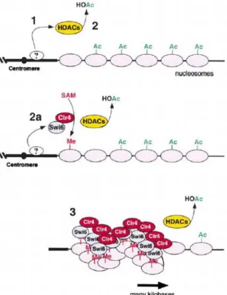
Figure 6. Model for step-wise assembly of silent chromatin domains in fission yeast. Following recruitment to DNA by protein(s) that have not yet been identified (step 1), histone deacetylases (HDACs, Clr3, and Clr6) deacetylate histone tails (step 2). The H3-specific methyltransferase, Clr4, then methylates lysine 9 of H3 and creates a binding site for the Swi6 protein (step 2a). Self-association of the Swi6 protein and subsequent rounds of modification and binding result in the spreading of the complex along nucleosomal DNA for several kilobases (step 3). Model adapted from Nakayama et al. (2001). Similar models have been proposed for mammalian HP1/SUV39H1 assembly (Bannister et al., 2001; Lachner et al., 2001). SAM, S-adenosyl-methionine; Me, methyl group on lysine 9 of histone H3; HOAc, acetate (Moazed, 2001).
Introduction
Figure 7. The Swi6/HP1 silencing complex is conserved in the fission yeast, S. pombe, and metazoans. The human HP1β protein has been shown to be associated with the methyltransferase protein SUV39H1. By analogy, Swi6 and the Drosophila HP1 are represented in association with SUV39H11 homologs Clr4 and Su(var)3-9; these interactions are strongly supported by genetic and colocalization experiments, but physical evidence for the association of Swi6 with Clr4 or HP1 with Su(var)3-9 is lacking. In S. pombe, HDACs and the zinc finger protein Rik1 are required for the association of Swi6 with silent chromatin, but it is unknown whether Rik1 and HDACs are physically associated with Swi6 or Clr4 (Moazed, 2001).
Originally identified in Drosophila melanogaster, HP1 belongs to a highly conserved family of chromatin proteins, with homologues that are found from fission yeast (Swi6, Chp2 and Chp1) to humans (HP1 α, HP1 β and HP1 γ) (Huisinga et al., 2006). These proteins contain an amino-terminal chromodomain, a short variable hinge region and, with the exception of Chp1, a chromoshadow domain. Each HP1 protein interacts with diverse factors that are involved in different aspects of heterochromatin structure and function. The diversification of HP1 isoforms is also indicated by their distinct localization patterns. Whereas HP1 α and HP1 β are distributed mainly at pericentric chromatin domains, HP1 γ is localized to discrete euchromatic sites (Huisinga et al., 2006). The binding of HP1 proteins to chromatin is believed to be highly dynamic (Cheutin et al., 2003; Festenstein et al., 2003). Histones and their modifications have crucial roles in the formation of heterochromatin (Jenuwein and Allis, 2001). Heterochromatin has a characteristic histone-modification profile, which is distinguished by hypoacetylation and H3K9 methylation; euchromatin is characterized by histone H4 acetylation and methylation of histone H3 at lysine 4 (H3K4me) (Cam et al., 2005; Grunstein, 1998; Litt et al., 2001; Nakayama et al., 2001; Noma et al., 2001). Histone methylation serves as a ‘molecular anchor’ recruiting proteins that either directly modify chromatin or
Introduction
recruit others that do so (Martin and Zhang, 2005). Given the multimerization of Swi6/HP1 through the chromoshadow domain (Brasher et al., 2000; Cowieson et al., 2000), and the ability of swi6/HP1 to bind to numerous proteins that are implicated in heterochromatin formation, including histone deacetylases (HDACs) (Lechner et al., 2005; Smothers and Henikoff, 2000; Yamada et al., 2005; Zhang et al., 2002a), it has been suggested that Swi6/HP1, when bound to methylated H3K9, serves as an assembly platform for chromatin-modifying factors that are involved in stabilization (maintenance) and spreading of heterochromatin (Hall et al., 2002; Yamada et al., 2005).
Evidences accumulated in the last few years indicate that the function of ORC extends beyond DNA replication. The N-terminal region of the largest subunit of ORC, the Orc1p, is required for transcriptional silencing at the HM loci but is dispensable for DNA replication (Fig. 8).
Figure 8. Molecular model for silencing at HM mating-type loci. Silencing is initiated by DNA-binding proteins [Rap1p and origin recognition complex (ORC), or Rap1p and Ku] that cooperate to recruit SIR protein complexes. These histone-interacting SIR complexes then assemble along adjacent nucleosomal
Introduction
The N-terminal silencing domain of Orc1p (BAH domain) shares ~50% amino acid identity with the N-terminal region of Sir3p (Bell et al., 1995). Unlike Sir3p, however, the Orc1p domain interacts with Sir1p (Triolo and Sternglanz, 1996) and in Drosophila, the corresponding region of dORC1 interacts with HP1 (Pak et al., 1997).
The clearest example of an alternate function is the role of ORC in the transcriptional repression of the silent mating type loci, HMR and HML, in S. cerevisiae (Fig. 8). Paradoxically, in this case ORC participates in the assembly of the chromatin conformation that eventually suppresses the activity of the origin (Vujcic et al., 1999).
The ability of ORC to promote the formation of transcriptionally silent, late-replicating, chromosomal domains has been recently demonstrated also for the yeast rDNA locus by means of the dynamic molecular combing technique (Pasero et al., 2002). In both cases, transcriptional silencing and origin inactivation depend on the interaction of ORC with Sir proteins. The ability of ORC to interact with heterochromatin markers and to direct their recruitment to silent portions of the genome seems to be evolutionary conserved. However, there is no evidence so far that this holds true also in mammalian cells. In
Xenopus and in Drosophila, Orc1p binds heterochromatin protein 1 (HP1), which is functionally analogous to Sir1p of budding yeast. In vitro studies have shown that both the yeast and the Drosophila Orc1p interact with Sir1/HP1 through their N-terminal portion (Pak et al., 1997; Zhang et al., 2002c).
This region overlaps the BAH domain that Orc1 proteins share with Sir3p and other chromatin associated proteins such as DNA methyl-transferases. Collectively these findings support the hypothesis that ORC could be involved also in the establishment and maintenance of chromatin domains.
Shareef and colleagues have identified a novel component of the HP1/ORC complex in Drosophila, named the HP1/ORC-associated protein (HOAP, Fig. 9) (Shareef et al., 2001). HOAP contains similarity to DNA sequence-specific HMG proteins and is shown to bind specific satellite sequences and the telomere-associated sequence in vitro. The protein is shown to have heterochromatin
Introduction
localization in both diploid interphase and mitotic chromosomes and polytene chromosomes. Moreover, the gene encoding HP1/ORC-associated protein was found to display reciprocal dose-dependent variegation modifier phenotypes, similar to those for mutants in HP1 and the ORC2 subunit.
Figure 9. Model for Drosophila heterochromatin assembly. The DNA-binding activities of ORC and the HP1/ORC-associated protein (HOAP) are proposed to recruit underphosphorylated HP1, followed by (or concomitant with) recruitment of histone deacetylation activities (HDAC) and Su(var)3-9 methyltransferase and highly phosphorylated HP1 (Shareef et al., 2003).
An interesting observation concerns the association of Orc1p with heterochromatin in mid and late G1. The association of ORC with transcriptionally silenced, late replicating portions of the genome has been observed in other organisms, where ORC seems to play an active role in the assembly of these chromatin conformations. In the yeast S. cerevisiae, ORC binding to an ARS element is required for the recruitment of Sir factors and hence for the transcriptional silencing of the HML locus (Vujcic et al., 1999). In
Drosophila mutations of Orc2p were shown to perturb HP1 localization (Huang et al., 1998; Pak et al., 1997). Finally, it has been reported that human Orc2p binds in vivo to α-satellite sequences that compose pericentric heterochromatin (Keller et al., 2002).
Introduction
that Swi6 recruits this kinase complex to activate replication origins within heterochromatic regions.
Introduction
2 The E2F/Rb complex
Transition through the mammalian cell cycle requires an interplay of transcription factors that coordinately induce or repress gene expression in a temporally defined manner.
The E2F transcription factor is known to play a pivotal role in mediating gene expression during cell proliferation (Takahashi et al., 2000). E2F activity consists of a heterodimer containing one of six factors (1, 2, 3, 4, E2F-5, and E2F-6) that pairs with a second subunit (DP-1 or DP-2) (Dyson, 1998). The transcriptional activation potential of E2F is counterbalanced by the retinoblastoma tumor suppressor protein (pRB) with which E2F tightly associates. E2F is under the control of the Rb (Flemington et al., 1993; Grana et al., 1998; Helin et al., 1993) and of CREB Binding Protein (CBP)(Ait-Si-Ali et al., 2000; Trouche and Kouzarides, 1996). E2F heterodimers not bound by the pRB family (free E2F) are thought to represent the active transcription factors. In fact, it has been proposed that different E2F heterodimers might activate particular sets of growth-related gene targets, and a number of ectopic expression studies have suggested that this could be the case (DeGregori et al., 1997; DeGregori et al., 1995).
The pRB pocket family of inhibitors consists of pRB and the related proteins p107 and p130. pRB associates with each member of the E2F family, except E2F-5 and E2F-6, whereas p107 binds E2F-4 exclusively, and p130 binds both E2F-4 and E2F-5 (Dyson, 1998).
Rb physically interacts with E2F's transactivation domain (Flemington et al., 1993; Helin et al., 1993; Ross et al., 1999). It is thought that the masking of this domain participates in E2F inhibition. In addition, a second mechanism is based
Introduction
complex SWI/SNF (Dunaief et al., 1994; Trouche et al., 1997) and DNA methyltransferase 1 (DNMT1) (Robertson et al., 2000).
The mechanisms by which the pRB family represses transcription have been the subject of considerable interest. The role of HDAC recruitment in repression by pRB is thought to inhibit gene expression by altering chromatin structure, and the decreased acetylation of histones is associated with transcriptionally inactive chromatin (Kornberg and Lorch, 1999). Moreover, the recruitment may be promoter-specific, however, as HDAC is not strictly required for transcriptional inhibition of all promoters (Luo et al., 1998; Ross et al., 1999).
2.1 The E2F/Rb complex and the cell cycle
Complex formation between E2F and pRB families is cell cycle dependent: although these proteins form tight physical interactions in early-to-mid-G1 phase, cyclin-dependent kinases phosphorylate the pRB family in late G1, liberating free E2F. Subsequent phosphorylation of specific E2F family members by cyclin A-associated kinases could down-regulate E2F activity after entry into S phase (Dynlacht et al., 1994; Dynlacht et al., 1997; Krek et al., 1994).
As a matter of fact, E2F controls one of the critical moment in the cell cycle, the G1/S transition, by regulating the transcription of families of genes whose products are either required for DNA synthesis or involved in the regulation of S phase entry (Johnson et al., 1993).
Another aspect of E2F function—that of a transcriptional repressor—has emerged, reflecting the importance of E2F–pRB family complexes. A repressive role for E2F was suggested by studies in which mutation of an E2F site in several different promoters (B-Myb, Cdc2, cyclin E, and E2F-1) led to increased expression in quiescent and G1 cells (Dyson, 1998). Expression of these genes is therefore thought to result primarily from relief of repression (derepression) in G1 phase, although it is likely that other transcription factors also contribute to activation at the G1/S transition. Genomic footprinting experiments with the B-Myb, cyclin A, and Cdc2 promoters further support this notion because potential E2F-binding sites in each promoter are occupied in quiescent and early G1 phase
Introduction
cells, when the promoters are repressed, and largely unoccupied during the G1/S.
Figure 10. A simplified view of the pRB–E2F pathway. In this rendition, pRB binds and inhibits E2F in G0 and early G1. In proliferating cells, pRB phosphorylation by cyclin D-Cdk4/Cdk6 releases E2F, which then induces genes that mediate S phase entry. In tumor cells, the pRB–E2F interaction is disrupted by mutation of the RB gene (X), by pRB binding to DNA tumor virus oncoproteins such as human papilloma virus E7, or by inappropriate pRB phosphorylation due to overexpression of D cyclins, loss of the p16INK4A inhibitor of Cdk4/Cdk6, or mutation or overexpression of the Cdk4 or Cdk6 genes (Cobrinik, 2005).
transition when the genes are actively transcribed (Huet et al., 1996; Tommasi and Pfeifer, 1995; Zwicker et al., 1996). The observation that E2F-1 knockout mice develop tumors may further support this negative role for E2F and may be
Introduction
Rb is regulated by phosphorylation: in non-cycling cells, or in early G1, Rb is hypophosphorylated and inhibits E2F activity; during G1, Rb is progressively phosphorylated by cyclin-CDK complexes (Harbour and Dean, 2000) and, as a consequence, loses its affinity for E2F. The release of Rb triggers the activation of E2F target genes, which allows the cells to proceed through the G1/S transition (Fig. 10).
2.2 The E2F/Rb complex and DNA replication
The initiation of DNA replication a the G1/S phase transition represents a key decision point in cell cycle control because the cell commits to duplication on traversing this boundary. The retinoblastoma protein (Rb) and the E2F transcription factors are crucial components of the cell machinery that control this G1/S phase transition. Fundamentally, Rb/E2F complex is known to regulate replication indirectly, by repressing genes that mediate S phase entry as well as genes that encode components of the replication machinery. Therefore, these transcriptional effects may be utilized to induce an intra-S phase block in response to DNA damage (Harrington et al., 1998; Knudsen et al., 2000; Kondo et al., 2001; Lan et al., 2002). Nevertheless, studies in Drosophila have shown that Rb/E2F complex could also directly affect DNA replication. These studies showed that the E2F1 and Rb homologs (dE2F1 and Rbf) bind the Drosophila
origin recognition complex (DmORC) and the chorion gene cluster origin of replication, and thereby limit the physiological amplification of this cluster in ovarian follicle cells (Bosco et al., 2001; Royzman et al., 1999) (Fig. 11). Furthermore, recent studies suggest that Rb also have replicative functions in vertebrates. Rb was found to affect the spatial organization of replication in primary mammalian cells and the presence of Rb was crucial for the production of specific focal replication structures (Barbie et al., 2004).
Rb is also connected to replication through its role in preventing genomic rereplication. Rb inhibits poliploidy in cells experienced an S phase DNA damage, G2/M arrest and M phase block (Harrington et al., 1998; Niculescu et al., 1998). Moreover, in cells irradiated in early S phase, Rb was associated with an early
Introduction
firing origin of DNA replication (Lamin B2) during the S phase block, and then bound to additional replication origins in the order in which they fired. The presence of Rb at origins, at about the time that they replicated, suggested that Rb might modify the origins in a manner that prevents rereplication (Avni et al., 2003). A second means by which Rb might suppress rereplication could be through effects on the replication licensing machinery, composed of MCM proteins, Cdc6, and others. This apparatus binds to replication origins in late M and G1, and prepares – or licenses – them to function after entry into S (Blow and Hodgson, 2002).
Licensing activity is normally suppressed in S, G2, and early M phase by cyclin A-Cdk activity (Yam et al., 2002). Rb might further suppress relicensing by interacting with MCM7 (Sterner et al., 1998), or by repressing genes that encode components of the licensing apparatus. In this regard, it is notable that Rb-deficient cells have increased expression of MCM2, 4, 5, 6, and 7 (Gladden and Diehl, 2003). Rb-deficient cells also have increased cyclin E, which could further promote relicensing and rereplication (Coverley et al., 2002; Spruck et al., 1999).
Introduction
Introduction
Figure 11. Model for the dE2F–Rbf regulation of endo cycles and DmORC activity during gene amplification. a, Drosophila follicle cells undergo endo cycles that cease at stage 9 or 10A of egg-chamber development. Mutations in the dDP DNA-binding domain or Rbf should inactivate both dDP–dE2F1–Rbf and dDP– dE2F2–Rbf complexes, and in these mutants endo cycles continue to occur. No amplification occurs in the dDP mutant, possibly because ectopic endo cycles occurring in all follicle cells compete critical replication factors away from chorion origins, inhibit initiation and lead to low levels of amplification. In the Rbf female-sterile mutant low levels of wild-type Rbf protein allows some cells to transiently exit endo cycles, but these cells cannot maintain the prolonged gap in stages 10–13 and eventually resume endo cycles. Those Rbf (mutant follicle cells that have established a gap phase can initiate amplification and later may resume endo cycles, seeming to do both amplification and endo cycles. b, dDP– dE2F1–Rbf–DmORC form complexes, some of which may be bound to DNA. Complexes bound to replication origins are inhibited from origin initiation by the dE2F1–Rbf–DmORC interaction. Release of Rbf from the bound complex permits DmORC to initiate origin firing when follicle cells receive a developmental signal to commence amplification. c, The dE2F1i2 truncation mutant protein cannot interact with Rbf or DmORC, but DmORC is bound to the chorion amplicon. As dE2F1i2 and Rbf are not in a complex they cannot downregulate DmORC activity, leading to overamplification. Similarly, in the Rbf mutant low levels of Rbf fail to limit the number of DmORC initiation events. d, In dE2F1i1 mutants deficient for DNA binding, dE2F sites become available for dDP–dE2F2–Rbf binding. DmORC does not localize in these mutants and amplification does not occur, possibly because dDP–dE2F2–Rbf repels DmORC localization from the replication origin (Bosco et al., 2001).
Moreover, pRB may prevent polyploidy by enforcing the normal expression of mitotic checkpoint proteins such as Emi1 and Mad2. The Emi1 and Mad2 genes are regulated by E2F, and the proteins are overexpressed in Rb-deficient cells or tumors (Hernando et al., 2004; Hsu et al., 2002).
Introduction
3 Protein acetylation
Histone acetylation promoted by histone acetyl-transferases (HATs) plays an important role in coordinating gene expression, cell-cycle progression and differentiation. Component of the cell cycle regulatory apparatus are both regulated and bind directly to HATs. Moreover transcription factors have been identified as substrates for HATs. Several are the enzymes, acetylases and deacetylases that can regulate transcription by modifying the acetylation state of histones or transcription factors and some of them are present in multisubunit complexes. Acetylation of histonic or nonhistonic proteins is a reversible process, and the balance between acetylation and deacetylation has been demonstrated to be important in regulating gene expression and it is thus linked to the control of cell fate. As a consequence, hyperacetylation of normally silenced regions or deacetylation of normally actively transcribed regions can lead to various disorders, including developmental and proliferative diseases.
Thus, protein acetylation, analogous to protein phosphorylation, may influence a wide range of biological processes, encompassing cellular proliferation, differentiation and tumorigenesis (Spencer and Davie, 1999).
3.1 Chromatin-modifying enzymes and histone acetylation
Chromatin structure is known to have profound effects on gene expression in eukaryotic cells. DNA in eukaryotes is tipically packaged in repeating arrays of nucleosomes, in which 146 bp of DNA are wrapped around a histone octamer. Each octamer includes four histone proteins (H2A, H2B, H3 and H4). Chromatin structure is dynamically regulated by proteins that remodel chromatin in an ATP dependent manner through post-translational modifications including phosphorylation, ADP ribosylation, methylation, ubiquitination and acetylation.
![Figure 9. Orc1p but not Orc2p binds to HP1α in vitro . (A) GST pull-down experiment performed by incubating 2 mg of either GST or GST HP1α immobilized on glutathione-agarose beads with in vitro translated [35S]-labeled Orc1p or Orc2p](https://thumb-eu.123doks.com/thumbv2/123dokorg/4781102.48334/101.892.184.703.368.778/figure-experiment-performed-incubating-immobilized-glutathione-translated-labeled.webp)
![Figure 3. Orc1 specifically interacts with Rb in vitro . GST pull-down experiment performed by incubating GST, GST-Rb or GST-E2F1 fusion proteins immobilized on gluthatione-agarose beads with in vitro translated [ 35 S]-labelled Orc1 or Orc2](https://thumb-eu.123doks.com/thumbv2/123dokorg/4781102.48334/127.892.252.646.358.644/specifically-interacts-experiment-performed-incubating-immobilized-gluthatione-translated.webp)