123
Miguel Peñarrocha-Diago
Ugo Covani
Luis Cuadrado
Editors
Atlas of Immediate
Miguel Peñarrocha-Diago • Ugo Covani
Luis Cuadrado
Editors
Atlas of Immediate Dental
Implant Loading
Editors
Miguel Peñarrocha-Diago Faculty of Medicine and Dentistry University of Valencia
Valencia Spain Luis Cuadrado
i2 Implantología Dental and Learning Center Madrid
Spain
Ugo Covani
Versilia General Hospital Lido di Camaiore Italy
ISBN 978-3-030-05544-8 ISBN 978-3-030-05546-2 (eBook) https://doi.org/10.1007/978-3-030-05546-2
© Springer Nature Switzerland AG 2019
This work is subject to copyright. All rights are reserved by the Publisher, whether the whole or part of the material is concerned, specifically the rights of translation, reprinting, reuse of illustrations, recitation, broadcasting, reproduction on microfilms or in any other physical way, and transmission or information storage and retrieval, electronic adaptation, computer software, or by similar or dissimilar methodology now known or hereafter developed.
The use of general descriptive names, registered names, trademarks, service marks, etc. in this publication does not imply, even in the absence of a specific statement, that such names are exempt from the relevant protective laws and regulations and therefore free for general use.
The publisher, the authors, and the editors are safe to assume that the advice and information in this book are believed to be true and accurate at the date of publication. Neither the publisher nor the authors or the editors give a warranty, expressed or implied, with respect to the material contained herein or for any errors or omissions that may have been made. The publisher remains neutral with regard to jurisdictional claims in published maps and institutional affiliations. This Springer imprint is published by the registered company Springer Nature Switzerland AG
v
Contents
Part I Biological Principles of Immediate Loading
Introduction to Immediate Loading in Implantology . . . 3
Enrica Giammarinaro, David Soto-Peñaloza, Javier Aizcorbe-Vicente, Miguel Peñarrocha-Diago, Ugo Covani, and David Peñarrocha-Oltra
Basic Bone Biology Healing During Osseointegration
of Titanium Dental Implants . . . 17
David Soto-Peñaloza, José Javier Martín-de-Llano, Carmen Carda-Batalla, Miguel Peñarrocha-Diago, and David Peñarrocha-Oltra
Histological Evaluation of Early and Immediately Loaded Implants Retrieved
from Human Jaws . . . 29
Giovanna Iezzi, Adriano Piattelli, Antonio Scarano, Marco Degidi, Jamil Awad Shibli, Carlo Mangano, and Vittoria Perrotti
Biomechanics and Occlusion in Immediate Loading . . . 49
Rubén Agustín-Panadero, Ana Orozco-Varo, Pablo Domínguez-Cardoso, Juan Carlos Bernabeu-Mira, David Soto-Peñaloza, and David Peñarrocha-Oltra
Part II Clinical Considerations for Diagnosis During Treatment Planning
General Diagnosis and Medical Evaluation . . . 71
Ugo Covani, Paolo Toti, David Soto-Peñaloza, Enrica Giammarinaro, Luis Cuadrado, and María Peñarrocha-Diago
Diagnosis and Planning in Immediate Loading: Implant Selection . . . 99
Ugo Covani, Enrica Giammarinaro, Simone Marconcini, Javier Aizcorbe-Vicente, and Miguel Peñarrocha-Diago
Diagnosis and Planning in Immediate Loading: Prosthetic Diagnosis . . . 111
Rubén Agustín-Panadero, Pablo Domínguez-Cardoso, Ana Orozco-Varo, Juan Carlos Bernabeu-Mira, and David Peñarrocha-Oltra
Diagnosis and Planning in Immediate Loading: Surgical Diagnosis . . . 123
Ugo Covani, Enrica Giammarinaro, Paolo Toti, David Soto- Peñaloza, Giovanni B. Menchini Fabris, and Simone Marconcini
Part III Immediate Restoration
Single- and Partial Multiple-Unit Provisional Restorations in the Esthetic Area . . . 139
Rubén Agustín-Panadero, Arturo Llobell-Cortell, Blanca Serra-Pastor, David Peñarrocha-Oltra, and Miguel Peñarrocha-Diago
vi
Immediate Loading with Fixed Full-Arch Prosthesis in the
Edentulous Patient: Treatment Protocol . . . 155
David Peñarrocha-Oltra, Juan Carlos Bernabeu-Mira, Ugo Covani, Alberto Fernández-Ruiz, and María Peñarrocha-Diago
Immediate Loading in Atrophic Jaws: Zygomatic Implants . . . 179
Miguel Peñarrocha-Diago, Javier Aizcorbe-Vicente, Alberto Fernández-Ruiz, Reginaldo Mario Migliorança, Blanca Serra-Pastor, and David Peñarrocha-Oltra
Immediate Loading in All-on-Four . . . 213
Miguel Peñarrocha-Diago, Javier Aizcorbe-Vicente, Vicente Ruz-Domínguez, David Soto-Peñaloza, and David Peñarrocha-Oltra
Immediate Loading of Mandibular Overdentures. . . 241
Javier Sanz-Alonso, Natalia Martínez-Rodríguez, and José Mª. Martínez-González
Part IV Digital Workflow Approaches for Immediate Loading
Guided Surgery and Immediate Loading . . . 253
Berta García-Mira
The i2 Protocol for Digital Immediate Loading in Totally Edentulous Patients:
The Basics . . . 293
Luis Cuadrado, Cristina Canals Salinas, Cristina Cuadrado Canals, Andrea Sánchez Becerra, and Luis Cuadrado Canals
The i2 Protocol for Digital Immediate Loading in Totally Edentulous Patients:
Non-guided Treatment Protocols . . . 329
Luis Cuadrado, Cristina Canals Salinas, Cristina Cuadrado Canals, Andrea Sánchez Becerra, and Luis Cuadrado Canals
The i2 Protocol for Digital Immediate Loading in Totally Edentulous Patients:
Guided Treatment Protocols . . . 357
Luis Cuadrado, Cristina Canals Salinas, Cristina Cuadrado Canals, Andrea Sánchez Becerra, and Luis Cuadrado Canals
vii
Rubén Agustín-Panadero Prosthodontics and Occlusion Unit, Department of Stomatology,
Faculty of Medicine and Dentistry, University of Valencia, Valencia, Spain
Javier Aizcorbe-Vicente Oral Surgery Unit, Department of Stomatology, Faculty of Medicine
and Dentistry, University of Valencia, Valencia, Spain
Andrea Sánchez Becerra i2 Implantología Dental and Learning Center, Madrid, Spain Juan Carlos Bernabeu-Mira Oral Surgery Unit, Department of Stomatology, Faculty of
Medicine and Dentistry, University of Valencia, Valencia, Spain
Cristina Cuadrado Canals i2 Implantología Dental and Learning Center, Madrid, Spain Luis Cuadrado Canals i2 Implantología Dental and Learning Center, Madrid, Spain
Carmen Carda-Batalla Department of Pathology and Health Research Institute of the
Hospital Clínico (INCLIVA), Faculty of Medicine and Dentistry, University of Valencia, Valencia, Spain
Ugo Covani Department of Surgical, Medical, Molecular and Critical Area Pathology,
University of Pisa, Pisa, Italy
Luis Cuadrado i2 Implantología Dental and Learning Center, Madrid, Spain Marco Degidi Private Practice, Bologna, Italy
Pablo Domínguez-Cardoso Prosthetic Unit, Department of Stomatology, Faculty of
Dentistry, University of Sevilla, Sevilla, Spain
Alberto Fernández-Ruiz Private Clinical Practice, Clínica Fernández, Ibiza, Spain
Enrica Giammarinaro Department of Surgical, Medical, Molecular and Critical Area
Pathology, University of Pisa, Pisa, Italy
Giovanna Iezzi Department of Medical, Oral and Biotechnological Sciences, University of
Chieti-Pescara, Chieti, Italy
Arturo Llobell-Cortell Oral Surgery Unit, Department of Stomatology, Faculty of Medicine
and Dentistry, University of Valencia, Valencia, Spain
Carlo Mangano Private Practice, Gravedona, Como, Italy
Simone Marconcini Department of Surgical, Medical, Molecular and Critical Area Pathology,
University of Pisa, Pisa, Italy
José Javier Martín-de-Llano Department of Pathology and Health Research Institute of the
Hospital Clínico (INCLIVA), Faculty of Medicine and Dentistry, University of Valencia, Valencia, Spain
viii
José Mª. Martínez-González Department of Medicine and Oral Surgery, Faculty of Dentistry,
Complutense University of Madrid, Madrid, Spain
Oral and Maxillofacial Surgery, University Complutense of Madrid, Madrid, Spain
Natalia Martínez-Rodríguez Department of Medicine and Oral Surgery, Faculty of Dentistry,
Complutense University of Madrid, Madrid, Spain
Giovanni B. Menchini Fabris Department of Surgical, Medical, Molecular and Critical Area
Pathology, University of Pisa, Pisa, Italy
Reginaldo Mario Migliorança Department of Implantology, Sao Leopoldo Mandic Institute
and Research Center, Campinas, SP, Brazil
Berta García Mira Oral Surgery Unit, Department of Stomatology, Faculty of Medicine and
Dentistry, University of Valencia, Valencia, Spain
Ana Orozco-Varo Prosthetic Unit, Department of Stomatology, Faculty of Dentistry,
University of Sevilla, Sevilla, Spain
María Peñarrocha-Diago Oral Surgery Unit, Department of Stomatology, Faculty of
Medicine and Dentistry, University of Valencia, Valencia, Spain
Miguel Peñarrocha-Diago Oral Surgery Unit, Department of Stomatology, Faculty of
Medicine and Dentistry, University of Valencia, Valencia, Spain
David Peñarrocha-Oltra Oral Surgery Unit, Department of Stomatology, Faculty of
Medicine and Dentistry, University of Valencia, Valencia, Spain
Vittoria Perrotti Department of Medical, Oral and Biotechnological Sciences, University of
Chieti-Pescara, Chieti, Italy
Adriano Piattelli Department of Medical, Oral and Biotechnological Sciences, University of
Chieti-Pescara, Chieti, Italy
Biomaterials Engineering, Catholic University of San Antonio of Murcia (UCAM), Murcia, Spain
Vicente Ruz-Domínguez Private Clinical Practice, Clínica Drs. Ruz, Montilla, Cordoba,
Spain
Cristina Canals Salinas i2 Implantología Dental and Learning Center, Madrid, Spain Javier Sanz-Alonso Department of Medicine and Oral Surgery, Faculty of Dentistry,
Complutense University of Madrid, Madrid, Spain
Antonio Scarano Department of Medical, Oral and Biotechnological Sciences, University of
Chieti-Pescara, Chieti, Italy
Blanca Serra-Pastor Prosthodontics and Occlusion Unit, Department of Stomatology,
Faculty of Medicine and Dentistry, University of Valencia, Valencia, Spain
Jamil Awad Shibli Department of Periodontology and Oral Implantology, Dental Research
Division, Guarulhos University (UnG), Guarulhos, SP, Brazil
David Soto-Peñaloza Oral Surgery Unit, Department of Stomatology, Faculty of Medicine
and Dentistry, University of Valencia, Valencia, Spain
Paolo Toti Department of Surgical, Medical, Molecular and Critical Area Pathology,
University of Pisa, Pisa, Italy
29 © Springer Nature Switzerland AG 2019
M. Peñarrocha-Diago et al. (eds.), Atlas of Immediate Dental Implant Loading, https://doi.org/10.1007/978-3-030-05546-2_3
Histological Evaluation of Early
and Immediately Loaded Implants
Retrieved from Human Jaws
Giovanna Iezzi, Adriano Piattelli, Antonio Scarano,
Marco Degidi, Jamil Awad Shibli, Carlo Mangano,
and Vittoria Perrotti
Abbreviations
BIC Bone-implant contact DLMS Direct laser metal sintering HA Hydroxyapatite
ZrO2 Zirconium implant
Take-Home Messages
• Retrieved dental implants are the only way to evaluate the short- and long-term response of human bone tissue and to corroborate the results of in vitro and of animal experi-mental studies. It is then essential to study well-integrated dental implants with surrounding human bone tissue. • Moderately rough implant surfaces are, probably, the best
in terms of bone response.
• Loading of the implant changes the microstructure of the peri-implant bone.
• Immediate loading does not create problems to the forma-tion of mineralized tissues at the interface with dental implants and has, probably, a beneficial effect on the peri- implant bone response.
• Osseointegration is a very dynamic ongoing process, and the peri-implant bone tends to become more organized over the years; these higher degrees of organization are reflected by the multiple remodeling areas within the mature, lamellar bone, indication of multiple remodeling cycles over the years of loading.
• Remodeling, very well-organized, mineralized, lamellar bone is found at the interface of retrieved implants even after three decades of loading.
• Bone remodeling is a prerequisite for dental implants to support functional loading in the long term.
• Bone-implant contact (BIC) and bone mechanical proper-ties tend to increase over time, and the bone tissue tends to adapt to loading to increase its biomechanics.
• In all well-integrated retrieved implants, excellent bone- implant contact is found, with mineralized, mature, lamel-lar bone in close and tight contact with the implant surface, at all regions of the implant perimeter, with no sign of migration of the epithelium or formation of con-nective tissue.
• In our specimens, the bone-implant contact percentage varied greatly from 32–37% to more than 90–95%. This fact means that implants may have a successful function over a wide range of degrees of osseointegration. Even implants with a low bone-implant contact percentage were stable, well-integrated, and able to bear loading con-ditions over the years.
• Mineralized bone was not found, in unloaded implants, at the base of the threads and, in loaded implants, at the tip of the threads.
• Osteocytes (the mechanosensors in the bone) were found in the peri-implant bone. Their number was significantly
G. Iezzi · A. Scarano · V. Perrotti
Department of Medical, Oral and Biotechnological Sciences, University of Chieti-Pescara, Chieti, Italy
e-mail: [email protected]; [email protected]; [email protected]
A. Piattelli (*)
Department of Medical, Oral and Biotechnological Sciences, University of Chieti-Pescara, Chieti, Italy
Biomaterials Engineering, Catholic University of San Antonio of Murcia (UCAM), Murcia, Spain
Villa Serena Foundation for Research, Città S. Angel, Pescara, Italy e-mail: [email protected]
M. Degidi
Private Practice, Bologna, Italy e-mail: [email protected]
J. A. Shibli
Department of Periodontology and Oral Implantology, Dental Research Division, Guarulhos University (UnG),
Guarulhos, SP, Brazil C. Mangano
Department of Dental Sciences, Dental School, San Raffaele University, Milan, Italy
30
higher in loaded implants when compared with unloaded implants.
• The number and thickness of bone trabeculae were sig-nificantly higher in loaded implants.
Introduction
Osseointegrated dental implants have been proven to have a high long-term success in several clinical indications with high survival and success rates, but some of them will still fail (Degidi et al. 2009a, b, 2010a; Erkapers et al. 2011; Salvi et al. 2004). In vitro studies can help in trying to find some answers (Gandolfi et al. 2015). Many animal studies have been performed to evaluate, for example, different implant macrogeometries, different implant surface topographies, different loading conditions, different bone qualities and quantities, etc. (Abrahamsson et al. 2004; Cesaretti et al.
2018; Han et al. 2014; Kuroshima et al. 2015; Piattelli et al.
1998, 2002, 2003; Quaranta et al. 2008; Steigenga et al.
2004; Yamamoto et al. 2014). All these studies are extremely valuable; they are, however, of a low evidence quality, and the results obtained from these studies could not be auto-matically transposed to a human situation. Accordingly, it is very important to evaluate retrieved human implants (Coelho et al. 2009; D’Avila et al. 2010; Degidi et al. 2003a, b, c,
2008, 2009c, 2010b; Di Stefano et al. 2006; Iezzi et al. 2007,
2009b, 2012, 2016; Mangano et al. 2009, 2015, 2017a, b; Piattelli et al. 2014; Proussaefs et al. 2002; Rocci et al. 2003; Romanos et al. 2005; Scarano et al. 2004, 2006; Shibli et al.
2008; Traini et al. 2014; Uehara et al. 2004). These implants can be removed due to a wide range of technical and biologi-cal problems (they are failing or have already failed), e.g., mobility, fracture, peri-implantitis, bone resorption, infec-tion, etc. (Mangano et al. 2015; Traini et al. 2014). They can also be obtained for other reasons such as psychological rea-sons, unrestorable prosthetics, misalignment, pain, dysesthe-sia, not optimal position from an esthetics and hygienic point of view, inability of an implant to meet changed prosthetic needs, or can be retrieved at autopsy (Proussaefs and Lozada
2004; Rocci et al. 2003). In all these latter cases, the retrieved implants present an excellent bone anchorage. Implants from humans can also be obtained as part of a research protocol approved by an Ethical Committee using experimental implants with reduced dimensions (micro-implants) or tem-porary implants to support an interim restorations (D’Avila et al. 2010; Mangano et al. 2009). In all these cases, the bone tissue anchorage is still present and allows an important source of biological evidence of bone-implant contact (BIC). The careful evaluation of all these different types of implants can also be extremely useful to help in understanding the failure modalities or the reactions of the peri-implant tissues (soft tissues and bone) (Albrektsson 2008; Coelho et al.
2009; Traini et al. 2014). The concept of osseointegration was based initially on a histological definition, but there are no histomorphometrical diagnoses of osseointegration because we do not know the precise values of the BIC per-centages that are required by an implant to meet the require-ment to be defined osseointegrated (Iezzi et al. 2016; Mangano et al. 2015). A histological and histomorphometri-cal evaluation of a large series of retrieved implants could help to give some answers in this field (Coelho et al. 2009; Iezzi et al. 2016). The Implant Retrieval Center of the University of Chieti-Pescara, Italy, has been working con-tinuously since mid-1988. In that time, a very large quantity of retrieved human implants have been treated to obtain thin ground sections and have been evaluated histologically and histomorphometrically.
In this chapter, the main focus will be on our long-term experience (about 30 years) on implants with different sur-face characteristics, on implants retrieved after different loading periods, on the long-term outcome of the hydroxy-apatite coating, on implants retrieved from grafted sites, on implants retrieved from post-extraction sites, on implants inserted in patients suffering from metabolic conditions as osteoporosis, on implants retrieved from smokers, on the peri-implant soft tissues, and finally on the peri-implant bone response to early and immediately loaded implants.
Dental Implant Surfaces
The initial healing period of an implant is the phase of the osseointegration process that is primarily affected by the sur-face condition of the implant (Abrahamsson et al. 2004; Degidi et al. 2009c, 2010c; Gandolfi et al. 2015; Mangano et al. 2017a, b; Piattelli et al. 2014). With roughened sur-faces, there is an increase of the bone-implant contact (BIC) percentage (Mangano et al. 2015, 2017a, b; Piattelli et al.
2002). The amount of BIC plays an extremely important role in long-term implant survival (Iezzi et al. 2016; Mangano et al. 2015; Sagirkaya et al. 2013).
When an implant is placed into a bone site, a cascade of biological events is initiated. There is a recruitment and migration of osteogenic cells to the implant surface. Then, new bone formation takes place, which results in the forma-tion of a mineralized interfacial matrix, followed by bone remodeling processes (Gandolfi et al. 2015; Iezzi et al. 2016; Mangano et al. 2015) (Fig. 1). It is important to emphasize, on the other hand, that the surface characteristics of an implant are not the only requirements to obtain a long-lasting implant anchorage. The implant material, bone quality and quantity, surgical technique, surface characteristics, implant design, and implant loading conditions are all related to the implant long-term success. The percentage of BIC may be employed to evaluate the stability of an implant, and values
31
higher than 50% appear to be satisfactory, even if lesser val-ues of BIC have been reported around stable implants after years or decades of loading (Coelho et al. 2009; Iezzi et al.
2016; Mangano et al. 2015). Torque removal value (RTV) has been used to describe the anchorage of an implant to the bone, and the higher the value, the greater the biomechanical strength of the bone-implant interface (Degidi et al. 2010b; Scarano et al. 2006) (Fig. 2). We have already said that results obtained in studies performed in humans are more reliable than the findings obtained in studies performed in animals or in vitro. Some studies can, however, be performed using an animal model (Cesaretti et al. 2018; Han et al. 2014; Piattelli et al. 1998; Quaranta et al. 2008; Vandamme et al.
2007), e.g., RTV evaluations of implants with different macro-and microgeometries, and in vitro studies can be use-ful in helping to understand the biological response of differ-ent types of cultured cells in contact with differdiffer-ent implant surface topographies (Gandolfi et al. 2015).
Each surface should be described by the combination of parameters representative of height and space (Albrektsson
2008). Albrektsson (2008) reported that surfaces showing a moderate roughness with an Sa value of 1.5 microns and a SDR value of 50% showed the best and strongest bone interlocking.
Machined Surfaces
Were the most commonly used in the past. These surfaces were also called “turned” or “smooth,” and microscopic observation under scanning electron microscopy revealed the presence of a slight roughness due to the grooves and ridges produced during the turning process. One of the main characteristics of the machined surfaces was that the bone growth pattern was characterized by “distance osteogenesis,” i.e., bone growth toward the implant surface (Fig. 3) (implan-topetal kind of bone growth) (Mangano et al. 2017b).
Sandblasted Surfaces
Were produced by blasting the metal with different types of blasting or gritting agents. This process was influenced by the number and the size of the particles used. The blasting procedure served to increase the irregularities of the implant surface of the implant, by using agents such as aluminum
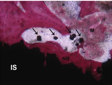
Fig. 1 Newly formed bone (NB) in close contact with the implant sur-face (IS). A rim of osteoblasts (arrows) is depositing osteoid matrix on the implant surface (contact osteogenesis). (Acid fuchsin-toluidine blue 200X)
Fig. 2 The newly formed bone (NB) grows inside the implant surface (IS) irregularities. Few small particles (arrows) detached from the implant surface can be observed. Far from the implant surface, a not yet mineralized matrix (M) is present. (Acid fuchsin-toluidine blue 100X)
Fig. 3 Machined surfaced implant. Bone (B) is growing toward the implants surface (IS): distant osteogenesis. In the marrow space, many blood vessels (*) are present close and far from the surface. (Acid fuchsin- toluidine blue 100X)
32
oxide (Al2O3) or titanium oxide (TiO2). The large variability
in surface appearance under scanning electron microscopy (SEM) of different implant surfaces is due to the different techniques employed in the blasting procedure. The sand-blasted surfaces have shown, in in vitro studies, a higher adhesion, proliferation, and differentiation of osteoblasts. Higher BIC values were found in histological studies that compared blasted and turned surfaces (Iezzi et al. 2012). Blasting procedures leave, however, blasting residual parti-cles on the surface of the implant, and this fact could modify the bone healing process (Piattelli et al. 2003). Some researchers think that aluminum ions could impair bone for-mation by a possible competitive action to calcium, while others suggested that histological data did not provide evi-dence to support the hypothesis that residual aluminum oxide particles on the implant surface could affect the osseo-integration of titanium dental implants (Piattelli et al. 2003). The bone growth pattern around blasted, rough surfaces is characterized by “contact osteogenesis,” i.e., the osteoblasts start depositing osteoid matrix directly on the implant sur-face (“implantofugal type of growth”) (Piattelli et al. 2002) (Fig. 4a, b). This type of bone growth could produce an ear-lier and a higher quantity of bone at the interface with the implant (Mangano et al. 2017a, b).
Plasma Sprayed Surfaces
These kinds of surfaces have been used in orthopedics since many decades. These implants were prepared by spraying heat molten metal on the titanium base, which resulted in a surface with irregularly sized and shaped valleys and peaks, pores, and cavities with an increase of the implant surface
area by 6–10 times. This surface topography, in which it was possible to observe the formation of bone into the coating, improved the implant fixation in bone, by a biomechanical interlock (Piattelli et al. 1998) (Fig. 5). One disadvantage of this type of surface could be the detachment of titanium par-ticles from the coating after implant insertion. The implica-tions of this occurrence were, however, not clear.
Acid-Etched Surfaces
They were introduced to modify the implant surfaces with-out the residues found after the blasting procedures, to have a more uniform surface treatment, and to control the loss of
a b
Fig. 4 (a) Newly formed bone (NB) filling the irregularities of the implant sandblasted surface (IS). (Acid fuchsin-toluidine blue 100X). (b) Close to the newly formed bone (NB), in tight contact
with the implant sandblasted surface (IS), few blood vessels (*) and many stromal cells (arrows) can be observed. (Acid fuchsin-toluidine blue 100X)
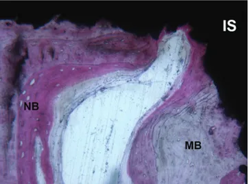
Fig. 5 Mature (MB) and newly formed bone (NB) are present in con-tact with the plasma-sprayed implant surface (IS) many years after implant placement. (Acid fuchsin-toluidine blue 100X)
33
metallic substance (Degidi et al. 2003c). Baths using chlo-ride (HCl), sulfuric (H2SO4), hydrofluoric (HF), and nitric
(HNO3) acids, in different combinations, have been used.
The acid-etching process was affected by the acid used, by the bath temperature, and by the etching time. The bone growth pattern was “contact osteogenesis” (Degidi et al.
2003c) (Fig. 6).
Sandblasted and Acid-Etched Surfaces
Surfaces obtained with a combined procedure of blasting (to produce a macro-texture) followed by acid etching (to produce a final microtexture). Sandblasted and acid-etched implants promoted a higher BIC at earlier time points compared to plasma-sprayed-coated implants. Sandblasted and acid-etched surfaces showed high osteoconductive properties and capabilities to induce cell proliferation (Iezzi et al. 2016) (Fig. 7).
Anodized Surfaces
These kinds of surfaces were obtained by modifying the structure of the superficial oxide layer of the implant surface without depositing grit particles. Anodized surfaces were prepared by applying a voltage on the titanium specimen immersed in an electrolyte. The resultant surface presented micropores of variable diameters (Rocci et al. 2003).
Hydroxyapatite (HA) Coatings
This kind of surface had a similar roughness and increase in surface area as that observed with titanium plasma spray
sur-faces (Fig. 8). A direct bonding to bone was observed, and the strength of the HA-to-bone interface was greater than that observed of titanium to bone and even greater than that seen in titanium plasma-sprayed surfaces and bone. In addi-tion, accelerated interfacial bone formation and maturation have been observed in dogs. Gap healing, i.e., the healing in the space between the implant and bone, could be enhanced by the HA coating. The advantages of an HA coating are increased surface area, increased roughness for initial stabil-ity, stronger bone-implant interface, faster healing at the interface, increased gap healing, and less corrosion of metal. The coating may, however, flake or crack upon insertion, especially into dense bone. The increased surface roughness could increase the risk of bacterial contamination should the coating be found outside the bone, e.g., in cases of peri-
Fig. 6 A layer of newly formed bone (NB) is growing within the thread of an acid-etched surfaced implant (IS) (contact osteogenesis) but
dis-tant from the trabecular bone. (Acid fuchsin-toluidine blue 100X) Fig. 7blasted and acid-etched implant (IS). Osteoblasts (arrows) are deposit- A thin bone trabeculae is forming on the concavity of a sand-ing not yet mineralized osteoid matrix (OM) (contact osteogenesis). In the marrow space, it is possible to observe small and large blood vessels (*). (Acid fuchsin-toluidine blue 100X)
Fig. 8 Newly formed bone (NB) in contact with an hydroxyapatite- coated (HA-c) implant (IS). (Acid fuchsin-toluidine blue 100X) Histological Evaluation of Early and Immediately Loaded Implants Retrieved from Human Jaws
34
implant crestal resorption. There is also an increased cost of the coating, compared with uncoated implants (Iezzi et al.
2009a; Proussaefs and Lozada 2004).
Zirconia
Zirconium oxide is used in implantology for its biocompati-bility, esthetics (its color is similar to the tooth), and mechan-ical properties. The ZrO2 implants are biocompatible,
bioinert, and radiopaque and present a high resistance to cor-rosion, flexion, and fracture; ZrO2 implants have been
reported to show a bone and soft tissue contact similar to that seen around titanium implants, and ZrO2 can be used to
pro-duce an entire implant, or as a coating (Scarano et al. 2004).
Bioceramic Molecular Impregnation
The surface properties in the nanometer range may modulate the characteristics of the protein layer adhesion to the implant surface, and the nanoscale structure of the extracellular matrix provides a natural web of nanofibers to support the cell structure. Dental implants with physical and bioceramic incorporation surface treatments at the nanometer range pre-sented higher BIC and torque values compared with rough implant surface topography at the micrometer level. The application of nanotechnology for the alteration of texture and chemistry in dental implant topography may result in different cell behaviors, i.e., from alterations in adhesion, orientation, mobility, and surface antigen display of the cells of the pre-osteogenic and osteogenic lineage. Moreover, fea-tures in the nanometer range may also affect the adsorption and conformation of integrin-binding proteins, modifying the availability of binding sites and the integrin signaling (Scarano et al. 2003).
Direct Laser Metal Sintering Implant Surface
Previous studies from our Laboratory have shown that direct laser metal sintering (DLMS) procedure produces structures with complex geometry that show better osteo-conductive properties (Mangano et al. 2009) (Fig. 9). Cells cultured on the DLMS implant surfaces showed a similar cell density to that observed on rough surfaces, but lower than that observed on machined surfaces. Moreover, it was shown that implants obtained through DLMS, having an elastic modulus closer to that of the bone, showed a better adaptation to the elastic properties of the bone (Fig. 10). DLMS implant topography not only minimizes stress-shielding effects, but also improves implants long-term success rates. These observations also suggested that
DLMS technique is an economical method for producing implants from commercially pure titanium or alloys (Mangano et al. 2009).
Implants Retrieved After Different Time
Periods
Bone undergoes remodeling, with a transformation of the initially produced woven bone into a bone with a lamellar configuration, showing a higher degree of organization (Coelho et al. 2009; Di Stefano et al. 2006; Iezzi et al. 2016; Kuroshima et al. 2015; Mangano et al. 2015; Vandamme et al. 2007, 2008). With the passing of time, a still higher
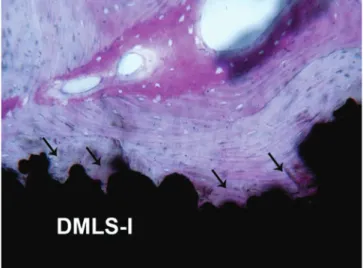
Fig. 9 Direct metal laser sintered implant (DMLS-I) the bone tis-sue grows inside the implant surface irregularities (arrows). (Acid fuchsin- toluidine blue 100X)
Fig. 10 The direct metal laser sintered surface (DMLS-I) is extremely irregular but enables bone growth around and within its indentations (arrows). (Acid fuchsin-toluidine blue 100X)
35
degree of organization of the peri-implant bone can be observed with the formation of many areas of remodeling (Coelho et al. 2009; Iezzi et al. 2009b, 2016) (Fig. 11). A submerged healing period of 3–4 months has been thought to be necessary to obtain mineralized bone at the interface of dental implants, and an earlier implant loading has been reported to determine the occurrence of fibrous tissue at the bone-implant interface. On the other hand, several research-ers have reported, in the last two decades, that in early and immediately loaded (IL) implants, placed in good-quality bone, it was possible to obtain a high level of osseointegra-tion, clinically, radiographically, and histologically similar to that of implants used with a standard submerged protocol (Cesaretti et al. 2018; Degidi et al. 2007a, b; Eccellente et al.
2010; Gapski et al. 2003; Iezzi et al. 2016; Linkow and Miller 2004; Romanos 2004) (Fig. 12). Cesaretti et al. (2018) found a higher BIC value in delayed implants when com-pared with immediately loaded implants. Same results were reported in a meta-analysis of Sagirkaya et al. (2013). On the other hand, previous experimental work done in our Laboratory has shown a higher quantity of bone in immedi-ately loaded implants when compared with control, sub-merged implants (Piattelli et al. 1998). Similar results were reported in a proof-of-principle human study (Degidi et al.
2009c). Moreover, very high implant survival rates for early and immediately loaded implants have been reported in the literature (Degidi et al. 2009a, 2010a). Many patients found the wearing of provisional prostheses rather uncomfortable, and most certainly the possibility to shorten the healing time without jeopardizing the dental implants long-term success would be beneficial for most of them. Immediate loading has been reported to be a viable and successful treatment option. However, the generalization from the results of the clinical trials to everyday routine dental practice should always be
made with extreme caution because in most trials the inclu-sion criteria were very strict, only very good candidates for the implant therapy were included, and the clinicians were high skilled. Primary implant stability and lack of micro-movement were considered to be the main factors involved in the success of IL implants (Degidi et al. 2010c). Macroretention offered by implant thread could reduce the risk of implant movements in the case of immediately loaded implants. Rigid splinting with minimal lateral forces decreased the amount of micromotion during the early heal-ing phase, givheal-ing the implant a higher tolerance to deleteri-ous micromotion. Healing processes were strongly influenced by the local mechanical loading history. In well-integrated implants retrieved from humans, it was possible to observe peri-implant lamellar bone organized in Haversian systems; these systems close to the implant surface were structured mainly in a parallel way, because the remodeling processes occurred, probably, from the implant surface in an outward direction (Iezzi et al. 2016, Mangano et al. 2015). Some authors found also that, under transmission electron micros-copy, the orientation of the collagen fibrils was parallel to the implant surface (Shah et al. 2014). Mechanical stimuli regu-lated cell division and differentiation and determined the tis-sue type and architecture. Many osteocytes were located closely to the implant surface, indicating their importance as mechanosensors (Piattelli et al. 2014; Shah et al. 2014) (Fig. 13). Well-controlled implant loading seemed to accel-erate the formation of mineralized tissues at the interface (Vandamme et al. 2007, 2008). Histological evidence of clinical successfully osseointegrated implants was rare in the
Fig. 11 Mature, lamellar bone (MB), with many remodeling areas lined by thin reversal lines (arrows), is observed in close contact with the implant surface (IS). (Acid fuchsin-toluidine blue 100X)
Fig. 12 A circular rim of osteoblasts (arrows) is depositing osteoid matrix (OM) in contact with the implant surface (IS) and with the newly formed bone (NB). Inside the osteoid matrix, few osteoblasts are entrapped and will turn into osteocytes. (Acid fuchsin-toluidine blue 200X)
36
literature, especially after a period of functional loading of more than 1 year (Coelho et al. 2009; Di Stefano et al. 2006; Iezzi et al. 2009a, b, 2012, 2016; Mangano et al. 2015; Piattelli et al. 2014; Proussaefs and Lozada 2002; Scarano et al. 2004; Traini et al. 2014). Moreover, it could also, per-haps, be useful to evaluate the healing evens at the interface after different time periods. The hardness and the elastic modulus of the bone tend to increase over time, and this fact could suggest that osseointegration is a very dynamic pro-cess with the occurrence of the adaptation of the bone to the functional loading stimuli in order to improve the overall bone biomechanics (Piattelli et al. 2014).
Histological data pertaining to IL implants demonstrate that IL did not produce untoward effect in the bone healing. Histological evidence showed that, even with shorter healing periods (4, 6, 8 weeks), it was possible to observe the forma-tion of mineralized tissue at the interface (Fig. 14) (Degidi et al. 2008, 2009c).
Even in poor bone sites, a high bone-implant contact per-centage was observed (Mangano et al. 2017b) (Fig. 15). In the spongious area, an almost continuous thin shell of newly formed bone usually covered the implant surfaces.
Mineralized tissues were found covering a large portion of the implant surface with no foreign body or inflammatory reactions visible. Bone remodeling was present in areas around the implants. The histological and histomorphomet-rical analysis on the interface of immediately loaded implants inserted in low-quality bone and retrieved from humans showed a high percentage of BIC (Mangano et al.
2017a, b).
In conclusion, the data from the observations of the inter-face of retrieved, clinically stable, immediately loaded implants showed that, independent of whether they were placed in the maxilla or the mandible and the implant design, the immediate loading allowed new bone formation at the
Fig. 13 Many osteocytes (arrows) are present at the level of the peri- implant bone, very close to the implant surface (IS). (Acid fuchsin- toluidine blue 100X)
Fig. 14 Newly formed bone (NB) lines the implant surface (IS) and is in turn lined by osteoid matrix (OM) newly deposited by a rim of osteo-blasts (arrows). Small newly formed trabeculae with wide osteocyte lacunae (OC) are present inside a marrow space (MS). (Acid fuchsin- toluidine blue 200X)
Fig. 15 Implant inserted in a low quality bone. It is possible to observe a thin bone lamella (BL) in contact with the implant surface (IS) and inside a large marrow space (MS), where a loose connective tissue is present. (Acid fuchsin-toluidine blue 100X)
37
interface of dental implants. High BIC percentages seemed to be possible in early and immediately loaded implants (Degidi et al. 2008; Mangano et al. 2017a, b).
Implants Inserted in Poor Bone Sites
An important parameter that influences the long-term suc-cess of oral implants is the bone quality of the implant bed. Posterior areas of the jaws have been avoided in implant den-tistry due to their poor bone quality, higher chewing forces, and presumed higher implant failure rates. In a meta-analysis of articles on human-retrieved implants conducted by Sagirkaya et al. (2013), these authors found that the mean BIC of implants in the mandible (70.97%) was higher than those in the maxilla (53.24%) and that the mean BIC in the anterior mandible (79.42%) was higher than that of the pos-terior mandible (69.14%). These authors concluded that the BIC of implants in the mandible was about 25% higher than that of implants located in the maxilla, due to a higher bone density in the mandible. They found also a 10% higher BIC in the anterior mandible than the posterior mandible and a 25–30% higher BIC in the anterior maxilla than the posterior maxilla (Sagirkaya et al. 2013), and they concluded that BIC was in a way related to the local bone density. Albrektsson (2008), in a retrospective study on more than 700 implants retrieved from humans, found that maxillary implants had a mean BIC of >50%, while the mandibular implants had a mean BIC of >75%. This same author underscored, however, the fact that we do not know if implants with a higher BIC have a higher percentage of long-term clinical success than implants with lower values of BIC. Soft bone-implant sites have been deemed by several researchers to be a great poten-tial risk situation, and most failures have been found in sites where the bone density from the start was low. Increasing the rate of early endosseous integration was a critical goal to achieve improved success rates. The surface microtexture of the implants has been shown increasingly to be of relevant importance in the early stages of osseointegration. Some microstructured surfaces have an improved characteristic of contact osteogenesis even in soft bone, with coverage of the implant surface by a bone layer as a base for intensive bone formation and remodeling. A high BIC was observed even in implants inserted in poor bone sites (Mangano et al. 2017a).
HA-Coated Implants
A coating of the titanium surface with a layer of hydroxyapa-tite (HA) has been proposed to get a higher osseointegration rate, a faster attachment to bone tissue and a stronger bond-ing to bone, a reduction in the healbond-ing time, a higher interfa-cial strength to bone, an enhancing of the load stress
distribution to the surrounding bone, and a better mainte-nance of the bone crest height. Concerns about the degrada-tion of the coating over the years have been raised: it has been speculated that the resorption of the HA could produce a space between implant and bone with a resultant mechani-cal instability. There is a risk for degradation of the coating, which can weaken the bone bond giving rise to implant fail-ure (Proussaefs and Lozada 2004). There have been some concerns about the long-term integrity of the coating in vivo and the fact that the dissolution and detachment of the coat-ing could expose the underlycoat-ing metal surface. It has been hypothesized that this fact could have adverse effects on interfacial bone-implant apposition and on the bone-implant interface stability. Moreover, the breakdown of the coating could produce particulate material with phagocytic response by macrophages or a foreign body reaction. Furthermore, implant failure has been associated with the loss of coating integrity, and studies of failed HA-coated orthopedic femoral stems have shown areas of coating degradation and separa-tion. Moreover, the duration of the advantage of the HA coat-ing is unclear. Coatcoat-ing dissolution and detachment from the titanium surface have been described histologically. There is some controversy whether the loss of the HA coating could be detrimental to the integration of the implant (Iezzi et al.
2009a). Porous HA is resorbed through physicochemical dis-solution and cell-mediated phagocytosis. Additional dissolu-tion may be due to the phagocytic and enzymatic acdissolu-tion of macrophages recruited to the surface. However, histological studies have shown that after many years of function, HA-coated implants continued to demonstrate adequate BIC percentages (Iezzi et al. 2009a). This fact seemed to support the view that an adequate stability of the HA-coated prostheses was maintained despite the coating loss. In some specimens, the almost complete resorption of the HA coating didn’t appear to have interfered with the osseointegration processes (Iezzi et al. 2009a). In these cases, the HA coating resorption, probably, did not have a great clinical signifi-cance because the implant was osseointegrated and was still providing an adequate function. Haversian systems were observed in close proximity to the implant surfaces in HA-coated implants, and this fact pointed to a physiologic remodeling activity of the peri-implant bone (Iezzi et al.
2009a). No foreign body reaction was observed associated with the HA particles that appeared to be detached from the coating (Iezzi et al. 2009a). These particles were always sur-rounded by bone.
Implants in Patients with Osteoporosis
Osteoporosis is a disease that influences the quality of bone tissue so that it may become susceptible to fracture. While animal studies have described the deleterious effect of
38
porosis on osseointegration, no clinical studies showed a clear association between implant failure and osteoporosis. The mechanism by which osteoporosis acted on peri-implant bone was based on the decrease in both cancellous bone vol-ume and BIC, consequently reducing bone tissue to support dental implants. However, in studies in humans, BIC was found to be similar for both osteoporosis and non- osteoporosis subjects (Shibli et al. 2008). In conclusion, the results of the histomorphometrical studies, in implants retrieved from humans, suggested that osteoporosis might not present an absolute contraindication for implant place-ment, at least, after osseointegration has been established (Shibli et al. 2008).
Immediate Post-extraction Implants
Subsequent to the removal of all teeth in the adult individual, the alveolar processes will undergo atrophy. Marked altera-tions of the height and width of the alveolar ridge will occur following single or multiple tooth extractions. The healing process following tooth removal apparently resulted in more pronounced resorption on the buccal than on the lingual/ palatal aspects of the ridge. So, after tooth extraction, the resorption and remodeling of the alveolar socket could result into a site that would be inadequate, from a dimensional point of view, for the implant placement. When an implant was placed into an extraction socket, osteogenic and osteore-sorptive responses were already initiated following extrac-tion, and this tissue could enhance the capacity for healing (Paolantonio et al. 2001). An immediate implant is one implant that was placed into an extraction socket at the same time the tooth was extracted. Immediate post-extraction implants have several advantages such as fewer surgical pro-cedures, preservation of bone volume, and shortening of the time needed until the implants could be restored (Degidi et al. 2007b). Additional advantages of the use of the imme-diate post-extraction implants were:
1. Shortening of the edentulous time period 2. Reducing the costs of treatment
3. Improve the psychological approach with the patient 4. Reduction of the comprehensive treatment time with less
surgical procedures and morbidity
5. Optimal esthetic result, with an easier definition of the implant position as a consequence of correct fixture posi-tion and angulaposi-tion
6. Improvement of biomechanics of the future restoration Several different human clinical studies have demonstrated that with immediate post-extraction implants, it was possible to obtain very high (more than 90%) long-term success per-centages. Moreover, many experimental studies have
con-firmed that a high percentage of bone-implant contact could be achieved on light microscopic level in animals, when using immediate post-extraction implants (Degidi et al.
2007b; Paolantonio et al. 2001). One major drawback in using immediately post-extraction implants was due to the lack of adaptation of the alveolar bone in the cervical portion of the implant. Soft tissues, creating problems in the osseoin-tegration of the implant, could fill this space. Almost always, when using immediate post-extraction implants, it was nec-essary to resort to guided bone regeneration techniques, with the use of biomaterials and membranes. However, in a histo-logical study aimed to evaluate the outcome of implantation in fresh extraction sockets without the use of membranes in humans in comparison with implants placed in healed, mature alveolar bone, no significant differences in the clini-cal and radiographic parameters were observed between the two experimental categories (Paolantonio et al. 2001). Bone resorption was not present in any area of the histological sec-tions (Paolantonio et al. 2001).
Implants Inserted in Grafted Sites
The successful outcome of a sinus augmentation procedure can be evaluated best by a histological examination of the events at the bone-implant interface. A successful implant osseointegration in sinus augmentation procedures should be characterized by a high quantity of newly formed bone at the implant interface, to provide enough bone for mechanical support and integration of the implants (Scarano et al. 2004). A bone substitute material should have the capability to allow the integration of loaded titanium implants. One of the most important questions about sinus augmentation proce-dures is if the regenerated bone obtained after the insertion of a graft is able to integrate dental implants. Other important questions are the extent of the surface of a dental implant placed into a grafted sinus, which will be surrounded by bone in direct contact with the implant surface, and to what extent functional ankylosis will be present and if the obtained implant osseointegration will remain stable over the long period, after functional loading (Fig. 16). Very high BIC was reported in implants retrieved from sinuses augmented after a period varying from some months to several years (Iezzi et al. 2007; Scarano et al. 2004). All these implants had osseointegrated and had remained osseointegrated after many years of functional loading (Iezzi et al. 2007; Scarano et al. 2004) (Fig. 17). It has been reported that grafted parti-cles in contact with the implant surface could reduce mechanical support for the dental implants. No contact was, however, observed between grafted particles and implant surfaces in most of the reported implants (Iezzi et al. 2007; Scarano et al. 2004). The continued presence of grafted par-ticles in the peri-implant bone did not seem to jeopardize the
39
integration of the implant because no contact between the grafted particles and the implant surface was observed, and a complete resorption of the grafted material did not seem to be a prerequisite needed to get formation of bone at the inter-face and implant osseointegration (Iezzi et al. 2007; Scarano et al. 2004). On the other hand, the lack of complete resorp-tion of the grafted material could even be advantageous in order to maintain the initial dimensions of the grafted area
with time. Other human histologic specimens retrieved from grafted sinuses after longer time periods will certainly help to clarify the question of the biomaterial resorption over time and of the potential of regenerated bone to achieve and main-tain osseointegration with dental implants. Provisional implants are helpful in helping the patients to avoid the inconveniences of wearing a denture and they can provide useful information after retrieval (Iezzi et al. 2007).
Implants in Smokers
The influence of smoking on peri-implant bone has been evaluated in a several histologic animal models. The major-ity of these studies agree that smoking had a detrimental effect on bone healing, BIC, and bone mineral density. Smoking delays the normal bone healing process by a mechanism that inhibits proliferation of precursor cells. Cigarette smoke is composed by over 4000 toxins that have the potential to undermine the peri-implant bone healing. Toxins such as nicotine, carbon monoxide, nitrosamines, benzenes, aldehydes, and hydrogen cyanide have been shown to affect essential processes of bone healing. Nicotine is a potent vasoconstrictor that not only reduces blood flow and nutrient delivery to the surgical implant site but also inhibits the proliferation of fibroblasts, red blood cells, and macrophages. Carbon monoxide decreases the oxygen-carrying capacity of red blood cells, while hydro-gen cyanide leads to hypoxia. In human-retrieved speci-mens, BIC% was found to be significantly lower in smokers (Fig. 18). A tendency toward slower wound repair has been suggested. Moreover, cigarette smoking reduced the rate of bone formation and increased the rate of bone destruction in postmenopausal women. Cigarette smoking seemed to suppress osteoprotegerin levels and might contribute
Fig. 16 Newly formed (NB) and mature bone (MB) match with the thread shape without gaps at the interface. A marrow space with loose connective tissue and remnants of partially resorbed synthetic hydroxy-apatite (HA) not in contact with the implant (IS) can also be observed. (Acid fuchsin-toluidine blue 100X)
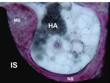
Fig. 17 Implant inserted in a grafted site. It is possible to observe newly formed bone (NB) in contact with the implant surface (IS). A residual grafted particle (GP) of heterologous origin is present far from the implant surface. (Acid fuchsin-toluidine blue 200X)
Fig. 18 Implant inserted in a smoker. A low-quality newly formed bone (NB) can be observed around the implant threads (IT). Only in a very small portion of the interface there is a contact between bone and implant surface (arrows). (Acid fuchsin-toluidine blue 40X)
40
toward the decreased peri-implant bone formation (Fig. 19). However, the precise mechanisms by which smoking exerted its deleterious effects on bone healing remain unclear (D’Avila et al. 2010).
Basic Concepts About Immediate Loading
The concept of immediate loading was proposed more than 50 years ago when the endosseous blade implants were intro-duced (Linkow and Miller 2004). Histological evidence of osseointegration in clinically successfully osseointegrated implants in man can be found in the literature (Coelho et al.
2009; Degidi et al. 2003a, b). Retrieved human implants are extremely important for long-term evaluation of implants subjected to functional loading (Di Stefano et al. 2006; Iezzi et al. 2012, 2016; Proussaefs et al. 2002). Immediate loading of dental implants was thought to produce a fibrous repair at the interface. Therefore, the most important question is if an implant with a high primary stability in bone can be immedi-ately loaded without formation of fibrous tissue at the inter-face. Primary stability seems to be a very important factor in
immediate loading protocols (Yamamoto et al. 2014). Stability of an implant has been found to be related to implant geometry, implant length, surface morphology, splinting of implants, control of occlusal functional loads, bone quality, size of the host recipient site, and lack of detrimental patient habits (e.g., bruxism) (Yamamoto et al. 2014; Mangano et al.
2017a). Several histological reports, in man and experimen-tal animals, have shown mineralized tissues at the interface in early and immediately loaded implants. Specifically, in monkeys (Piattelli et al. 1998; Quaranta et al. 2008), dogs (Cesaretti et al. 2018), rabbits (Han et al. 2014; Kuroshima et al. 2015; Vandamme et al. 2007), rats (Yamamoto et al.
2014), and humans (Degidi et al. 2008; Mangano et al.
2017a), it was possible to observe mineralized tissues at the bone-implant interface in early and immediately loaded implants. Immediate loading allows immediate restoration of esthetics and functions, reduces the morbidity of a second surgical intervention, and facilitates the functional rehabili-tation increasing patient acceptance and satisfaction (Degidi et al. 2010a). Functional loading, in experimental studies, appeared to stimulate bone apposition and accelerate implant osseointegration (Kuroshima et al. 2015). Mechanical load-ing increased osseointegration, bone volume, and bone min-eral density; moreover, the quality of the peri-implant bone was changed with a higher quantity of osteocytes and a dif-ferent alignment and degree of direction of the peri-implant bone collagen fibers (Kuroshima et al. 2015). Wolff stated that there was a direct link between mechanical loading and bone form; Wolff’s law would imply that increased stresses acted as a stimulus to new bone formation, while reduced stress tended to produce bone loss (Traini et al. 2014). It is, however, necessary to proceed cautiously with the possibil-ity of transferring to man the histologic results obtained in animal experimentation, due to the different loading condi-tions. Only few histological reports of clinically stable early or immediately loaded implants in man can be found in the literature (Degidi et al. 2003a, b, 2008; Di Stefano et al.
2006; Iezzi et al. 2009b; Mangano et al. 2017a; Romanos et al. 2005). Nowadays, the immediate loading treatment concept can be successfully used in implant dentistry. Immediate loading of endosseous oral implants is a well- established, evidence-based concept. There is some variance among the different studies with the exact definition of the term “immediate loading,” as some research groups avoid occlusal contacts in the temporary restoration placed immediately after surgery. Other authors have demonstrated the use of an immediate placement procedure for restoring single teeth in the esthetic zone focusing on the stability of hard and soft tissues and recommended a careful elimination of all occlusal contacts after surgery (Degidi et al. 2009a). In MEDLINE, such concepts should be associated with “imme-diate restoration” or “imme“imme-diate temporization” (imme“imme-diate non-occlusal functional loading) and not with an immediate
Fig. 19 Soft tissue (ST) is present inside the implant thread (IT). The new peri-implant woven bone (WB) does not contact the implant sur-face. (Acid fuchsin-toluidine blue 100X)
41
functional (occlusal) loading. To more accurately compare research findings, the term “immediate loading,” in recent literature, should be used to indicate only implant-supported restorations with occlusal contacts in place immediately after implant surgery (Degidi et al. 2009b).
Loading Effects on Osseointegration
A submerged healing period of about 3–4 months has been thought to be necessary to obtain mineralized bone at the interface of dental implants, and an earlier implant loading has been reported to determine the occurrence of fibrous tis-sue at the bone-implant interface (Degidi et al. 2003a). An immediate loading protocol implies healing under loading and was thought to involve the risk of fibrous tissue encapsu-lation (Vandamme et al. 2008). On the other hand, several researchers have reported, in the last two decades, that in early and immediately loaded implants, placed in good qual-ity bone, it was possible to obtain a high level of osseointe-gration, clinically and radiographically similar to that of implants used with a standard submerged protocol, and very high implant survival rates for immediately loaded implants have been reported in the literature (Eccellente et al. 2010) (Fig. 20a, b). Histologic comparisons have been reported in animal studies on implants that were immediately loaded versus implants with a delayed loading. The results of the BIC are variable with some papers showing more BIC, some less and some a similar value as the non-loaded side (Cesaretti et al. 2018; Quaranta et al. 2008; Sagirkaya et al. 2013). Albrektsson (2008), in a histological and histomorphometri-cal evaluation of more than 700 dental implants retrieved from man, found that unloaded, sleeping implants had a 10% lower BIC than the loaded implants. Piattelli et al. (1998) have histologically and histomorphometrically found a
higher density in the bone around immediately loaded implants (screw-shaped, TPS-coated) in comparison to unloaded implants in the maxilla and mandible of Macaca
fascicularis. According to this study, the histomorphometri-cal analysis demonstrated that in test implants, the BIC per-centage was 67.3% in the maxilla vs. 73.2% in the mandible; in the unloaded implants, these percentages were 54.5% and 55.8%, respectively. Moreover, the bone around the immedi-ately loaded implants tended to have a more compact appear-ance. The microstructure of the bone seems to be able to adapt to different loading forces (Kuroshima et al. 2015; Romanos 2015) (Fig. 21). Greater bone density was found when the implants were immediately loaded (Kuroshima et al. 2015; Romanos 2015), and mechanical forces seemed
a b
Fig. 20 (a) Around a loaded implant (LI), bone tissue shows different stages of maturation marked by reversal lines (arrows). (Acid fuchsin- toluidine blue 200X). (b) Polarized light microscopy image showing
well-organized, lamellar bone (B) after many years of loading. (Acid fuchsin-toluidine blue 200X)
Fig. 21 Implant loaded for a very long period (30 years). Many remod-eling areas (arrows) are present near the implant surface (IS), in direct contact with the metal. Bone formed in different time periods has differ-ent staining affinities (the bone formed more recdiffer-ently (NB) has a higher staining affinity for the dyes). (Acid fuchsin-toluidine blue 100X) Histological Evaluation of Early and Immediately Loaded Implants Retrieved from Human Jaws
42
to be able to increase the mineral content of the bone by 34% with the formation of a dense lamellar bone in a peri-implant location (Romanos 2015). The density of the peri-implant bone and the BIC was found to be higher in loaded implants (Kuroshima et al. 2015; Romanos 2015). Yamamoto et al. (2014), in an experimental study in rat tibiae, found that bone metabolic activity was higher, during the period of wound healing, when load was applied. These authors also found that the peak of metabolism in immediately loaded implants was much lower than that found in delayed implants: this fact could mean that immediate loading of implants with a high primary stability might represent a much safer proce-dure than delayed early loading (Yamamoto et al. 2014). There are also histological and histomorphometrical reports showing similar levels of BIC percentages after immediate loading of oral implants with a progressive thread design, placed in the posterior part of the mandible of monkeys using different types of loading in comparison with delayed loaded implants (Quaranta et al. 2008). In contrast to the BIC levels, there is a higher bone density surrounding the immediately loaded implants than the density around implants loaded with the classical protocol (delayed loaded implants) in monkeys (Quaranta et al. 2008). The definition of osseointe-gration is clinical and is based mainly on implant stability. However, clinical stability alone is insufficient to demon-strate the presence of osseointegration, i.e., the presence of mineralized tissues at the interface with dental implants (Degidi et al. 2003a, 2007b; Iezzi et al. 2009b). Only the biopsy of human-retrieved implants allows a precise evalua-tion of the events occurring at the interface (Degidi et al.
2008, 2009a).
The range of BIC that is necessary for an implant to be osseointegrated is unknown (Iezzi et al. 2016; Romanos et al. 2005), and different values have been reported in the literature from as low as 25% to as high as 50% (Degidi et al. 2010b; Di Stefano et al. 2006; Scarano et al. 2006). Increased BIC may provide an earlier and better anchorage, thus allowing for an earlier functional loading of implants (Abrahamsson et al. 2004). Histological evidence of clinical successfully osseointegrated implants is rare in the litera-ture, especially after a period of functional loading of more than 1 year (Coelho et al. 2009; Iezzi et al. 2016; Romanos et al. 2005; Uehara et al. 2004) because there are not many possibilities to get retrieved implants in humans. Removal and histological evaluation of implants due to fracture or other reasons (orthodontic, psychological, esthetic, hygienic) can give extremely important data, from the scien-tific point of view, as they could be useful to evaluate the healing events, thus the bone response, at the interface after different time periods (Uehara et al. 2004). Furthermore, such histological data demonstrate that different kinds of implant systems using various surfaces, inserted in different
bone qualities in the maxilla or the mandible, can be equally successful in establishing osseointegration. Important fac-tors in immediately loaded implants are primary stability, related to the thread design and the surface microstructur-ing, bone quality, and reduction of micromotion (i.e., splint-ing) (Romanos 2004).
Primary Stability
Good implant stability decreases the distortional strains in the newly forming tissues and improves the chances of neo- osteogenesis at the interface; on the contrary, a poor stability of the implants has been shown to determine an important distortional strain with fibrous tissue formation at the inter-face (Degidi et al. 2010c). A higher removal torque value (RTV) of dental implants might lead to a more predictable use of short implants and to a support of prosthesis with a smaller number of implants and allows shorter healing peri-ods (Degidi et al. 2007a).
Macro-/Microstructure
An implant should have a retentive shape. Over the years, many different types of implants have been proposed and used, i.e., blades, screws, and root-form implants. Screws seem to behave in a better mechanical way than cylindrical implants without threads (Romanos 2015).
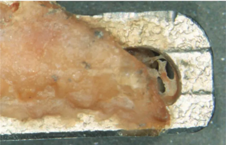
Blades
In blade implants, a high failure rate was reported due to their non-retentive shape (Fig. 22). This has been attributed to the formation of connective tissues around their surface
Fig. 22 A retrieved blade partially surrounded by hard tissues G. Iezzi et al.
43
(Proussaefs et al. 2002). Although the presence of mineral-ized tissues at the interface with blade implants has been reported (Di Stefano et al. 2006; Proussaefs et al. 2002), the view that blade implants cannot integrate still persists (Proussaefs et al. 2002). Blades are the immediately loaded implants with the longest clinical history, so their histologi-cal evaluation has a historihistologi-cal value and may certainly have some applications to root-form implants (Proussaefs et al.
2002) (Fig. 23). In blade implants retrieved after 13 and 21 years of function, mature bone in tight contact with the implant surface was seen around most of the implant surface (Proussaefs et al. 2002). The response of the bone tissue appeared not to be disturbed by the stresses and strains trans-mitted at the interface.
Screws
The screw has a large mechanical retention and greater abil-ity to transfer compressive forces to the peri-implant osseous tissue and to produce lower shear stresses at the interface (Gapski et al. 2003). Screw design not only minimizes micromotion of the implant, but also makes a significant contribution to the initial stability of the implant during placement (Steigenga et al. 2004); therefore threaded implants present considerable advantages compared with press-fit implants for the immediate loading protocol. Macroretention offered by implant thread can reduce the risk of implant movements in the case of immediately loaded implants. Additionally, the threads increase the surface area of the implant (Gapski et al. 2003). In the early stages of healing, many blood vessels were observed within the threads (Fig. 24a, b), as well as new bone formation was appreciated few weeks after the implant insertion (Fig. 25). In conclusion, threads are also used to maximize the initial contact, improve the initial stability, enlarge the implant sur-face area, and favor dissipation of interfacial stresses (Steigenga et al. 2004). A high quantity of mineralized tissue was found in an immediately loaded screw, retrieved after several years of function (Iezzi et al. 2009b).
Root-Form Implants
Very high success and survival rates have been reported in immediately loaded dental root-form implants (Degidi et al.
2003c; Proussaefs and Lozada 2004), and immediately
Fig. 23 Trabecular bone (TB) at the interface with the implant surface (IS) in a blade retrieved after about 20 years of loading. (Acid fuchsin- toluidine blue 40X)
b a
Fig. 24 (a) Intense angiogenic (*) and osteogenic activities are evi-dent. Only a small bone trabecula (arrow) can be seen in contact with the implant surface (IS) and connecting through a bridge of osteoid matrix (OM) to the trabecular bone (TB). (Acid fuchsin-toluidine
blue 100X). (b) High-power image showing dense connective tissue (CT) and newly formed blood vessels (*) between the implant sur-face (IS) and the newly formed bone (NB). (Acid fuchsin-toluidine blue 400X)