University of Foggia
Department of the Sciences of Agriculture, Food and Environment (SAFE)
Doctoral Thesis
in
Management of Innovation in Agricultural and Food Systems of the
Mediterranean Region
Study of Chemical, Biochemical and Technological
characteristics of Gluten Friendly
TMgrains of cereals and
derived products
PhD Candidate: Loretta Landriscina Supervisor: Carmela Lamacchia
ii List of Contents
Abstract 1
Aim and outlines of the thesis 2
Chapter 1 General Introduction 4
Chapter 2 Changes in wheat kernel proteins induced by microwave treatment
microwave treatment 38
Chapter 2 Impact of gluten friendlyTM technology on wheat kernel endosperm
and gluten protein structure in seeds by light and electron microscopy 58
Chapter 4 Chemical, Rheological and Sensorial Evaluation of Gluten FriendlyTM 79 Flour
Chapter 5 Impact of Gluten FriendlyTM Bread on the Metabolism and function of in Vitro Gut Microbiota in Healthy Human and Coeliac subjects 95
Chapter 6 In Vitro Fermentation of Gluten FriendlyTM Bread in Healthy Human 125 and Coeliac subjects: impact on Gut Microbiota
Chapter 7 General Discussion 140
List of publications 155
1
Abstract
This thesis aimed to investigate chemical, biochemical and technological characteristics of Gluten FriendlyTM grains of cereals and derived products.
“Gluten Friendly™” is a patented technology (Italian patented method n°: 0001414717, also filed under the Patent Cooperation Treaty, application no. PCT/IB2013/000797 and published in Europe as EP 2903453 A1 and titled “Detoxification method of gluten proteins from cereal grains”) that allows to obtain gluten detoxified flours suitable for the preparation of bakery products and pasta made from wheat. The “Gluten Friendly™” technology implies the application of microwave energy for a few seconds to hydrated wheat kernels before milling to reach a high temperature for a short amount of time aiming to combine the nutritional and technological properties of wheat proteins with safety for coeliac sufferers and other gluten-sensitive subjects. This innovation relies in a wise combination of temperatures with other parameters (moisturizing, evaporation, resting, etc.,) that are clearly described in the patent and non-patent literature quoted in this thesis and does not cause proteins to be denatured but a change in their configuration that makes them not recognizable by specific antibodies and able to produce further pleiotropic effects on man. In particular, “Gluten Friendly™” technology causes a rearrangement of the secondary and tertiary structure of the gluten proteins, involving a different spatial conformation of the sequences, including the so-called antigenic. So the antigenic capacity of gluten it is abolished and the immunogenicity in vitro of the most common epitopes involved in coeliac disease is reduces. Moreover, proteins structure rearrangements induced by “Gluten Friendly™” involve the exposure of charges that may allow a new kind of aggregation among different classes of wheat endosperm proteins, only through hydrophobic and/or ionic interactions visible at the immunofluorescent microscopy. Although, the rearrangement of the secondary and tertiary structure and the exposure of charges determines the solubility of proteins in “Gluten Friendly™” flours increasing the electrostatic repulsion and breaking the hydrogen the flours are able to form dough and leaven and produce bread. The sensory qualities of “Gluten Friendly™”bread is comparable to that of the Control Bread in terms of appearance, taste, aroma, color and bread texture. Furthermore, experiments carried out in vitro on healthy and coeliac human faecal microbiota pinpointed that “Gluten Friendly™”bread prolonged the survival of L.acidophilus and exerted an antibacterial effect towards S.aureusand S. Typhimurium. Moreover, “Gluten Friendly™” bread modulated the intestinal microbiota in vitro, promoting changes in lactobacilli and bifidobacteria members in coeliac subjects. A final multivariate approach combining both viable counts and metabolites
2
suggested that “Gluten Friendly™” bread could beneficially modulate the coeliac gut microbiome. These results were confirmed by an in vitro study performed by a three-stage continuous fermentative system on the intestinal microbiota and metabolites composition of healthy and coeliac individuals for 30 days.
Aim and outline of the thesis
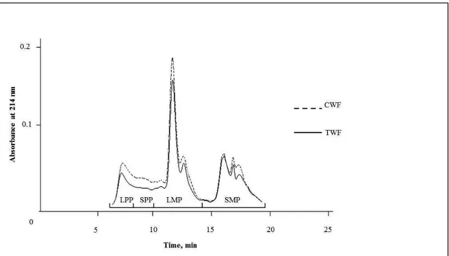
This thesis is aimed on the study of chemical, biochemical and technological characteristics of Gluten FriendlyTM grains of cereals and derived products. Following the general introduction in Chapter 1, Chapter 2 describes the effect of “Gluten Friendly™” (GF) technology on flour protein fractions, by size exclusion high-performance liquid chromatography (SE-HPLC), sodium dodecyl sulphate–polyacrylamide gel electrophoresis (SDS–PAGE) and R5-sandwich ELISA. Chapter 3 describes the impact of Gluten-FriendlyTM technology (Italian priority
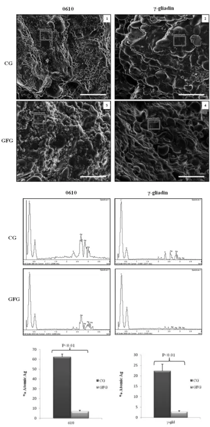
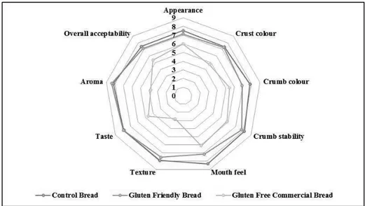
patent n_ 102015000084813 filed on 17th December 2015) on wheat kernel endosperm morphology and gluten protein structure, using SEM, light and immunofluorescent microscopy. Microscopy was combined with immunodetection with specific antibodies for gliadins, c-gliadins, LMW subunits and antigenic epitopes to gain a better understanding of the technology at a molecular level. In Chapter 4 the chemical, rheological and pasting properties of Gluten Friendly Flours was investigated. In this study, attempt was made to evaluate technological proterties of Gluten Friendly Flours in the production of bread by analyzing the rheological properties of the flour and dough, as well as baking qualities and sensorial evaluation of the resulting bread.
Chapter 5 describes the in vitro response of healthy and coeliac human faecal microbiota to gluten-friendly bread (GFB). Thus, GFB and control bread (CB) were fermented with faecal microbiota in pH-controlled batch cultures. The effects on the major groups of microbiota were monitored over 48 h incubations by fluorescence in situ hybridisation. Short-chain fatty acids (SCFAs) were measured by high-performance liquid chromatography (HPLC). Furthermore, the death kinetics of Lactobacillus acidophilus, Bifidobacterium animalis subsp. Lactis, Staphilococcus aureus, and Salmonella Typhimurium in a saline solution supplemented with GFB or CB were also assessed. Chapter 6 aimed to investigate the in vitro effects of GF bread (GFB) and control bread on the intestinal microbiota and metabolites composition of healthy and coeliac individuals in a validated three-stage continuous fermentative system simulating the proximal, transverse and distal parts of human colon (vessel 1, 2 and 3, respectively),
3
resemble the complexity and diversity of the intestinal microbiota. Main bacterial groups of the faecal microbiota were evaluated using 16S rRNA-based fluorescence in situ hybridization (FISH). Potential effects on microbial metabolism were also studied measuring short chain fatty acids (SCFAs) by HPLC analysis.
To finalize, Chapter 7 is the general discussion on the obtained results of the patented method, conclusions and future works.
4
Chapter 1
5 1. Wheat
Cereals constitute the most important source of food worldwide and cereal products form the basis of the human diet in most countries. Cereal crops are also an important source of nutrition for animals and, moreover, they have many other important economic and ecological features (production of oils, starch, flour, sugar, malt and alcoholic beverages, renewable energy, etc.) [1, 2]. Cereals such as maize, rice, wheat, sorghum, millet, barley and oats are particularly important as a staple food in many countries, but wheat stands out from the others. Wheat is the world`s leading cereal and wheat grains are the major source of energy and proteins in human nutrition. Although wheat is grown on more land area than any other crop, it is only the third cereal crop in total world production, behind maize and rice [1]. Nevertheless, wheat is the most widely cultivated food crop for several reasons. Wheat is well adapted to harsh environments and, therefore, it can be grown in wide range of elevations, climatic conditions and soil fertility [3, 4]. It can be safely stored for a long period, easily transported and processed into various types of food. Wheat has high content of gluten proteins (wheat storage proteins) enabling to form a strong, cohesive dough that retain gas and produce light baked products. On that account, most breads, even rye and oat bread, are made with a portion of wheat flour. Wheat was one of the first domesticated cereals originated in the Middle East, area that stretches from Israel and Lebanon into Syria, Turkey, Iran and Iraq [1, 5]. Cultivation and repeated harvesting and sowing of wild grass grains led to the selection of mutant forms, which was the important part of wheat domestication. The cultivation started about 11,000 years ago, in the Middle East and by 5,000 years ago, wheat was cultivated from England to China [1]. In the 1600`s the wheat was introduced in America by Spaniards. Development of reaping and threshing machines in 1800`s (steam engine) and 1900`s (internal combustion engine) resulted in increased farmer productivity and wheat field became larger [1].
Wheat belongs to the grass family Ponceau, the tribe Triticale and the genus Tritium. The genus Tritium comprise several species and subspecies, some of which are cultivated and some are uncultivated, or rarely so. However, the botanical classification of wheat varieties is not unified. The most economically important species is Tritium ostium also known as common or bread wheat [1, 6]. This species, which is mainly used for baked products such as bread, covers up to 95 % of the wheat grown today. Remaining 5 % is mostly durum wheat (Tritium durum), traditionally used for pasta products [3, 7]. Other wheat species (e.g., spelt wheat) are grown in limited quantities, mostly for specialized use. Within a species, wheat varieties can be further classified by planting season (winter vs. spring wheat), hardness of the grain (hard vs. soft wheat), and the color of the grain (red, white or amber) [1, 8]. According to these criteria, all wheat species fall into one of the six major classes (classification system used in the USA): Hard Red Winter, Durum wheat, Hard Red Spring, Hard
6
White, Soft Red Winter and Soft White. Winter wheat varieties are sown late September to November (they lie dormant during the winter) and harvested early in the summer. Whereas spring wheat varieties are sown February to April and harvested late in the summer. Spring wheat is significantly less yielding than the winter wheat but it usually offers very high quality bread making wheat. Hard wheat grains have higher gluten content than soft wheat varieties. The hardest wheat is durum, whose flour is used for macaroni, spaghetti and other pasta products. The soft wheat grains have higher starch-to-protein ratio compared to hard wheat varieties [1]. In general, hard wheat flour is used for bread and soft wheat flour for pastry. Nevertheless, many flours are carefully blended mixtures of both hard and soft wheat varieties designed for certain purpose.
Wheat grain (also known as a seed or kernel) is the most economically important part of the wheat plant, because it constitutes main source of people food. The grain is, in fact, the fruit of the wheat, botanically known as caryopsis [2]. It consists of three distinct parts: bran, endosperm and germ (Fig. 1). Bran, the outer coat consisting of seven cell layers, is separated from the flour during most milling processes. Bran surrounds germ and the endosperm and protects the grain from weather, insects, soil, and bacteria. It accounts for about 14 % of the grain weight [9]. Bran has low protein content but is rich in indigestible cellulose material called dietary fibers. It also contains B-vitamins and trace minerals. Bran is included in whole-wheat flour. The endosperm is the largest and most important part of the grain comprising about 83 % of the total grain mass, and consists mostly of starch (about 75 % of the endosperm mass). The endosperm contains the greatest proportion of carbohydrates (in starch granules), proteins, iron and B-vitamins. The starchy endosperm is the only material of wheat grain, from which the white flour is made [4, 9, 10]. Germ is the embryo or sprouting section of the seed comprising about 2.5 % of the grain weight and is usually separated from the flour during most milling processes because of fat content (about 10 %) that limits the flour quality. Fats can easily go rancid, which limits shelf life of the flour. Germ is present in whole-wheat flour. Besides lipids, germ further contains minimal quantities of proteins, and a greater share of B-vitamins and trace minerals [9].
1.2 Wheat grain proteins
Amongst the cereals, wheat is unique because wheat flour has the ability to form dough suitable for production of bread and wide range of baked products. Wheat flour is also widely used in starch and gluten industry, etc. Bread making quality is determined, in particular, by protein composition of the wheat grain. Within the cereal group, wheat has higher protein content than rice or corn. The mature wheat grains contain 8-20 % proteins, which are mostly present in the endosperm [4]. Although some of these proteins are important structural or metabolic proteins, majority of wheat grain proteins
7
(gluten proteins) are storage proteins that function as a source of nitrogen during the grain germination.
Fig. 1. Schematic diagram of the wheat grain illustrating its major parts.
Cereal proteins located in the endosperm constitute a heterogeneous group of proteins. At the beginning of the 20th Century, T. B. Osborne classified these proteins into four groups upon their solubility in a series of solvents [11]. The following protein fractions are subsequently extracted from the milled grain: albumins, water-soluble proteins; globulins, soluble in diluted salt solutions.; prolamins, soluble in aqueous alcohols (wheat prolamins are called gliadins); glutelin’s, soluble in dilute alkali or acid (wheat glutelin’s are called glutenin’s). This classification is still in use despite the number of factors, such as fineness of grain milling, vigor and time of shaking, concentration of extraction solutions used, and the extraction temperature, which influencing the extractability of particular protein fraction. Moreover, each of mentioned fractions is a complex mixture of different proteins, which overlap in their solubility. This is particularly observed in gliadin and glutenin fractions. Thus, all four fractions can be contaminated by any proteins from other fractions [12, 13]. Recently, the wheat grain proteins are more often classified based on biological characteristics of the proteins together with their chemical and genetic relationships. Nevertheless, the Osborne`s classification system was a major milestone in the development of cereal chemistry. Water-soluble albumins and salt-soluble globulins constitute up to 20 % of total wheat grain proteins. These proteins are involved, particularly, in metabolic processes (e.g., starch synthesis and degradation) as the enzymes (e.g., α- and β-amylases) or enzyme inhibitors (e.g., α-amylase inhibitor), and minority of albumins and globulins are structural proteins [12, 14]. Albumins and globulins can also play a role
8
as nutrient reserves for germinating embryo. They are, in contrast to gliadins and glutenin’s, rich in essential amino acids, mainly lysine [12, 15]. Due to the amino acid composition both these protein groups are important with respect to nutritional requirements of humans and monastic animals as well. On the other hand, albumins and globulins are minority fractions of wheat grain proteins and, therefore, the wheat grain does not provide as high amount of nutritionally indispensable amino acids as do animal protein-based foods [12, 16]. However, the wheat grain proteins are complementary to other food proteins (e.g. animal products, legume) in terms of nutritional value. Molecular weight (MW) pattern of albumins and globulins is ranging from 2,000 to 106,000 and is predominantly divided into two relatively wide regions with MW from 23,000 to 66,000 and 2,000-16,000 [13]. In general, albumins and globulins have lower MW than gliadins and glutenin’s. Among the varieties both albumins and globulins show very similar protein pattern and, consequently, these proteins are not suitable for wheat varieties identification [13]. The most important protein fractions of wheat grain, in many respects, are gliadins and glutenin’s, known under general name gluten proteins or gluten, eventually. The investigation of wheat grain proteins, their relationships, metabolism and structures is an important research area, because these proteins impact the flour and nutritional quality [14]. There are several promising studies and approaches (including proteomics and genomics) focused to improvement of both grain yield and the end-use quality of wheat [3-5, 10, 14, 17-21].
1.3 Gluten proteins
Gluten is insoluble, viscoelastic and cohesive mass that remains after removal starch and water-soluble components from the wheat dough by water. Gluten proteins, conventionally subdivided into gliadins and glutenin’s, are the major storage proteins of the wheat grain that make up about 80 % of total wheat grain proteins [4, 14, 22]. Both gliadins and glutenin’s play a key role in dough rheology and bread making quality. In dough, gliadins are responsible for viscosity and extensibility whereas glutenin’s are cohesive and distinctively affect the elasticity (dough strength). Thus, qualitative and quantitative constitution of gluten proteins is a crucial factor for dough formation and baking properties [25-27]. This is one of the major reasons for which the gluten proteins are the most investigated proteins of wheat grain. While gliadins are monomeric proteins (single polypeptide chains), glutenin’s are present as oligo- and polymers (multiple polypeptide chains), where the glutenin subunits are linked 19 together by interchain desulphated bonds [10, 12, 28]. Gluten proteins are unique in terms of their amino acid composition. They have extremely high content of glutamine and proline and, on the contrary, low content of essential amino acids such as lysine. Another very important amino acid (besides the proline and glutamine) is cysteine, because it determines the structure and functionality of gluten [29]. Cysteines are mostly present in oxidized state and form
9
either intrachain desulphated bonds within a single protein (both gliadins and glutenin’s) or interchain desulphated bonds between proteins (glutenin’s) [28]. It has been found that number of these bonds increase in heated gluten [30]. Both intra- and interchain desulphated bonds, as well as non-covalent (hydrogen, hydrophobic) interactions are involved in the formation of gluten polymer complex. Gluten proteins are classically divided into monomeric gliadins and polymeric glutenin’s. However, after reduction of desulphated bonds individual glutenin subunits are soluble in aqueous alcohols as well as gliadins. For this reason, both gliadins and glutenin’s can be classified as prolamins [10, 12, 17, 40]. More recently, based on the structural and evolutionary relationships, the gluten proteins are divided into three broad groups (Fig. 2). Sulphur-poor prolamins include the ω-gliadins, the LMW-GS and α/β- and γ- gliadins belong to the Sulphur-rich prolamins and the HMW prolamins include the HMW-GS. From the biological point of view the role of gluten proteins is to provide a store of carbon, nitrogen and Sulphur to support grain germination and seedling growth. Furthermore, these proteins determine bread making quality of the wheat. Finally, gluten proteins are also known as an environmental factor triggering celiac disease (CD) in genetically-predisposed individuals [43, 44].
Both fractions are important contributors to the rheological properties of dough, though their functions are divergent. Hydrated gliadins have little elasticity and are less cohesive than glutenin’s, and contribute mainly to the viscosity and extensibility of the dough. In contrast, hydrated glutenin’s are both cohesive and elastic and are responsible for dough strength and
elasticity [24,25]. A proper mixture of both fractions is essential for the quality of the end product. However, of particular importance are the glutenin polymers, and it is well established that strong (i.e., highly viscos-elastic) doughs contain high proportions of HMW glutenin pol ymers [24].
Numerous studies are consistent with the hypothesis that the HMW subunits form an elastomeric polymer network which provides a “backbone” for interactions with other glutenin subunits and with gliadins (Figure 3) [26,27]. There is no doubt that this network is mainly stabilized by inter-chain desulphated bonds [28,29]. Additional covalent bonds formed during dough making are tyrosine-tyrosine and they-tyrosine crosslinks between gluten proteins [30,31,32]. However, the covalent structure of the gluten network is superimposed by non-covalent bonds (hydrogen bonds, ionic bonds, hydrophobic bonds) [33].
10
11
Figure 3. A structural model for wheat gluten in which the HMW subunits provide a disulphide -bonded
backbone that interacts with other gluten proteins by disulphide bonds (LMW subunits) and non -covalent interactions (gliadins).
1.4 Gliadins
Gliadins are basically present as monomers and they are classically divided into four groups upon their mobility at low pH in gel electrophoresis (GE): α-, β-, γ- and ω-gliadins (in order of decreasing mobility) [12, 28, 31]. However, electrophoretic mobility of particular gliadin subgroups does not correspond to amino acid composition of these proteins. The α- and β- gliadins are closely related and, consequently, they fall into one group, α/β-type. On that account the gliadins are usually divided into three major groups: α/β-, γ- and ω-gliadins. Gliadins can be also classified in accordance to their amino acid composition and MW into four different types: α/β-, γ-, ω1,2- and ω5-gliadins [28, 32] (Figure 4). The ω-gliadins are minor components of gliadin fraction and they distinctively differ from other gliadins. The ω-gliadins lack cysteine (so there is no possibility to form desulphated bonds) and they are also characterized by higher proportions of glutamine, proline and phenylalanine compared to other gliadins [12, 28, 32, 33]. These three amino acids account approximately 80 % of the total amino acid composition. Based on different amino acid composition and MW, the ω-gliadins are further categorized into ω1,2- and ω5-gliadins. Within ω-gliadins, the ω5-type have higher contents of glutamine and phenylalanine and higher MW than ω1,2-type. The MWs of ω-gliadins are between 40,000 and 75,000. The primary sequence of these proteins consists almost entirely of octapeptide repetitive motifs (e.g., PQQPFPQQ) rich in glutamine and proline [12, 28, 31]. The α/β- and γ-gliadins have lower MWs that are overlapping (MW ranging from 28,000 to 40,000). In addition, they have lower proportion of glutamine and proline than ω-gliadins. On the other hand, the content of glutamine and proline remains high. The α/β- and γ-gliadins are relatively high in leucine and tyrosine compared to ω-gliadins [12, 28]. Each of these gliadin subgroups has different N- and
C-12
terminal domains. N-terminal domain is largely formed by repetitive sequences, and is unique for each type. With respect to C-terminal domain, and γ-gliadins are homologous. Most of α/β-gliadins contain six and most of γ- α/β-gliadins eight cysteine residues. As a result, they form three and four intrachain desulphated bonds, respectively [12, 28, 29]. Some gliadins have an odd number of cysteine residues due to point mutations [28]. Those gliadins are linked together or to glutenin’s and form oligomers with MW ranging from around 100,000-500,000. Such oligomers are called high molecular weight (HMW) gliadins. These complexes are solely formed by α/β-gliadins, γ-gliadins and low molecular weight (LMW) glutenin’s [28]. Substitution, insertion or deletion of amino acid residues are the common events occurring in each gliadin type [28, 34]. Although the gliadin composition is characteristic to variety and growing conditions, it can be generalized that α/β- and γ-gliadins occur in much higher proportions than ω-γ-gliadins. The gliadin proteins are coded by genes located on the short arms of group 1 (1A, 1B and 1D) and 6 (6A, 6B and 6D) chromosomes [12]. Genes located in the three loci (Gli-A1, Gli-B1 and Gli-D1) of the group 1 chromosomes code for all ω-gliadins and most of γ-gliadins. While all α-gliadins, most of β-gliadins and some γ-gliadins are encoded by genes located in the three loci (Gli-A2, Gli-B2 and Gli-D2) of the group 6 chromosomes [12, 35]. Variation at each of these loci provides a large number of proteins.
Figure 4. Classification of gluten proteins
1.5 Glutenin’s
The glutenin fraction is formed by highly polymerized protein molecules linked together by interchain desulphated bonds. Native glutenin is HMW protein complex soluble only in dilute alkali or acid. However, after reduction of desulphated bonds, individual glutenin subunits are soluble in aqueous alcohols analogous to gliadins [10, 12, 28, 31]. MW of aggregated glutenin subunits is ranging from about 50,000 to more than 10 million (Figure 4). Glutenin complex is the major determinant of dough
13
properties and the amount of glutenin’s in wheat flour correlates with dough strength. Glutenin’s are classified into two distinct groups differing in MWs: LMW glutenin subunits (LMW-GS) and HMW glutenin subunits (HMW-GS). LMW-GS constitute predominant proteins within glutenin fraction. They represent about 20 % of total gluten proteins [28]. Despite of their abundance, they have been much less investigated than HMW-GS in the past. It was mainly due to overlapping between LMW-GS and gliadins in analysis by one-dimensional sodium dodecylsulphate polyacrylamide gel electrophoresis (1D-SDS PAGE). Nevertheless, modern analytical techniques and approaches involving PCR (polymerase chain reaction) analysis, mass spectrometry (MS) and proteomics, has enabled better research of LMW-GS [36-38]. With respect to MW and amino acid composition the LMW-GS are related to α/β- and γ-gliadins. LMW-GS also have two distinct N- and C-terminal domains. N-terminal domain consists of repetitive sequences rich in glutamine and proline, while C-terminal domain is homologous to C-C-terminal domain of α/β- and γ-gliadins [28]. LMW-GS contain eight cysteine residues, which are considered to be involved in the formation of both intra- and interchain disulphide bonds [28, 36]. For steric reasons, some cysteine residues are not able to form intrachain disulphide bonds and, consequently, interchain disulphide bonds between different gluten proteins are generated. It was found that only two cysteine residues, one located at the N-terminal end and the second one located near the C-terminal end, form interchain disulphide bonds [28, 39]. LMW-GS can be divided into B-, C- and D-type, based on their SDS-PAGE mobilities and isoelectric points (pI) [12, 37]. The B-type is the most abundant and middle-weight group within the LMW-GS. The C-type is faster and the D-type is slower compared to the B-type. The LMW-GS are encoded by genes at the Glu-A3, Glu-B3 and Glu-D3 loci on the short arm of chromosomes 1A, 1B and 1D, respectively [12, 36]. The synthesis of some LMW-GS is also controlled by genes on the group 6 chromosomes [12]. HMW-GS are minor components within the gluten proteins family in terms of quantity but are major determinants of gluten elasticity. They account for about 10 % of gluten proteins [10, 28, 40]. HMW-GS are encoded at the Glu-A1, Glu-B1 and Glu-D1 loci located on the long arms of group 1 chromosomes (1A, 1B and 1D) [12]. All bread wheat cultivars have six HMW subunit genes, where each locus includes two genes linked together encoding two different types of HMW-GS: the x-type (1Ax, 1Bx and 1Dx) and the y-type (1Ay, 1By and 1Dy) subunit [10, 12, 28, 39-42]. However, some of these genes are not expressed, resulting in variations in HMW-GS number. HMW-GS vary in different wheat cultivars from three to five. It has been found that dough properties are strongly influenced by the quantity and the type of HMW-GS [10, 28, 40, 42]. The x-type subunits affect gluten elasticity more significantly than the y-type subunit. Generally, the x-type subunits have lower electrophoretic mobility in SDS-PAGE and higher MW (ranging from 83,000-88,000) than the y-type subunits (with MW from 67,000-74,000) [28, 40]. Structurally, HMW-GS consist of three
14
domains, small non-repetitive N- and C-terminal domains, and a large repetitive central domain [28, 39, 40]. Almost all cysteine residues are situated in the N- and C-terminal domains. Central domain comprises repetitive hexapeptides (consensus PGQGQQ), nonapeptides (consensus GYYPTSPQQ) and tripeptides (consensus GQQ). The hexapeptides and nonapeptides are present in both x- and y-type subunits, while the tripeptide motifs are present only in the x-y-type subunits [12, 40, 42]. Both LMW-GS and HMW-GS genes exhibit extensive allelic variations.
Figure 5. Gluten proteins: fractions and technological properties
From the biological point of view the role of gluten proteins is to provide a store of carbon, nitrogen and sulphur to support grain germination and seedling growth. Furthermore, these proteins determine bread making quality of the wheat (Figure 5). Finally, gluten proteins are also known as an environmental factor triggering celiac disease (CD) in genetically-predisposed individuals [43, 44]. 2. Nutritional Quality of Wheat Flour
The therapy with gluten-free products, besides the risk of nutrient deficiency and metabolic syndrome, as described above, entails the difficulty of maintaining the cure over time. Reduced palatability and taste of gluten-free food create enormous limitations in the diet of patients. To solve these issues, numerous studies are currently devoted to the use of in vitro detoxified flour or flour from ancient wheat cultivars, in the formulation of pasta and baked goods. Worldwide, the number of people who eat wheat for a substantial part of their diet reaches several billions. Because of the high content of starch, (about 60%–70% of the whole grain and 65%–75% of white flour) wheat is often considered no more than a source of calories. Despite its relatively low protein content (usually 8%–15%), wheat still provides as much protein for human and livestock nutrition as the total soybean crop (as calculated in reference [45]. However, the lysine content of wheat is low and varies
15
significantly from grain to flour [46]. Grain of high protein content has very low content of lysine approximately 30 mg g−1 protein [47]. Wheat is a source of minerals such as Zn (20–30 mg Kg−1) and
Fe (30–36 mg Kg−1), contributing to 44% of the daily intake of iron (15% in bread), and 25% of the
daily intake of zinc (11% in bread) in the UK [48] Wheat is also a source of selenium which varies widely from about 10 µg Kg−1 to over 2000 µg Kg−1(FAO/WHO, 2001; [49]). The concentration of selenium in wheat is largely determined by the availability of this element in the soil. Wheat produced in Western Europe may contain only one-tenth of the selenium that is present in wheat grown in North America. Wheat also contains a range of components with established health benefits that are concentrated, or solely located, in the bran. In addition, the following components are either present in low amounts, or completely absent, and with a large variation in their concentrations.
2.1 Detoxification of Wheat Gluten
Numerous studies are currently devoted to prepare pasta and baked goods made from wheat flours modified in order to eliminate, or reduce, the immune toxicity of gluten proteins (detoxification process). A first method, using endopeptidase of bacterial origin during the preparation of wheat flour dough, results in the complete degradation of gluten peptides including those that are strongly immune toxic for celiacs [50]. Such an approach, carrying out a total destruction of the gluten network, reduces the technological properties (viscoelasticity) of dough and, consequently, of pasta or baked goods, unless the flour is integrated with structuring agents, as pre-gelatinized starch, emulsifiers or hydrocolloids.
Another method to detoxify gluten proteins uses the specific transdamidation of toxic epitopes done by the tissue-transglutaminase of microbial origin (Streptomyces mobaraensis) in the presence of lysine methyl ester [51]. This method has the great advantage of blocking the immunogenicity of T cell epitopes (as demonstrated in an in vitro assays using intestinal T cell from celiacs), and more importantly, it keeps intact the gluten network and preserves the technological properties of the flour. Furthermore, this procedure uses an enzyme largely employed in the food industry for improving the texture of foods. A preliminary 90-days trial made with CD subjects in remission consuming bread slices with transamidated gluten indicated that only a subgroup of celiacs exhibited clinical symptoms compared to subjects consuming the toxic gluten [52]. The researchers have now implemented the transamidation reaction in order to reach protection in the great majority of CD volunteers that eat detoxified wheat flour.
3. Celiac Disease
16
Celiac disease is a chronic intestinal disorder induced in sensitive individuals by protein complex called gluten, which is found in wheat and, to a lesser extent in barley, rye and possibly oats [53-54]. This condition is known under several names: coeliac disease, coeliac sprue, non-tropical sprue, endemic sprue, gluten enteropathy or gluten-sensitive enteropathy, gluten intolerance and idiopathic steatorrhea. CD is also defined as autoimmune mediated enteropathy of small intestine in genetically-predisposed individuals. Ingestion of food containing gluten proteins causes inappropriate immune response leading to small intestine inflammation with typical destruction of villous structure (Figure 6), which results to malabsorption of nutrients [44]. As the almost all nutrients (amino acids, carbohydrates, minerals, vitamins, etc.) are absorbed in the small intestine, the damage of the gut villi leads to a wide range of symptoms in CD: weight loss, chronic diarrhea, bone pain, fatigue, anemia, delayed growth, etc. The symptoms can differ person to person, which complicates the diagnosis of CD. Moreover, many symptoms of CD are similar to those of other diseases, e.g. intestinal infections, Crohn`s disease, chronic fatigue syndrome, etc. [54, 55-65].
It is generally accepted that CD is the most common chronic inflammatory intestinal disease, which confirm recent studies [57-63]. CD has been recognized as a world-wide health problem because many foods are based on cereals that contain proteins toxic to patients with CD. The prevalence of all forms of CD is 1:100-300 in general population [53, 54, 59, 61-63]. Until now the only effective treatment of CD is based on strict elimination of mentioned cereals from diet of celiac patients for a lifetime.
Fig. 6. Small intestine biopsy: A - normal small intestine, B - total villous atrophy of CD patient.
17
The first reference about CD dates back to the second century A.D., when Greek physician Aretaeus of Cappadocia reported the first acceptable description of CD [44, 62, 72, 73]. The name “sprue” was firstly used in 18th century and is derived from Dutch word spruw, meaning aphthous disease, due to high prevalence of aphthous mouth ulcers in celiac patients. Findings of Aretaeus of Cappadocia were translated from Greek to English for the Sydenham Society of England in 1856 by Francis Adams [72, 73]. Adams introduced the term “coeliac”, recently more often spelled as celiac. In 1888 Dr. Samuel Gee, a paediatrician, presented the scientific paper called “On the Coeliac Affection”, which was the first modern-day description of CD [61, 63, 72-74]. He also mentioned the importance of diet control in CD. However, he did not mention anything about avoiding of cereals from human diet. Dutch paediatrician, Willem Dicke, observed that condition of individuals with CD improved significantly during the cereal shortage of World War II. When the cereal supplies were restored, the patients with CD relapsed. He noted that removal of wheat from diet of celiac patients resulted in complete disappearance of all symptoms of CD [61-62, 65, 56, 75- 73]. He reported his findings in the thesis in 1950 [69]. In 1954 British physician Paulley described the intestinal lesion (villous atrophy) in patients with CD [44-54]. The development of small intestine biopsy in 1950`s, confirmed the small intestine as target organ in CD [62, 68, 70]. The discovery of genetic, immune and molecular mechanisms in CD has distinctively increased the knowledge about this disorder in the last 25 years.
3.3 Immunopathogenesis
Celiac disease is multifactorial disorder with genetic predisposition. The disease is strongly associated with human leukocyte antigen (HLA) genes. HLA genes, the most polymorphic human genes, are located within the gene cluster called major histocompatibility complex (MHC) on chromosome 6 [55, 56, 71, 72]. The MHC region covers over 200 genes and many of them are related to the function of immune system. In humans, this region is divided into three classes: MHC I, MHC II and MHC III. While the MHC class I molecules are expressed by almost all nucleated cells of the human body, the expression of the MHC class II molecules is restricted to professional antigen-presenting cells (APCs) of the immune system, such as B cells, macrophages and dendritic cells. MHC class III region covers a diverse group of genes, many of which are involved in the immune system. The MHC class I and II molecules act as recognition molecules and antigens in the immune system. These molecules bind peptide fragments derived from pathogens and present them to antigen-specific cells. CD is primarily associated with the MHC class II in the loci HLA-DQ. The HLA-DQ alleles encode specific HLA-DQ2 or HLA-DQ8 molecules (heterodimers) that bind the gluten derived peptides and present them to antigen-specific T cells [65]. The HLA-DQ2 heterodimer is formed by α chain encoded by the allele HLA-DQA1*05 and β chain encoded by the allele HLA-DQB1*02. Up to 95% individuals
18
with CD carry HLA-DQ2 heterodimer encoded either in cis (in DR3 homozygous individuals) or in trans (in DR5/DR7 heterozygous individuals) configuration [44, 55, 72, 74-79]. Majority of remaining patients with CD carry the HLA-DQ8 heterodimer formed by α chain encoded by the allele HLADQA1*03 and β chain encoded by the allele HLA-DQB1*0302 [44, 72, 77]. Although, the frequency of HLA-DQ2 in European population is high (20-30%), only minority of these individuals (approximately 40%) will develop CD. This means that HLA-DQ is necessary but not sufficient for development of CD. Due to this fact, the HLA testing is not useful as screening test for CD [44, 55, 72]. On the other hand genetic testing for HLA-DQ can be used as an effective test excluding CD in any individual. The next investigations have been focused to identify additional non-HLA genes associated with CD, and more recently, the linkage of CD with some others chromosomes was found [58, 72, 76-80]. It is well known that ingestion of gluten proteins in genetically susceptible individuals results to inappropriate T cell mediated immune response taking place in two compartments, the lamina propria (CD4+ T cells) and the epithelium (CD8+ T cells) [58]. T lymphocytes (cells) together with B lymphocytes (cells), the special types of leukocytes, are central cellular elements of adaptive immunity. B cells are involved in the humoral immune response, whereas T cells are involved in cell-mediated immune response [81, 82]. All mature lymphocytes (both B and T cells) carry receptor molecules on their surface that recognize specific target (Fig. 6). B lymphocytes carry B cell receptors (BCR) and T lymphocytes carry T cell receptors (TCR). TCR is a disulfide heterodimer consists of one α− and one β−chain. The other form has one γ− and one δ− chain [75, 82]. T cells are able to recognize pathogen only when the antigens (small fragments of the pathogen) were processed and presented in combination with a receptor called MHC molecules (also known as HLA molecules). On the other hand, BCR on the B cell surface is, in fact, an antibody (immunoglobulin) recognizing whole pathogen without antigen processing. If each lineage of B cells expresses different antibodies then the complete set of BCRs represents all the antibodies that the human body can produce [81, 82]. There are two major subtypes of T cells: cytotoxic (killer) T cells (Tc) and helper T cells (Th), carrying either CD8 or CD4 co-receptor molecules on their surface, respectively (Fig. 6). Tc cells (CD8+ T lymphocytes) only recognize antigens bound to class I MHC molecules, while Th cells (CD4+ T lymphocytes) only recognize antigens bound to class II MHC molecules [81]. The function of minor subtype of T cells, γδ T cells, has not been clarified. Furthermore, Th cells are divided functionally into Th1 and Th2 by the pattern of cytokines (bioactive polypeptides influencing behaviour of target cell) that these cells produce. Th1 cells are characterized by production of interferon-γ (a cytokine), which is linked to mucosal damage in CD patients [58, 72, 75-86]. Cytokines associated with Th2 cells are involved in activation of B cells to produce antibodies. CD4+ T lymphocytes that can be isolated from the intestinal mucosa of untreated patients suffering from
19
CD are not found in the small intestine of healthy individual or in patients with CD on a gluten-free diet (GFD). This implies that CD4+ T lymphocytes play a central role in the pathogenesis of CD, although the actual mechanisms responsible for mucosal damage are not understood well yet [58]. The HLA class II molecules DQ2 and DQ8, which are found on APCs, bind gluten derived peptides with variable length (epitopes) and present them to CD4+ T lymphocytes in the lamina propria of the small intestine (Fig. 6). When the interactions between gluten derived peptides, T cells and HLA-DQ molecules were determined, it was found that HLADQ2 and -DQ8 molecules have preference for peptides with negatively charged residues. Seeing that thegluten proteins contain only few amino acids with negative charge, it was secret for a long time by which means the HLA-DQ2 and -DQ8 molecules bind peptides generated from gluten proteins in the intestinal tract. This question was solved after discovery of tissue transglutaminase (tTG) as the major target autoantigen of autoantibodies present in the sera of patients with untreated CD [85-88]. tTG is a calcium-dependent enzyme present in most cells of human body, which is released during cellular damage (e.g., small intestine inflammation in CD). It cross-links glutamine residues in one protein with lysine residues of other protein in order to control the tissue damage. In the absence of an appropriate substrate (protein with lysine residues), the tTG activity leads to conversion of glutamine residue to glutamic acid. Moreover, it was found that tTG deamidates only selected glutamine residues of the gluten-derived peptides. The spacing between proline and targeted glutamine residues seems to play a key role in the tTG deamidation specificity. But only certain deamidated gluten-derived peptides have increased affinity to HLA-DQ2 and -DQ8 molecules [88-94]. However, deamidation is not necessary requirement for T cell stimulation in CD [58, 72]. Gluten proteins are known as an important environmental factor triggering CD in genetically-predisposed individuals. These proteins have several unique features. They are characterized by extremely high content of the amino acids proline and glutamine. High proline content makes them exceptionally resistant to proteolytic digestion by gastric, pancreatic and intestinal proteases within the gastrointestinal tract, because those enzymes have no prolyl endopeptidase activity. This results to relatively large gluten-derived peptides with a high content of proline and, in particular, glutamine amino acids. Such peptides represent suitable substrates for tTG [72, 86, 94]. As was mentioned above gluten constitutes a highly complex mixture of heterogeneous proteins divided into two groups depending upon their solubility – prolamins and glutelins. Several studies confirm that both these groups of proteins contain sequences responsible for stimulation of celiac mucosal T cells (epitopes) [60, 85, 95-97]. Development of sophisticated analytical techniques, including MS, in last fifteen years has brought considerable progress in identifying of amino acid sequences of the gluten peptides (epitopes) that may trigger CD.
20
Majority of recent studies have been focused on determination of toxic peptides (in terms of CD) of wheat storage proteins, especially gliadins. There is a large number of T cell stimulatory epitopes in wheat gluten proteins (approximately 50) that were found on the basis of in vivo and in vitro testing of natural, synthetic and recombinant peptides from wheat [99-106]. As result from recent studies, all toxic peptides have (besides the glutamine) high content of the proline, which was found to be an essential for immunogenic properties of gluten peptides [95]. Although some studies described certain tetrapeptide sequences (like PSQQ and PQQQ) essential for toxicity, other studies reported immunogenic peptides, which do not contain any of these sequences. In 2002 Shan et al. described the 33-mer peptide (within the 266 aminoacid long sequence of α2-gliadin) as an immunodominant gluten peptide, which is resistant to degradation by gastric, pancreatic and intestinal proteases. After deamidation by tTG, the 33- mer peptide is extremely stimulatory all gliadin-specific T cells [105-107]. This peptide (sequence LQLQPFPQPQLPYPQPQLPYPQPQLPYPQPQPF, underlined Q residues correspond to tTG deamidation sites) contains several short sequences, in part overlapping, previously described as the T cell stimulatory epitopes (Figure 7).
These epitopes have lower T cell stimulatory activity than the 33-mer peptide, which corresponds with finding that large gluten peptides containing several HLA-DQ binding sequences have greater T cell stimulatory activity than small peptides containing single HLA-DQ binding sequence [72, 107]. Finally, gluten proteins contain many peptides with T cell stimulatory capability. While some of gluten-derived peptides are capable to elicit T cell responses in almost all CD patients (those peptides are immunodominant), others appear to be less immunogenic.
Generally, CD is caused by dietary of wheat gluten proteins (gliadins and glutenins) and related prolamin proteins of barley (hordeins) and rye (secalins) in susceptible individuals. Although barley and rye have not been studied as extensively as wheat, few studies have been done on the toxicity of barley and rye proteins for patients with CD [92, 106]. Oat and its toxicity for CD patients is widely discussed question in recent years. Oat prolamins (avenins) are considered to be less harmful than related proteins of wheat, barley and rye. Moreover, oat is taxonomically more distantly related to wheat, barley and rye (Fig. 7). Traditionally, oat has been excluded from the GFD. Nevertheless, there are some studies indicated that oat is harmless for CD patients [107]. Other studies show that the moderate amounts of oat in GFD is harmless for majority of CD patients [67]. On the other hand, even if the oat is well tolerated by many CD patients, there are still patients intolerant to ingestion of oat. In addition, the commercial oat products are often contaminated by wheat, barley and rye. For this reason the oat should be excluded from the diet of CD patients [108, 109]. Finally, 30 other members of grass family (e.g., rice, maize, sorghum and millet) are considered to be safe in relation to CD.
21
Figure 7. Pathogenesis of Celiac Sprue (Farrell and Kelly, 2002).
3.4 Clinical manifestations
Clinical manifestations of untreated CD are highly variable, involving multiple organ systems, and may present at any age. Nevertheless, they present most commonly in early childhood (usually in infants less than two years of age) after introduction of cereals to the diet. The second peak age of diagnosis is around forty years [61, 63]. Typical gastrointestinal manifestations include diarrhea, weight loss, failure to grow, vomiting, abdominal pain, bloating and distension, anorexia and constipation. Gastrointestinal symptoms may be, however, absent or less pronounced. Extraintestinal (non-gastrointestinal) manifestations such as iron deficiency anemia, fatigue and malaise, dermatitis herpetiformis, osteoporosis, infertility, vitamin deficiencies and neurological symptoms, are most present in adulthood [58, 59, 109]. Iron deficiency anemia has been found the most common clinical manifestation in adults with CD. CD is usually divided into four clinical subtypes [109]: Classical CD represents gastrointestinal malabsorption symptoms. The diagnosis is confirmed by both
22
serological testing and biopsy. Symptoms improve on GFD. Classical CD is a common presentation in the childhood. CD with atypical symptoms is characterized by predominance of extraintestinal manifestations with few or no gastrointestinal symptoms. At the present time it is the most common presentation of CD. The diagnosis is established by serologic testing and biopsy. As with classical CD, symptoms improve on GFD. Silent CD refers to individuals who are apparently asymptomatic, but have positive serological test and villous atrophy revealed by biopsy. These individuals are usually detected via screening of high risk groups and villous atrophy may be detected, eventually, by endoscopy and biopsy performed for another reason. Latent CD is defined by positive serological test, but negative biopsy results. These individuals are asymptomatic at the time of diagnosis, but later may develop symptoms and/or histological changes. Untreated CD is associated with a number of diseases and complications [110]. Recent screening studies have shown that autoimmune diseases occur more frequently in patients with CD in comparison to general population. For example, the prevalence of CD in patients with type 1 diabetes mellitus is up to 8 % [111]. Another example of autoimmune condition associated with CD is thyroiditis. Other associated diseases are IgA deficiency, hepatitis, rheumatoid arthritis, Down`s syndrome, Crohn`s disease, epilepsy, and many others [111]. In addition, patients with CD are at increased risk of malignancy and mortality [112-114]. Specific manifestation of CD is refractory sprue. This state is defined as a symptomatic severe villous atrophy of the small intestine with no improvement after a strict GFD [115]. Refractory sprue can be associated with a number of complications such as enteropathy-associated T cell lymphoma, ulcerative jejunoileitis, and collagenous sprue. Patients with refractory sprue typically undergo treatment with corticosteroids and immunosuppressants. The ultimate treatment, in the case of patients who do not respond to treatment with corticosteroids and immunosuppressants, is total parenteral nutrition. 4.2.5 Diagnosis CD was originally considered to be relatively uncommon disorder. However, recent screening studies based on serologic tests have revealed that prevalence of CD approaching 1 % of the population [45, 51, 53, 55, 104]. The “celiac iceberg” is a common model illustrating the epidemiology of CD. The tip of iceberg constitutes individuals with clinically recognized disease. Greater proportion of the iceberg corresponds to patients with silent and latent CD, and to individuals having the heritage but with normal intestinal histology [105, 108]. Definitive diagnosis of CD is based on 3 key parameters: (1) positive serologic test, (2) characteristic findings on the small intestine biopsy and (3) clinical and serological improvement after the GFD [47, 49, 55, 63]. Small intestine biopsy is the gold standard in diagnosis of CD. The histological findings on the biopsy include varying degrees of villous atrophy, crypt hyperplasia, and increased intraepithelial lymphocytes. As the pathological interpretation of intestinal biopsies is a major pitfall in the diagnosis of CD, the multiple biopsies need to be obtained to confirm histological findings. Histological
23
changes of small intestine are not specific for CD and may respond to other conditions such as tropical sprue, giardiasis, or acute viral gastroenteritis. Therefore, the biopsy alone is not sufficient for diagnosis of CD.
Currently available serologic tests include immunoglobulin A endomysial antibody (IgAEMA), IgA tissue transglutaminase antibody (IgA-tTG), IgA and IgG antigliadin antibodies (IgA-AGA and IgG-AGA). EMA is detected using an immunofluorescent assay, which is qualitative assay, so results are either positive or negative. tTG is detected using an enzyme-linked immunosorbent assay (ELISA). Although the results of EMA and tTG tests can vary depending on laboratory and severity of CD, both these tests are highly sensitive and specific [110, 111]. In CD patients with IgA deficiency the IgG-EMA and IgG-tTG tests should be performed. Antigliadin antibody (IgA-AGA and IgG-AGA) tests are less sensitive and specific compared to IgA-EMA and IgA-tTG antibodies. Therefore, the use of antigliadin antibodies as a screening test is no longer recommended, except in special clinical circumstances. Currently, the only effective treatment of CD is life-long adherence to strict GFD. Elimination of wheat, barley, rye and oats from the diet usually results in symptomatic and histological improvement. Most patients show rapid improvement of symptoms within weeks, while histological improvement can take several months to years. Response to GFD depends on a number of factors and, therefore, varies from one patient to another [44, 50]. However, some patients do not respond to GFD. Those patients should undergo systematic evaluation. The most common reason is an incomplete adherence to GFD. It should be noted that the complete elimination of gluten is not easy task. Gluten is present in many commercial and processed foods, which is not always declared properly. For example, wheat flour is widely used as a thickener in many commercial products. In patients who still have the symptoms despite the strict GFD is very important to rule out alternative disorder such as irritable bowel syndrome, chronic infections, pancreatic insufficiency, etc. Another reason for why some patients do not respond to GFD is refractory CD [115]. In addition to gluten restriction, the newly diagnosed patients having clinically evident malabsorption should be initially treated with appropriate supplements (e.g., multivitamin, iron, calcium, vitamin D) to correct deficiency states [62]. As the strict GFD is very difficult to achieve and maintain, some other approaches are being studied in order to reduce the necessity of GFD. One of the most promising areas of current research is peptidases, especially microbial prolyl endopeptidases [119-122]. Prolyl endopeptidases represent a unique class of serine proteases with the ability to cleave peptides at internal proline residues. These enzymes can degrade gluten to non-toxic components. Therefore, prolyl endopeptidases have a great potential to treat CD by reducing or eliminating the immunostimulatory peptides from the small intestine. Another alternative to GFD are breeding programs (and possibly genetic engineering) aimed at generating cereal varieties acceptable for
24
individuals suffering from CD [123]. This, however, can interfere with the fact that gluten proteins play a key role in dough properties. Recently was described decapeptide from durum wheat (sequence QQPQDAVQPF) able to inhibit the abnormal immune response triggered by gliadin peptides in CD [123]. This finding suggests an alternative therapeutic strategy for CD based on peptides naturally occurring in toxic cereals.
4. Gut microbiota in gut homeostasis
At birth, we are colonized with a complex community of microbes that reaches up to a density of 1 × 10¹² bacterial cells per gramme of content in the adult colon (Figure 8). These microbes live in a symbiotic relationship with the host and are key determinants of health and disease by influencing nutrient absorption, barrier function and immune development. Even though the bacterial load in the colon is markedly higher than in the small bowel, evidence exists that the microbiota of the small bowel is in closer contact with the host because of a loose mucus layer, and that it has a critical role in shaping the immune system and inducing the production of antimicrobial peptides that in turn affect the colonic microbiota [124]. A comprehensive study of the gut microbiota using culture-independent approaches determined that, unlike previously thought, the small intestine harbours facultative and strict anaerobes [125]. Although less complex than the microbiota of the colon, Clostridium spp., Streptococcus spp. and coliforms are dominant groups in the small intestine. Moreover, this study indicated that the small intestinal microbial community rapidly responds metabolically to dietary changes [125].
Studies using germ-free mice have demonstrated the importance of the microbiota on the development of host physiology and a functional immune system (Figure 9). In addition to gut morphological and functional differences, germ-free animals have immature organized lymphoid tissues [126-128], as well as reduced numbers of intestinal CD4+ T cells, small intestinal type 17 T
helper (TH17) cells [129], colonic regulatory T (TREG) cells [130]and T-cell receptor
(TCR)αβ+ intraepithelial lymphocytes (IELs) [131,132] compared with colonized mice.
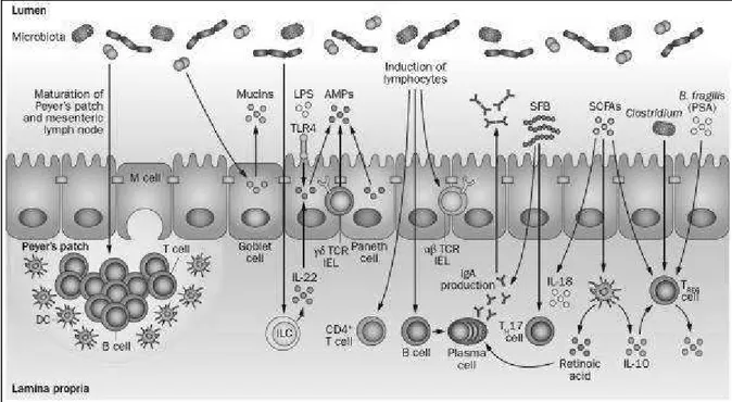
Differentiation of B cells to IgA-producing plasma cells is also dependent on the microbiota [133,134]. Differentiation of B cells to IgA-producing plasma cells is also dependent on the microbiota. [133,134] Signals from the microbiota induce production of antimicrobial peptides (AMPs), such as RegIIIγ, from Paneth cells, γδTCR+ IELs and epithelial cells [135-137]. The gut
microbiota also stimulates the release of mucins from goblet cells [126] The microbiota might also be critical for the development of various innate lymphoid cell (ILC) subsets, and for the production of IL-22 from group 3 ILCs [138], which in turn stimulates AMP release from epithelial cells [139,140]. Thus, microbiota–host interactions are key in the development of normal functional and immune responses to gut luminal antigens.
25
Figure 8. Development of the gut microbiota. The composition and density of the microbiota varies along the
length of the intestine as well as with age. a | Differences in microbial composition and density are observed along the length of the gastrointestinal tract, with much lower densities and greater variability in the proximal intestine. b | The gut microbiota fluctuates over the first 2–3 years of life, with high interindividual variability and low diversity, but becomes more stable over time.
When studied individually, particular members of the gut microbiota can differentially modulate host responses (Figure 9). For example, flagellin, a bacterial structure that stimulates Toll-like receptor (TLR)5, can stimulate RegIIIγ production from epithelial cells via IL-22 release from dendritic cells [141]. Flagella are associated with pathogenicity by promoting bacterial motility, cell adhesion and biofilm formation, constitute a virulence factor that can modulate host immune responses and are present in bacteria such as Escherichia coli and Salmonella [142,143].
Furthermore, segmented filamentous bacteria (SFB) are potent inducers of TH17 cells in mice
[140,141]. A murine community of eight commensals, or altered Schaedler flora, induce balanced immune responses, which includes TREG cells as well as TH17 cells, but to a lesser extent than
SFB. On the other hand, monocolonization of mice with Clostridium or Bacteroides fragilis induces colonic TREG-cell differentiation [146-148]. Bacterial products, such as B. fragilis-derived
polysaccharide A or short-chain fatty acids (SCFAs; for example, acetic acid, propionic acid and butyric acid), have also been shown to induce TREG cells [152].
Products of bacterial metabolism (SCFAs) have also been shown to induce IL-18 production from epithelial cells and promote tolerogenic dendritic cells, which produce IL-10 and retinoic acid [153].
26
Figure 9. Gut microbiota shapes host immunity. The gut microbiota induces maturation of the gastrointestinal
lymphoid tissue (Peyer's patches, MLN). Signals from the microbiota induce production of AMPs, such as RegIIIγ, from Paneth cells, γδTCR+ IELs and epithelial cells. Microbial signals can also stimulate the development of ILC subsets, including IL-22-producing ILCs. Flagellin or LPS can stimulate AMP production from epithelial cells via IL-22 or TLR4, respectively. The gut microbiota also stimulates the release of mucins from goblet cells, and microbes influence the development of T-cell subsets, including CD4+ T cells, αβTCR+ IELs, and are critical for the induction of IgA-producing plasma cells. and SCFAs stimulate T
REG -cell differentiation. SCFAs can also promote IL-18 production from IL-18 production from epithelial -cells and promote IL-10 and retinoic acid production from DCs, which in turn promotes differentiation of TREG cells and IgA-producing plasma cells.
Overall, these studies suggest that induction of immune responses by the gut microbiota is influenced not only by the presence or absence of live bacteria (germ-free versus colonized conditions), but also by the relative abundance of particular members of the microbiota and their by-products. Thus, given the importance of host–microbial interactions on host immunity and physiology, disruptions in gut microbiota composition or function (dysbiosis) might have important implications for health and disease. Indeed, dysbiosis has been described in a number of chronic inflammatory diseases [153-154]. However, the overall contribution of dysbiosis from disruption of homeostasis to disease development is not well understood.
4.1 The gut microbiota in coeliac disease
Approximately 30% of the general population carry the HLA-DQ2/8 coeliac disease susceptibility genes; however, only 2–5% of these individuals will go on to develop coeliac disease, suggesting that additional environmental factors contribute to disease development [155]. The additional factors that influence coeliac disease development are unknown, but might include alterations in the intestinal
27
microbiota. Indeed, some studies have demonstrated that patients with active coeliac disease have altered faecal and duodenal microbiota compositions compared with healthy individuals, which is partially restored after treatment with a gluten-free diet. Specifically, changes in the abundance of Firmicutes and Proteobacteria have been detected in children and adults with active coeliac disease [156-157]. Other studies have reported decreases in the proportion of protective, anti-inflammatory bacteria such as Bifidobacterium, and increases in the proportion of Gram-negative bacteria such as Bacteroides and E. coli, in patients with active coeliac disease [160]. Increases in the number of Staphylococcus and Clostridium [161] and decreases in Lactobacillus spp. have also been reported in children with coeliac disease. Altered diversity and altered metabolic function (SCFAs) of the microbiota have also been reported in patients with coeliac disease [164]. A study demonstrated that the microbial composition of the gut in patients with coeliac disease was associated with the clinical manifestation of disease. The gut microbiota in patients experiencing gastrointestinal symptoms was dominated by Proteobacteria, whereas the microbiota of patients with dermatitis herpetiformis or individuals experiencing dyspepsia (controls) was dominated by Firmicutes [157]. Increases in the number of Proteobacteria were also detected in patients with coeliac disease who were experiencing persistent symptoms, despite having normal histology and adhering to a gluten-free diet [165]. Together, these studies demonstrate that there are differences in microbial composition between patients with coeliac disease and healthy individuals as controls; however, the literature has not revealed a typical `coeliac microbiota signature'. This scenario is not unlike other chronic inflammatory gastrointestinal diseases, such as IBD or IBS, for which evidence supports an association between altered microbial composition and disease states [165]. However, consensus across studies with respect to the specific changes involved is lacking and a disease-specific microbial signature has not yet been defined [166-168] 8figure 10). Differences in the age of the study population (children versus adults), methodology (fluorescence in situ hybridization-PCR, denaturing gradient gel electrophoresis, 16s ribosomal RNA sequencing), sampling technique (biopsy versus faecal sample), length of gluten-free diet and the clinical presentation of disease could contribute to inconsistent findings in the literature. These differences make it difficult to compare across studies and determine whether the gut microbiota contributes to coeliac disease development or progression, or whether it is simply a consequence of the disease. Moreover, the exact mechanisms through which the gut microbiota might influence coeliac disease onset or progression is unknown, but could include activation of innate immune system, modulation of the epithelial barrier, or exacerbation of the gliadin-specific immune response.
28
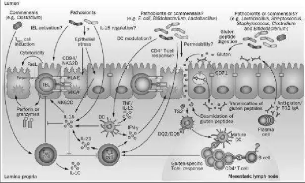
Figure 10. Potential microbial modulation of coeliac disease pathogenesis. Gluten peptides in the small
intestinal lumen translocate the epithelial barrier, via transcellular or paracellular mechanisms and are deamidated by tissue TG2 in the lamina propria. Deamidated gliadin peptides are taken up by lamina propria DCs, inducing a proinflammatory gluten-specific CD4+ T-cell response, characterized by IFN-γ and IL-21 production, and anti-gliadin and anti-TG2 antibody production by B cells in genetically predisposed hosts. Activation of the innate immune response is also a key initial step in coeliac disease.
5. “Gluten Friendly™” Technology
“Gluten Friendly™” technology is a method for the detoxification of gluten proteins from grains of cereals, in particular from the grains of wheat, aimed to obtain detoxified flours for the preparation of bread and pasta products, from wheat, preferably suitable for the alimentation of patients with celiac disease, but also adequate for its organoleptic characteristics and for its aspect, for the alimentation of the whole population. This is an Italian patented method n°: 0001414717, also filed under the Patent Cooperation Treaty, application no. PCT/IB2013/000797 and published in Europe as EP 2903453 A1 and titled “Detoxification method of gluten proteins from cereal grains”. This technology implies the application of microwave energy for a few seconds to hydrated wheat kernels before milling to reach a high temperature for a short amount of time so to induce changes in proteins that reduce the immunogenicity in vitro of the most common epitope involved in cealic disease (Lamacchia et al., 2015aItalian Patented Method N. 0001414717 (2015)[169]. Metodo per la detossificazione delle proteine del glutine dalle granaglie dei cereali. Inventors: Lamacchia, C., Di Luccia, A., Gianfrani, C (Lamacchia et al., 2015b; Lamacchia, C., Di Luccia, A. & Gianfrani C. (2015). Method for the detoxification of gluten proteins from grains of cereals. BioVaria Congress,
29
Europe’s top technologies, Munchen, Germany, https://www.biovaria.org/past-event/ technologies/)[170] preserving the technological properties of the flour.
The “Gluten Friendly™” technology is based on the analysis of recent studies in which Lamacchia and others (2010) have reported that, when the high temperatures are applied to the caryopsis of wheat, the proteins undergo changes that are not similar to those seen in model systems, consisting only of gluten (Schofield et al., 1983; Singh and MacRitchie, 2004), nor to those seen in the pasta during the drying cycles. In particular, albumins and globulins are not incorporated in the polymers of high molecular weight but coagulate and interact with gliadins forming an aggregate of molecular weight intermediate to that of gliadins and albumins and globulins revealed as a new peak called "Intermediate Protein" (IP) peak. The participation of omega-gliadins to these changes suggests that the interaction between the proteins takes place not only through the formation of disulfide bonds but also through the formation of covalent bonds involving tyrosine residues.
The researchers Lamacchia and others (2010) explained this phenomenon on the basis of the fact that in the caryopsis of wheat, gluten is not yet formed and gluten proteins are deposited in different protein bodies (Rubin et al., 1992, Krishnan et al., 1986; Lending et al., 1989). In a recent study, Tosi and others (2009) confirmed, in fact, that the HMW are particularly abundant in the innermost layer of the caryopsis of wheat (endosperm) and practically absent in the subaleuronic layer which, however, is rich in gliadins and LMW. This pattern of deposition is maintained throughout all the development phase of the caryopsis of wheat and continues even after the merger of protein bodies and the formation of the starchy matrix.
Therefore, the segregation of gluten proteins in protein bodies when they are in the caryopsis and the application of high temperatures in this stage before the milling, would allow such proteins of experiencing structural changes such as not to make them recognizable anymore by intestinal transglutaminase, thereby blocking the waterfall of inflammatory cytokine.The technology has since been further improved (Italian priority patent n_: 102015000084813. Method for the detoxification of gluten proteins from grains of cereals and related medical uses filed on 17th December 2015. Inventor: Lamacchia C.).