Journal of the American Heart Association
ORIGINAL RESEARCH
Serum Activity Against G Protein–Coupled
Receptors and Severity of Orthostatic
Symptoms in Postural Orthostatic
Tachycardia Syndrome
Isabella Kharraziha, MD; Jonas Axelsson, MD, PhD; Fabrizio Ricci, MD, PhD; Giuseppe Di Martino, MD; Margaretha Persson, PhD; Richard Sutton, MBBS, DSc; Artur Fedorowski, MD, PhD*; Viktor Hamrefors, MD, PhD*
BACKGROUND: Postural orthostatic tachycardia syndrome (POTS) is characterized by excessive heart rate increase on standing and orthostatic intolerance. Previous data indicate autoimmune involvement. We studied serum activity against G protein– coupled receptors in relation to symptoms in patients with POTS and controls using a commercial cell-based assay.
METHODS AND RESULTS: Forty-eight patients with POTS (aged 28.6±10.5 years; 44 women) and 25 healthy individuals (aged 30.7±8.6 years; 21 women) were included. The 10-item Orthostatic Hypotension Questionnaire (OHQ) was completed by 33 patients with POTS and all controls. Human embryonic kidney 293 cells overexpressing one G protein–coupled receptor: adrenergic α1 receptor, adrenergic β2 receptor, cholinergic muscarinic type 2 receptor, and opioid receptor-like 1 were treated with sera from all patients. Receptor response was analyzed using a β-arrestin–linked transcription factor driving transgenic β-lactamase transcription by fluorescence resonance energy transfer method. Receiver operating characteristic curves were constructed. G protein–coupled receptor activation was related to OHQ indices in linear regression models. Sera from pa-tients with POTS activated all 4 receptors to a higher degree compared with controls (P<0.01 for all). The area under the curve was 0.88 (0.80–0.97, P<0.001) combining all 4 receptors. Adrenergic α1 receptor activation associated with OHQ composite score (β=0.77 OHQ points per SD of activity, P=0.009) and with reduced tolerability for prolonged standing (P=0.037) and walking for short (P=0.042) or long (P=0.001) periods. All 4 receptors were associated with vision problems (P<0.05 for all).
CONCLUSIONS: Our results indicate the presence of circulating proteins activating adrenergic, muscarinic, and nociceptin re-ceptors in patients with POTS. Serum-mediated activation of these rere-ceptors has high predictive value for POTS. Activation of adrenergic α1 receptor is associated with orthostatic symptoms severity in patients with POTS.
Key Words: adrenergic receptors ■ autoimmunity ■ G protein–coupled receptors ■ orthostatic intolerance ■ postural orthostatic tachycardia syndrome
P
ostural orthostatic tachycardia syndrome (POTS) is a disorder of unknown cause characterized by orthostatic intolerance and increased heart rate (HR) of 30 beats per minute during orthostasis, in the absence of orthostatic hypotension.1 In additionto orthostatic intolerance, patients with POTS may
experience debilitating symptoms only partly related or unrelated to orthostasis, including light-headed-ness, nausea, blurred vision, fatigue, mental confusion (“brain fog”), chest pain, and gastrointestinal problems.2
Syncope may occur although presyncopal symp-toms are more common. Several potential underlying
Correspondence to: Isabella Kharraziha, MD, Department of Clinical Sciences, Lund University, Malmö, Clinical Research Centre, Box 503 32, 202 13 Malmö, Sweden. E-mail: [email protected]
Supplementary Materials for this article are available at https://www.ahajo urnals.org/doi/suppl/ 10.1161/JAHA.120.015989 *Dr Fedorowski and Dr Hamrefors are co-senior authors.
For Sources of Funding and Disclosures, see page 9.
© 2020 The Authors. Published on behalf of the American Heart Association, Inc., by Wiley. This is an open access article under the terms of the Creative Commons Attribution-NonCommercial-NoDerivs License, which permits use and distribution in any medium, provided the original work is properly cited, the use is non-commercial and no modifications or adaptations are made.
JAHA is available at: www.ahajournals.org/journal/jaha
mechanisms have been suggested for POTS including autonomic denervation, hypovolemia, hyperadrenergic stimulation, and autoantibodies against adrenergic re-ceptors.2–4 However, none of these proposed
mecha-nisms has yet led to effective treatment.
It has been observed that some patients develop POTS following an infection. This observation has given rise to the hypothesis of an autoimmune-me-diated cause of POTS. The fact that the majority of patients with POTS are women, who are more sus-ceptible to developing autoimmune diseases, further supports the autoimmune hypothesis.2 Additionally,
nearly one fourth of patients with POTS have positive antinuclear antibodies and a higher prevalence of au-toimmune diseases such as Hashimoto disease and Sjögren syndrome.5 Finally, we and others have
pre-viously demonstrated presence of antibodies against adrenergic and cholinergic GPCRs (G protein–coupled receptors) in POTS.6–9
GPCRs constitute a large family of receptors that detect molecules outside the cell and activate inter-nal siginter-naling pathways, ultimately leading to cellular responses.10,11 Over time, antibodies against GPCRs
have been linked to a spectrum of conditions, includ-ing POTS.10,12 Thus, we aimed to expand previous
observations by studying serum activity against spe-cific GPCRs, cardiovascular ADRA1 (adrenergic α1 receptor), ADRB2 (adrenergic β2 receptor), CHRM2 (cholinergic muscarinic type 2 receptor), and noci-ception-related OPRL1 (opioid receptor-like 1) in pa-tients with POTS and matched controls, and relate the serum-mediated activation to specific orthostatic symptoms.
METHODS
The data that support the findings of this study are available from the corresponding author upon reason-able request.
Study Population
A total of 48 patients with POTS and 25 healthy con-trols were recruited for study from a tertiary referral center incorporating both syncope unit and cardio-vascular autonomic laboratory at Skåne University Hospital, Malmö, Sweden. The patients and con-trols were recruited between January and December 2018. All patients with POTS had confirmed diagno-ses by one of us with special expertise in POTS (A.F.). Blood samples were collected from all 73 patients and sent for analysis at the Center for Apheresis and Stem Cell Handling at Karolinska University Hospital in Stockholm, Sweden. Of the 73 participants, 48 (33 POTS and 25 controls) performed orthostatic tests and answered the Orthostatic Hypotension Questionnaire (OHQ) during their blood sample collection visit at the Clinical Research Unit at Skåne University Hospital in Malmö. Blood samples from the other 15 patients with POTS were collected and sent to Karolinska University Hospital from local hospitals and primary care facilities
CLINICAL PERSPECTIVE
What Is New?
• Serum of patients with postural orthostatic tachycardia syndrome (POTS) demonstrates activity against cardiovascular and nociceptive G protein–coupled receptors, and such activa-tion is highly predictive of POTS diagnosis.
• Serum-mediated adrenergic α1 receptor
activ-ity is associated with the severactiv-ity of orthostatic symptoms in POTS, independently of the ortho-static hemodynamic response.
What Are the Clinical Implications?
• Our findings provide new insights in thepatho-physiology of POTS and prompt further re-search on a possible autoimmune involvement in POTS.
• Measurement of G protein–coupled receptor activity may be added as a diagnostic tool for POTS, even though the optimal panel of recep-tors and specific cutoff values are yet to be determined.
• POTS is likely to be a heterogeneous disease, and it remains to be explored whether detec-tion of autoimmune G protein–coupled receptor activity may identify different subtypes of POTS, potentially responding to different treatments.
Nonstandard Abbreviations and Acronyms
ADRA1 adrenergic α1 receptor
ADRB2 adrenergic β2 receptor
CHRM2 cholinergic muscarinic type 2 receptor
FRET fluorescence resonance energy transfer
GPCR G protein–coupled receptor
HR heart rate
IVIG intravenous immunoglobulin
OHDAS Orthostatic Hypotension Daily Activity Scale
OHQ Orthostatic Hypotension Questionnaire
OHSA Orthostatic Hypotension Symptom
Assessment
OPRL1 opioid receptor-like 1
POTS postural orthostatic tachycardia
syndrome
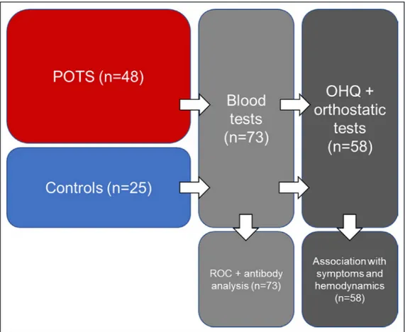
around Sweden, which is why these 15 participants did not complete the OHQ or orthostatic test at the time of their blood sample collection visit. All patients with POTS had a previous positive tilt test, which qualified them as confirmed cases in this study. Controls did not perform a formal tilt testing. Active standing tests were performed in both patients and controls during the study visit when the blood samples were collected and the study questionnaire was filled in. Controls’ active standing test results were negative. The study population is depicted in Figure 1. All patients provided written informed consent. The study was approved by the regional ethical review board in Lund (DNR 08/82 and 17/295) and all procedures were performed in ac-cordance with the Declaration of Helsinki.
Orthostatic Hypotension Questionnaire
The OHQ is a questionnaire that has been previously validated and used for orthostatic hypotension13 buthas also been used for quantification of POTS-related symptoms.2,14 The OHQ is divided into 2 subgroups:
Orthostatic Hypotension Symptom Assessment (OHSA) and Orthostatic Hypotension Daily Activity Scale (OHDAS). OHSA consists of 6 questions: (1)
dizziness, light-headedness, feeling faint, or feeling like you might blackout; (2) problems with vision (eg, blur-ring, seeing spots, and tunnel vision); (3) generalized weakness; (4) fatigue; (5) trouble concentrating; and (6) head/neck discomfort. OHDAS consists of 4 questions that assess the impact of symptoms on daily activities (standing for long and short duration, walking for short or long duration). The recall period is “over the past week.” The items are scored on a scale from 0 to 10, with 0 indicating no symptoms and 10 indicating the worst possible symptoms. The composite OHQ score is calculated by averaging the OHSAS and the OHDAS. Activities that are marked as zero or “cannot be done for other reasons” at baseline are not included in the scoring system. In this study, the OHQ was translated into Swedish and validated by an expert in the field of health status assessment.
Measurement of GPCR Activity
Sera from all patients with POTS and controls were analyzed by a fluorescence resonance en-ergy transfer (FRET)–based reporter system (Tango GeneBLAzer, Thermo Fisher Scientific) based on a β2 -arrestin–linked transcription factor driving transgenic
Figure 1. Study population flow chart.
Orthostatic Hypotension Questionnaire (OHQ) and orthostatic test were completed by 33 patients with postural orthostatic tachycardia syndrome (POTS) and 25 controls at the time of blood sampling. ROC indicates receiver operating characteristic.
β-lactamase transcription. The FRET-based method has been previously described in greater detail.15
Human embryonic kidney 293 cells overexpressing one of the GPCRs, ADRA1, ADRB2, CHRM2, and OPRL1, were plated and allowed to reattach during 48 hours. Cells were treated with 10% sera diluted in Roswell Park Memorial Institute medium for 5 hours, followed by addition of the FRET substrate, incuba-tion for 60 minutes, and quantificaincuba-tion analysis in a CLARIO Star multipurpose plate reader. GPCR activ-ity was measured as the ratio between emission of cleaved and noncleaved FRET substrate. The selec-tion of the ADRB2 over the ADRB1 was based on our previous results,8 which showed that the
combina-tion of the 2 adrenergic receptors, α1 and β2, pro-vides the highest discriminative efficacy in regard to patients with POTS (≈94%).
Statistical Analysis
OHQ scores were compared according to the me-dian of serum activation, using independent sam-ples Student t test. Receiver operating characteristic curves were constructed to analyze the predictive value of GPCR activity for POTS. A logistic model with all 4 GPCRs as POTS predictors was performed and a predicted value for every individual was calcu-lated. Quantification of the activation of the GPCRs was log-transformed and related to the OHQ com-posite and individual items scores in age-adjusted linear regression models. In addition, the relationship between GPCR and the OHQ scores were tested in linear regression models, including the change in HR and systolic blood pressure after 3 minutes of orthostatic test as additional covariates. Data were analyzed using SPSS software version 25 (IBM). A 2-sided P value <0.05 was considered significant for
all tests. P values are displayed unadjusted for mul-tiple testing; however, all results were interpreted ac-counting for multiple testing.
RESULTS
Study Population Characteristics
The mean age in the POTS and control groups was 28.6±10.5 years and 30.7±8.6 years, respectively. Among patients with POTS, 44 (91.7%) were women, and among controls, 21 (84%) were women. Mean OHQ score was 6.36±1.68 in patients with POTS and 0.67±1.03 in con-trols (P<0.001). A total of 36 patients with POTS were treated with HR-regulating and/or vasoactive agents by the time of the completion of the symptom questionnaire and when blood was drawn (Table S1). By inclusion cri-teria, no control patients were taking HR-regulating or vasoactive medications. Three patients with POTS and 2 controls reported use of levothyroxine for hypothyroidism (P for difference in proportions between groups=0.799). Exclusion of these 5 patients from the analyses did not substantially change the results, which is why they are included in the following results. Study population char-acteristics are shown in Table 1.
Receptor Activity in Patients With POTS
Compared With Controls
The mean receptor activity was significantly higher in patients with POTS compared with controls for all 4 receptors (Figure 2A through 2D; Table S2). The obtained area under the curve was 0.88 (0.80–0.97, P<0.001) when analyzing all 4 receptors (Figure 3A). The area under the curve when analyzing receptors individually was 0.72 (0.58–0.85, P<0.001) for ADRA1 was 0.76 (0.64–0.88, P<0.001), for ADRB2 was 0.73 (0.60–0.87, P<0.001) for CHRM2, and 0.75 (0.62– 0.88, P<0.001) for OPRL1, respectively (Figure 3B through 3E). Some GPCR activity was seen in all patients with POTS and all controls. However, 42 patients with POTS (87.5%) had at least one value above the 75th percentile in control patients for the respective receptor (Figure S1). Correlations for re-ceptor activity in patients with POTS and controls are shown in Tables S3 and S4.
Association Between Receptor Activity
and OHQ Score in Patients With POTS
The OHQ composite score was higher in those pa-tients with above the median serum ADRA1 activation (P=0.043), but not in ADRB2, CHRM2, or OPRL1 in patients with POTS (Table 2). There were no significant associations in the control group.Serum ADRA1 activation was associated with the OHQ composite score (β=0.77, OHQ points per SD of Table 1. Characteristics of the Study Population
Patients With POTS (n=48) Controls (n=25) P Value Age, y 28.6±10.5 30.7±8.6 0.394 Women, n. 91.7 84.0 0.320 SBP, mm Hg 116.73±12.46* 113.60±8.33 0.283 HR, beats per min 69.39±12.27* 65.14±9.87 0.161 ΔSBP, 3 min −1.00±7.91† 1.24±5.97 0.252
ΔHR, 3 min 26.32±11.75‡ 17.64±9.72 0.005 OHQ score 6.36±1.68* 0.67±1.03 < 0.001
Age, heart rate (HR), and systolic blood pressure (SBP) are expressed as mean±SD, whereas the proportion of women and different medications are expressed as percentages of total within each group. ΔHR and ΔSBP refer to the change in HR and SBP from supine to 3 minutes of active standing. P values denote P for independent samples t test or chi-square test, respectively. OHQ indicates Orthostatic Hypotension Questionnaire; and POTS, postural orthostatic tachycardia syndrome.
Missing values: *n=15; †n=19; ‡n=17.
activity; P=0.009), whereas there were no significant associations among controls (P=0.953). The associa-tion between ADRA1 and total OHQ also remained sig-nificant after adjusting for increase in HR and decrease in systolic blood pressure after 3 minutes (P=0.031). ADRA1 activation was associated with symptoms during prolonged standing (P=0.037) and walking for short (P=0.042) or long (P=0.001) periods. All 4 receptors were associated with a higher score for vision problems (ADRA1, P<0.001; ADRB2, P=0.011; CHRM2, P=0.014; and OPRL1, P=0.003). In addition, OPRL1 activity was associated with symptoms during prolonged walking (P=0.035). The activity of ADRA1, ARB2, CHRM2, and OPRL1 was not associated with OHQ composite score in controls. The full results are shown in Table S5.
A detailed clinical assessment revealed that 2 pa-tients in the control group had treated hypothyroidism and another patient had rheumatoid arthritis. However, results did not differ when we excluded these 3 pa-tients from the control group (data not shown).
DISCUSSION
We report that sera from patients with POTS activate the 4 GPCRs, ADRA1A, ADRB2, CHRM2, and OPRL1, to a significantly higher degree compared with sera from controls. We also show that such GPCR activity is highly predictive of POTS, as indicated by receiver operating characteristic analyses. The ADRA1 activity is associ-ated with severity of orthostatic symptoms, measured by the OHQ score, in patients with POTS and this associa-tion is partly independent of the hemodynamic response during orthostasis. Moreover, activity in all 4 GPCRs cor-relates specifically with symptoms of disturbed vision.
Role of GPCRs in POTS
The present results are in line with previous research indicating that there may be autoimmune involvement in POTS, targeting specific GPCRs. Previous studies have detected various autoantibodies in POTS.6,7,9,16,17
In the present study, activity towards specific GPCRs rather than presence of autoantibodies was measured. The FRET-based method detects changes in recep-tor conformation, which can be caused by allosteric or orthosteric binding of a ligand or antibody. The hy-pothesis is that these conformational changes seen in the present study could be caused by autoantibodies. After binding to GPCR, autoantibodies can yield stimu-latory and inhibitory effects.11 Previous studies have
detected increased levels of autoantibodies against ADRA1 and ADRAB1/2 in patients with POTS com-pared with healthy controls.6,7 Interestingly, IgG from
POTS was found to shift the ADRA1 dose-response curve to the right after phenylephrine administration, indicating a partial antagonistic effect on ADRA1. This Figure 2. Receptor activation (y axis) shown as the
ratio between emitted light from cleaved substrate and noncleaved substrate.
A, ADRA1 (adrenergic α1 receptor) activation in patients with
postural orthostatic tachycardia syndrome (POTS) and controls. B, ADRB2 (adrenergic β2 receptor) activation in patients with
POTS and controls. C, CHRM2 (cholinergic muscarinic 2 receptor) activation in patients with POTS and controls. D, OPRL1 (opioid-receptor-like 1) activation in patients with POTS and controls. P values denote the difference between mean values, using independent samples t test for the log-transformed receptor activity. Please note that by design, one extreme outlier in the POTS group with a value of 14.838 for ADRA1 activity and 26.709 for CHRM2 activity, respectively, is not displayed in the figures.
could potentially block the effects of endogenous nor-epinephrine on ADRA1, which, in turn, would lead to impaired vasoconstriction and increased baroreceptor activation, consecutively leading to increased sym-pathetic activity. In contrast, the ADRB1/2 effect was found to be the opposite and shifted the ADRB1/2
dose-response curve to the left after isoproterenol ad-ministration; hence, the IgG from POTS had a stimu-latory effect on ADRB1/2. The relatively unprotected ADRB1/2 would respond to increased sympathetic activity and circulating catecholamines with reflex tachycardia.6,7 These inhibitory and stimulatory effects Figure 3. The predictive value of specific G protein–coupled receptors (GPCRs) in postural orthostatic tachycardia syndrome (POTS).
Receiver operating characteristic (ROC) curves for all 4 GPCRs (A) and the individual receptors (B through E) for the diagnosis of POTS in the 73 patients. A, ROC curve for all 4 GPCRs (ADRA1 [adrenergic α1 receptor], ADRB2 [adrenergic β2 receptor], CHRM2
[cholinergic muscarinic 2 receptor], and OPRL1 [opioid receptor-like 1]) for the diagnosis of POTS. B, ROC curve for ADRA1 for the diagnosis of POTS. C, ROC curve for ADRB2 for the diagnosis of POTS. D, ROC curve for CHRM2 for the diagnosis of POTS. E, ROC curve for OPRL1 for the diagnosis of POTS. AUC indicates area under curve.
0 50 100 0 50 100 100 - Specificity, % Se ns itiv ity, % AUC 0.88 95%CI 0.80-0.97 A 0 50 100 0 50 100 100 - Specificity, % Se nsi tiv ity , % ADRA1 AUC 0.72 95%CI 0.58-0.85 B 0 50 100 0 50 100 100 - Specificity, % Se nsi tiv ity , % ADRB2 AUC 0.76 95%CI 0.64-0.88 C 0 50 100 0 50 100 100 - Specificity, % Sen si tivi ty , % CHRM2 AUC 0.73 95%CI 0.60-0.87 D 0 50 100 0 50 100 100 - Specificity, % Sen si tiv ity, % OPRL1 AUC 0.75 95%CI 0.62-0.88 E
provide an interesting pathophysiological explanation for the cardiovascular effects associated with upright posture in patients with POTS.
The predictive value of GPCR activity for a diag-nosis of POTS was analyzed using receiver operat-ing characteristic, showoperat-ing an excellent prediction of the diagnosis in POTS (area under the curve, 0.88) when combining all 4 receptors. The area under the curve for the individual receptors were between 0.72 and 0.76. These findings indicate that measurement of GPCR activity may be added as a diagnostic tool for POTS, even though the optimal panel of receptors and specific cutoff values are yet to be determined. POTS is likely to be a heterogeneous disease, and it remains to be explored whether detection of autoim-mune GPCR activity may identify different subtypes of POTS, potentially responding to different treatments.
Orthostatic Symptoms in Relation to
Receptor Activity
A correlation between the severity of symptoms and the presence of autoantibodies in POTS has been ob-served in a previous study by Gunning et al. This study used a method of calculating symptom severity in OH, another form of orthostatic intolerance with overlap in clinical presentation, revealing a weak correlation with all 9 receptor subtypes of autoantibody concentration (ADRA1/2, ADRB1/2, and CHRM1–5) and the severity of orthostatic symptoms.9 The same study indicated
that 89% of patients with POTS had autoantibodies against ADRA1 whereas antibodies against other adr-energic and muscarinic antibodies were less prevalent. Interestingly, there was a tendency that subtypes of both adrenergic (α2, β1, and β2) and muscarinic
recep-tor antibodies were not detected in the sera of patients with POTS, unless autoantibodies were expressed against the α1 adrenergic receptor subtype.9 In our
study, ADRA1 activity was more strongly associated
with the orthostatic symptoms compared with ADRB2, CHRM2 and OPRL1. Thus, ADRA1 may play a particu-larly important role in POTS. It should be emphasized that in the study by Gunning et al,9 the authors applied
a different antibody detection method, ELISA, which does not include the whole cell assay but only specific isolated epitopes derived from GPCRs.
In addition to adrenergic receptors, we included CHRM2 and OPRL1. CHRM2 autoantibodies were first detected in patients with Chagas disease and have later been found in patients with dilated cardiomyopathy.11
The CHRM2 was reported to have a negative chrono-tropic effect in cultured cardiomyocytes.18 In our study,
CHRM2 activity was greater in patients with POTS com-pared with controls but did not correlate with symptom severity. This is in contrast to a previous study, where symptoms correlated with all 5 different muscarinic re-ceptors.9 The strongest correlation in that study;
how-ever, was seen in CHRM4, which was not included in our study.9 In contrast to adrenergic and muscarinic
recep-tors, there are no published studies regarding the role of OPRL1 in POTS and other syndromes of orthostatic intolerance. The OPRL1 is involved in pain perception in humans.19 Thus, OPRL1 may provide a clue to the
symptom of chronic pain at various locations, which is often described by patients with POTS.2 OPRL1
activ-ity was greater in patients with POTS compared with controls and was associated with vision problems and walking for long distances. However, the associations were not strong. It is still unclear whether the increased activities seen in CHRM2 and OPRL1 are caused by stimulatory or inhibitory effects and the specific role of these 2 receptors in POTS is yet to be further explored.
As demonstrated in the present study, there was strong association between vision disturbances and all 4 GPCRs in patients with POTS. Disturbed vision is a common symptom of orthostatic intolerance. The retina is more susceptible to hypoperfusion than the brain, because of the presence of the intraocular Table 2. Severity of Symptoms in Relation to Receptor Activity in Patients With POTS (n=33)
ADRA1 Above the Median ADRA1 Below the Median P Value
OHQ score 6.94±1.18 5.74±1.95 0.043*
ADRB2 Above the Median ADRB2 Below the Median
OHQ score 6.42±1.41 6.14±1.88 0.638*
CHRM2 Above the Median CHRM2 Below the Median
OHQ score 6.62±1.30 5.95±1.93 0.260*
OPRL1 Above the Median OPRL1 Below the Median
OHQ score 6.39±1.67 6.32±1.74 0.904
The composite Orthostatic Hypotension Questionnaire (OHQ) score in relation to specific receptor activation. ADRA1 indicates adrenergic α1 receptor;
ADRB2, adrenergic β2 receptor; CHRM2, cholinergic muscarinic 2 receptor; OPRL1, opioid receptor-like 1; and POTS, postural orthostatic tachycardia
syndrome.
Missing values: *n=1.
pressure, which adds an impediment to eye perfusion that is not present in cerebral circulation.20 However,
the decrease in blood pressure when upright in pa-tients with POTS is usually only modest or nonexistent, unless the patient experiences presyncope/syncope, caused by vasovagal reflex activation.3 The autonomic
nervous system influences numerous ocular functions such as controlling pupil size, accommodation of the lens, regulation of ocular blood flow, and intraocular pressure.21 This may explain why patients with
auto-nomic dysfunction, such as those with POTS, may experience impaired vision. Interestingly, OPRL1 was also associated with impaired vision. Of interest in rela-tion to our findings, opioid receptors have been impli-cated in the regulation of iris function22 and regulation
of intraocular pressure,23 which could explain why
pa-tients with increased activity in OPRL1 might have vi-sion disturbances.
Immunomodulatory Treatments in
Patients With POTS
The present results support the hypothesis that POTS is an autoimmune disease, which, in turn, may suggest that some patients with POTS could benefit from im-munomodulatory therapies. Controlled treatment trials are in progress to determine whether immunomodula-tory therapies may be effective in certain POTS sub-groups.12 At this time, a few case reports have been
published describing improvements in POTS symp-toms after intravenous immunoglobulin (IVIG), rituxi-mab, autologous adipose stem cell infusions, and plasmapheresis in highly selected cases with comor-bid autoimmunity.24–28 A case report on a 32-year-old
patient with POTS demonstrated a positive response to plasma exchange and improved OHQ score.29
A retrospective study30 including 38 patients with
dysautonomia of various types, including 26 patients with POTS, reported that 83.5% improved while taking IVIG. The mean time to the first sign of response was 5.3 weeks and the study reported no serious adverse events. GPCRs were not analyzed but antiphospho-lipid antibodies and novel Sjögren antibodies were often found to be present and correlated with a high response rate to IVIG. There is increasing evidence that IVIG is safe and effective in a subset of patients with autonomic disorders and evidence of autoimmunity. According to this study,29 a 4-month IVIG trial should
be considered in severely affected patients who are refractory to lifestyle and pharmacological therapies. Taken together, these above reports on efficacy of im-munomodulatory therapy in select patients with POTS lack both systematic assessment of serum positivity for autoantibodies against GPCRs as entrance crite-rion and randomization of patients to a placebo-con-trolled arm.
Limitations
Our study has a number of important limitations. First, the sample size is small and our findings should be externally validated in larger cohorts. Second, the OHQ was previously validated in orthostatic hypoten-sion and not specifically in POTS. In addition to symp-toms of orthostatic intolerance, patients with POTS may demonstrate a number of additional symptoms, including cognitive impairment, gastrointestinal prob-lems, and unexplained pain,2 which are not specifically
captured by the OHQ. Third, as already described in the Methods section, we did not measure ADRB1 ac-tivity. Fourth, the majority of patients with POTS were treated with HR-regulating and/or vasoactive medica-tions when completing symptom questionnaire and when the blood samples were drawn. However, these patients still reported significant symptoms, as indi-cated by the OHQ. Finally, as previously mentioned, receptor activity rather than antibodies was meas-ured in the current study, and it is possible that the conformational changes observed in the GPCR could be the result of something other than autoantibodies. However, since several previous studies have identi-fied antibodies against these GPCRs, it may be per-missible to assume that the increased activity in the specific receptors is a consequence of autoimmune disease.
CONCLUSIONS
Serum of patients with POTS demonstrates activity against cardiovascular and nociceptive GPCRs and such activation is highly predictive of POTS diagnosis. Serum-mediated ADRA1A activity is associated with the severity of orthostatic symptoms in patients with POTS, indepen-dently of the orthostatic hemodynamic response. These findings provide new insights into the pathophysiology of POTS and prompt further research on possible autoim-mune involvement in patients with POTS.
ARTICLE INFORMATION
Received January 31, 2020; accepted June 25, 2020. Affiliations
From the Department of Clinical Sciences, Lund University (I.K., F.R., M.P., R.S., A.F., V.H.), Department of Internal Medicine, Skåne University Hospital, Malmö, Sweden (I.K., M.P., V.H.); Department of Stem Cell Therapy and Apheresis, Karolinska University Hospital, Stockholm, Sweden (J.A.); Department of Neuroscience, Imaging and Clinical Sciences, Institute for Advanced Biomedical Technologies, (F.R.), and Department of Medicine and Ageing Sciences (G.D.M.), "G. d’Annunzio" University, Chieti, Italy; National Heart and Lung Institute, Imperial College, Hammersmith Hospital Campus, London, United Kingdom (R.S.); and Department of Cardiology, Skåne University Hospital, Malmö, Sweden (A.F.).
Acknowledgments
We would like to thank Jenny Persson-Tholin and her colleagues at the Clinical Research Unit at Skåne University Hospital in Malmö for their de-voted assistance in this study.
Sources of Funding
This study was supported by grants from the Swedish Heart-Lung Foundation, Solidex, the Swedish Heart and Lung Association, the Medical Faculty of Lund University, ALF Funds, Skåne University Hospital Funds, the Crafoord Foundation, Ernhold Lundströms Research Foundation, Region Skåne, Hulda and Conrad Mossfelt Foundation, and Anna-Lisa and Sven Eric Lundgren Foundation for Medical Research.
Disclosures
Fedorowski reports personal fees from Medtronic Inc and Biotronik outside the submitted work. Hamrefors reports an educational congress grant from Boston Scientific Inc outside of the submitted work. Sutton reports per-sonal fees and other from Medtronic Inc. and St. Jude Medical Inc. (Abbott Laboratories) outside the submitted work; is a member of the Speakers’ Bureau at St. Jude Medical/Abbott Inc.; is a shareholder in Boston Scientific Inc. and Edwards Lifesciences Inc; and performs consultancy for Medtronic Inc. The remaining authors have no disclosures to report.
Supplementary Materials Tables S1–S5
Figure S1
REFERENCES
1. Schondorf R, Low PA. Idiopathic postural orthostatic tachycardia syn-drome: an attenuated form of acute pandysautonomia? Neurology. 1993;43:132–137.
2. Fedorowski A. Postural orthostatic tachycardia syndrome: clin-ical presentation, aetiology and management. J Intern Med. 2019;285:352–366.
3. Sheldon RS, Grubb BP, Olshansky B, Shen WK, Calkins H, Brignole M, Raj SR, Krahn AD, Morillo CA, Stewart JM, et al. 2015 Heart Rhythm Society expert consensus statement on the diagnosis and treatment of postural tachycardia syndrome, inappropriate sinus tachycardia, and vasovagal syncope. Heart Rhythm. 2015;12:e41–e63.
4. Arnold AC, Ng J, Raj SR. Postural tachycardia syndrome—diagnosis, physiology, and prognosis. Auton Neurosci. 2018;215:3–11.
5. Blitshteyn S. Autoimmune markers and autoimmune disorders in patients with postural tachycardia syndrome (POTS). Lupus. 2015;24:1364–1369.
6. Li H, Yu X, Liles C, Khan M, Vanderlinde-Wood M, Galloway A, Zillner C, Benbrook A, Reim S, Collier D, et al. Autoimmune basis for pos-tural tachycardia syndrome. J Am Heart Assoc. 2014;3:e000755. DOI: 10.1161/JAHA.113.000755
7. Fedorowski A, Li H, Yu X, Koelsch KA, Harris VM, Liles C, Murphy TA, Quadri SMS, Scofield RH, Sutton R, et al. Antiadrenergic autoimmunity in postural tachycardia syndrome. Europace. 2017;19:1211–1219. 8. Yu X, Li H, Murphy TA, Nuss Z, Liles J, Liles C, Aston CE, Raj SR,
Fedorowski A, Kem DC. Angiotensin II type 1 receptor autoantibodies in postural tachycardia syndrome. J Am Heart Assoc. 2018;7:e008351. DOI: 10.1161/JAHA.117.008351
9. Gunning WT III, Kvale H, Kramer PM, Karabin BL, Grubb BP. Postural orthostatic tachycardia syndrome is associated with elevated G-protein coupled receptor autoantibodies. J Am Heart Assoc. 2019;8:e013602. DOI: 10.1161/JAHA.119.013602
10. Luft FC. Activating autoantibodies and cardiovascular disease. Physiology (Bethesda). 2013;28:254–261.
11. Wallukat G, Schimke I. Agonistic autoantibodies directed against G-protein-coupled receptors and their relationship to cardiovascular diseases. Semin Immunopathol. 2014;36:351–363.
12. Vernino S, Stiles LE. Autoimmunity in postural orthostatic tachycardia syndrome: current understanding. Auton Neurosci. 2018;215:78–82. 13. Kaufmann H, Malamut R, Norcliffe-Kaufmann L, Rosa K, Freeman R.
The Orthostatic Hypotension Questionnaire (OHQ): validation of a novel symptom assessment scale. Clin Auton Res. 2012;22:79–90. 14. Wells R, Spurrier AJ, Linz D, Gallagher C, Mahajan R, Sanders P, Page
A, Lau DH. Postural tachycardia syndrome: current perspectives. Vasc Health Risk Manag. 2018;14:1–11.
15. Hanson BJ, Wetter J, Bercher MR, Kopp L, Fuerstenau-Sharp M, Vedvik KL, Zielinski T, Doucette C, Whitney PJ, Revankar C. A homo-geneous fluorescent live-cell assay for measuring 7-transmembrane receptor activity and agonist functional selectivity through beta-arrestin recruitment. J Biomol Screen. 2009;14:798–810.
16. Li J, Zhang Q, Liao Y, Zhang C, Hao H, Du J. The value of acetylcho-line receptor antibody in children with postural tachycardia syndrome. Pediatr Cardiol. 2015;36:165–170.
17. Watari M, Nakane S, Mukaino A, Nakajima M, Mori Y, Maeda Y, Masuda T, Takamatsu K, Kouzaki Y, Higuchi O, et al. Autoimmune postural ortho-static tachycardia syndrome. Ann Clin Transl Neurol. 2018;5:486–492. 18. Wallukat G, Nissen E, Morwinski R, Muller J. Autoantibodies against the
beta- and muscarinic receptors in cardiomyopathy. Herz. 2000;25:261–266. 19. Al-Hasani R, Bruchas MR. Molecular mechanisms of opioid receptor-de-pendent signaling and behavior. Anesthesiology. 2011;115:1363–1381. 20. Wieling W, Thijs RD, van Dijk N, Wilde AA, Benditt DG, van Dijk JG.
Symptoms and signs of syncope: a review of the link between physiol-ogy and clinical clues. Brain. 2009;132:2630–2642.
21. McDougal DH, Gamlin PD. Autonomic control of the eye. Compr Physiol. 2015;5:439–473.
22. Murray RB, Adler MW, Korczyn AD. The pupillary effects of opioids. Life Sci. 1983;33:495–509.
23. Drago F, Panissidi G, Bellomio F, Dal Bello A, Aguglia E, Gorgone G. Effects of opiates and opioids on intraocular pressure of rabbits and humans. Clin Exp Pharmacol Physiol. 1985;12:107–113.
24. Adamec I, Bilic E, Lovric M, Habek M. Postural orthostatic tachycar-dia syndrome (POTS) as presenting symptom of CIDP. Neurol Sci. 2016;37:1163–1166.
25. Weinstock LB, Brook JB, Myers TL, Goodman B. Successful treatment of postural orthostatic tachycardia and mast cell activation syndromes using naltrexone, immunoglobulin and antibiotic treatment. BMJ Case Rep. 2018;2018:bcr2017221405.
26. Hendrickson JE, Hendrickson ET, Gehrie EA, Sidhu D, Wallukat G, Schimke I, Tormey CA. Complex regional pain syndrome and dysau-tonomia in a 14-year-old girl responsive to therapeutic plasma ex-change. J Clin Apher. 2016;31:368–374.
27. Numan MT, Kamdar A, Young J, Butler IJ. Autologous adipose stem cell therapy for autonomic nervous system dysfunction in two young patients. Stem Cells Dev. 2017;26:391–393.
28. Blitshteyn S, Brook J. Postural tachycardia syndrome (POTS) with an-ti-NMDA receptor antibodies after human papillomavirus vaccination. Immunol Res. 2017;65:282–284.
29. Wells R, Hissaria P, Elliott AD, Sanders P, Page A, Baumert M, Lau DH. Plasma exchange therapy in postural tachycardia syndrome: a novel long-term approach? Am J Med. 2020;133:e157–e159.
30. Schofield JR, Chemali KR. Intravenous immunoglobulin therapy in re-fractory autoimmune dysautonomias: a retrospective analysis of 38 pa-tients. Am J Ther. 2019;26:570–582.
Supplemental Material
POTS (n=45) Droxidopa 33.3 Beta-blockers 35.6 Procoralan 37.8 Midodrin 6.7 Orstanorm 4.4
Displayed as % of POTS subjects. No control subjects reported use of heart rate regulating or
vasoactive medications. Data about current medications were missing for three POTS patients.
to healthy controls.
POTS (n = 48) Controls (n = 25) p-value
ADRA1 1.726 ± 2.157 * 0.923 ± 0.871 * 0.006
ADRB2 5.890 ± 5.641 *** 2.256 ± 1.615 * <0.001
CHRM2 1.861 ± 3.940 *** 0.883 ± 1.135 * 0.007
OPRL1 0.955 ± 0.857 ** 0.640 ± 0.992 0.009
Data was analysed with Independent samples t-test. Log-transformation was applied to calculate
the p-values. Receptor activity is shown as the ratio between emitted light from cleaved substrate
and non-cleaved substrate and calculated as mean value
± SD of the mean. Missing values: *n=1;
**n=2; ***n=3. ADRA1: adrenergic alpha-1; ADRB2: adrenergic beta-2; CHRM2: muscarinic
type-2; OPRL1: opioid-receptor-like 1.
ADRA1 ADRB2 CHRM2 OPRL1
ADRA1 Pearson Correlation 1 0.206 0.938** 0.418**
p-value 0.181 < 0.001 0.004
N 47 44 44 45
ADRB2 Pearson Correlation 0.206 1 0.445** 0.530**
p-value 0.181 0.002 < 0.001
N 44 45 44 45
CHRM2 Pearson Correlation 0.938** 0.445** 1 0.481**
p-value < 0.001 0.002 0.001
N 44 44 45 45
OPRL1 Pearson Correlation 0.418** 0.530** 0.481** 1
p-value 0.004 < 0.001 0.001
N 45 45 45 46
**. Correlation is significant at the 0.01 level (2-tailed).
ADRA1 ADRB2 CHRM2 OPRL1
ADRA1 Pearson Correlation 1 0.139 0.490* 0.610**
p-value 0.549 0.024 0.002
N 23 21 21 23
ADRB2 Pearson Correlation 0.139 1 -0.368 0.262
p-value 0.549 0.101 0.228
N 21 23 21 23
CHRM2 Pearson Correlation 0.490* -0.368 1 0.082
p-value 0.024 0.101 0.711
N 21 21 23 23
OPRL1 Pearson Correlation 0.610** 0.262 0.082 1
p-value 0.002 0.228 0.711
N 23 23 23 25
*. Correlation is significant at the 0.05 level (2-tailed).
**. Correlation is significant at the 0.01 level (2-tailed).
patients.
Dependant VariableIndependent Variable
B (per SD) p-value Adjusted p-value
OHQ total ADRA1 0.768 0.009 0.031
OHQ total ADRB2 0.176 0.599 0.851
OHQ total CHRM2 0.290 0.364 0.541
OHQ total OPRL1 0.472 0.118 0.188
Vision ADRA1 1.653 < 0.001 0.014 Vision ADRB2 1.336 0.011 0.021 Vision CHRM2 1.199 0.014 0.046 Vision OPRL1 1.490 0.003 0.005 Dizziness ADRA1 0.358 0.349 0.234 Dizziness ADRB2 -0.219 0.586 0.338 Dizziness CHRM2 -0.049 0.897 0.673 Dizziness OPRL1 0.030 0.937 0.948 Weakness ADRA1 0.607 0.093 0.343 Weakness ADRB2 -0.100 0.795 0.609 Weakness CHRM2 0.233 0.521 0.720 Weakness OPRL1 0.292 0.433 0.481 Fatigue ADRA1 0.196 0.583 0.546 Fatigue ADRB2 -0.352 0.363 0.254 Fatigue CHRM2 -0.245 0.493 0.449 Fatigue OPRL1 -0.018 0.960 0.869 Concentration ADRA1 0.529 0.264 0.702 Concentration ADRB2 -0.042 0.934 0.792 Concentration CHRM2 -0.076 0.871 0.682 Concentration OPRL1 0.271 0.570 0.625
Head discomfort ADRA1 0.699 0.186 0.323
Head discomfort ADRB2 0.592 0.318 0.797
Head discomfort CHRM2 0.481 0.384 0.600
Standing long ADRA1 0.867 0.037 0.041
Standing long ADRB2 -0.003 0.994 0.985
Standing long CHRM2 0.277 0.518 0.553
Standing long OPRL1 0.637 0.136 0.144
Standing short ADRA1 0.990 0.056 0.193
Standing short ADRB2 -0.866 0.112 0.093
Standing short CHRM2 -0.069 0.896 0.744
Standing short OPRL1 -0.198 0.712 0.762
Walking long ADRA1 1.690 0.001 0.023
Walking long ADRB2 0.523 0.388 0.516
Walking long CHRM2 0.773 0.173 0.377
Walking long OPRL1 1.181 0.035 0.042
Walking short ADRA1 1.117 0.042 0.107
Walking short ADRB2 -0.001 0.999 0.290
Walking short CHRM2 0.468 0.402 0.694
Walking short OPRL1 0.480 0.393 0.635
OHSA ADRA1 0.575 0.031 0.109 OHSA ADRB2 0.257 0.371 0.765 OHSA CHRM2 0.236 0.363 0.634 OHSA OPRL1 0.401 0.143 0.276 OHDAS ADRA1 0.960 0.009 0.022 OHDAS ADRB2 0.078 0.849 0.930 OHDAS CHRM2 0.299 0.436 0.533 OHDAS OPRL1 0.543 0.159 0.191
Linear regression was performed to calculate the association between severity of symptoms and receptor activity. All analyses were adjusted for age. Adjusted p-value is defined as the p-value adjusted for the increase in heart rate from baseline to 3 minutes and for the decrease in systolic blood pressure from baseline to three minutes during orthostatic tests. B-values are shown as standard deviation from mean value. OHQ, orthostatic hypotension questionnaire; ADRA1, adrenergic receptor alpha 1; ADRB2, adrenergic receptor beta 2; CHRM2, Cholinergic receptor muscarinic 2; OPRL1, opioid receptor like 1; OHSA, Orthostatic Hypotension Symptom Assessment; OHDAS, Orthostatic Hypotension Daily Activity Scale.
Heat map in which red and blue boxes indicate POTS patients with a greater or lower GPCR
activity than the 75
thpercentile of controls respectively. ADRA1, adrenergic receptor alpha 1;
ADRB2, adrenergic receptor beta 2; CHRM2, Cholinergic receptor muscarinic 2; OPRL1, opioid
receptor like 1.
ADRA1>75 ADRB2>75 CHRM2>75 OPRL1>75
0 1 0 1 0 1 0 1 0 1 0 1 0 1 0 1 1 1 1 0 0 1 1 0 0 1 1 0 1 1 1 1 0 1 1 1 0 1 0 1 0 1 1 1 0 1 1 1 0 1 1 1 1 1 1 0 0 1 0 0 0 0 1 0 0 0 1 1 1 1 0 0 1 0 1 1 1 0 1 0 1 0 1 1 1 0 0 0 0 0 0 0 0 0 0 1 0 0 0 0 0 1 1 1 0 0 0 1 0 1 1 1 1 1 1 1 1 1 1 1 1 0 1 1 1 1 1 1 1 1 1 1 1 1 1 1 1 1 1 1 0 0 0 0 1 1 1 1 1 0 1 1 1 1 1 0 0 0 0 0 1 1 1 1 0 1 1 0 1 1 1 1 0 0 1 0 1 0 0 1 1 1 1