Contents lists available atScienceDirect
IJP: Parasites and Wildlife
journal homepage:www.elsevier.com/locate/ijppawZoonotic nematodes of wild carnivores
Domenico Otranto
a,∗, Peter Deplazes
baDipartimento di Medicina Veterinaria, Universita’ degli Studi di Bari, 70010, Valenzano, Italy
bInstitute of Parasitology, University of Zürich, 8057, Zürich, Switzerland
A R T I C L E I N F O Keywords: Parasitic zoonosis Nematodes Wild carnivores Baylisascaris Capillaria Dirofilaria Onchocerca Thelazia Toxocara Trichinella A B S T R A C T
For a long time, wildlife carnivores have been disregarded for their potential in transmitting zoonotic nema-todes. However, human activities and politics (e.g., fragmentation of the environment, land use, recycling in urban settings) have consistently favoured the encroachment of urban areas upon wild environments, ultimately causing alteration of many ecosystems with changes in the composition of the wild fauna and destruction of boundaries between domestic and wild environments. Therefore, the exchange of parasites from wild to do-mestic carnivores and vice versa have enhanced the public health relevance of wild carnivores and their po-tential impact in the epidemiology of many zoonotic parasitic diseases. The risk of transmission of zoonotic nematodes from wild carnivores to humans via food, water and soil (e.g., genera Ancylostoma, Baylisascaris, Capillaria, Uncinaria, Strongyloides, Toxocara, Trichinella) or arthropod vectors (e.g., genera Dirofilaria spp., Onchocerca spp., Thelazia spp.) and the emergence, re-emergence or the decreasing trend of selected infections is herein discussed. In addition, the reasons for limited scientific information about some parasites of zoonotic concern have been examined. A correct compromise between conservation of wild carnivores and risk of in-troduction and spreading of parasites of public health concern is discussed in order to adequately manage the risk of zoonotic nematodes of wild carnivores in line with the‘One Health’ approach.
1. Introduction
For a long time, zoonotic diseases transmitted to humans have primarily been attributed to livestock throughout food consumption with a few exceptions such as in the case of rabies and echinococcosis. In addition, the emergence or the re-emergence of zoonotic infections (e.g., West Nile Virus) has emphasized the importance of wildlife in spreading new agents to humans (Polley, 2005). Since the industrial revolution in the mid 18th century, human activities and politics (e.g., industrialization, urbanization, fragmentation of the environment, land use) have consistently favoured at different extent and in almost all latitudes, the encroachment of urban areas upon wild environments. This has ultimately caused imbalance and alteration of many ecosys-tems with major changes in the composition of the wild fauna and destruction of boundaries between domestic and wild territories (Thompson et al., 2010;Liccioli et al., 2015). As a consequence of the new ecological scenarios, there has been an exchange of parasites mainly from wild to domestic animals but also vice versa (Thompson et al., 2009,2010). It has been estimated that about 43% of the zoo-notic viral, protozoal, bacterial, and fungal human infections naturally originated from carnivore hosts (Cleaveland et al., 2001). This fact enhances the public health relevance of wild carnivores and their
potential impact in the epidemiology and transmission of many in-fectious diseases (Polley, 2005). Parasites shared by wild and domestic carnivores in Europe and their transmission have been reviewed else-where, specifically for protozoa and other tick-borne pathogens (Otranto et al., 2015a) as well as helminths and arthropods (Otranto et al., 2015b). Some of these parasites are of zoonotic concern and humans may become infected by hand-to-mouth contact after exposure to a contaminated environment (e.g., Echinococcus spp.) or throughout contaminated food, water (e.g., Giardia), ingestion of intermediate (e.g., Toxoplasma, Angiostrongylus costaricensis) or paratenic hosts (i.e., Toxocara), or direct contact (i.e., Sarcoptes). Another important source of infection is represented by arthropods living in the environment such as ticks and mosquitoes. With the spread of allochthonous carnivore species, such as raccoons (Procyon lotor) or raccoon dogs (Nyctereutes procyonoides), new epidemiological patterns for zoonoses have been introduced to previously non-endemic countries. For example, the raccoon dog originates from eastern Asia and represents an emerging allochthonous species in Europe. Since the successful control of rabies in the 1990s, a westward expansion of this canid has been observed in Europe, and today there are stable, large populations from western Russia and the Baltic States through Poland into eastern Germany with the tendency of dispersion all over Europe (Laurimaa et al., 2016a). The
https://doi.org/10.1016/j.ijppaw.2018.12.011
Received 6 December 2018; Received in revised form 29 December 2018; Accepted 31 December 2018 ∗Corresponding author.
E-mail address:[email protected](D. Otranto).
2213-2244/ © 2019 The Authors. Published by Elsevier Ltd on behalf of Australian Society for Parasitology. This is an open access article under the CC BY-NC-ND license (http://creativecommons.org/licenses/BY-NC-ND/4.0/).
raccoon dog established in large populations in Central and Eastern Europe (e.g., the hunting statistic for raccoon dogs in Germany was around 31,000 for the season 2017/2018; Anonymus, 2019) posing challenges in the control of zoonoses such as rabies, alveolar echino-coccosis, trichinellosis (Kauhala and Kowalczyk, 2011) and many others. In fact, the parasitic fauna of European raccoon dogs corre-sponds to that of the red fox with a minimum of 32 helminth species documented, of which 19 have a certain zoonotic potential (Laurimaa et al., 2016a). On the other hand, the raccoon originating from North America introduced as definitive host the extremely virulent neuro-tropic ascarid species, Baylisascaris procyonis to Europe and Japan (Bauer, 2013;Mackenstedt et al., 2015). Furthermore, the invasion of urban areas by indigenous carnivores such as foxes in European cities (Deplazes et al., 2004) or coyotes in Northern America (Liccioli et al., 2015) created new infection risks in highly populated environments. Similarly, the resettlement of wolves and golden jackals in Central and Northern Europe opened new pathways of zoonotic transmission, al-though the zoonotic risk is minimal due to the small population size. In this article, we discuss the risk of zoonotic transmission of nematodes from wild carnivores to humans (Tables 1 and 2) and the emergence, re-emergence or the decreasing trend of selected infections under current and future ecological scenarios.
2. Vector borne transmitted nematodes 2.1. Dirofilarioses
Amongst zoonotic vector-borne nematodes, the genus Dirofilaria (Spirurida, Onchocercidae) is the best known, primarily because of the worldwide distribution of the two main species, Dirofilaria immitis and Dirofilaria repens, the causative agents of canine heartworm diseases and subcutaneous dirofilariosis, respectively (Table 1). In areas where these filarioids are endemic, specifically in Europe, Asia, Africa and America for D. immitis and Europe for D. repens (Dantas-Torres and Otranto, 2013), they have also been diagnosed in wild carnivores (McCall et al., 2008;Penezić et al., 2014; Ionică et al., 2016). In ad-dition, a further 27 species have been described within the genus Dir-ofilaria and 15 are of questionable validity (Canestri Trotti et al., 1997). Almost all of the above mentioned species parasitize wild carnivores (e.g., foxes, coyotes, wolves, sea lions, harbour seals, laboratory ferrets, bears, muskrats, raccoons and bobcats), localizing in their
subcutaneous tissues (reviewed inCanestri Trotti et al., 1997; Dantas-Torres and Otranto, 2013) with the exception of D. immitis. These fi-larioids are transmitted by mosquitoes belonging to the genus Culex, Aedes, Ochlerotatus, Anopheles, Armigeres and Mansonia, according to the geographical area, whereas, Aedes vexans, Culex pipiens and Aedes al-bopictus have been implicated as the most common vectors (Cancrini et al., 2007;Capelli et al., 2013). While biting carnivores, the vector may transmit Dirofilaria spp. third stage larvae also to accidental hosts, such as humans, causing symptoms of various severity according to the localization of migrating larvae into different organs and to the in-dividual immune responsiveness of the patient. In humans, larvae or adult nematodes of D. immitis and D. repens are primary localized in the subcutaneous tissues, but also in pulmonary vessels and in the central nervous system, causing mainly asymptomatic outcome of disease or rarely fatal symptoms (McCall et al., 2008;Genchi et al., 2011;Otranto and Eberhard, 2011). In addition, Dirofilaria ursi of bears, Dirofilaria subdermata of porcupines and Dirofilaria striata of wild cats and bobcats have occasionally been diagnosed in humans (Otranto and Eberhard, 2011). Dirofilaria ursi has furthermore commonly been reported in the American black bear (Ursus americanus Pallas, 1780) across North America, and in the Asiatic black bear (Ursus thibetanus japonicus Cu-vier, 1823) in Japan (Uni, 1983;Yokohata et al., 1990;Duffy et al., 1994). The latter mentioned Dirofilaria species, vectored by black flies, may reach a microfilariae prevalence up to 21% in blood from body cavities of hunted bears (Michalski et al., 2010), therefore indicating that a higher prevalence of the infection may occur, if counting the number of non-filaremic animals. For long time, Dirofilaria tenuis, a common parasite of raccoons in the United States, has been considered as a causative agent of human infection (Canestri Trotti et al., 1997). This parasite has been diagnosed in a case of a wrist lesion leading to median nerve compression pathology in a healthy young woman (Ramirez et al., 2013). Certainly, current knowledge about the number of Dirofilaria spp. living in wild carnivores and their zoonotic potential is limited mainly in geographical areas where there is a large variety of wild canids and felids, such as in southern and Central America. For instance, a live nematode surgically extracted from the anterior eye chamber of a patient from Amazon forest in Brazil was morphologically similar, but molecularly distinct, from D. immitis infecting dogs in the same area. Thisfinding suggests the occurrence of a closely related zoonotic Dirofilaria species in wild mammals in Brazil (Otranto et al., 2011a). Nevertheless, most reported zoonotic cases are caused by D. Table 1
Vector-borne nematode zoonoses of potential wild carnivore origin: geographical distribution, definitive hosts and ways of transmission.
Nematode species: zoonosis Vector species Geographical
distribution
Potential definitive hosts D: domestic
W: wild (in bolt species with major transmission) Onchocercidae (filarioids)
Dirofilaria repens: subcutaneous and ocular dirofilariosis
Aedes, Anopheles, Culex Europe, Asia, Africa D: dog, cat
W: red fox (Vulpes vulpes), wolf (Canis lupus), coyotes (Canis latrans), least weasel (Mustela nivalis)
Dirofilaria immitis: mainly pulmonary dirofilariosis of low pathogenicity
Aedes, Anopheles, Culex Worldwide D:dog, cat
W: wild cat (Felix silvestris), fox, ocelot (Leopardus pardalis), mountain lion (Felis concolor), clouded leopard (Neofelis neburosa), snow leopard (Uncia uncia), Bengal tiger (Panthera tigris), lion (Panthera leo), coyote, jackals (Canis aureus), wolf
Dirofilaria ursi Blackflies North America, Japan W: bear (Ursus americanus, Ursus thibetanus japonicas)
D. tenuis Aedes, Anopheles, Canada, USA W: raccon
Dipetalonema arbuta Aedes, Taeniorhynchus Canada, USA W: porcupine
Dipetalonema sprenti Mosquitoes Canada, Oregon, USA W: beaver
Onchocerca dewittei japonica Simulium spp. Japan W: bear
Onchocerca lupi: ocular onchocercosis Simulium spp.?? Europe, USA, Iran D:dogs, cats
W: wolf Thelazidae
Thelazia callipaeda: ocular thelaziosis Phortica spp. China, South East Asia,
Europe, USA
D:dogs, cats, rabbits
W:fox, wild cat, wolf, beech marten (Martes foina), hare
T. californiensis: ocular thelaziosis Fannia canicularis? UDA, Canada? D:dogs, cats, rabbits
Table 2 Food- water-and soil-borne nematode zoonoses of potential wild carnivore origin: geographical distribution, primary de fi nitive host and ways of transmission. Nematode species: zoonosis Geographical distribution Potential de fi nitive hosts D: domestic W: wild (in bolt species with major transmission) Parasite stage transmitted to humans/way of transmission (L: larva) Ascaridida, Ascarididae (ascarids) Toxocara canis: visceral, ocular larva migrans Worldwide D: dog, feral dog W: red fox (Vulpes vulpes), golden jackal (Canis aureus ), wolf (Canis lupus), raccoon dog (Nyctereutes procyonoides), coyotes (Canis latrans ) L3 (in egg), rarely L3 in paratenic host tissues/per os Toxocara cati (Syn T. mystax ): Visceral, ocular larva migrans Worldwide D: cat, feral cat (Felis catus ) W: European wild cat (Felis s. silvestris ), Iberian lynx (Lynx pardinus ), Eurasian lynx (Lynx lynx ), bobcat (Lynx rufus ) L3 (in egg)/per os Baylisascaris procionis : Visceral, ocular and neural larva migrans North America, Asia, focally Central and East Europ, Japan D: dog W: Raccoon (Procyon lotor ), main host L3 (in egg)/per os Strongylida, Ancylostomatidae (hookworms) Uncinaria stenocephala: cutaneous larva migrans Worldwide D: dog W: red fox , raccoon dog, golden jackal, wolf Free L3/active skin invasion Ancylostoma caninum: cutaneous larva migrans, rarely eosinophilic enteritis Worldwide, predominantly in tropical and subtropical areas D: dog W: red fox, raccoon dog, golden jackal, wolf, bobcat (Lynx rufus ), Canadian lynx (Lynx canadensis ) Free L3/active skin invasion A. ceylanicum: intestinal ancylostomosis Asia, Australia, South America D: dog , cat W: dingo (Canis lupus dingo ) Free L3/active skin invasion A. braziliense: cutaneous larva migrans South and Nord America, Malaysia, Indonesia D: dog, cat W: bobcat (Lynx rufus ), grey fox (Urocyon cinereoargenteus ) other Free L3/active skin invasion Strongylida, Angiostrongylidae Angiostrongylus costaricensis : abdominal angiostrongylosis South and North America D: dog W: white-nosed coati (Nasua narica ) L3 in slug and snail or in their mucus/ per os Rhabditida, Strongyloides Strongyloides stercoralis: Strongyloidosis Worldwide, predominantly in tropical and subtropical areas D: dog W: red fox, grey fox (Urocyon cinereoargenteus) Free L3/active skin invasion Strongyloides procyonis : larva migrans cutanea and a short-lived intestinal infection USA, Japan W: raccoon Free L3/active skin invasion Strongylida, Gnathostomatidae Gnathostoma spinigerum: transient gastrointestinal symptoms, cutaseous, visceral, gnathosomosis, South East Asia, Central Africa Wild and domestic canidae, felidae and other carnivores L3 in fi sh, frogs, snakes, meet (e.g. wild boar, poultry) or with L1 with coprepodes in water/per os Gnathostoma binucleatum: cutaseous, visceral, gnathosomosis Latin America and USA D: dog, cat W: wild felids L3 in fi sh …… Enoplida, Trichuridae, Trichinellidae Capillaria (Syn. Eucoleus ) aerophila: Rare, pulmonary infection Europe W: red fox, raccoon dog , wolf, golden jackal, European wild cat, beech marten (Martes foina ), wild felids L 1 in egg/per os Trichinella spp. (e.g. Trichinella britovi, T. nativa, T. nelsoni, T. spiralis, T. pseudospiralis ) Worldwide, Numerous domestic and wild animals L1 in meet/per os
immitis and D. repens, the prevalent species in dog and cat populations, probably because they live in close contact to humans. While both the latter mentioned Dirofilaria spp. develop to adults in domestic dogs and some wild canids, producing abundant and persistent microfilaremia in their blood stream, cats and wild felids are no suitable hosts for these parasites and may develop only low-level, transient microfilaremia (McCall et al., 2008). Dirofilaria immitis does usually not develop to the sexual mature adult stage in humans, whereas circulating microfilariae derived from patent D. repens infections have increasingly been re-ported in human patients from Europe and Asia (Damle et al., 2014; Genchi and Kramer, 2017;Blaizot et al., 2018). The overall increasing number of human cases of D. repens infection in Europe and, in parti-cular, towards eastern and northern European countries (e.g., up to 1465 human cases in Ukraine from 1996 to 2012; (Sałamatin et al., 2013) has raised the interest of veterinarians and medical physicians working in thefield of Public Health about the infection not only in dogs, but also in wild carnivores (Capelli et al., 2018). While in endemic areas frequent chemoprophylactic treatments of domestic dogs reduce the overall prevalence of the infection, wild canids might play a crucial role in the maintenance of the infection. Another reason for the spread of human dirofilarioses caused by D. repens may be the expansion of vector habitats (e.g., more favourable environmental and climate con-ditions), a less efficient immune reactions to the parasite located in subcutaneous tissues in the human host and the limited diagnostics and treatment approaches in dogs as primary host. Nonetheless, the in-volvement of wild canids in spreading human dirofilarioses has never been properly investigated, although they could play a major epide-miological role, mainly because they are out of any preventative control strategies. Wild carnivores (Table 1) living in zoological gardens and free ranging‘safari’ parks were diagnosed with D. immitis at necropsy (McCall et al., 2008;Penezić et al., 2014), however, microfilariae have exclusively been found in a leopard (Panthera pardus pardus) died in a zoological park in a highly endemic area of northern Italy (Mazzariol et al., 2010). In contrast, coyotes (Canis latrans), jackals (Canis aureus), wolves (Canis lupus) and foxes (Vulpes vulpes) living in their natural habitat have been found positive for D. immitis adults at necropsy (re-viewed inOtranto et al., 2015b). For example, up to 18.52% of golden jackals from the southern part of Romania have been found positive for D. immitis at the necropsy, suggesting a major role of this animal species in the transmission and the maintenance of natural disease foci of dirofilariosis (Ionică et al., 2016). In addition, the spatial distribution of red foxes and golden jackals infected with D. immitis was similar to that of dogs in the same time period (Bacsadi et al., 2016). Heartworm were also detected in 37.2% of 212 coyotes collected from 28 counties in Florida, US), with a higher prevalence in adults (45.6%) than juveniles (29%) (Aher et al., 2016). Some of the species of wild carnivores mentioned above were also microfilaremic such as in the case of coy-otes in the USA and foxes in Italy with a prevalence ranging from 16%
to 35% (Miller et al., 2009;Paras et al., 2012) and of 2.3% (Magi et al., 2008), respectively. Microfilariemia indicate the potential for those hosts to serve as a source of infection for mosquitoes. Nonetheless, the high prevalence of D. immitis detected in 9.6% of jackals from Bulgaria (Kirkova et al., 2011), 7.4% from Hungary (Tolnai et al., 2014), and up to 12.7% from Serbia (Penezić et al., 2014), suggests that also these animal species play a role in the maintenance of infection. Similarly, the epidemiological role of foxes needs to be clarified, particularly because of the report of up to 32.3% animals positive for D. immitis in irrigated habitats of Ebro Valley (Spain) (Gortázar et al., 1998). A high prevalence of D. immitis dirofilariosis (i.e., 46%) was recorded in Aus-tralian dingoes, but not in sympatric living domestic dogs, suggesting the indirect exchange of parasites between dogs and dingoes (Smout et al., 2018). The evidence of adult D. repens at necropsy (i.e., in the subcutaneous tissue) is more difficult than D. immitis (i.e., in the pul-monary arteries and in heart right ventricle) resulting in fragmentary available data about the distribution of this nematode in wild carni-vores. Incidentally, the same applies to all onchocercids species loca-lizing in the subcutaneous tissues of carnivores (e.g. Cercopithifilaria bainae, Cercopithifilaria grassii and O. lupi). The presence of D. repens DNA has recently been detected in spleen samples of red foxes, golden jackals, one grey wolf and one least weasel (Mustela nivalis), suggesting their potential role in the epidemiology of disease (Ionică et al., 2017). Adults of D. repens were furthermore subcutaneously diagnosed in two wolves and a red fox from central Balkan (i.e., Macedonia and Serbia) (Ćirović et al., 2014).
2.2. Thelaziosis
From the 16 species of Thelazia eyeworms, which are localized in the conjunctiva, lachrymal ducts and surrounding structures of animal hosts, two species, namely Thelazia callipaeda and Thelazia californiensis Price, 1930 (Spirurida, Thelaziidae) parasitize a wide range of animal hosts including carnivores, lagomorphs and humans. While these ne-matodes cause mild clinical symptoms in infested animals (e.g., con-junctivitis, epiphora, ocular discharge, keratitis) and rarely severe corneal ulcers and blindness, the zoonotic potential of T. callipaeda is of concern, mainly in rural areas of Southeast Asia (Shen et al., 2006; Otranto and Dutto, 2008) where, according to some molecular in-vestigations, the parasite was originated (Otranto et al., 2005). Scien-tific information about T. californiensis is limited and uncertain also for the scarce number of reports from Western USA (California) in dogs, cats, coyotes, bears and humans (Doezie et al., 1996). In a unique case, T. californiensis adult nematodes were morphologically and genetically diagnosed in a Swiss patient after a stay in Northern California (Grimm and Deplazes, personal communication). Information about the vector of this eyeworm species is lacking, however, the little houseflies Fannia canicularis and Fannia benjamini (Diptera, Fannidae) have been
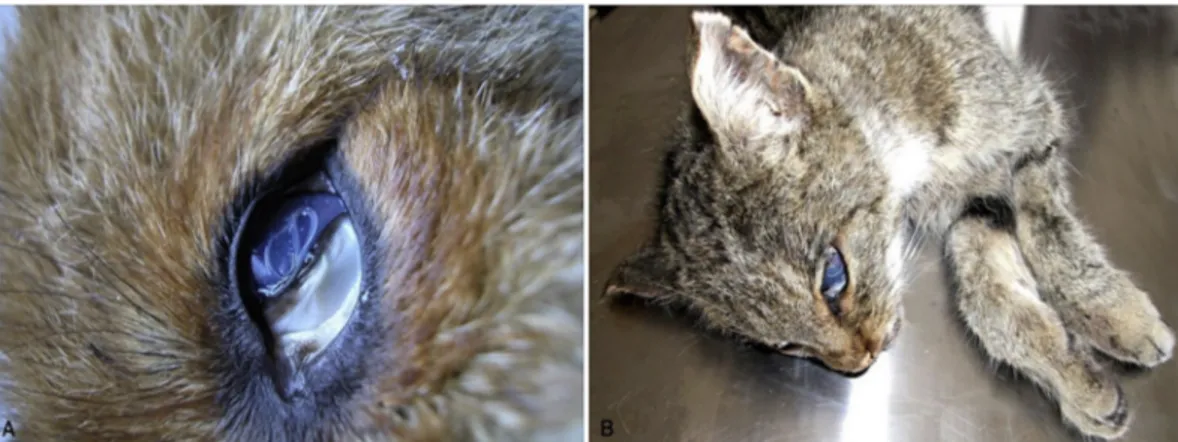
implicated in its transmission (Burnett and Wagner, 1958). Conversely, T. callipaeda lives in the orbital cavities and associated host tissues of dogs and cats as well as a large range of wild carnivore species (e.g., foxes, wolves, beech martens and wild cats;Fig. 1A and B) and lago-morphs (Otranto et al., 2009). This parasite is well known by the sci-entific community due to its distribution in a large number of geo-graphical areas and to its zoonotic impact. Accordingly, the common name “oriental eyeworm” issuing from the primary occurrence of T. callipaeda in the Far Eastern countries (i.e., Indonesia, Thailand, China, Korea, Myanmar, India, and Japan) (seeAnderson, 2000) is obsolete. Indeed, over the last 20 years, this parasite has been identified as an agent of ocular disease in many regions of Europe (Otranto et al., 2013a) primarily in dogs and also in many other animal species, in-cluding humans. Since the primary report in Italy, T. callipaeda has rapidly expanded its distribution throughout Europe from western to eastern Portugal, Spain, France, Switzerland, Germany, Bosnia and Herzegovina, Croatia, Romania, Bulgaria, Hungary, Greece and Slo-vakia (Rossi and Bertaglia, 1989;Otranto et al., 2003;Dorchies et al., 2007; Malacrida et al., 2008;Magnis et al., 2010;Miró et al., 2011; Vieira et al., 2012; Hodžić et al., 2014; Mihalca et al., 2015;Colella et al., 2016;Čabanová et al., 2017; Papadopoulos et al., 2018). The expansion of the parasite across domestic and wild animal species throughout Europe is in accordance with increased numbers of reported human cases (Otranto and Dutto, 2008;Fuentes et al., 2012;Paradzik et al., 2016; Tasic-Otasevic et al., 2016). Nowadays, T. callipaeda is considered in the diagnosis of helminthic infection of human eyes along with the better-known Dirofilaria spp. and, the less known, Onchocerca lupi (Otranto and Eberhard, 2011). Undoubtedly, the relatively short time in which T. callipaeda has been identified in many European countries in the eyes of domestic carnivores clearly indicate that canine thelaziosis is an emerging vector borne helminth. Dogs are supposedly the primary host of this nematode and they can be considered sentinel animals because of the localization of T. callipaeda on the conjunctiva mucosa, which is one of the main anatomical sites diagnosed by prac-titioners during general clinical examinations. The role of vectors, re-servoir hosts and the environment in explaining the emergence of thelaziosis is difficult to assess. For example, foxes (Fig. 1A) are also heavily infected by T. callipaeda with prevalence up to 27.71% in Bosnia Herzegovina (Hodžić et al., 2014;Sargo et al., 2014), 29.38% in Romania (Ionică et al., 2018) and 49.3% in southern Italy (Otranto et al., 2009). In addition, the pan European distribution of the vector Phortica variegata (Drosophilidae, Steganinae) is crucial to explain the spread of T. callipaeda (Máca and Otranto, 2014). Accordingly, thela-ziosis has been reported from many areas of Europe identified in a Genetic Algorithm for Rule-Set Prediction model, which was based on ecological field data collected for this lachryphagous drosophilid (Otranto et al., 2006a). The success of this predictive model further indicates the importance of observational field studies, even in the modern concept of veterinary parasitology. Since wild canids (i.e., foxes, beech martens, wolves) and felids (i.e., wild cats) live in forested, meadow hilly areas (e.g., around oak woods, Quercus cerris) and in environments characterized by high relative humidity (e.g. around rivers) and undergrowth, they are exposed to the vector (Otranto et al., 2006a). Therefore, wild animals species may represent proper hosts for T. callipaeda in endemic as well as in non-endemic areas (Mihalca et al., 2016), such as in UK, where merely imported cases of canine thelaziosis have been reported so far (Graham-Brown et al., 2017), although the vector is endemic (Palfreyman et al., 2018). The circulation of eye-worms within different animal species and its recent introduction to Europe is corroborated by the occurrence of a unique genetic haplotype of T. callipaeda amongst all specimens collected from wild and domestic animals (Otranto et al., 2005, and all other subsequent studies). Data above suggest that wild carnivores contribute to the dissemination of infection within their natural territories, which range from 10 to 30 km for foxes (Niewold, 1980;Doncaster and Macdonald, 1991) and up to 800 km for wolves (Mech, 1970). In addition, both canid species are
more active during dawn and dusk (Fedriani et al., 1999) when P. variegata is prevalent during summer months (Otranto et al., 2006b). 2.3. Onchocercosis
The genus Onchocerca (Spirurida, Onchocercidae) encompasses species parasitizing ungulates, with the only exception of Onchocerca volvulus and O. lupi, which primarily parasitize humans and carnivores, respectively (Bain et al., 2013;Lefoulon et al., 2017). The public health impact of O. volvulus in Africa is estimated to about 25 million in-dividuals annually infected in the Middle East in Yemen and Americas (e.g., Mexico, Guatemala, Ecuador, Colombia, Venezuela, and Brazil), making this infection one of the most important neglected tropical diseases (http://www.cdc.gov/parasites/onchocerciasis/gen_info/faqs. html). Conversely, the zoonotic potential of O. lupi has only recently been recognised (Otranto et al., 2011b) and, since then, human cases have been reported in Europe, Middle East regions and US (Mowlavi et al., 2014;Otranto et al., 2015c;Cantey et al., 2016). Since thefirst report of the parasite in a Caucasian wolf (Canis lupus) from Gruziya (Georgia, former USSR,Rodonaja, 1967), which was the main reason for naming the nematode as O. lupi, the infection has been diagnosed in dogs and cats from Portugal, Spain, Germany, Greece, Hungary, and Romania (Széll et al., 2001;Komnenou et al., 2002;Hermosilla et al., 2005;Faísca et al., 2010;Maia et al., 2015;Tudor et al., 2015;Hodžić et al., 2017), as well as in the U.S. and Canada (Labelle et al., 2011, 2013;Otranto et al., 2015c).
Nonetheless, the largest bulk of above-mentioned reports derives from clinical cases of pet animals presented for surgical removal of nodules, or suffering for conjunctivitis, chemosis, lacrimation and periorbital swelling (Komnenou et al., 2002; Sréter and Széll, 2008). The acute presentation of the infection represents only the tip of the iceberg for the parasite endemicity in canine population, as it was de-monstrated by the high prevalence (8.4%) of microfilaremic, but healthy dogs from Greece and Portugal (Otranto et al., 2013b). Therefore, considering the difficulties of diagnosing microfilariae from skin biopsy sediments in necropsied wild animals, data on O. lupi in-fection in wildlife is inexistent. Nonetheless, the involvement of wild carnivores cannot be ruled out mainly in areas where human, but not canine cases have been reported, such as in Iran (Mowlavi et al., 2014), Tunisia (Otranto et al., 2012) and Turkey (Otranto et al., 2011b). Fi-nally, the lack of data about the insect species acting as vector of this barely known onchocercid, greatly impairs the information about its distribution in wild carnivores and the potential spread of the infection. Up to now, wild-caught Simulium tribulatum have been found molecu-larly positive for O. lupi and this species has been suggested as putative vector in California (Hassan et al., 2015).
3. Soil-transmitted nematode zoonoses 3.1. Toxocarosis
Ascarids (Ascaridida, Ascarididae) of carnivores are worldwide-distributed, large (10–15 cm in length) intestinal nematodes, some of which have a zoonotic potential (Table 2). Their life cycle is direct but can include paratenic hosts as a source of infection for carnivorous definitive or dead-end hosts. Definitive hosts defecate thick-shelled eggs which embryonate in the environment under suitable conditions (i.e., 28–33 °C in laboratory conditions) within 2–6 weeks to infectious third stage larvae (L3). The biology of Toxocara canis, Toxocara cati and B. procyonis differs significantly in their definitive hosts (Deplazes et al., 2016). Toxocara canis transmission in canids is highly efficient by the in utero infection of the offspring, through the transplacental transmission of reactivated somatic larvae (Schnieder et al., 2011). Furthermore, juvenile and adult foxes can become infected or re-infected by preda-tion of paratenic hosts, mainly rodents and birds, or by ingespreda-tion of infectious T. canis eggs (Saeed et al., 2005). The relatively high
intestinal infection rate in adult foxes (prevalence between 10 and 40%, see below) in endemic areas suggests a low level of immune protection for intestinal infections and reinfections. Generally, higher prevalence of patent T. canis infections are observed in foxes younger than six months of age (Luty, 2001;Saeed et al., 2006;Reperant et al., 2007), indicating that the transplacental transmission is common. Also the seasonality may play a role in the occurrence of toxocarosis, with a higher prevalence of intestinal T. canis infections in fox cubs during spring (73%) and summer (65%), compared to fall (37%) and winter (42%) (Reperant et al., 2007). The prevalence of patent T. canis infec-tions varies in European foxes between 9 and 65% with data available for Denmark (48–65%,Saeed et al., 2006;Al-Sabi et al., 2014); Swit-zerland (44% in Geneva and 47% in Zurich,Reperant et al., 2007;Hofer et al., 2000); Austria (48%,Duscher et al., 2014); Italy (9–53%,Magi et al., 2009; Fiocchi et al., 2016); Ireland (20%,Stuart et al., 2013); Lithuania (41%,Bružinskaitė-Schmidhalter et al., 2012); Estonia (30%, Laurimaa et al., 2016b); Poland (11%,Borecka et al., 2013); the Slovak Republic (43%,Miterpáková et al., 2009). Also in other endemic areas high prevalence of T. canis in foxes have been documented (e.g., Aus-tralia, New South Wales, up to 35.2%,Ryan, 1976; Japan, 71%,Sato et al., 1999; Iran, 32%,Meshgi et al., 2009; Canada, 32.5% in juveniles, 15.1% in adults,Wapenaar et al., 2013).
Although the biology of T. canis has not been studied in detail in other wild carnivores than those mentioned above, many of them are suitable natural hosts for T. canis. For example, the prevalence of T. canis intestinal infections in raccoon dogs varies according to the geo-graphical areas being 13% in Denmark (Al-Sabi et al., 2014), 18% in Germany (Thieß et al., 2001), 4.7% in Estonia (Laurimaa et al., 2016a), 16% in Poland (Osten-Sacken et al., 2017) and 18% in Lithuania (Bružinskaitė-Schmidhalter et al., 2012). However, when data on T. canis infection is available for foxes and racoon dogs (e.g., Lithuania and Denmark), the prevalence in raccoon dogs is lower as compared to foxes (Bružinskaitė-Schmidhalter et al., 2012;Al-Sabi et al., 2014). The latter observation could be explained by the fact that raccoon dogs decrease their activity during the‘hibernation’ period in winter and tend to defecate in latrines, which may also reduce their potential to spread parasite eggs.
In golden jackals, prevalence of T. canis intestinal infections was documented in Hungary (20%,Takács et al., 2014) and Iran (27.2%, Vafae Eslahi et al., 2017). Toxocara canis also infects wolves with dif-ferent prevalence in Italy (17%,Guberti et al., 1993; 33%,Fiocchi et al., 2016), Germany (5%,Bindke et al., 2017; 11%,Lesniak et al., 2017), Belarus (21%,Shimalov and Shimalov, 2000), Estonia (8%,Moks et al., 2006), Poland (6.9%, Borecka et al., 2013) and Spain (6%,Segovia et al., 2003). No wolves infected with T. canis were diagnosed in Sweden (Al-Sabi et al., 2018). In Australien wild dogs (dingoes) the prevalence of T. canis was up to 46% from Wet Tropics region around Cairns, Far North Queensland (Smout et al., 2013), and 27.8% of 18 dingoes from Fraser Island (Queensland) with close ntcoact with human habitation were infected (Mackenstedt et al., 2015). Finally, up to 24% in the coyotes from Southeastern region of Nebraska and Shenandoah area of Iowa in USA (Redman et al., 2016) and, again, to 3.5% in ju-veniles and 1.1% in adults from Prince Edward Island, Canada (Wapenaar et al., 2013).
In felids, T. cati (syn. Toxocara mystax) is mainly transmitted by predation of paratenic hosts or ingestion of infectious eggs. Infection with large numbers of T. cati eggs during the last third of gravidity in female cats results in the trans-mammary transmission of infective larvae to the kittens (Coati et al., 2004), however, the epidemiological significance of this way of transmission for wild felids is not confirmed (Fig. 2). In Europe, T. cati is highly prevalent in the domestic cat po-pulations that have free access to the environment and to potentially infected paratenic hosts (Giannelli et al., 2017). Furthermore, large populations of feral or stray cats represent the main reservoir for the maintenance of the T. cati life cycle. A variety of wild felids are sus-ceptible to T. cati intestinal infections, with a report in an Iberian lynx
(Lynx pardinus) in Spain, and very high prevalences in Eurasian lynxes (Lynx lynx) in Finland (98%,Deksne et al., 2013) or in Poland (7 of 11 animals examined,Kołodziej-Sobocińska et al., 2018), or bobcats (Lynx rufus) in USA, Midwest (70%,Hiestand et al., 2014). Furthermore, this parasite was also endemic in wild cats in Sicily Island (southern Italy) with 43.8% of 121 cat scats positive for T. cati eggs (Napoli et al., 2016). In addition, T. cati was the most prevalent ascarid found in the Siberian tiger (Panthera tigris altaica), although the epidemiological role of this animal species is unknown (Moskvina et al., 2018).
Human Toxocara infections are predominantly acquired from in-gestion of embryonated eggs by hand-to-mouth contact after activities in contaminated environments such as sandpits, parks or other places where carnivores have defecated. The transmission has been also hy-pothetically linked to water or foodborne sources. The actual im-portance of wildlife contributing to environmental contamination with ascarid eggs is still unknown. Since the 1940th in Great Britain and, later on, in the periurban areas throughout Europe, a steady increase in populations of foxes has been reported (Scott et al., 2014). These highly reproductive fox populations present annually a new highly susceptible generation of cubs with high T. canis worm burdens, contributing sig-nificantly to the environmental contamination with eggs. Within the fox home ranges, which can be much smaller in urban areas (Deplazes et al., 2004), a complex pattern of spatial faeces distribution can be observed with significantly higher density both on the borders of ploughedfields and on road verges (Guislain et al., 2007). Furthermore, marking of territories along roads, pavements and feeding places in private gardens may contribute to the environmental contamination with zoonotic helminths (e.g. Toxocara and Echinococcus eggs). Based on the complex wild animal ecology, we can assume that foxes may play a major role in the maintenance of the wildlife cycle of T. canis and strengthening a continuous transmission to humans and pet dogs. Si-milarly, a study of host density of sympatric cats, dogs and foxes in the city of Bristol (UK) in relationship to the environmental contamination with Toxocara eggs revealed that dog puppies “dominate total egg output”, although “foxes could take over as the primary source of eggs environmental contamination” (Morgan et al., 2013). However, the authors further discussed that the relative contribution to the egg contamination of dogs, cats and foxes might differ among specific lo-cations dependant on animal densities and their access to the public. Furthermore, involvement of large feral or stray domestic dogs and cat populations might again have a high epidemiological impact in the contamination with eggs of Toxocara spp. (Otranto et al., 2017). Al-though Toxocara eggs have been found in the hair coat of dogs and foxes, the importance of the direct transmission to human has been discussed controversially in the past as, in most of the cases, eggs were Fig. 2. Anterior edge of Toxocara cati from the gut of a wild cat. The short and wide cervical algae give the typical appearance of an arrow.
not fully larvated (Deplazes et al., 2011;Merigueti et al., 2017). Fur-thermore, an experimental study confirms that T. canis eggs did not fully embryonate on the coat of dog puppies but during the same ob-servation time in their surrounding (Nagy et al., 2011). Epidemiological studies further confirm that pet ownership was not associated with increased human seroprevalence for toxocarosis (Lee et al., 2014). Fi-nally, humans could also become infected by eating row tissues of T. canis paratenic host as experimentally documented for raw liver or other viscera of pigs or chickens (Taira et al., 2004). The relative contribution of wild carnivores in such a rather“exotic” way of trans-mission might be strongly related to certain cultural settings, as it was discussed for eating habits in East Asia (e.g. South Korea and Japan) through ingestion of raw beef, lamb, chicken or ostrich liver (Akao and Ohta, 2007).
Clinically toxocarosis is categorised as a visceral, neurological and ocular larva migrans syndrome that can affect millions of children and adolescent populations worldwide. Many reviews claim this disease to be neglected, under-reported or under-diagnosed (Deplazes et al., 2011; Lee et al., 2014; Chen et al., 2018). Asymptomatic and self-limiting infections are probably frequent based on high serological prevalence often associated with eosinophilia. Series of clinical cases or serological studies estimate burdens of disease in diverse geographic regions worldwide, but especially in impoverished communities. A compre-hensive literature review focussing on clinical cases (Chen et al., 2018) documented worldwide a total of 823 cases of ocular toxocarosis (i.e., 282 in Europe, 317 in Asia,five in Australia, 218 in Latin America, and few from other areas), but interestingly, the highest number has been reported in Japan and Korea, France, Brazil and the USA. In 99 cases, neurotoxocarosis was diagnosed (32 in Europe, 32 in Asia, 20 in Americas and few from other areas) and up to 247 visceral larva migrans cases documented. Despite the worldwide distribution of T. canis in dog and fox populations, cases are not equally distributed in the endemic areas indicating differences in clinical and diagnostic management of patients. For example reports of human cases in Austria are rare, but Auer (2011) estimated for this country several hundred undiagnosed overt cases per year.
3.2. Baylisascariosis
Baylisascaris procyonis (Fig. 3) is an ascarid endemic in North America. Main definitive hosts of B. procyonis are raccoons (Procyon lotor) and related procionids, however, also in dogs patent infections can develop (Kazacos, 2001). Definitive hosts acquire the infection by ingesting larvated eggs or by predation of paratenic hosts, which often display neurological disease. Raccoons originating from North America have been imported into Europe (i.e., Germany, Russia and Poland) and Japan many decades ago (Bauer, 2013;Mackenstedt et al., 2015). In Germany, it is believed that several independent introductions of the
raccoon occurred, which then widespread throughout the country (Fischer et al., 2015;Osten-Sacken et al., 2018), representing the lar-gest population outside America with an impressive hunting statistic of about 172,000 individuals for the season 2017/2018 (Anonymus, 2019). It has been estimated that up to 700 raccoons found per square mile live in the U.S. ( https://study.com/academy/answer/how-many-raccoons-are-there-in-the-world.html).
Baylisascaris infections have been documented in around 70% of raccoons in Central Germany (Bauer, 2013), even in the intestine of 24 raccoons (75%) originating from the urban area of Leipzig ( Rentería-Solís et al., 2018), and in the faeces of animals from a zoo in Denmark (Al-Sabi et al., 2015) and Poland (Karamon et al., 2014). In addition, B. procyonis, Toxocara and Toxascaris eggs have been recently documented in the intestine of raccoons illegally imported to Norway (Davidson et al., 2013). Furthermore, B. procyonis was found in captive raccoons in Japan (Page, 2013) and in 50% of those examined in Costa Rica (Baldi et al., 2016). Similar as for Toxocara spp., human infections with B. procyonis eggs occurs via ingestion of larvated eggs in contaminated areas, mainly represented by the animal's defecation sites and latrines (Page et al., 1999). Baylisascaris procyonis causes visceral and neuro-logical larva migrans, which can particularly be severe because of the increased growth of the larvae compared to Toxocara spp. during its migration (reviewed byKazacos, 2001). Human cases of baylisascar-iosis are relatively rare; however, many cases show severe clinical complications. Since 1973, at least 28 cases of human baylisacariosis have been reported in the USA (Gavin et al., 2005;Sircar et al., 2016) and most of them occurred in children. So far, only a single human baylisascariosis case outside America has been reported in Germany in a patient presented with unilateral neuroretinitis syndrome, who had purchased a raccoon from a local zoo (Küchle et al., 1993). Similar to toxocarosis, epidemiological investigations suggest that subclinical in-fections with B. procyonis may occur in human patients. Relatively high seroprevalence in the population have been found in endemic areas; for example in Chicago, 8% of 389 children without a history of disease (Brinkman et al., 2003) or healthy individuals was found seropositive after exposure to raccoons or their faeces (Conraths et al., 1996). Fi-nally, experimental evidence in Mongolian jirds (Sato et al., 2004) supports the zoonotic potential of Baylisascaris transfuga, a parasite for example found frequently in brown bears (Ursus arctos) in Slovakia, therefore, the zoonotic role of this species should be further in-vestigated (Štrkolcová et al., 2018).
3.3. Strongyloides
Strongyloides stercoralis (Rhabditida: Strongyloididae) is soil-trans-mitted species with zoonotic potential (Thamsborg et al., 2017), it oc-curs in humans, other primates and dogs mainly in tropical and sub-tropical areas (Bisoffi et al., 2013;Paradies et al., 2017). However, the
Fig. 3. Baylisascaris procionis anterior end with typical lips (A), and male posterior end with spicules (B) and papillae (F). Scale bars = 220μm (A), 600 μm (B), 900μm (C), 200 μ (D, E), 100 μm (F).
parasite is also present in Europe, but data on the epidemiology of canine strongyloidosis is missing (Iatta et al., 2018). Strongyloides spp. have been sporadically diagnosed in wild carnivores and were mostly allocated to S. stercoralis (Thamsborg et al., 2017), however, ata based on morphological identification of larvae from environmental samples without genetic analyses have to be critically interpreted. A Strongy-loides spp. prevalence of 0.7% was recorded in foxes from the Nether-lands (Borgsteede, 1984), the Slovak Republic (1.6%, Miterpáková et al., 2009). A Strongyloides sp. has also been documented in grey foxes (Urocyon cinereoargenteus Schreber, 1775) in dry tropical highlands of central Mexico (Hernández-Camacho et al., 2011). Even if S. stercoralis may cause a serious disease especially for chronically infected and immunocompromised hosts (Bisoffi et al., 2013), sporadic infections in wild carnivores or in domestic dogs have so far not been associated as a major zoonotic risk for humans. Strongyloides procyonis was reported for thefirst time in Louisiana and was demonstrated to cause experimental creeping eruption and a short-lived intestinal infection in inoculated human volunteers (Little, 1965). Although the zoonotic role has never been demonstrated in naturally infected human patients, the parasitic females of S. procyonis were identified in 66 (28.3%) of 233 raccoons collected in Japan, indicating the potential zoonotic risk in particular environments (Sato et al., 2006).
3.4. Hookworm infections
Hookworms (Strongylida, Ancylostomatidae) display a high patho-genic potential due to their blood-feeding behaviour. A recent sys-tematic review of the literature on hookworms demonstrated that at least 68 hookworm species parasitize the gastrointestinal tract of wildlife mammals, black bears, red foxes, and bobcats harboured the highest diversity of hookworm species (Seguel and Gottdenker, 2017). The parasites have been studied mainly in red foxes and coyotes, probably because they are commonly hunted/culled (Seguel and Gottdenker, 2017). The occurrence and the prevalence of hookworm species depend on peculiarities in local humidity, temperature and host population density, which induce a dynamic scenario in their infections of animal-human-wildlife. For example, Ancylostoma caninum has worldwide distribution mainly in tropical and subtropical areas in do-mestic dogs, but it occurs also in wild canids and felids but infects humans as cutaneous larva migrans (CLM) and rarely intestinally causing eosinophilic enteritis (Bowman, 2011;Mclaughlin et al., 1993). Ancylostoma braziliense occurs in dogs, cats and various wild animals, and in humans as CLM (Bowman, 2011). Ancylostoma ceylanicum in-testinal infections have been mainly documented in humans and dogs in Asia (Traub, 2013) and in wild dingoes in Northern Australia (Smout et al., 2013,2018;Mackenstedt et al., 2015). Domestic cats and wild felids are usually infected by Ancylostoma tubaeforme worldwide, but its zoonotic potential has so far not been documented (Bowman, 2011). In bobcats from the USA, three species: A. tubaeforme, A. caninum and A. braziliense have been documented (Hiestand et al., 2014). Finally, Un-cinaria stenocephala is worldwide distributed in dogs and wild canids, but rarely in cats and wild felids (Deplazes et al., 2016). The life cycle of the hookworm species is direct, with intestinal females excreting non-larvated, thin-shelled eggs with the faeces. Under suitable environ-mental conditions, L1 hatch from the eggs and moult to second stage larvae (L2) and L3 within 5–8 days, however, the development can be delayed at temperatures below 15 °C. Cutaneous and peroral infections with free-living L3 of A. caninum have been described for definitive, paratenic or dead-end hosts. Furthermore, in definitive hosts, trans-mission of parasitic L3 with milk or the ingestion of paratenic hosts (e.g., rodents) is possible (Deplazes et al., 2016). Uncinaria stenocephala transmission occurs predominantly by ingestion of L3 from the en-vironment or paratenic hosts; however, L3 of this species can also pe-netrate the skin (Gibbs, 1961). In foxes, high prevalence of U. stenoce-phala infections have been reported all over Europe: Denmark 60–86% (rev.Al-Sabi et al., 2014), Spain 58–71% (Criado-Fornelio et al., 2000;
Segovia et al., 2004), Switzerland 64–78% (Hofer et al., 2000;Reperant et al., 2007), Ireland 38% (Stuart et al., 2013), the United Kingdom 68% (Richards et al., 1995), Lithuania 99% (Bružinskaitė-Schmidhalter et al., 2012), Poland 19% (Borecka et al., 2009), the Slovak Republic 7% (Miterpáková et al., 2009), the Netherlands 60% (Borgsteede, 1984), Croatia 26% (Rajković-Janje et al., 2002), France 68% (Petavy and Deblock, 1980), Portugal 77% (Eira et al., 2006), Italy 39–75% (Di Cerbo et al., 2008;Magi et al., 2009;Fiocchi et al., 2016) and Slovenia 59% (Vergles Rataj et al., 2013). Ancylostoma caninum is worldwide endemic especially in canids, in Europe mainly in Southern and Southeastern regions, however, this species has also been described in foxes in central Europe up to Denmark. Interestingly, this species has not been documented in several studies in foxes from Italy, Slovenia and Portugal (Eira et al., 2006;Magi et al., 2009;Vergles Rataj et al., 2013) where, conversely, high prevalence with U. stenocephala was found. A low A. caninum prevalence (1.4%) has been found in France (Auvergne) as compared with U. stenocephala (68%) (Petavy and Deblock, 1980), comparably, in Italy (Emilia-Romagna) prevalence of 1.8% for A. caninum and 75.4% for U. stenocephala were reported (Fiocchi et al., 2016). Similarly, the prevalence of U. stenocephala (68.6%) in Denmark was higher that A. caninum (0.6%) in 1040 foxes (Saeed et al., 2006). Accordingly, a very high prevalence of U. steno-cephala (68%) was also detected in 843 rural and urban red foxes from the UK without indication of occurrence of A. caninum (Richards et al., 1995). In Tunisia, one of 9 foxes was infected with A. caninum, 4 with U. stenocephala (Lahmar et al., 2014). Investigations in Australian red foxes (n = 930), New South Wales found prevalence for U. stenocephala and A. caninum of 30.6% and 7.5%, respectively (Ryan, 1976). Con-versely, in Iran prevalence in foxes reached up to 5.4% for A. caninum and up to 8.1% for U. stenocephala (Meshgi et al., 2009) and in the Slovak Republic the average prevalence of U. stenocephala (i.e., 6.9%) was lower than A. caninum (i.e., 18.1%) in 1198 foxes. Finally, the highest prevalence of A. caninum (up to 32%) was determined in areas with a high proportion of irrigated soil (Miterpáková et al., 2009). A single study reported the prevalence of A. caninum in foxes (12%) from Spain without records of U. stenocephala (Lledó et al., 2015), though identification data have not been discussed.
Raccoon dogs seem to be highly susceptible to U. stenocephala with a prevalence of 99% in 85 necropsied animals in Lithuania, 65% in 74 animals in Germany (Thieß et al., 2001; Bružinskaitė-Schmidhalter et al., 2012). Both A. caninum and U. stenocephala were detected in golden jackals, in Hungary with prevalence of 45% and 40%, in Tunisia with 10% and 68%, respectively (Lahmar et al., 2014). Wolves are susceptible to both hookworm species in Italy (Guberti et al., 1993) and Poland (Borecka et al., 2013). Based on the hookworm prevalence re-corded in European foxes, this species contributes significantly to the transmission of U. stenocephala whereas, foxes are not as frequently infected with A. caninum and therefore, based on the limited data available, the contribution of foxes to A. caninum transmission cannot be conclusively determined. However, in particular epidemiological situations, wild canids such as wild dogs (dingoes and dingo hybrids) may contribute to Ancylostoma spp. transmission. High prevalence of A. caninum (up to 100%) was found in dogs and sympatric dingoes in an Aboriginal community in the Wet Tropics of Australia, with A. ceyla-nicum being diagnosed in around 11% of the same canid population (Smout et al., 2013, 2018; Mackenstedt et al., 2015). Furthermore, coyotes are highly susceptible for A. caninum and U. stenocephala in-fections (Seguel and Gottdenker, 2017) and, based on the invasion and increase of populations of this animal species in urban areas in North America (Liccioli et al., 2015), they may represent a risk factor for the occurrence of hookworms and other helminths, such as Echinococcus multilocularis. Based on their relative low population sizes, other large canids (e.g., wolf, jackal) and also wild felids play probably an insig-nificant role in the transmission of hookworms. However, in one study, documenting high prevalence of hookworms in bobcats throughout the state of Florida (A. tubaeforme, 11%; A. caninum, 18%; A. braziliense,
19%; Ancylostoma pluridentatum, 29%) Mclaughlin et al. (1993) sug-gested that this wild felid could serve as a zoonotic reservoir of hook-worms.
The migration of hookworm (Ancylostoma, Uncinaria) larvae through the human skin may result in CLM, also known as ‘creeping eruptions’ (Bowman et al., 2010), though the zoonotic role of Uncinaria spp. is unclear (Bowman et al., 2010). However, in the UK, CLM was attributed to U. stenocephala because A. caninum is not endemic (Beattie and Fleming, 2002). Cases of CLM have been reported from several European countries (e.g., the UK, Germany, Italy and Serbia) with four cases described from Germany, whereas some of the patients spent time in a shelter together with domestic and wild animals during the flooding of the river Elbe (reviewed byBowman et al., 2010). In any case, all patients had a history of visiting lakesides and riverbanks around urban areas (Klose et al., 1996), where wildlife species were prevalent, potentially contributing to the contamination of such en-vironments. In addition, dog-owners used to walk in those areas, therefore favouring the transmission of hookworms among wildlife and dogs (and vice versa) and, ultimately to humans (Smith et al., 2014). 3.5. Respiratory capillariosis
Capillaria aerophila (syn. Eucoleus aerophilus) (Enoplida, Capillaridae) is a lungworm of wild and domestic carnivores, but it has also rarely been described in humans. Eggs are excreted with the faeces of the definitive hosts, and the infective L1 develops inside the eggs within 40–60 days (Conboy, 2009). Infections occur by ingestion of larvated eggs; but facultative or transport hosts, such as earthworms can be involved in the transmission (Otranto et al., 2015b). Prevalence in European red foxes are usually high; reaching 97% in Lithuania (Bružinskaitė-Schmidhalter et al., 2012); 88% in Norway (Davidson et al., 2006); 74.1% in Denmark (Saeed et al., 2006); 76.2% in Poland (Karamon et al., 2018); 67–75% in Germany (Schug et al., 2018); 66% in Hungary (Sréter et al., 2003); 49% in Serbia (Ilić et al., 2016); 46.8% in The Netherlands (Borgsteede, 1984) and 41.8% in Italy (Magi et al., 2015). Capillaria aerophila has been also reported in European wildcat, wolf, jackal, beech marten (Martes foina) and raccoon dog with pre-valence rates ranging from 5% (jackals), 33.3% (wild cats), and 32% in raccoon dogs (rev. Bružinskaitė-Schmidhalter et al., 2012; Otranto et al., 2015b). These epidemiological data strongly support the hy-pothesis, that wild carnivores act as main definitive hosts for the C. aerophila transmission. Respiratory C. aerophila infections in humans can mimic the clinical and radiographic findings of pulmonary neo-plasia, with bronchitis, cough, mucoid sputum, haemoptysis, fever, dyspnoea and eosinophilia (Lalosevic et al., 2008). Human cases have been described in Ukraine, Russia, Morocco, Iran and France (reviewed inDi Cesare et al., 2012). The last documented case in humans was in Serbia, where the prevalence of infection in foxes is high (84%) (Lalosevic et al., 2008,2013).
4. Other nematodes 4.1. Trichinellosis
Within the genus Trichinella (Trichuroidea, Trichinellidae), 12 spe-cies and additional several genotypes are recognised with a diverse host range, geographical distribution, ecology and histopathological features (encapsulated or not encapsulated). Trichinella spp. have a peculiar life cycle which completes in a single host and transmission occurs through the ingestion of infected meat containing larvae (Gottstein et al., 2009). Trichinella spp. are amongst the most prevalent parasites of predatory and scavenger wild carnivores (Pozio and Zarlenga, 2013). Zoonotic transmission has been documented for most Trichinella spp. with the exception of Trichinella papuae and Trichinella zimbabwensis. Trichinella britovi is the most prevalent species among wild carnivores as well as wild boars (the main source of human infection) in Europe, Western Asia, North and Western Africa (Gottstein et al., 2009). In humans, the clinical course of the disease is characterized by an enteral (i.e., adult worms in the intestinal mucosa) and a parenteral (i.e., larvae invading the host muscles) phase (Gottstein et al., 2009). The parenteral phase may occur with high degree of severity and it is characterized by fever, myalgia, periorbital swelling and eosinophilia. The ocular signs (i.e., periorbital oedema, conjunctivitis, macular and retinal haemorrhage) appear after about three weeks from the infection and they are caused by both the effect of migrating larvae and the allergic reactions (Otranto and Eberhard, 2011). The diagnosis in humans is based on clinical signs, corroborated by the anamnesis of enteral syndromes linked to history of ingesting raw or inadequately cooked meat and by serologic confirmatory tests.
The diagnosis of trichinellosis in animals is based on the evidence of larvae in muscles at meat inspections. Muscle distribution of Trichinella spp. larvae in production animals and wildlife can differ significantly. Experimental studies indicate that larvae of different Trichinella spp. establish in high numbers in foxes (Fig. 4), localizing primarily in the tongue, extremities and diaphragm (encapsulating species), and in the diaphragm (non-encapsulating species) (Kapel et al., 2005).
Importantly, refined diagnostic methods may increase the sensi-tivity in diagnosing Trichinella infection in muscles of foxes, for example by artificial digestion of frozen muscles, combined with larva isolation by a sequential sieving method (SSM). Using the SSM, dead Trichinella larvae were detected in one frozen muscle sample from a red fox (0.067 larvae per gram) during a survey from the Netherlands (n = 369), suggesting a decrease in Trichinella spp. prevalence in Dutch red foxes from 3.9% to 0.27%, withinfifteen years (Franssen et al., 2014). In 47 trichinellosis outbreaks from 1995 to 2002 (i.e., 864 human cases) in Russia, the consumption of pork meat was implicated in only 35.8% of the cases during 1998–2002, compared to 80% in 1995–1996 (Ozeretskovskaya et al., 2005). The remaining cases were due to in-gestion of bear (Ursus arctos) (39.5%), badger (Meles meles) (10.6%), or even dog (11.9%) meat. Interestingly, clinically severe cases of disease derived from the consumption of pork (7.7%) and bear (7.9%) meat with a significantly higher number of moderate cases from pork than from bear meat (Ozeretskovskaya et al., 2005). Due to the habits of the aboriginal Siberian population of feeding bear meat the incidence of clinically severe cases of trichinellosis is high in those populations (Ozeretskovskaya et al., 2005). A great success in controlling the in-fection in humans has been achieved in the USA and Europe by de-termine ad hoc regulations on food inspection and safety as well as feeding practices in pigs, which has reduced the perpetuation of Tri-chinella spp. domestic cycle. Conversely, pigs continue to be responsible for human infection in many countries and wild game meat represents a global threat to the perpetuation of trichinellosis when meat is ingested without proper cooking. Indeed, a range of wild animals can harbour infection with Trichinella larvae maintaining the sylvatic cycle (Hurníková et al., 2007). Due to their high population level, red foxes and raccoon dogs are the more suitable reservoir of the parasite in Fig. 4. Encapsulated larvae of Trichinella britovi in the striated muscular tissue
Europe. A prevalence of Trichinella up to 21.5% and a mean larval density of 10.5 larvae per gram were recorded at the muscle digestion method of 121 foxes from Romania (Imre et al., 2015). Of the Trichinella larvae identified, 96% were T. britovi and only 4% were Trichinella spiralis, suggesting a major role of the fox as a reservoir of T. britovi (sylvatic cycle), but minor importance in the epidemiology of T. spiralis in the domestic cycle (Imre et al., 2015). Due to their small population densities, other wild carnivores such as martens, badgers, bears, lynxes, wild cats, wolves, etc. play probably a secondary role in the transmis-sion of Trichinella spp. to humans. Ways of transmistransmis-sion from wild carnivores to humans can occur with undercooked meet (bear, rarely other species) (Rostami et al., 2017) or by illegal use of wild carnivore meet for meat products (Gottstein et al., 2009). Carnivores are at the end of the food chain and therefore probably often dead-end hosts. However, transmission from carnivores can occur by predation (foxes or raccoon dogs hunted by larger carnivores), by scavenging (e.g., wild boars) but also rarely by free ranging pigs on dead animals, such as road kills or carcasses left in the environment by hunters (Duscher et al., 2014;Gottstein et al., 2009. Trichinellosis outbreaks due to T. britovi and Trichinella murrelli have been identified to be horsemeat-related. Horses can become accidentally infected by grazing in pastures con-taminated with infected small animal and rodent carcasses or through hay contaminated with pieces of rodents (Bruschi and Dupouy-Camet, 2014), but also illegal protein sources, including wild animal meet, have been claimed to be source of infections of horses used for meat production (Gottstein et al., 2009).
4.2. Angiostrongylosis
Angiostrongylus costaricensis (Strongylida, Angiostrongylidae) is a nematode species causing severe or fatal zoonosis in North and South America. Besides the wide range of rodents (i.e., cotton rat Sigmodon hispidus) as most important definitive hosts, white-nosed coati (Nasua narica) and the domestic dog can act as potential definitive hosts ex-creting L1 with their faces (Alfaro-Alarcón et al., 2016). Definitive hosts and humans may become infected ingesting L3 in raw or undercooked intermediate hosts (slug and snails) or with vegetables contaminated with mucus of them containing L3 (Spratt, 2015). Around 200 human cases of human angiostrongylosis have been described mainly in chil-dren (Romero-Alegría et al., 2014). However, based on serological studies, the number of human cases might be much higher (e.g., 600 cases in Costa Rica per year), though human cases of A. costaricensis have also been reported from Africa (Zaire) and Europe.
4.3. Gnathostomosis
Gnathostoma spinigerum Owen 1836, is the most widespread zoo-notic species in South East Asia, China, Japan, the Indian subcontinent and Central Africa. Definitive hosts with gastric nodules are domestic cats and dogs, but also various wild felids feeding on mammals, reptiles, amphibians and aquatic prey have been occasionally diagnosed with gastric nodules (Eshwaran et al., 2003;Shrivastav et al., 2011;Boo Liat, 1976). Gnathostoma binucleatum has been reported from northern Latin America and occasionally the USA, with dogs, cats, wild felids as de-finitive hosts.
Humans acquire the infection mainly from L3 in rawfish, but also from raw frogs, snakes, wild boar, poultry, but possible also from L1 copepods in water. The larvae migrate to the skin and subcutaneous tissue causing the typical migratory swellings (cutaneous disease in the majority of cases), occasionally the larvae may penetrate into deeper tissues (viscera, the lungs, eyes, ears, gastrointestinal and genitourinary systems and rarely, but often fatally, the Central Nervous System (Herman and Chiodini, 2009). Gnathostoma spinigerum infections have been recognised as an emerging cause of eosinophilic en-cephalomeningitis (Diaz, 2010) and ocular infections (Lenka et al., 2016). Cutaneous gnathostomosis developes within an incubation time
of weeks up to 12 months as larva migrans in cutaneous nodules. The importance of the wild carnivores (e.g., felids) in the zoonotic trans-mission or as reservoir of Gnathostoma spp. is not well elucidated in the literature; however, probably the so farfindings in rather exotic and rare felid species do not support an important zoonotic contribution of wildlife carnivores.
5. Conclusions
For long time, wildlife carnivores have been disregarded for their potential in transmitting zoonotic nematodes. Nonetheless, increasing numbers of case reports suggest their role in the maintenance and spread of such infections due to a complex range of interactions fa-vouring their contact with humans. Amongst others, factors are linked to i) human habits (e.g., increased outdoor activities such as camping and tracking in forested or wild environments), ii) environmental en-croachment in urban areas (such as the tropical forest in the peripheries of the Brazilian towns) and, iii) the altered ecology of wild animals (e.g., fox or coyotes populations in urban settings). On the other hand, improved diagnostic strategies including DNA analyses of clinical ma-terial enabled the specific diagnosis of even exotic parasitic infections. In addition, insect vectors have no boundaries in spreading zoonotic nematode infections to domestic or wild carnivore reservoirs, as long as theyfind suitable ecological conditions. Due to the fact that a large number of zoonotic helminths are shared among wild and domestic carnivores, the actual importance of wildlife contributing to environ-mental contamination is difficult to assess. This is the case of foxes in shedding ascarid eggs. In addition, the limited scientific information about some parasites, the limited access to biological samples or the correct diagnostic tools may also impair a clear picture of the infection in animals and their zoonotic risks. This is substantiated by the fact that knowledge about the occurrence of zoonotic nematodes amongst wild carnivores mostly relies on necropsy studies, which not always offer the most appropriate samples for diagnosing some parasites (e.g., micro-filariae from skin biopsy sediments for O. lupi). Since urban and rural/ forested ecosystems are often without frontiers and inter-related, the study of zoonotic parasites in wild carnivores requires a multifaceted approach whit the involvement of biologists, epidemiologists, ecolo-gists and veterinary and medical microbioloecolo-gists and parasitoloecolo-gists. Such an approach could contribute to a better understanding regarding the population dynamics of carnivore hosts and their parasites in con-tinuously changing ecosystems. For example, the introduction or in-vasion of exotic carnivore species in a given habitat may increase the transmission of zoonotic nematodes. It is the case of raccoon dogs and raccoons. Since their introduction into Europe, the populations of these animal species well adapted and increased in Europe and they showed to be suitable hosts for almost all the zoonotic nematodes herein re-ported (Kauhala and Kowalczyk, 2011;García et al., 2012). The pro-cyonids have caused the introduction and spread of“exotic” parasites, such as B. procyonis in Central European countries, with a potential risk of spill-over to other autochthonous species. These facts have never been considered as a concern into international or national legislation on the conservation of biodiversity (92/43/EEC), which should require a population control of invading animal species in order to reduce the risk of spreading exotic parasites. Indeed, with the exception of rabies in foxes, no other zoonotic infectious agents of wild carnivores are considered in the international legislation aiming to protect human health. This might also be due to the fact that in the EU Member States, wildlife management and conservation authorities are not responsible for animal health but National Veterinary Services being in charge for health and food safety in domestic animals. Therefore, the impact of diseases on wildlife population dynamics is often underestimated and assessed through research projects or local surveys. Conversely, wildlife diseases are included in the international reporting system for animal health of the World Organization for Animal Health (OIE) (World An-imal Health Information Database–WHAIS, available at:http://www.