Tricyclic thienopyridine–pyrimidones/thienopyrimidine–pyrimidones
as orally efficacious mGluR1 antagonists for neuropathic pain
T. K. Sasikumar
a,*, Li Qiang
a, Duane A. Burnett
a, William J. Greenlee
a, Cheng Li
a, Larry Heimark
a,
Birendra Pramanik
a, Mariagrazia Grilli
b, Rosalia Bertorelli
b, Gianluca Lozza
b, Angelo Reggiani
b aSchering-Plough Research Institute, 2015 Galloping Hill Road, Kenilworth, NJ 07033, USA
b
Schering-Plough Research Institute, San Raffaele Science Park, Via Olgettina, 58, 20132 Milan, Italy
a r t i c l e
i n f o
Article history: Received 12 March 2009 Revised 21 April 2009 Accepted 23 April 2009 Available online 3 May 2009
Keywords: Tricyclic thienopyridine–pyrimidone Thienopyrimidine–pyrimidone Triazafluorenone mGluR1 antagonist Neuropathic pain
a b s t r a c t
Introduction of small unsaturated alkylamino groups at the 4-position of the A-ring of the tricyclic frame-work (triazafluorenone) afforded extremely potent and selective mGluR1 antagonists with desirable properties. Compounds 11q and 11s are active in the SNL pain model with ED50s 3.3 and 6.4 mg/kg respectively. Metabolic outcome of propargyl amino moiety was studied.
Ó 2009 Published by Elsevier Ltd.
Glutamate is the major excitatory neurotransmitter in the
cen-tral nervous system and mediates its actions via activation of both
ionotropic and metabotropic receptor families. The metabotropic
glutamate receptors (mGluRs) form a family of eight subtypes
(mGlu1 to mGlu8) and are assigned to three groups based on their
structure, coupling to effector mechanisms and pharmacology.
Group I mGluRs (mGluR1 and mGluR5) are post synaptic receptors
while Group II (mGluR2 and mGluR3) and Group III (mGluR6,
mGluR7 and mGluR8) are located presynaptically. It has been
found that the Group I mGluRs play key roles in the central
sensi-tization of pain and other neurologic disorders.
1–6Over the last
decade, there have been tremendous advances in the development
of small molecules that selectively activate or inhibit specific mGlu
receptor subtypes. Given below are some of the recent examples
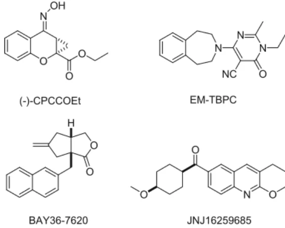
from literature (
Fig. 1
).
7–11Compound 1 was identified from our high throughput screening
as a potent mGluR1 antagonist that was active in an in vivo model
for pain (rat spinal nerve ligation, SNL).
12A recent paper describing
the SAR around the tricyclic frame work was published.
13They
have not disclosed any unsaturated alkyl substitutions on the
A-ring of the tricyclic structure. Our attempt to replace the dimethyl
amino moiety was exceptionally fruitful.
14We found that small
unsaturated alkyls gave extremely potent mGluR1 antagonists.
SAR studies involving A-ring and N-aryl modifications and some
in vivo results are discussed in this Letter (
Fig. 2
).
Compounds of this type were synthesized according to
Scheme
1
. Commercially available ethylcyano malonate was condensed
with dimethylformamide–dimethylacetal (DMF–DMA) followed
by acid catalyzed cyclization gave pyridine derivative 4 in good
yields. Upon reaction with POCl
3on 4 gave 5 which was cyclized
to a common intermediate 6. Heating a solution of 6 in DMF with
0960-894X/$ - see front matter Ó 2009 Published by Elsevier Ltd. doi:10.1016/j.bmcl.2009.04.104
* Corresponding author. Tel.: +1 908 740 4373; fax: +1 908 740 7164. E-mail addresses: [email protected], thavalakulamgar.sasikumar@ spcorp.com(T.K. Sasikumar). O N O O OH O O H N N N O NC N O O O (-)-CPCCOEt BAY36-7620 EM-TBPC JNJ16259685
Figure 1. Noncompetitive antagonists of mGluR1.
Contents lists available at
ScienceDirect
Bioorganic & Medicinal Chemistry Letters
j o u r n a l h o m e p a g e : w w w . e l s e v i e r . c o m / l o c a t e / b m c l
DMF–DMA afforded compound 7 which was cyclized to the
tricy-clic compound 8 in good yields. During this course of our studies,
we found that heating a solution of 6 with desired amine in
tri-ethylorthoformate in the presence of acetic acid gave the cyclized
compound in very high yields.
15Demethylation of 8 followed by
triflation of the phenol afforded 10 in good yields. Intermediate
10 was reacted with unsaturated alkylamines to afford the final
compounds in excellent yields. We used allyl, methallyl and
prop-argyl amines in this current SAR study. Similar chemistry was
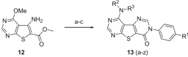
em-ployed in the construction of thienopyrimidine–pyrimidone
analogs as shown in
Scheme 2
. The known amino ester 12
16was
converted to the final targets in three steps with good overall
yields. The final step is the displacement of methoxy group in
the pyrimidine nucleus with appropriate amine in polar solvents
such as DMSO to get compounds 13a–z.
The mGluR1 inhibitory potencies of the newly synthesized
tri-cyclic compounds are summarized in
Table 1
. Initially we turned
our attention to the allyl substitution on the A-ring of the pyridine
nucleus. Human mGluR1 IC
50and rat K
iare shown throughout in
this manuscript. Allylamino compounds 11a–i are generally well
tolerated as shown in
Table 1
. Compound 11a showed human
IC
50value of 2 nM with a rat K
iof 1.1 nM. Other substitution on
the aromatic ring such as 4-Me, 4-Br, and 4-F are very well
toler-ated. However the 4-chloro substitution (compound 11c) gave only
moderate activity on the human receptor but showed excellent
po-tency in the rat assay. Similar results were obtained with the 4-Br
derivative (11i). Disubstitution on the aromatic ring is well
toler-ated in this scaffold. N-Methyltoler-ated compounds such as 11g and
11h are also tolerated. It has been found that all these tricyclic
derivatives are inactive in the mGluR5 assay.
Next, we studied the effect of substitution on the allyl group.
Methallyl derivatives generally afforded active analogs with
2–3-fold less mGluR1 affinity as shown in
Table 2
. Disubstitution on
the aromatic ring (compounds 11n, 11o and 11p) generally
de-creases the human inhibitory potency. Compounds 11k and 11l
exhibited subnanomolar activity in rat mGluR1 assay. Methallyl
substitution was also very effective in the pyrimidine series (
Table
3
). Simple phenyl substitution (compound 13f)
17provided good
human affinity but a moderate rat K
ivalue. It was interesting to
note that the 4-pyridyl analog displayed poor activity at both the
human and rat receptors. Unlike in the case of pyridine analogs,
disubstitution in the pyrimidine series is very well tolerated. For
example, compound 13l, showed human IC
50of 8.9 nM and rat K
iof 2.3 nM.
Replacement of the dimethylamino group of our lead
pound with propargylamine afforded an extremely potent
com-pound 11q with human IC
500f 0.9 nM and a rat K
iof 0.6 nM as
S X Y N N N O R1 N R2 R3 S N N N O N R1 = Aryl R2 = unsaturated alkyls R3 = H or Me X = C or N; Y = C or N 1 OMe A B CFigure 2. General SAR plans.
S N N N O OMe R1 S N N N O OTf R1 S N O NH2 O OMe Cl N OMe CN NC CN EtO NC CN MeO N N OH OMe CN S N N N O N R1 R2 R3 2 3 4 5 6 7 10 a b c d j 11 (a-x) S N O N O OMe N e S N N N O OH R1 f h i 8 9 g
Scheme 1. Reagents and conditions: (a) DMF–DMA, MeOH, 80 °C; (b) HOAc (80%), 130 °C; (c) POCl3, Et3N, 100 °C; (d) HSCH2CO2Me, NaOMe, DMF, 80 °C; (e) DMF–DMA, DMF,
100 °C; (f) 4-Cl-aniline (R1= Cl), HOAc, toluene, 110 °C; (g) 4-Cl-aniline (R1= Cl), HOAc, CH(OEt)
3, 160 °C; (h) BBr3, CH2Cl2; (i) NPhTf2, CH2Cl2; (j) R2R3NH, DMSO. S N N O NH2 O OMe S N N N N O N R1 R2 R3 12 13 (a-z) a-c
Scheme 2. Reagents and conditions: (a) DMF–DMA, DMF, 100 °C; (b) 4-Cl-aniline (R1
= Cl), HOAc, toluene, 110 °C; (c) R2
R3
NH, DMSO.
Table 1
mGluR1 receptor binding for compounds 1, 11a–i
S N N N O R1 N R2 11 (a-i) Compd R1 R2 h-mGluR1 IC50a(nM) r-mGluR1 Kia(nM) 1 9.5 7.9 11a 4-MeO-Ph H 2 1.1 11b 4-Me-Ph H 11 2.7 11c 4-Cl-Ph H 60 1.1 11d 4-Br-Ph H 12.6 0.7 11e 4-F-3-MeO-Ph H 6.8 14 11f 4-F-Ph H 4.4 7.7 11g 4-Cl-Ph Me 1.1 5.4 11h 4-MeO-Ph Me 6.9 4.1 11i 4-Br-Ph Me 66 3.2 aThe IC
50and Kidata are an average of at least three measurements, performed
on human mGlu1/5 and rat mGlu1 receptors, respectively. The standard error was 10%, and variability was less than twofold from assay to assay. h-mGluR5 IC50> 3
l
M.shown in
Table 4
. 4-Me, 4-MeO, and 4-Br substitution on the N-aryl
group afforded similarly highly potent compounds. N-Methylation
lowered affinity 4–5-fold in all cases except 4-BrPh derivative
(11x).
Similar results were obtained in the A-ring pyrimidine series as
evidenced from the
Table 5
. Generally, mono substitution at the
para substitution of the N-aryl ring is generally well tolerated.
Compounds 13n, 13o, 13q and 13s attained single digit nanomolar
at the human mGluR1 assay. However disubstituted compounds
such as 13x and 13z exhibited a large decrease in potency as
evi-denced from
Table 5
.
Having achieved excellent potency against mGluR1 receptors,
we shifted our attention toward measuring the pharmacokinetics
of these types of molecules. Data for representative compounds
11q and 11s are shown in
Table 6
.
Compounds 11q and 11s showed moderate rat AUC with high
brain to plasma ratio as demonstrated in
Table 6
. These
com-pounds were inactive in the hERG assay. It has been found that
compounds 11q and 11s were active in the in vivo pain model
(rat spinal nerve ligation, SNL)
12with ED
50
values of 3.3 and
6.4 mg/kg, respectively, when dosed orally. The metabolic pathway
in rat for high clearance compound 11q was further investigated
using microsome incubation. The metabolic details are shown in
Figure 3
. The metabolism of acetylenic compounds commonly used
in the formulation of pharmaceuticals have been investigated
pre-viously using
13C NMR and mass spectrometry.
19,20Compound 11q
Table 2
mGluR1 receptor binding for compounds 11j–p
S N N N O R1 NH 11 j-p Compd R1 h-mGluR1 IC50a(nM) r-mGluR1 Kia(nM) 11j 4-Cl-Ph 10.6 1.2 11k 4-Me-Ph 16.4 0.2 11l 4-MeO-Ph 10.4 0.4 11m 4-Br-Ph 43 1.5 11n 3-F-4-MeO-Ph 103 13
11o 3-(2,3-Dihydrobenzo[b] [1,4]Dioxin-6-yl) 196 13 11p 3-(Benzo[d][1,3]dioxol-5-yl) 241 14
a
The IC50and Kidata are an average of at least three measurements, performed
on human mGlu1/5 and rat mGlu1 receptors, respectively. The standard error was 10%, and variability was less than twofold from assay to assay. h-mGluR5 IC50> 3
l
M.Table 3
mGluR1 receptor binding for compounds 13a–m
S N N N N O R1 NH 13 (a-m) Compd R1 h-mGluR1 IC50a(nM) r-mGluR1 Kia(nM) 13a 4-Cl-Ph 7.7 2.2 13b 4-Me-Ph 12.4 2.9 13c 4-MeO-Ph 9.6 7.5 13d 4-Br-Ph 14.5 3.8 13e 3-F-4-MeO-Ph 72 68 13f Ph 4.6 25 13g 4-Py 955 1000 13h 4-F-Ph 10.7 38 13i 3-Cl-Ph 53 62 13j 3-(Benzo[d]thiazol-5-yl) 16 43 13k 3-(Benzo[d]thiazol-6-yl) 17 49 13l 3-(Benzo[b]thiophen-5-yl) 8.9 2.3 13m 3-(2,3-Dihydrobenzo[b] [1,4]dioxin-6-yl) 33 64 a The IC
50and Kidata are an average of at least three measurements, performed
on human mGlu1/5 and rat mGlu1 receptors, respectively. The standard error was 10%, and variability was less than twofold from assay to assay. h-mGluR5 IC50> 3
l
M.Table 4
mGluR1 receptor binding for compounds 11q–x
S N N N O R1 N R2 11 (q-x) Compd R1 R2 h-mGluR1 IC50a(nM) r-mGluR1 Kia(nM) 11q 4-Cl-Ph H 0.9 0.6 11r 4-Me-Ph H 1.5 1.6 11s 4-MeO-Ph H 2.1 3.5 11t 4-Br-Ph H 1.8 0.7 11u 3-F-4-MeO-Ph H 12.5 28 11v 4-Cl-Ph Me 3.8 18 11w 4-MeO-Ph Me 10.8 27 11x 4-Br-Ph Me 1.9 8 a
The IC50and Kidata are an average of at least three measurements, performed
on human mGlu1/5 and rat mGlu1 receptors, respectively. The standard error was 10%, and variability was less than twofold from assay to assay. h-mGluR5 IC50> 3
l
M.Table 5
mGluR1 receptor binding for compounds 13n–z
S N N N N O R1 NH 13 (n-z) Compd R1 h-mGluR1 IC50a (nM) r-mGluR1 Kia (nM) 13n 4-Cl-Ph 3.0 4.4 13o 4-Me-Ph 3.9 8.3 13p 4-MeO-Ph 13 25 13q 4-Br-Ph 6.8 29 13r 3-F-4-MeO-Ph 212 77 13s 4-F-Ph 8.2 39 13t 2-F-4-MeO-Ph 85 32 13u 3-(2,3-Dihydrobenzo[b] [1,4]dioxin-6-yl) 106 160 13v 3-(Benzo[d][1,3]dioxol-5-yl) 137 997 13w 3-(Benzo[d]thiazol-5-yl) 45 65 13x 3-(Benzo[d]thiazol-6-yl) 209 107 13y 3-(Benzofuran-5-yl) 21 63 13z 3-(Benzo[b]thiophen-5-yl) 219 69 a
The IC50and Kidata are an average of at least three measurements, performed
on human mGlu1/5 and rat mGlu1 receptors, respectively. The standard error was 10%, and variability was less than 2-fold from assay to assay. h-mGluR5 IC50> 3
l
M.was incubated with rat liver microsome for 0–24 h and the
metab-olites were identified by LC–MS methodology. The oxidation of the
carbon adjacent to the amine lead to the intermediate 14 followed
by degradation to the final molecules 15 and 16. LC–MS peak at
13 min with an observed mass of 383 is attributed to the
interme-diate 14. LC–MS analysis showed a peak at 3.9 min. with a mass of
401 could be water addition product (17) to intermediate 14 as
shown in
Figure 3
. The parent amino compound 15 displayed in
LC–MS at 11.3 min. This retention time of the amine 15 was
inde-pendently confirmed via analysis of pure synthetic sample.
Com-pound 15 was found to be the major metabolite in the rat bile.
The side product of this whole sequence, compound 16, is a known
glutathione scavenger described in the literature.
19In order to avoid the metabolic issues, we introduced methyl
groups adjacent to the amino group. Unfortunately
monomethyla-tion and dimethylamonomethyla-tion of our lead compounds afforded
200–300-fold less active compounds (19–21) as shown in
Figure 4
. Identical
results were obtained for compounds with different substitution
on the right hand side aromatic ring.
In summary, we have achieved a large number of single digit
nanomolar mGluR1 antagonists in the tricyclic series. This
excel-lent potency sometimes extrapolates to good in vivo efficacy as
seen in compounds 11q and 11s. These types of compounds show
moderate PK and high brain plasma ratio. It has also been found
that these compounds are inactive in the hERG assay. Further data
will be published elsewhere.
Acknowledgments
We thank Drs. Deen Tulshian, Julius Matasi and Peter Korakas
for helpful discussions. We also thank Lisa Broske’s group for
ani-mal dosing and Sam Wainhaus’ group for PK sample analysis.
References and notes
1. Higgins, G. A.; Miczek, K. A. Psychopharmacology 2005, 179, 1. 2. Kew, J. N. C.; Kemp, J. A. Psychopharmacology 2005, 179, 4.
3. Steckler, T.; Lavreysen, H.; Oliveira, A. M.; Aerts, N.; Van Craenendonck, H.; Prickaerts, J.; Megens, A.; Lesage, A. S. J. Psychopharmacology 2005, 179, 198. 4. Niswender, C. M.; Jones, C. K.; Conn, P. J. Curr. Top. Med. Chem. 2005, 5, 847. 5. Kunishima, N.; Shimada, Y.; Tsuji, Y.; Sato, T.; Yamamoto, M.; Kumasaka, T.;
Nakanishi, S.; Jingami, H.; Morikawa, K. Nature 2000, 407, 971.
6. Constantino, G.; Macchiarulo, A.; Pellicciari, R. Bioorg. Med. Chem 2001, 9, 847. 7. Layton, M. E. Curr. Top. Med. Chem 2005, 5, 859.
8. Ott, D.; Floersheim, P.; Inderbitzin, W.; Stoehr, N.; Francotte, E.; Lecis, G.; Richert, P.; Rihs, G.; Flor, P. J.; Kuhn, R.; Gasparini, F. J. Med. Chem. 2000, 43, 4428.
9. Malherbe, P.; Kratochwil, N.; Knoflach, F.; Zenner, M.-T.; Kew, J. N. C.; Kratzeisen, C.; Maerki, H. P.; Adam, G.; Mutel, V. J. Biol. Chem. 2003, 278, 8340. 10. Carroll, F. Y.; Stolle, A.; Beart, P. M.; Voerste, A.; Brabet, I.; Mauler, F.; Joly, C.; Antonicek, H.; Bockaert, J.; Müller, T.; Pin, J. P.; Prézeau, L. Mol. Pharmacol. 2001, 59, 965.
11. Lavreysen, H.; Wouters, R.; Bischoff, F.; Pereira, S. N.; Langlois, X.; Blokland, S.; Somers, M.; Dillen, L.; Lesage, A. S. J. Neuropharmacology 2004, 47, 961. 12. Varty, G. B.; Grilli, M.; Forlani, A.; Fredduzzi, S.; Grzelak, M. E.; Guthrie, D. H.;
Hodgson, R. A.; Sherry, X. L.; Nicolussi, E.; Pond, A. J.; Parker, E. M.; Hunter, J. C.; Higgins, G. A.; Reggiani, A.; Bertorelli, R. Psychopharmacology 2005, 179, 207. Table 6
PK Profile and in vivo activity of selected compounds
S N N N O NH R Parameters R = Cl (11q) R = OMe (11s) Human mGluR1 IC50(nM) 0.9 2.1 Rat mGluR1 Ki(nM) 0.6 3.5 Human mGluR5 IC50(nM) >3000 >3000 Rat SNL ED5012 3.3 mg/kg po 6.4 mg/kg po Caco-2 permeability 430 nm/s 690 nm/s Efflux substrate No No Rat PK, (10 mg/kg), AUC (ng.h/mL)18 427 682 Brain conc. @ 6 h (ng/g) 67 257 Brain/Plasma 1.8 2.5 Clearance (mL/min/kg) 30 22 Bioavailability (%) 16 34 S N N N O NH Cl N S N N O NH Cl OH S N N N O NH Cl OH (11q) C18H11ClN4OS (RT = 14.1 Min) Mol. Wt.: 366.82 (14) C18H11ClN4O2S (RT = 13.0 Min) Mol. Wt.: 382.82 H OH (17) C18H13ClN4O3S (RT = 3.9 Min) Mol. Wt.: 400.84 S N N N O NH Cl OH O S N N N O NH2 Cl (15) C15H9ClN4OS (RT = 11.3 Min) Mol. Wt.: 328.78 O H [O] H2O + (16) (18)
Figure 3. Metabolism of propargyl group (Mass analysis of rat bile samples, 0–24 h).
S N N N O NH R1 R2 19; R1 = Me, R2 = H; h-IC 50 = 314 nM
20; R1 = Me, R2 = Me; h-IC
50 = 171 nM
21; R1 = Et, R2 = Et; h-IC
50 = 358 nM
α
13. Recently, scientists from Abbott Laboratories disclosed similar mGluR1 antagonists based on the tricyclic triazafluorenone core structure. See: Zheng, G. Z.; Bhatia, P.; Daanen, J.; Kolasa, T.; Patel, M.; Latshaw, S.; Kouhen, O. F. E.; Chang, R.; Uchic, M. E.; Miller, L.; Nakane, M.; Lehto, S. G.; Honore, M. P.; Moreland, R. B.; Brioni, J. D.; Stewart, A. O. J. Med. Chem. 2005, 48, 7374.
14. Our efforts in the modification of tricyclic structure is being published in the following patents, WO 2006/002051 and US 2006/167029. Also see: Wu, W-L.; Burnett, D. A.; Domalski, D.; Greenlee, W. J.; Li, C.; Bertorelli, R.; Fredduzzi, S.; Lozza, G.; Veltri, A.; Reggiani, A. J. Med. Chem 2007, 50, 5550. Biology materials and methods were described in this Letter.
15. Typical one step synthesis of tricyclic compound is described as follows: Compound 6 (2 g, 0.0084 mol) was suspended in 15 mL triethyl orthoformate and treated with acetic acid (2 mL) and 4-chloroaniline (2 g, 0.0156 mol). The contents were heated in a sealed tube at 160 °C for 16 h. The reaction mixture was cooled to room temperature and the precipitated product was washed several
times with ether. The product was dried in vacuo to get 2.5 g of compound 8 (R1= Cl) as white solid.1H NMR (CDCl
3): d 8.65 (d, 1H), 8.29 (s, 1H), 7.56 (d,
2H), 7.40 (d, 2H), 6.93 (d, 1H), 4.17 (s, 3H). Mass Spectrum (M+1): m/z calcd for
C16H11ClN3O2S+= 344.03, found m/z = 344.2.
16. Clark, J.; Shahhet, M. S. J. Heterocycl. Chem. 1993, 30, 1065. 17. Spectral data for compound 13f:1
H NMR (CDCl3): d 8.62 (s, 1H), 8.27 (s, 1H),
7.91 (s, 1H), 7.58 (m, 3H), 7.45 (m, 2H), 4.99 (s, 1H), 4.93 (s, 1H), 4.32 (d, 2H), 1.86 (s, 3H). Mass Spectrum (M+1
): m/z calcd for C18H16N5OS+= 350.11, found
m/z = 350.2.
18. Korfmacher, W. A.; Cox, K. A.; Ng, K. J.; Veals, J.; Hsien, Y.; Wainhaus, S.; Broske, L.; Prelusky, D.; Nomeir, A.; White, R. E. Rapid Commun. Mass. Spectrom. 2001, 15, 335.
19. Banijamali, A. R.; Xu, Y.; DeMatteo, V.; Strunk, R. J.; Sumner, S. J. J. Agric. Food Chem. 2000, 48, 4693.
20. DeMaster, E. G.; Sumner, H. W.; Kaplan, E.; Shirota, F. N.; Nagasawa, H. T. Toxicol. Pharmacol. 1982, 65, 390.