Creative Commons Non Commercial CC BY-NC: This article is distributed under the terms of the Creative Commons Attribution-NonCommercial 4.0 License (http://www.creativecommons.org/licenses/by-nc/4.0/) which permits non-commercial use, reproduction and distribution of the work without further permission provided the original work is attributed as specified on the SAGE and Open Access pages (https://us.sagepub.com/en-us/nam/open-access-at-sage).
https://doi.org/10.1177/0394632017740976 International Journal of
Immunopathology and Pharmacology 2017, Vol. 30(4) 383 –394 © The Author(s) 2017 Reprints and permissions:
sagepub.co.uk/journalsPermissions.nav DOI: 10.1177/0394632017740976 journals.sagepub.com/home/iji
Introduction
Programmed cell death or apoptosis is a common cell death process involved in the normal growth, differentiation, and development of many tissue systems, including nervous system.1 In addition, apoptosis is involved in the death of motor neurons in diverse spinal motor neuron degenerative dis-eases, such as amyotrophic lateral sclerosis (ALS),
Conditioned medium from human
gingival mesenchymal stem cells protects
motor-neuron-like NSC-34 cells against
scratch-injury-induced cell death
Thangavelu Soundara Rajan1, Francesca Diomede2,
Placido Bramanti1, Oriana Trubiani2 and Emanuela Mazzon1
Abstract
Neuronal cell death is a normal process during central nervous system (CNS) development and is also involved in the death of motor neurons in diverse spinal motor neuron degenerative diseases. Here, we investigated the neuroprotective effect of secretory factors released from human gingival mesenchymal stem cells (hGMSCs) in mechanically injured murine motor-neuron-like NSC-34 cells. The cells were exposed to scratch injury and the markers for apoptosis and oxidative stress were examined. Immunocytochemistry results showed that proapoptotic markers cleaved caspase-3 and Bax were elevated while anti-apoptotic protein Bcl-2 was suppressed in scratch-injured NSC-34 cells. Oxidative stress markers SOD-1, inducible nitric oxide synthase (iNOS), Cox-2, and proinflammatory cytokine tumor necrosis factor alpha (TNF-α) were activated. Conditioned medium (CM) derived from hGMSCs (hGMSC-CM) significantly blocked the cell death by suppressing SOD-1, iNOS, TNF-α, cleaved caspase-3, and Bax. Bcl-2 and anti-inflammatory cytokine anti-interleukin 10 (IL-10) were increased in hGMSC-CM-treated injured cells. Moreover, hGMSC-CM treatment upregulated neurotrophins anti-brain-derived neurotrophic factor (BDNF) and NT3. Western blot data of hGMSC-CM revealed the presence of neurotrophins nerve growth factor (NGF), NT3, anti-inflammatory cytokines IL-10, and transforming growth factor beta (TGF-β), suggesting their positive role to elicit neuroprotection. Our results propose that hGMSC-CM may serve as a simple and potential autologous therapeutic tool to treat motor neuron injury.
Keywords
apoptosis, gingival mesenchymal stem cells conditioned medium, in vitro CNS injury model, inflammation, neurotrophic factors, NSC-34 cells, oxidative stress
Date received: 7 July 2017; accepted: 12 October 2017
1IRCCS Centro Neurolesi “Bonino-Pulejo,” Messina, Italy 2 Stem Cells and Regenerative Medicine Laboratory, Department
of Medical, Oral and Biotechnological Sciences, “G. d’Annunzio” University of Chieti-Pescara, Chieti, Italy
Corresponding author:
Emanuela Mazzon, IRCCS Centro Neurolesi “Bonino-Pulejo,” Via Provinciale Palermo, contrada Casazza, 98124 Messina, Italy. Email: [email protected]
spinal muscular atrophy, and spinal cord injury (SCI).2–4 Pharmacological intervention to modulate apoptosis and other related essential pathological cellular and molecular cascades, including excito-toxicity, oxidative stress, and inflammation, is nec-essary to ameliorate these motor neuron degenerative diseases.
Adult mesenchymal stem cells (MSCs) have displayed a wide range of neuroprotective effects in various preclinical and clinical investigations against motor neuron degenerative diseases.5–7 The neuroprotective effects produced by MSCs have been attributed partly to the paracrine activity of their soluble secreted factors, including inflamma-tory cytokines and neurotrophins.8 MSCs are derived from various adult tissues and particularly neural crest-originated adult MSCs from human oral tissues have received considerable interest owing to the less invasive method used in dental tissue explants collection and their capacity to be a simple autologous MSCs resource tool.9 Human dental MSCs are derived from oral tissues, such as gingiva, periodontal ligament, dental pulp, pulp of human exfoliated deciduous teeth, apical papilla, and dental follicle.10 In addition to the application in regenerative dentistry,11 we and other groups have demonstrated the neuroprotective and anti-inflammatory properties of dental MSCs and their secretory molecules in various in vivo and in vitro disease models, including multiple sclerosis, Alzheimer’s disease, and Myasthenia gravis.12–17 Notably, human gingival MSCs (hGMSCs) are promising in regenerative medicine. Indeed, hGM-SCs in comparison with other sources of MhGM-SCs are abundant, easy to isolate, and possess remarkable immunomodulatory properties.18,19
In this study, we have investigated whether con-ditioned medium (CM) from healthy hGMSCs may exert neuroprotection in mechanically injured motor-neuron-like NSC-34 hybrid cells. These cells resulted from the fusion of motor-neuron-enriched primary mouse embryonic spinal cord cells and mouse neuroblastoma. These cells pos-sess motor neuron morphology, exhibit many physiological properties of motor neurons, includ-ing neurotrophins synthesis, neurites formation, and acetyl choline synthesis, and thus are a recog-nized model to investigate the pathophysiology of motor neurons.20–22 In our study, motor-neuron-like NSC-34 cells were subjected to manual scratch injury, treated with hGMSC-CM, and
injury-mediated apoptotic, oxidative stress, and inflammatory markers were examined.
Materials and methods Ethical statement
Experimental protocol for human gingival tissues collection used in this study was approved by the Medical Ethics Committee at the Medical School, “G. d’Annunzio” University, Chieti, Italy (no. 266/17.04.14). Each donor has signed the formal consent form.
hGMSCs culture establishment
All donors were unaffected by any systemic and oral diseases. The gingival tissues were collected from oral cavity without inflammation. The tissues were then subjected for de-epithelialization and were washed several times with 1× phosphate buff-ered saline (PBS) (Li StarFish, Milan, Italy). Consequently, the tissues were cultured in serum free, chemically defined medium for the growth of human MSCs (TheraPEAK™ MSCGM-CD™ BulletKit; Lonza, Basel, Switzerland) under stand-ard cell culture conditions. Medium was replaced with fresh medium twice a week. Explants-derived adhered cells were grown until 80%, detached using Triple Select (Li StarFish, Milan, Italy), and subcultured for further experiments. The cytofluor-imetric evaluation of stem cell markers has been carried out as previously reported by Libro et al.23 hGMSCs mesengenic differentiation
hGMSCs were differentiated in adipogenic lineage using a protocol previously described by Diomede et al.24 Briefly, expanded cells at 100% confluence were maintained for three cycles of induction/ maintenance stimulated with adipogenic supple-mented media (Lonza) differentiation. After 28 days of induction, the cells were fixed in 10% formalin for 15 min and washed with dH2O. Subsequently, the cells were stained with Oil Red O (ORO) working solution (300 mg of ORO/100 mL of isopropanol) for 5 min and counterstained with hematoxylin.
For osteogenic induction, hGMSCs were treated as previously reported by Trubiani et al.25 In brief, cells were incubated with osteogenic supplemented media (Lonza). After 3 weeks, cells
were stained with Alizarin Red S solution to visu-alize calcium deposition and extracellular matrix (ECM) mineralization.
Chondrogenic differentiation procedure of hGMSCs was performed as previously described by Rajan et al.26 After 28 days of induction proce-dure, the pellets were fixed in 4% paraformalde-hyde at 4°C for 24 h, dehydrated in an ascending series of ethanol (40%, 70%, 90%, and 100% etha-nol; 20 min/step), and embedded in paraffin. Sections (3 μm) were cut and stained with 1% Alcian Blue (pH 2.5; Sigma–Aldrich, Milan, Italy) for 5 min and observed by means of light micros-copy (Leica Microsystem, Milan, Italy).
Preparation of CM
hGMSCs at passage number 2 were plated at the density of 1.5 × 10⁴ cells/cm2 in TheraPEAK™ MSCGM-CD™ and incubated for 3 days. Then, the CM of hGMSCs (hGMSC-CM) was collected and processed for two-step centrifugation in order to remove cell debris as follows: first one at 1200 r/ min for 5 min and the second one at 3000 r/min for 3 min. For protein quantification, 1 mL of hGMSC-CM was added with 3 mL of cold acetone, incu-bated for 1 h at −20°C, and centrifuged at 16,000 r/ min for 12 min at 4°C. The resulted pellet was resuspended in radioimmunoprecipitation assay (RIPA) buffer and quantified for protein concentra-tion using BIO-RAD PROTEIN ASSAY Kit (Bio-Rad Laboratories GmbH, Germany).27
NSC-34 cell culture
The murine motor-neuron-like NSC-34 cells (Cellutions Biosystems Inc., Cedarlane, Canada) were cultured in high glucose Dulbecco’s modified Eagle’s medium (DMEM) (Sigma–Aldrich, Co., USA) combined with 10% fetal bovine serum (FBS) (Sigma–Aldrich, Co., USA). The cells were maintained in an incubator using the following parameters: 37°C temperature and 95% air/5% CO2 atmosphere. Cells with passage less than 28 were used for the experiments.
Scratch injury
Mechanical injury in NSC-34 cells was attained by scratch injury as reported earlier with minor modifications.28–31 Briefly, cells were grown in DMEM supplemented with 10% FBS to achieve
80% cell density. Twelve-millimeter coverslips in six-well plates were used for experiments. Then, the FBS containing DMEM was removed and the cells were exposed to serum free DMEM for 12 h. Subsequently, cells were injured by manual scratches (a total of four scratches; two scratches in horizontal direction and two scratches in verti-cal direction) using a 1 mL pipette tip, which pro-duced a sum of nine quadrants. Space between each scratch area was 2 mm. After the scratch injury, medium with floated cells was removed and the cells were incubated with fresh serum free medium for 24 h. Then, the medium was elimi-nated and the injured cells were incubated with fresh serum free DMEM, serum free DMEM com-bined with unconditioned MSCGM-CD, or serum free DMEM combined with hGMSC-CM (1 mg/ mL) for 24 h. Control cells with no scratches were also included. Then, the cells were either fixed or collected for further studies.
Trypan blue exclusion assay
In order to assess cell viability, NSC-34 cells were stained with Trypan blue solution (Thermo Scientific, USA) according to manufacturer’s instructions. Briefly, cells from each group were harvested and resuspended in 1× PBS. Cells were mixed with filtered 0.4% Trypan Blue reagent at 1:1 ratio and incubated for 3 min at room tempera-ture. Then, approximately 10–20 μL of cells were loaded on a hemocytometer and were immediately quantified for non-blue cells (live) and blue cells (dead). Percentage of dead cells (number of dead blue cells divided by total number of cells) was calculated from three separate experiments.
Eosin and hematoxylin staining
Eosin and hematoxylin (E&H) staining was per-formed to stain cytoplasm and nucleus, respec-tively.32 NSC-34 cells on 12-mm coverslips (Thermo SCIENTIFIC, Germany) were fixed using PBS–4% paraformaldehyde (Chem Cruz, USA) for 30 min at ambient temperature. After thrice 1× PBS washes, cells on coverslips were stained using Hematoxylin Harris (Bio-Optica, Italy) for 1 min at room temperature and washed with tap water. Subsequently, cells were stained with eosin (Bio-Optica, Italy) for 5 min at room temperature and washed with distilled water. Then, coverslips were serially dehydrated using ethanol
(Carlo Erba Reagents, France) with the following percentage: 50%, 70%, 80%, 96%, and 100%. Later, coverslips were treated with xylene (J.T.Baker, The Netherlands). Microscopy was car-ried out using LEICA DM 2000 microscope con-nected with LEICA ICC50 HD camera. All micrographs are representative of three independ-ent experimindepend-ents.
Immunocytochemistry
In order to perform immunocytochemical staining, NSC-34 cells fixed with 4% paraformaldehyde were incubated with 3% hydrogen peroxide (Sigma–Aldrich, USA) in room temperature for 15 min followed by blocking with normal horse serum (Vector Laboratories, Burlingame, CA) and Triton X-100 in room temperature for 20 min. Then, the motoneurons were incubated with the following primary antibodies at 4°C for overnight: anti-superoxide dismutase 1 (SOD-1; Abcam, USA; 1:50 dilution), anti-inducible nitric oxide synthase (iNOS; Cell Signaling Technology, USA; 1:100 dilution), anti-cyclooxygenese-2 (Cox-2; Santa Cruz Biotechnology Inc., USA; 1:50 dilu-tion), anti-tumor necrosis factor alpha (TNF-α; Santa Cruz Biotechnology Inc., USA; 1:50 dilu-tion), anti-interleukin 10 (IL-10; Santa Cruz Biotechnology Inc., USA; 1:50 dilution), B-cell lymphoma 2 (Bcl-2; Santa Cruz Biotechnology Inc., USA; 1:50 dilution), Bcl-2-associated X pro-tein (Bax; Santa Cruz Biotechnology Inc., USA; 1:50 dilution), cleaved caspase-3 (Cell Signaling Technology, USA; 1:100 dilution), anti-brain-derived neurotrophic factor (BDNF; Santa Cruz Biotechnology Inc., USA; 1:50 dilution), and anti-neurotrophin 3 (NT3; Santa Cruz Biotechnology Inc., USA; 1:50 dilution). Then, the cells were exposed to biotinylated universal secondary anti-body and streptavidin ABComplex-horseradish peroxidase (HRP) as per manufacturer’s instruc-tions (Vectastain ABC Kit, Vector Laboratories, USA). Immunostaining was developed with DAB KIT (Vector Laboratories, USA) (immunoreaction produced brown color). Counterstaining was per-formed with nuclear fast red staining (pink color). Cells were analyzed using a light microscope (LEICA DM 2000 combined with LEICA ICC50 HD camera). Images (n = 3 images from each group) were acquired for densitometry evaluation to calculate the percentage of positive staining
using LEICA Application Suite V4.2.0 software. Data were acquired from three separate experi-ments done in duplicates.
Western blot
Proteins present in hGMSC-CM were quantified by Bio-Rad Protein Assay Kit (Bio-Rad, USA). Proteins were separated using 15% sodium dodecyl sulfate-polyacrylamide gel electrophoresis (SDS-PAGE). The proteins in the gel were transferred onto polyvinylidene difluoride (PVDF) membranes (Immobilon-P, Millipore, USA). After blotting, the PVDF membranes were stained with Ponceau S solution (SERVA, Denmark) to assess protein sepa-ration. Then, the membranes were incubated in blocking solution (5% skimmed milk in 1× PBS) at ambient temperature for 1 h. After blocking, the membranes were incubated with the following anti-bodies for overnight at 4°C: nerve growth factor (NGF) (1:250; Abcam, USA), NT3 (1:250; Santa Cruz Biotechnology Inc., USA), IL-10 (1:250; Santa Cruz Biotechnology Inc., USA), and trans-forming growth factor beta (TGF-β) (1:500; Abcam, USA). Subsequently, membranes were incubated with appropriate HRP-conjugated secondary anti-bodies IgG (1:2000; Santa Cruz Biotechnology Inc., USA). Finally, the membranes were subjected for protein detection using chemiluminescence (enhanced) solution kit (Luminata Western HRP Substrates, Millipore, USA). Protein band images were obtained using imaging software ChemiDoc™ MP (Bio-Rad, USA). Representative blot images from three separate analyses are given.
Statistical data analysis
Statistical analysis of the data was accomplished using GraphPad Prism version 6.0 program (GraphPad Software, La Jolla, CA). Statistical sig-nificance was measured by one-way analysis of variance (ANOVA) and post hoc Bonferroni test for multiple comparison. P-value less than 0.05 was considered statistically significant. Results are expressed as mean ± SD.
Results
hGMSCs characterization
hGMSCs have been characterized using flow cytometry to evaluate the expression profile of
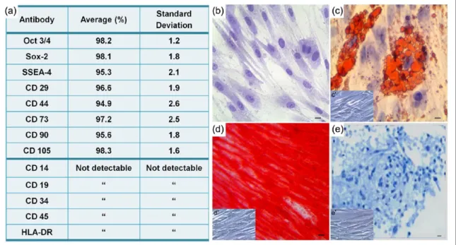
the principal mesenchymal markers. Cells did not express any hematopoietic marker (CD14, CD19, CD34, and CD45), but expressed a variety of mesenchymal markers (CD29, CD73, CD90, and CD105), surface adhesion molecule (CD44), and the stemness markers Oct 3⁄4, Sox-2, and SSEA-4. Consistent with a stem cell profile, these cells stained negative for human leukocyte antigen-antigen D related (HLA-DR) (Figure 1(a)). Plastic-adherent GMSCs, stained with toluidine blue solution, showed a fibroblastic morphology and ovoidal nuclei with one or two nucleoli (Figure 1(b)). hGMSCs were induced to adipo-genic, osteoadipo-genic, and chondrogenic commitment to assay their capability to differentiate into mes-engenic lineage. To evaluate the adipogenic dif-ferentiation of hGMSCs, the cellular monolayer was stained with ORO and observed by light microscopy. Cells showed numerous intracellular lipid droplets at cytoplasmic level (Figure 1(c)). hGMSCs induced to osteogenic differentiation showed a characteristic arrangement and several areas of mineralization, as evidenced by Alizarin Red S staining (Figure 1(d)). hGMSCs pellets cultured under chondrogenic conditions showed strong Alcian Blue staining (Figure 1(e)),
indicating abundant ECM proteoglycans and glycosaminoglycans.
hGMSC-CM decreases cell death produced by scratch injury in NSC-34 cells
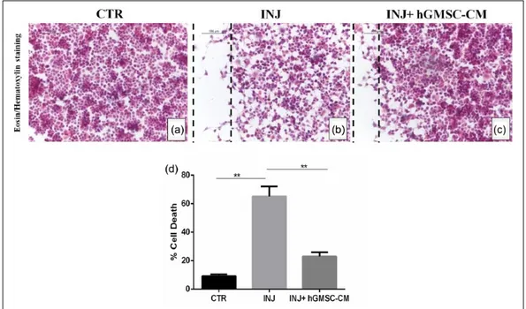
E&H staining showed considerable cell loss in scratch injury area (Figure 2(b)), while injured cells treated with hGMSC-CM displayed a mild increase in cell density (Figure 2(c)). Trypan blue assay demonstrated severe cell death in injured cells. However, treatment with hGMSC-CM sig-nificantly reduced the cell death in injured cells (Figure 2(d); P < 0.05). Injured cells treated with unconditioned MSCGM-CD did not reduce the cell death (data not shown).
hGMSC-CM inhibits apoptosis in injured NSC-34 cells
Later, we investigated the modulation of apoptosis induction in injured cells. Immunohistochemistry data showed that apoptosis was notably elevated in injured cells. Proapoptotic markers cleaved cas-pase-3 (Figure 3(b)) and Bax (Figure 3(f)) were increased, while anti-apoptotic marker Bcl-2 (Figure 3(j)) was absent in injured cells. Interestingly,
Figure 1. hGMSCs characterization. (a) Cytofluorimetric analysis of hGMSCs culture. (b) Plastic-adherent hGMSCs stained with
toluidine blue solution. (c) The adipogenic potential of GMSCs was analyzed using lipid droplets stained with Oil Red O solution. (c′) Undifferentiated hGMSCs. (d) Alizarin Red S staining highlighted mineralization after osteogenic induction. (d′) Undifferentiated hGMSCs. (e) Alcian Blue staining evaluated hGMSCs capability to differentiate in chondrogenic lineage. (e′) Undifferentiated hGMSCs. Bars: 10 µm.
Figure 2. hGMSC-CM decreases cell death produced by scratch injury in NSC-34 cells. Eosin/hematoxylin staining displayed
substantial loss of NSC-34 cells in scratch area (b), while injured cells treated with hGMSC-CM showed a mild increase in cell density (c). Control cells without injury (a). Black dotted lines indicate the area of scratch injury. Magnification: 20×; scale bar: 100 µm. Trypan blue assay demonstrated severe cell death in injured cells. However, treatment with hGMSC-CM significantly reduced the cell death in injured cells (d).
**P < 0.05 CTR versus INJ, **P < 0.05 INJ versus INJ + hGMSC-CM. CTR: uninjured cells; INJ: scratch-injured cells; INJ + hGMSC-CM: scratch-injured cells treated with hGMSC-CM.
Figure 3. hGMSC-CM inhibits apoptosis in injured NSC-34 cells. Immunohistochemistry results showed that in scratch-injured
NSC-34 cells located in injury area as well as located proximally to injury area, marked positive staining was observed for proapoptotic markers cleaved caspase-3 (b) and Bax (f). Anti-apoptotic protein Bcl-2 was absent in injured cells (j). hGMSC-CM treatment significantly suppressed apoptosis activation in injured cells. hGMSC-hGMSC-CM-treated injured cells displayed negative staining for cleaved caspase-3 (c) and Bax (g). In addition, hGMSC-CM treatment significantly resumed Bcl-2 expression in injured cells (k). Uninjured control cells showed negative staining for cleaved caspase-3 (a) and Bax (e) and positive staining for Bcl-2 (i). Magnification: 40×; scale bar: 50 µm. Black dotted lines indicate the area of scratch injury. Densitometric quantification of cleaved caspase-3 (d), Bax (h), and Bcl-2 (l).
****P < 0.0001 CTR versus INJ, ****P < 0.0001 INJ versus INJ + hGMSC-CM. CTR: uninjured cells; INJ: scratch-injured cells; INJ + hGMSC-CM: scratch-injured cells treated with hGMSC-CM. ND: not detectable.
hGMSC-CM treatment significantly blocked the apoptosis. Marked reduction of cleaved caspase-3 and Bax was observed in injured cells treated with hGMSC-CM (Figure 3(c) and (g), respectively). Moreover, Bcl-2 expression was significantly enhanced in hGMSC-CM-treated injured cells (Figure 3(k)). Densitometric quantification of cleaved caspase-3, Bax, and Bcl-2 is shown in Figure 3(d), (h), and (l), respectively. These results demonstrate the anti-apoptotic role of hGMSC-CM in NSC-34 cells exposed to scratch injury.
hGMSC-CM reduces oxidative stress in injured NSC-34 cells
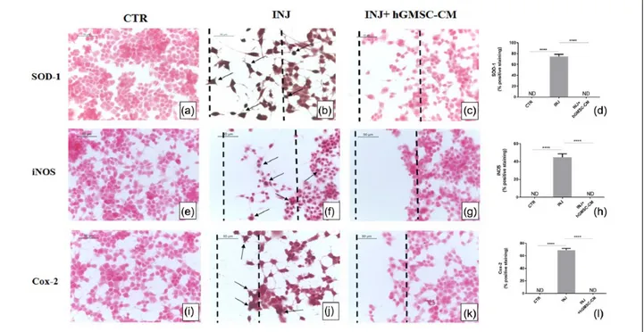
Scratch injury produced severe oxidative stress in NSC-34 cells. Immunohistochemistry results dis-played negative staining for oxidative stress mark-ers SOD-1, iNOS, and Cox-2 in NSC-34 cells without injury (Figure 4(a), (e), and (i), respec-tively), while marked positive staining was noticed for these markers in cells located in injury area as well as in cells located proximally to injury area (Figure 4(b), (f), and (j), respectively). On the other hand, treatment with hGMSC-CM totally suppressed the expression of SOD-1, iNOS, and Cox-2 (Figure 4(c), (g), and (k), respectively). Densitometric quantification of SOD-1, iNOS, and Cox-2 is shown in Figure 4(d), (h), and (l), respectively. These data indicate that GMSC-CM strongly suppresses oxidative stress produced by scratch injury in motor-neuron-like NSC-34 cells. hGMSC-CM suppresses inflammation in injured NSC-34 cells
Next, we investigated the inflammatory response occurred by scratch injury. Immunohistochemistry data revealed the induction of inflammation in injured cells. Proinflammatory cytokine TNF-α was absent in uninjured cells (Figure 5(a)) while positive staining was observed in cells injured with scratches (Figure 5(b)). hGMSC-CM treatment completely blocked TNF-α expression in injured cells (Figure 5(c)). Interestingly, anti-inflamma-tory IL-10 was markedly increased in hGMSC-CM-treated injured cells (Figure 5(g)), while its expression was undetectable in both uninjured cells (Figure 5(e)) and injured cells without treat-ment (Figure 5(f)). Densitometric quantification of TNF-α and IL-10 is shown in Figure 5(d) and (h),
respectively. These results suggest the immuno-suppressive effect of hGMSC-CM in NSC-34 cells affected by scratch injury.
hGMSC-CM augments neurotrophic factors in injured NSC-34 cells
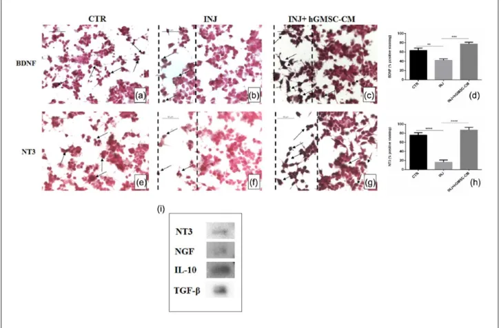
Then, we investigated whether hGMSC-CM could stimulate the expression of neurotrophic factors after injury. We found a basal level expression of neurotrophic factors BDNF and NT3 in uninjured cells (Figure 6(a) and (e), respectively) while their expression was markedly reduced in injured cells (Figure 6(b) and (f), respectively). On the other hand, hGMSC-CM treatment significantly aug-mented BDNF and NT3 expression in injured cells (Figure 6(c) and (g), respectively). Densitometric quantification of BDNF and NT3 is shown in Figure 6(d) and (h), respectively. These findings suggest that hGMSC-CM may provide neuropro-tection in scratch-injured NSC-34 cells by elevat-ing the level of BDNF and NT3.
hGMSC-CM contains trophic factors NT3 and NGF and immunosuppressors IL-10 and TGF-β Finally, we studied the presence of trophic factors and immunosuppressive cytokines present in hGMSC-CM. Western blot data reveal the pres-ence of NGF, NT3, IL-10, and TGF-β in GMSC-CM (Figure 6(i)), which suggest that the neuroprotec-tive effect of hGMSC-CM in injured NSC-34 cells might be, at least in part, due to the presence of these growth factors and immunomodulatory cytokines. Ponceau S staining of Western blot membrane of hGMSC-CM is provided as Supplement Figure 1.
Discussion
Apoptotic loss of motor neurons is a key pathologi-cal feature in motor neuron degenerative dis-eases.2–4 Moreover, in these neurodegenerative diseases, motor neuronal apoptosis is accompanied with other crucial cellular and molecular pathways. These include oxidative stress, excitotoxicity, and inflammation.33–36 MSCs-based treatment has emerged as a potential regenerative therapy to encounter motor neuron degenerative diseases.5,7 Using in vitro injury-induced cell death model, in this study, we demonstrated the neuroprotective
Figure 4. hGMSC-CM reduces oxidative stress in injured NSC-34 cells. Immunohistochemistry results showed that in
scratch-injured NSC-34 cells located in injury area as well as located proximally to injury area, marked positive staining was observed for oxidative stress markers SOD-1 (b), iNOS (f), and Cox-2 (j). However, treatment with hGMSC-CM completely suppressed the expression of these oxidative stress markers. Injured cells treated with hGMSC-CM displayed negative staining for SOD-1 (c), iNOS (g), and Cox-2 (k). Similar negative staining was noticed in uninjured control cells for SOD-1 (a), iNOS (e), and Cox-2 (i). Magnification: 40×; scale bar: 50 µm. Black dotted lines indicate the area of scratch injury. Densitometric quantification of SOD-1 (d), iNOS (h), and Cox-2 (l).
****P < 0.0001 CTR versus INJ, ****P < 0.0001 INJ versus INJ + hGMSC-CM. CTR: uninjured cells; INJ: scratch-injured cells; INJ + hGMSC-CM: scratch-injured cells treated with hGMSC-CM. ND: not detectable.
Figure 5. hGMSC-CM suppresses inflammation in injured NSC-34 cells. Immunohistochemistry results showed positive staining
for proinflammatory cytokine TNF-α in scratch-injured NSC-34 cells (b). However, hGMSC-CM treatment significantly blocked the activation of TNF-α. Negative staining was noticed for TNF-α in injured cells treated with hGMSC-CM (c). Similar negative staining was observed in uninjured control cells (a). Anti-inflammatory cytokine IL-10 was significantly upregulated in hGMSC-CM-treated injured cells (g), while negative staining was observed in both uninjured (e) and injured cells (f). Magnification: 40×; scale bar: 50 µm. Black dotted lines indicate the area of scratch injury. Densitometric quantification of TNF-α (d) and IL-10 (h).
****P < 0.0001 CTR versus INJ, ****P < 0.0001 INJ versus INJ + hGMSC-CM. CTR: uninjured cells; INJ: scratch-injured cells; INJ + hGMSC-CM: scratch-injured cells treated with hGMSC-CM. ND: not detectable.
efficacy of hGMSC-CM in motor-neuron-like NSC-34 cells.
Scratch injury in NSC-34 cells produced severe deleterious cellular responses, such as apoptosis oxidative stress and inflammation, which eventu-ally resulted in increased cell death. Proapoptotic markers cleaved caspase-3 and Bax were consider-ably elevated in cells subjected to scratch injury. Likewise, oxidative stress markers SOD-1, iNOS, Cox-2, and proinflammatory cytokine TNF-α were enhanced in injured neurons. Apoptosis induction, cleaved caspase-3, Bax, and SOD-1 activation, and Bcl-2 reduction have been observed in scratch-injured mouse motor-neuron-like NSC-19 cells and in rodent primary cortical and cerebellar neu-ronal cells.28,29,37,38 Conversely, we found signifi-cant protection against cell death in injured cells
treated with hGMSC-CM. Expression of cleaved caspase-3, Bax, SOD-1, iNOS, and Cox-2 was abolished in hGMSC-CM-treated injured NSC-34 cells. Moreover, anti-apoptotic protein Bcl-2 and anti-inflammatory IL-10 were augmented. Anti-inflammatory and antioxidative properties of hGMSCs have been demonstrated in experimental colitis and oral mucositis models.39,40 In these models, modulation of reactive oxygen species, including iNOS, Cox-2, and IL-10 cytokine, was observed after the administration of hGMSCs. In addition to IL-10, induction of a transmembrane protein FAS ligand has also been suggested to the immunomodulatory response of hGMSCs.41 In this study, we reported that hGMSC-CM could signifi-cantly block apoptosis, oxidative stress, inflamma-tion, and eventually the cell death in injured motor
Figure 6. hGMSC-CM augments neurotrophic factors in injured NSC-34 cells and expression of NT3, NGF, IL-10, and
TGF-β in hGMSC-CM. Immunohistochemistry results indicated that in scratch-injured NSC-34 cells neurotrophins BDNF (b) and NT3 (f) were considerably downregulated. However, hGMSC-CM treatment significantly resumed their expressions. Marked immunopositivity was noticed in hGMSC-CM-treated injured cells for BDNF (c) and NT3 (g). Basal positive staining was observed in uninjured control cells for BDNF (a) and NT3 (e). Magnification: 40×; scale bar: 50 µm. Black dotted lines indicate the area of scratch injury. Densitometric quantification of BDNF (d) and NT3 (h). Western blot data showed the presence of NGF, NT3, IL-10, and TGF-β in hGMSC-CM (i).
**P = 0.0023 CTR versus INJ, ***P = 0.0001 INJ versus INJ + hGMSC-CM, ****P < 0.0001 CTR versus INJ, ****P < 0.0001 INJ versus INJ + hGMSC-CM. CTR: uninjured cells; INJ: scratch-injured cells; INJ + hGMSC-CM: scratch-injured cells treated with hGMSC-CM. ND: not detectable.
neurons. Altogether, these data suggest that hGM-SCs and their secretory products may possess con-siderable therapeutic applications in regenerative medicine.
Scratch injury in NSC-34 cells resulted in the reduction of neurotrophic factors. Neurotrophins BDNF and NT3 were downregulated in injured cells, while treatment with hGMSC-CM signifi-cantly upregulated their levels. Protective effect of BDNF and NT3 in injured neurons and nerve fibers has been elucidated in preclinical studies.37,42–44 In a recent study, it has been demonstrated that both hGMSCs and induced neural precursor cells from hGMSCs showed neuroprotective effect in a crush-injury model of rat sciatic nerve.45 Our findings, together with the above-mentioned findings, sug-gest the therapeutic application of hGMSCs in neu-rodegenerative diseases. Based on our data, we assume that the neuroprotection exerted by hGMSC-CM in injured NSC-34 cells may be, at least in part, via elevating the level of BDNF and NT3.
In our study, we evaluated the presence of trophic factors and inflammatory suppressors in hGMSC-CM. We found that hGMSC-CM contains neuro-trophic factors NGF and NT3. In addition, anti-inflammatory cytokines IL-10 and TGF-β were also present in CM derived from hGMSCs. We pro-pose that the neuroprotective and anti-inflammatory effects of hGMSC-CM may partly depend on the presence of NGF, NT3, IL-10, and TGF-β. Further study using in vivo motor neuron degenerative dis-ease models shall provide more knowledge on the potential translational application of hGMSC-CM as an autologous therapeutic tool in patients affected with motor neuron degenerative diseases.
In summary, our in vitro study showed that CM derived from hGMSCs provided neuroprotection in scratch-injured motor-neuron-like NSC-34 cells by suppressing apoptosis, oxidative stress, and inflammation, and by activating neurotrophic fac-tors expression. In addition, our results demon-strated the presence of NGF, NT3, IL-10, and TGF-β in hGMSC-CM, which might play a vital role to elicit multifaceted cell survival responses against mechanical injury. Our results shed a light on the potential therapeutic application of hGMSC-CM in motor neuron degenerative diseases.
Acknowledgements
T.S.R., O.T., and E.M. conceived the study and discussed the data. T.S.R. carried out in vitro and biochemical
experiments and wrote the first draft of the manuscript. F.D. and O.T. performed human gingival mesenchymal stem cells culture establishment and maintenance, charac-terization, and conditioned medium extraction. E.M. per-formed immunocytochemical analysis. P.B. provided critical comments for the manuscript preparation. All authors revised and approved the final version of the manuscript.
Declaration of conflicting interests
The author(s) declared no potential conflicts of interest with respect to the research, authorship, and/or publication of this article.
Funding
This study has been supported by current research funds 2016 of IRCCS Centro Neurolesi “Bonino-Pulejo,” Messina, Italy.
References
1. Meier P, Finch A and Evan G (2000) Apoptosis in development. Nature 407: 796–801.
2. Sathasivam S and Shaw PJ (2005) Apoptosis in amyo-trophic lateral sclerosis—What is the evidence? The
Lancet Neurology 4: 500–509.
3. Soler-Botija C, Ferrer I, Gich I, et al. (2002) Neuronal death is enhanced and begins during foetal develop-ment in type I spinal muscular atrophy spinal cord.
Brain 125: 1624–1634.
4. Lu J, Ashwell KW and Waite P (2000) Advances in secondary spinal cord injury: Role of apoptosis. Spine 25: 1859–1866.
5. Hajivalili M, Pourgholi F, Kafil HS, et al. (2016) Mesenchymal stem cells in the treatment of amyo-trophic lateral sclerosis. Current Stem Cell Research
& Therapy 11: 41–50.
6. Villanova M and Bach JR (2015) Allogeneic mesen-chymal stem cell therapy outcomes for three patients with spinal muscular atrophy type 1. American
Journal of Physical Medicine & Rehabilitation 94:
410–415.
7. Dasari VR, Veeravalli KK and Dinh DH (2014) Mesenchymal stem cells in the treatment of spinal cord injuries: A review. World Journal of Stem Cells 6: 120–133.
8. Konala VBR, Mamidi MK, Bhonde R, et al. (2016) The current landscape of the mesenchymal stromal cell secretome: A new paradigm for cell-free regen-eration. Cytotherapy 18: 13–24.
9. Aly LAA (2015) Stem cells: Sources, and regenera-tive therapies in dental research and practice. World
Journal of Stem Cells 7: 1047–1053.
10. Sharpe PT (2016) Dental mesenchymal stem cells.
11. Bansal R and Jain A (2015) Current overview on den-tal stem cells applications in regenerative dentistry.
Journal of Natural Science, Biology, and Medicine 6:
29–34.
12. Trubiani O, Giacoppo S, Ballerini P, et al. (2016) Alternative source of stem cells derived from human periodontal ligament: A new treatment for experi-mental autoimmune encephalomyelitis. Stem Cell
Research & Therapy 7: 1.
13. Rajan TS, Giacoppo S, Trubiani O, et al. (2016) Conditioned medium of periodontal ligament mes-enchymal stem cells exert anti-inflammatory effects in lipopolysaccharide-activated mouse motoneurons.
Experimental Cell Research 349: 152–161.
14. Rajan TS, Giacoppo S, Diomede F, et al. (2016) The secretome of periodontal ligament stem cells from MS patients protects against EAE. Scientific Reports 6: 38743.
15. Park Y-J, Cha S and Park Y-S (2016) Regenerative applications using tooth derived stem cells in other than tooth regeneration: A literature review. Stem
Cells International 2016: 9305986.
16. Ahmed NM, Murakami M, Hirose Y, et al. (2016) Therapeutic potential of dental pulp stem cell secretome for Alzheimer’s disease treatment: An in vitro study. Stem Cells International 2016: 8102478. 17. Ulusoy C, Zibandeh N, Yildirim S, et al. (2015)
Dental follicle mesenchymal stem cell administration ameliorates muscle weakness in MuSK-immunized mice. Journal of Neuroinflammation 12: 231.
18. Mekhemar MK, Adam-Klages S, Kabelitz D, et al. (2017) TLR-induced immunomodulatory cytokine expres-sion by human gingival stem/progenitor cells. Cellular
Immunology, Jan 10. pii: S0008-8749(17)30007-2. doi:
10.1016/j.cellimm.2017.01.007.
19. Fawzy El-Sayed KM and Dorfer CE (2016) Gingival mesenchymal stem/progenitor cells: A unique tis-sue engineering gem. Stem Cells International 2016: 7154327.
20. Cashman NR, Durham HD, Blusztajn JK, et al. (1992) Neuroblastoma x spinal cord (NSC) hybrid cell lines resemble developing motor neurons. Developmental
Dynamics 194: 209–221.
21. Matusica D, Fenech MP, Rogers M-L, et al. (2008) Characterization and use of the NSC-34 cell line for study of neurotrophin receptor trafficking. Journal of
Neuroscience Research 86: 553–565.
22. Maier O, Bohm J, Dahm M, et al. (2013) Differentiated NSC-34 motoneuron-like cells as experimental model for cholinergic neurodegeneration. Neurochemistry
International 62: 1029–1038.
23. Libro R, Scionti D, Diomede F, et al. (2016) Cannabidiol modulates the immunophenotype and inhibits the activation of the inflammasome in human gingival mesenchymal stem cells. Frontiers in
Physiology 7: 559.
24. Diomede F, Rajan TS, Gatta V, et al. (2017) Stemness maintenance properties in human oral stem cells after long-term passage. Stem Cells International 2017: 5651287.
25. Trubiani O, Piattelli A, Gatta V, et al. (2015) Assessment of an efficient xeno-free culture system of human periodontal ligament stem cells. Tissue
Engineering Part C, Methods 21: 52–64.
26. Rajan TS, Scionti D, Diomede F, et al. (2017) Gingival stromal cells as an in vitro model: Cannabidiol modu-lates genes linked with amyotrophic lateral sclerosis.
Journal of Cellular Biochemistry 118: 819–828.
27. Giacoppo S, Thangavelu SR, Diomede F, et al. (2017) Anti-inflammatory effects of hypoxic-preconditioned human periodontal ligament cells secretome in an experimental model of multiple sclerosis: A key role of IL-37. FASEB Journal, Aug 23. pii: fj.201700524R. doi: 10.1096/fj.201700524R.
28. Citron BA, Zhang SX, Smirnova IV, et al. (1997) Apoptotic, injury-induced cell death in cultured mouse murine motor neurons. Neuroscience Letters 230: 25–28.
29. Payette DJ, Xie J, Shirwany N, et al. (2008) Exacerbation of apoptosis of cortical neurons follow-ing traumatic brain injury in par-4 transgenic mice.
International Journal of Clinical and Experimental Pathology 1: 44–56.
30. Zhao Y, Luo P, Guo Q, et al. (2012) Interactions between SIRT1 and MAPK/ERK regulate neuronal apoptosis induced by traumatic brain injury in vitro and in vivo. Experimental Neurology 237: 489–498. 31. Han Z, Chen F, Ge X, et al. (2014) miR-21
allevi-ated apoptosis of cortical neurons through promoting PTEN-Akt signaling pathway in vitro after experimen-tal traumatic brain injury. Brain Research 1582: 12–20. 32. Giacoppo S, Gugliandolo A, Trubiani O, et al. (2017)
Cannabinoid CB2 receptors are involved in the pro-tection of RAW264.7 macrophages against the oxi-dative stress: An in vitro study. European Journal of
Histochemistry 61: 2749.
33. McCombe PA and Henderson RD (2011) The role of immune and inflammatory mechanisms in ALS.
Current Molecular Medicine 11: 246–254.
34. Foran E and Trotti D (2009) Glutamate transporters and the excitotoxic path to motor neuron degenera-tion in amyotrophic lateral sclerosis. Antioxidants &
Redox Signaling 11: 1587–1602.
35. Jia Z, Zhu H, Li J, et al. (2012) Oxidative stress in spinal cord injury and antioxidant-based intervention.
Spinal Cord 50: 264–274.
36. Zhang N, Yin Y, Xu S-J, et al. (2012) Inflammation & apoptosis in spinal cord injury. Indian Journal of
Medical Research 135: 287–296.
37. Ma Y-H, Zeng X, Zhang K, et al. (2012) A new in vitro injury model of mouse neurons induced by mechani-cal scratching. Neuroscience Letters 510: 14–19.
38. Ma J, Shui S, Han X, et al. (2016) microRNA-22 atten-uates neuronal cell apoptosis in a cell model of trau-matic brain injury. American Journal of Translational
Research 8: 1895–1902.
39. Zhang Q, Shi S, Liu Y, et al. (2009) Mesenchymal stem cells derived from human gingiva are capable of immunomodulatory functions and ameliorate inflam-mation-related tissue destruction in experimental coli-tis. Journal of Immunology 183: 7787–7798.
40. Zhang Q, Nguyen AL, Shi S, et al. (2012) Three-dimensional spheroid culture of human gingiva-derived mesenchymal stem cells enhances mitigation of chemotherapy-induced oral mucositis. Stem Cells
and Development 21: 937–947.
41. Xu X, Chen C, Akiyama K, et al. (2013) Gingivae con-tain neural-crest and mesoderm-derived mesenchymal stem cells. Journal of Dental Research 92: 825–832.
42. Chen H and Weber AJ (2001) BDNF enhances retinal ganglion cell survival in cats with optic nerve dam-age. Investigative Ophthalmology & Visual Science 42: 966–974.
43. Zhang JY, Luo XG, Xian CJ, et al. (2000) Endogenous BDNF is required for myelination and regeneration of injured sciatic nerve in rodents. European Journal of
Neuroscience 12: 4171–4180.
44. Ernfors P, Duan ML, ElShamy WM, et al. (1996) Protection of auditory neurons from aminoglycoside toxicity by neurotrophin-3. Nature Medicine 2: 463– 467.
45. Zhang Q, Nguyen P, Xu Q, et al. (2017) Neural progenitor-like cells induced from human gingiva-derived mesenchymal stem cells regulate myelination of Schwann cells in rat sciatic nerve regeneration.