Murine obscurin and Obsl1 have functionally
redundant roles in sarcolemmal integrity,
sarcoplasmic reticulum organization, and muscle
metabolism
Jordan Blondelle
1,7
, Valeria Marrocco
1,7
, Madison Clark
1
, Patrick Desmond
1
, Stephanie Myers
1
, Jim Nguyen
1
,
Matthew Wright
1
, Shannon Bremner
2
, Enrico Pierantozzi
3
, Samuel Ward
2
, Eric Estève
1,4
,
Vincenzo Sorrentino
3
, Majid Ghassemian
5
& Stephan Lange
1,6
Biological roles of obscurin and its close homolog Obsl1 (obscurin-like 1) have been
enig-matic. While obscurin is highly expressed in striated muscles, Obsl1 is found ubiquitously.
Accordingly, obscurin mutations have been linked to myopathies, whereas mutations in Obsl1
result in 3M-growth syndrome. To further study unique and redundant functions of these
closely related proteins, we generated and characterized Obsl1 knockouts. Global Obsl1
knockouts are embryonically lethal. In contrast, skeletal muscle-speci
fic Obsl1 knockouts
show a benign phenotype similar to obscurin knockouts. Only deletion of both proteins and
removal of their functional redundancy revealed their roles for sarcolemmal stability and
sarcoplasmic reticulum organization. To gain unbiased insights into changes to the muscle
proteome, we analyzed tibialis anterior and soleus muscles by mass spectrometry,
unco-vering additional changes to the muscle metabolism. Our analyses suggest that all obscurin
protein family members play functions for muscle membrane systems.
https://doi.org/10.1038/s42003-019-0405-7
OPEN
1Division of Cardiology, School of Medicine, University of California, San Diego 92093 CA, USA.2Department of Orthopedic Surgery, School of Medicine,
University of California, San Diego 92093 CA, USA.3Molecular Medicine Section, Department of Molecular and Developmental Medicine, University of Siena, Siena 53100, Italy.4Université Grenoble Alpes, HP2, Grenoble 38706, France.5Department of Chemistry and Biochemistry, University of California,
San Diego 92093 CA, USA.6Wallenberg Laboratory, Department of Molecular and Clinical Medicine, Institute of Medicine, University of Gothenburg,
Gothenburg 413 45, Sweden.7These authors contributed equally: Jordan Blondelle, Valeria Marrocco. Correspondence and requests for materials should be addressed to S.L. (email:[email protected])
123456789
G
iant muscle proteins have been known for a long time to
play important functions for skeletal and cardiac
devel-opment and function, as well as for pathology of
myo-pathies. Among the best characterized of these cellular giants are
titin (also called connectin; approx. 3.5 MDa), and obscurin
(approx. 800 kDa). Similar to titin, proteins of the obscurin
protein family combine structural with signaling functions.
Obscurin, the biggest mammalian member of this protein family,
consists
of
serially
arranged
immunoglobulin-like
and
fibronectin-type III domains that are interspersed with signaling
domains
1,2. Unlike titin, obscurin contains in addition to its
protein kinase domains also a calcium/calmodulin-binding IQ
motif, as well as a RhoGEF domain triplet (SH3-DH-PH
domains). Extensive splicing of the obscurin gene results in at
least three main transcripts, the giant obscurin-A and obscurin-B
splice isoforms, and the obscurin kinase only (also known as
KIAA1639 or Obsc-kin), which originates from a separate
pro-moter
3. Recent reports indicate the presence of smaller obscurin
splice variants
4, their expression in non-muscle tissues, and
important functions for tumorigenicity and metastasis
5–7. Other
members of the obscurin protein family are the ubiquitously
expressed obscurin-like 1 (Obsl1)
8as well as the striated muscle/
atrial preferentially expressed protein kinase (Speg, also known as
Apeg)
9. Evolutionary, all members of the obscurin family are
thought to have originated from one ancestral gene
2,8. This idea is
supported by the fact that invertebrates like Caenorhabditis
ele-gans have one obscurin family ortholog called unc-89
10. In
addition, Obsl1 and Speg show sequence similarity to the
obscurin N and C terminus, respectively, and at least for Obsl1
also a certain degree of functional redundancy
8,11.
Knockout models for obscurin and Speg have been helpful to
delineate biological functions of these genes/proteins. While the
knockout for obscurin resulted in a mild skeletal myopathy,
changes to the sarcoplasmic reticulum (SR) and membrane
fra-gility after exercise
12–14, Speg knockouts displayed a prominent
dilated cardiomyopathy, disruption of the junctional SR
mem-brane, and centronucleolar myopathy
15–17.
Lately, it emerged that mutations in the human Obsl1 result in
3M-growth syndrome in affected patients. On the molecular level,
many of the human Obsl1 mutations are thought to result in
nonsense-mediated decay of its messenger RNA (mRNA) and
ultimately loss of the protein. However, owing to the extensive
splicing displayed by Obsl1
8(Supplementary Fig. 1a), detailed
investigations into which isoforms are affected/unaffected and their
respective expression levels in patient tissues remain to be done.
The sarcomeric proteins titin and myomesin-1 have been
identified as interaction partners for both obscurin and Obsl1.
Titin offers two binding sites to obscurin: the titin C-terminal
Ig-domain M10 interacts with obscurin Ig-Ig-domain 1
11, while titin
domains Z9–Z10 were identified to bind to obscurin Ig domains
Ig48-Ig49 (also called Ig58-Ig59, depending on the obscurin splice
isoform)
2. Interaction of obscurin with the titin C terminus is the
predominant binding site in mature myofilaments, giving rise to
the prominent M-band colocalization of obscurin. The titin
binding site in Ig-domain 1 of obscurin is evolutionary conserved
for Obsl1 Ig-domain 1, albeit with a higher affinity compared to
obscurin
18. Differences in the side chains in obscurin vs. Obsl1
that generate the titin interaction interface and account for the
differential binding affinity also contribute to the slightly different
intracellular sorting of obscurin vs. Obsl1. Mutations in titin
Ig-domain M10 that are known to cause limb-girdle muscular
dystrophy 2J in affected patients were shown to disrupt the
interaction with obscurin or Obsl1
11. Indeed, biochemical
ana-lyses of the various titin mutations found in titin Ig-domain M10
indicated that the severity of the muscular dystrophy correlates
with the degree of loss of interaction to obscurin or Obsl1.
The functional redundancy between Obsl1 and obscurin can
also be seen in their association with myomesin-1
11. Recent
advances in the co-crystallization of this interaction revealed that
binding of myomesin-1 to obscurin or Obsl1 Ig3 is necessary for
proper folding of their Ig domains in a hitherto unprecedented
trans-complementation mechanism
19.
Another well-characterized binding site for a muscle-specific
isoform of ankyrin-1 (sAnk1.5) is located within the obscurin-A
isoform C terminus
20,21. Complex formation between obscurin,
sAnk1.5, and tropomodulin-3 was demonstrated to be important
for SR architecture and function
12,22,23, and stability of sAnk1.5
itself. Indeed, we demonstrated that loss of obscurin leads to
increased sAnk1.5 turnover in a cullin-3/KCTD6-dependent
manner
24. Intriguingly, the functional property that obscurins
may regulate the stability and turnover of their interaction
part-ners may be conserved within this protein family, as well as
evolutionary: Obsl1 interacts with cullin-7
25,26, and dysfunction
of this E3-ligase complex by mutations in cullin-7 or Obsl1 have
been linked to the development of 3M-growth syndrome in
patients
27,28. Moreover, the invertebrate obscurin homolog
unc-89 directly interacts with cullin-1, and regulates myosin
filament
organization in a MEL-26/cullin-3- and MEI-1
(katanin)-dependent way
24,29.
In this study, we set out to further investigate biological functions
for obscurin proteins for skeletal muscles, with a special emphasis to
uncover functional redundancies between obscurin and Obsl1. As
Obsl1 is ubiquitously expressed and its mutations lead to a growth
disorder in affected patients, we also investigated the phenotype of
global Obsl1-knockout mice. Our studies uncovered that global
Obsl1 knockouts are embryonically lethal, while skeletal
muscle-specific Obsl1 knockouts show a mild phenotype similar to
obscurin knockouts. Only deletion of both proteins in skeletal
muscles uncovered their role for SR, cellular calcium storage and
handling, dystrophin–sarcoglycan (DSG) complex, and
sarco-lemmal integrity, as well as changes to the muscle metabolism.
Results
Global knockout mice for Obsl1 are embryonically lethal. We
generated the Obsl1 conditional knockout mouse to test the effect
that ablation of Obsl1 would have on the whole organism, and on
skeletal muscle organization and function. Due to its extensive
splicing
8and the presence of alternative start codons, we decided
to place coding exons 1 through 4 of the murine Obsl1 locus
within LoxP sites (Supplementary Fig. 1a). This strategy should
result in the total ablation of any Obsl1 isoform (Supplementary
Fig. 1b). By crossing gene-targeted mice, with protamine-Cre
recombinase mice
30, we generated global Obsl1 knockouts.
Sur-prisingly, global ablation of Obsl1 in mice results in embryonic
lethality before embryonic day E8 (Table
1
), while mice
hetero-zygous for Obsl1 developed normally. These results are in
con-trast to reports on human patients carrying various mutations in
Obsl1, which are thought to result in nonsense-mediated decay of
its mRNA in a majority of cases. Clinically, these patients present
with the development of 3M-growth syndrome
28,31.
Obsl1 and/or obscurin are not required for sarcomerogenesis
and skeletal muscle development. To overcome the lethality, we
crossed conditional Obsl1 mice with mice expressing Cre
recombinase under control of the myogenin promoter
32. These
mice do not develop gross morphological abnormalities or display
premature death. However, it was shown that obscurin and Obsl1
display functional redundancy for two of their common
inter-action partners, namely myomesin-1 (Myom1) and titin’s
C-terminal domain M10
11. Loss of obscurin was shown to result in
animals at baseline
12. To investigate if the lack of a more
pro-nounced skeletal muscle phenotype might be due to a rescue by
Obsl1, we generated skeletal muscle-specific double knockouts
(dKO; Fig.
1
a). dKO mice were viable, fertile, and developed
normally. Deletion of obscurin led to slightly increased protein
levels in Obsl1 (normalized protein levels 2.5 ± 0.1 in obscurin vs.
1.0 ± 0.02 in CTL; p < 0.001, n
= 4), while obscurin expression
was unchanged in Obsl1-knockout TA muscles (Fig.
1
a,
Supple-mentary Fig. 1c). Microscopic analysis of myofilament structure
using sarcomeric
α-actinin 2 indicated normal sarcomerogenesis
(Fig.
1
b, c, Supplementary Fig. 1d, h). Loss of Obsl1 or obscurin
(Obsc) does not alter localization of the other obscurin protein
family member in tibialis anterior (TA) muscles (Fig.
1
b, c).
However, we noticed that two knockout validated Obsl1
anti-bodies recognizing different epitopes within the protein displayed
differential localizations of Obsl1 within the sarcomere. An
antibody that was raised against N-terminal Ig-domain 1 of the
protein
11displayed M-band and Z-disc localization (Fig.
1
c),
while a commercially available antibody that recognizes
Ig-domain 14 of Obsl1 localized almost exclusively to Z-discs
(Supplementary Fig. 1d).
Loss of unc-89 in Caenorhabditis elegans and in Drosophila
results in the disruption of myosin lattice organization, partially due
to loss of the RhoGEF activity in the invertebrate obscurin
homolog
33,34. While searching for novel interaction partners for
Obsl1 by yeast two-hybrid screening, we identified the C terminus
of myosin (MyH6), dystonin (Dst), and
filamin-C (FlnC) as
putative binding partners (Supplementary Fig. 1a, lower panel).
Both dystonin and FlnC were previously identified in another
high-throughput affinity assay as putative Obsl1 interaction partners
35.
Analysis of myosin binding to Obsl1 by co-immunoprecipitation
validated their association (Supplementary Fig. 1e). However,
looking at protein expression and localization for some of the newly
and previously reported putative interaction partners like
myome-sin-1, no overt changes in protein levels, organization, and
subcellular localization were observable in immunoblots or
immunofluorescence images of longitudinal TA sections
(Supple-mentary Fig. 1f, g). In addition, unlike in C. elegans or Drosophila,
loss of obscurin and/or Obsl1 did not result in markedly altered
Z-disc and M-band structures (Supplementary Fig. 1h).
We next wondered what impact the loss of Obsl1 and/or
obscurin would have on muscle mass and cross-sectional muscle
area. Normalized muscle masses for TA, soleus (Sol) and
gastrocnemius (Gas), was unchanged in 4-month-old animals.
We observed only a small increase in the mass of extensor
digitorum longus (EDL) muscle of Obsl1-knockout animals
(Fig.
2
a). No significant changes were observed in the number of
centralized nuclei in TA muscle at 4 months of age (Fig.
2
b, c),
confirming earlier observations for muscles from obscurin
knockouts
12. Study of cross-sectional areas in sections of TA
muscle revealed increases in
fiber sizes in Obsl1 and
obscurin-knockout mice, probably responsible for the slight increase in
muscle mass of the Obsl1 animals, while dKO mice displayed
Table 1 Genotype analysis of global Obsl1-knockout mice
Genotype Number and % of embryos/animals in genotyping analysis Expected Mendelian ratios
E7–E8.5 E10.5–E13.5 E17–E18 P21
Control (+/+) 7 (30%) 15 (44%) 4 (33%) 37 (36%) 25% Heterozygous (+/−) 16 (70%) 19 (56%) 8 (67%) 66 (64%) 50% Knockout (−/−) 0 (0%) 0 (0%) 0 (0%) 0 (0%) 25% Obsl1 obscurin-like 1 CTL Obsl1
a
b
c
CTL Obsc Obsl1 dKO CTL Obsc Obsl1 dKO Obsc lQ-64 α-Actinin ObsI1-Ig1 α-Actinin 200 800*
200 Obscurin Ponceau (myosin band)Obsc Obsl1 dKO
Fig. 1 Expression and localization of obscurin and Obsl1 (obscurin-like 1) in tibialis anterior (TA) muscles.a Expression levels of obscurin and Obsl1 in TA muscles from obscurin-knockout (Obsc), Obsl1 skeletal muscle-knockout (Obsl1), and double-muscle-knockout (dKO) mice, compared to controls (CTL). Asterisk denotes non-specific cross-reactivity of the secondary antibody with myosin. Myosin band in Ponceau stain as a loading control. b, c Sarcomeric localization of obscurin (b, IQ-64 epitope antibody) or Obsl1 (c, Ig1 epitope antibody) in Obsc, Obsl1, and dKO, as well as control (CTL) TA muscles. Arrows indicate Z-disc localizations as indicated byα-actinin 2 counterstain, while arrowheads signify the sarcomeric M-band. Please note that some background staining caused by non-specific cross-reactivity of the antibodies is observed in knockout tissues. Scale bars= 10 µm
decreases in
fiber sizes, specifically of fibers with high
cross-sectional areas (Fig.
2
d, e).
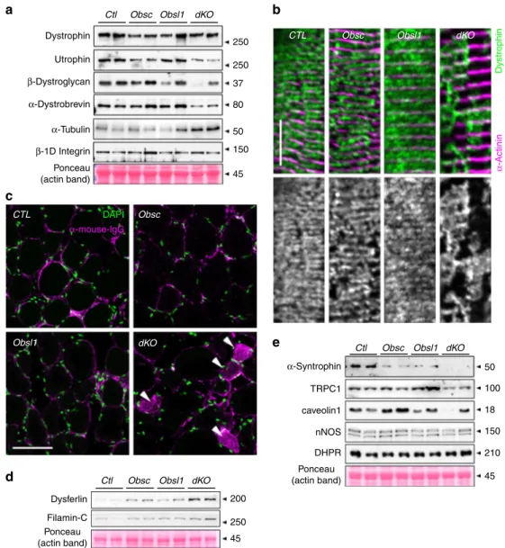
Loss of obscurin/Obsl1 impacts the DSG complex and its
associated proteins. Obscurin has been reported to be important
for sarcolemmal integrity, and loss of the protein results in
changes to the subsarcolemmal microtubule network, increased
membrane fragility, and reduced running performance of
mice
13,14. We investigated if changes to sarcolemmal integrity
and the DSG complex and its associated proteins are exacerbated
in Obsl1 and/or dKO mice. Loss of both obscurin and Obsl1
reduced protein levels of some DSG components, while levels of
tubulin increased slightly (p
= 0.06). In addition, overall levels of
dystrophin and
β1D- integrin remained unchanged (Fig.
3
a,
Supplementary Figs. 2a, 7a). However, analysis of
sub-sarcolemmal dystrophin localization in longitudinal sections of
TA muscles revealed a patchy and abnormal distribution of the
protein in obscurin knockouts, but more so in dKO mice,
indi-cating enhanced membrane fragility even at baseline (Fig.
3
b,
Supplementary Fig. 2b). Indeed, when investigated for the
pre-sence of mouse immunoglobulins in immunofluorescence assays,
a significant portion of muscle fibers in dKO mice stained positive
(Fig.
3
c, Supplementary Fig. 2c). Increased membrane fragility in
dKO muscles also leads to significantly enhanced expression of
dysferlin (Dysf) and FlnC, proteins involved in repair and
com-pensatory mechanisms (Fig.
3
d, Supplementary Figs. 2d, 7a).
The DSG complex also regulates expression and function of
sarcolemma-based membrane channels and signaling.
Specifi-cally,
α-syntrophin has been demonstrated to establish a link
between dystrophin/the DSG complex and sarcolemmal ion
channels, such as Trpc1 as well as caveolae components and
neuronal nitric oxidase (nNOS) signaling. When investigated, we
noticed reduced caveolin-1 and Trpc1 levels in TA muscles of
dKO mice, while nNOS remained unchanged (Fig.
3
e,
Supple-mentary Figs. 2e, 7a). Levels of L-type calcium channel remained
also comparable with controls, indicating potential changes to
DSG-associated, but not T-tubule-based ion channels.
Proteome analysis of knockout muscles. To reveal quantitative
changes to the muscle proteome between control,
obscurin-knockout, Obsl1-obscurin-knockout, or dKO mice, we isolated proteins
from TA and Sol muscles. We specifically focused on changes
between control and dKO muscles to eliminate compensatory
mechanisms between Obsl1 and obscurin that could potentially
obfuscate results. Of the 1503 and 1597 detected proteins in TA
and Sol of all genotypes, we identified 260 and 181 proteins that
were significantly deregulated between dKO and control muscles,
respectively (Fig.
4
a, b, Supplementary Figs. 8, 9, Supplementary
Data File 1). Gene ontology (GO) term and pathway analysis of
significantly changed proteins from all investigated muscles, or
Sol and TA alone revealed enrichment of oxidative
phosphor-ylation, TCA (tricarboxylic acid) cycle, and electron transport
Fibres with centralized
nuclei (%) CTL Obsc Obsl1 dKO (807) (581) (611)(845) 5 0 10 15 20 25 Cross-sectional area ( μ m 2) CTL Obsc Obsl1 dKO (661) (409) (509) (442)
* ******
***
EDL/TL (mg/mm) CTLObsc Obsl1 dKO
GC/TL (mg/mm)
CTL
Obsc Obsl1 dKO
Sol/TL (mg/mm) CTL Obsc Obsl1 dKO TA/TL (mg/mm) 40 120 100 80 60 7 6 5 4 3 4 3 2 1 0 35 30 25 20 CTL Obsc Obsl1 dKO DAPI WGA CTL Obsc Obsl1 dKO CSA of fibres (μm2) <1 1–1.5 1.5–5 2–2.5 2.5–3 3–3.5 3.5–4 4–4.5 4.5–5 5–5.5 5.5–6 >6
a
b
c
d
e
10,000 8000 6000 4000 2000 0 25 20 15 10 5 0 Number of fibres (%) CTL (n = 667) Obsc (n = 409) Obsl1 (n = 509) dKO (n = 442)Fig. 2 Loss of obscurin and/or Obsl1 (obscurin-like 1) does not influence muscle weights, and has little impact on cross-sectional area and muscle regeneration.a Muscle weight to tibia-length ratios of tibialis anterior (TA), soleus (Sol), gastrocnemius (GC), and extensor digitorum longus (EDL) muscles in control (CTL), obscurin knockout (Obsc), Obsl1 skeletal muscle-knockout (Obsl1), and double-knockout mice (dKO). Sample sizes (n) are indicated in thefigure; ***p < 0.001 vs. CTL. b–e Wheat germ agglutinin (WGA)- (magenta) and 4′,6-diamidino-2-phenylindole (DAPI)- (green) stained immunofluorescence of frozen TA cross-sections (b) used to determine fibers with centralized nuclei (c) and cross-sectional fiber areas for each group (d, e). Scale bar= 200 µm. Muscle from three mice per group were analyzed, counted fibers (n) and p values are indicated in figure (c–e). *p < 0.05, ***p < 0.001 vs. CTL. Box and whisker plots in c and d depict 5–95 percentile, averages (as cross) and outliers
chain, as well as muscle contraction processes (Supplementary
Fig. 3a–c, Supplementary Data Files 2–4). Among the
sig-nificantly upregulated proteins in TA muscles of dKO mice are
FlnC and Dysf, corroborating results obtained in our immunoblot
analyses (Figs.
3
d,
4
c, Supplementary Fig. 2d). Comparison of
significantly changed proteins between TA and Sol revealed 15
proteins as commonly upregulated and 33 proteins as commonly
downregulated in both analyzed muscle types, including
calsequestrin-2 (Casq2), Hspb1 (also known as Hsp27), myotilin,
or calmodulin-1 (Fig.
4
b, Supplementary Data File 1).
Intrigu-ingly, we also noticed several proteins that were differentially
regulated between Sol and TA, including
αB-crystallin (Fig.
4
c, d,
Supplementary Figs. 2f, 7b). Indeed, when studied in immunoblot
assays of protein lysates from TA and Sol muscles from all
groups, only TA muscle of dKO mice showed significantly
increased
αB-crystallin levels, while αB-crystallin levels in Sol
muscles remained largely unchanged (Fig.
4
e, Supplementary
Figs. 2f, 7b). These muscle type-specific differences may be
reflective of the differential expression levels of obscurin splice
isoforms observed in these muscles, and/or on the muscle type
(fast vs. slow twitch)
12,36.
Removal of obscurin/Obsl1 alters SR protein levels. Our
enrichment analysis revealed several proteins involved in muscle
contraction, calcium handling, and signaling as significantly
changed in dKO muscles. Among them, the SR proteins
sar-coendoplasmic reticulum Ca
2+ATPase (Serca), sarcalumenin,
triadin, JP-45 and junctin were found to be deregulated in dKO
muscles (Fig.
5
a). We corroborated suspected changes to the SR
and its associated proteins by immunoblot analyses, and found a
significant reduction of Serca levels, while ryanodine receptors
Ctl Dystrophina
c
d
e
b
Dystrophin 250 250 37 80 50 150 50 100 18 150 210 45 200 250 45 45Ctl Obsc Obsl1 dKO
Ctl Obsc Obsl1 dKO
DAPI β-Dystroglycan β-1D Integrin Dysferlin Filamin-C Ponceau (actin band) Ponceau (actin band) α-Dystrobrevin α-Syntrophin TRPC1 caveolin1 nNOS DHPR Ponceau (actin band) α-Tubulin α-mouse-lgG α -Actinin Utrophin
Obsc Obsl1 dKO
CTL
CTL
Obsl1
Obsc
dKO
Obsc Obsl1 dKO
Fig. 3 Loss of obscurin and Obsl1 (obscurin-like 1) impacts dystrophin–sarcoglycan (DSG) complex and its associated proteins, membrane integrity and membrane repair mechanisms.a Expression levels of DSG complex proteins dystrophin, utrophin,β-dystroglycan, and α-dystrobrevin, as well as α-tubulin andβ1D-integrin in whole protein lysates of tibialis anterior (TA) muscles of control (CTL), obscurin-knockout (Obsc), Obsl1 skeletal muscle-knockout (Obsl1), and double-knockout (dKO) mice. Ponceau-stained actin band is shown as a loading control.b Subsarcolemmal dystrophin localization in longitudinal sections of CTL, Obsc, Obsl1, and dKO TA muscles. Sarcomericα-actinin 2 was used as counterstain. Scale bar = 10 µm. c Mouse immunoglobulin (magenta) and 4′,6-diamidino-2-phenylindole (DAPI) stain (green) of frozen cross-sections of CTL, Obsc, Obsl1, and dKO TA muscles. Arrowheads indicate immunoglobulin G (IgG)-positive musclefibers. Scale bar = 200 µm. d, e Protein expression levels of dysferlin and Filamin-C (d), as well as DSG-associated proteinsα-syntrophin, Trpc1, caveolin-1, neuronal nitric oxidase (nNOS), andL-type calcium channel DHPRα-2 subunit (e) in whole
(RyRs) were upregulated (Fig.
5
b, Supplementary Figs. 2g, 7c).
We also investigated levels of sAnk1.5, which was shown to be a
binding partner for the obscurin-A C terminus. As shown before,
loss of obscurin resulted in a decrease in sAnk1.5 levels due to
increased turnover of the protein. Loss of Obsl1 did not affect
sAnk1.5 levels, while muscles from dKO mice displayed sAnk1.5
levels comparable with those found in obscurin knockouts
(Fig.
5
b, d). Our proteome data also indicated changes to
calcium-binding proteins located within the lumen of the SR. We
tested protein levels of Casq1, Casq2, and sarcalumenin by
immunoblot analyses of whole muscle lysates. Sarcalumenin
levels decreased significantly between control and dKO muscles,
while Casq2 protein levels increased, and Casq1 remained
unchanged (Fig.
5
c, e, Supplementary Figs. 2g, 7c).
These data indicate changes to cellular calcium handling in
dKO muscles, which may be reflected in defective excitation
contraction coupling and muscle contraction. To investigate if
these molecular changes alter muscle physiology, we studied EDL
twitch parameters in all groups (Table
2
). EDL muscles of
obscurin-knockout and dKO mice displayed increased
time-to-peak (TtP) values compared to muscles from control and Obsl1
knockouts (Fig.
5
f). However, half-relaxation times remained
unchanged (Fig.
5
g).
Metabolic changes in obscurin/Obsl1-knockout mice. A
sur-prising result of the enrichment analysis was the large number of
significantly changed proteins belonging to metabolic pathways
and the mitochondrial electron transport chain (Fig.
6
a).
Speci-fically, we found increased levels of Gbe1 (1,4-α-glucan branching
enzyme 1) and Stbd1 (starch binding domain 1) in dKO Sol and
TA muscles. Both proteins are involved in glycogen metabolism,
increasing solubility of glycogen by branching, and its breakdown
through glycophagy, respectively. Conversely, the rate
determin-ing enzyme responsible for glycogen breakdown, muscle glycogen
phosphorylase, was significantly decreased in dKO mice. We also
found significantly decreased levels of monoamine oxidase A and
B (Maoa, Maob) in TA muscles of dKO mice, while Sol muscle of
dKO mice had increased levels of catalase, perhaps indicative of
increased oxidative stress. Another
finding was that Gapdh
pro-tein levels were decreased in dKO mice, a result that we also
consistently observed in Obsl1-knockout cells and tissues
TA Σ260 significantly deregulated proteins Sol Σ181 significantly deregulated proteins
260 from a total of 1503 detected proteins 181 from a total of 1597 detected proteins
Downregulated (132) Upregulated (128) TA αB crystallin Ponceau (actin band) 20 45
Ctl Obsc Obsl1 dKO Ctl Obsc Obsl1 dKO
Sol Downregulated (121) Upregulated (60)
log2 fold change
Obsc Obsl1 Moab Cryab 150 100 50 Nol3 Tpm4 Obsc Srl Cryab Ybx2 Casq2 150 100 50 –2 0 2
log2 fold change
–2 0 2 Xirp1 Hspb1 Significance Significance Dysf FlnC TA upregulated Common upregulated Common downregulated 106 0 0 0 0 33 0 0 0 81 15 45 0 7 99 CASQ2 Max
a
b
c
e
d
Min CAT GBE1 HSD17B4 HSPB1 IVD MYH11 PBXIP1 ANKRD2 CRYAB NES NME2 PVALB SERBP1 VIM RPL35A RPL4 RPL6 RPL8 STBD1 YBX2 MYOTABHD11 EEF1B2 MAP2K6 NDUFB11 PRDX3 TNNC2
ADSL FAHD2A MYH2 NDUFB9 PRDX6 UBE2L3
AKR1B10 GSTM1 NDUFA13 NDUFC2 PTER YWHAG
CALM1 GSTM5 NDUFA4 NDUFV2 SELENBP1
CHCHD3 LETM1 NDUFA8 NOL3 SMYD2
CLYBL LGALS1 NDUFB10 OBSCN
Obsc Obsl1 dK Obsc
O CTL Obsl1 dK O CTL SPR Sol upregulated Sol down-regulated TA down-regulated Significantly changed proteins (CTL vs. dKO) Significantly changed proteins in TA (CTL vs. dKO) Significantly changed proteins in Sol (CTL vs. dKO)
Fig. 4 Proteome analysis of soleus (Sol) and tibialis anterior (TA) muscles. a Hierarchical clustering of significantly altered proteins identified in TA and Sol muscles of control (CTL), obscurin-knockout (Obsc), skeletal muscle-specific Obsl1 (obscurin-like 1)-knockout (Obsl1), and double-knockout (dKO) mice. b Grouping of significantly changed proteins in both muscles identified 15 proteins as common upregulated, while 33 proteins were found to be common downregulated between Sol and TA. The analysis also identified seven proteins that are differentially regulated between TA and Sol muscles. All commonly and differentially regulated proteins are identified in the boxes. c, d Volcano plot of significantly altered proteins between control (CTL) and dKO TA (c) and Sol (d) muscles. e Protein levels ofαB-crystallin in whole TA and Sol muscle lysates from CTL, Obsc, Obsl1, and dKO mice. Ponceau-stained actin band is shown as a loading control
(Supplementary Figs. 1b, 4a, 4b), necessitating the use of total
protein, actin or myosin, for normalization. Most intriguingly,
however, almost all identified mitochondrial electron transport
chain proteins were downregulated in dKO mouse Sol and TA
muscles (Fig.
6
b, Supplementary Fig. 4c). Analysis of whole TA
and/or Sol muscle extracts using the oxphos antibody cocktail or
a Uqcrb antibody substantiated slightly or significantly decreased
levels of mitochondrial complex I, II, III, IV, and V proteins in
dKO mice (Fig.
6
c, Supplementary Fig. 4d–f). Analysis of Prdx3
protein levels, a mitochondrially targeted peroxiredoxin family
member
37that was also detected in our proteome analysis as
significantly altered, indicating that the effect of Obsl1/obscurin
loss on the mitochondria may go well beyond electron transport
chain proteins (Fig.
6
c, Supplementary Fig. 4e, f). Further analysis
of the proteome data revealed that downregulation of electron
transport chain proteins is more associated with loss of obscurin
than Obsl1 (Supplementary Fig. 4c). However, only the loss of
both proteins, obscurin and Obsl1, resulted in the substantial
reduction in mitochondrial proteins. It remains to be
demon-strated if these changes are mirrored on the physiological level,
and if dKO muscles display altered reactive oxygen species (ROS)
levels caused by mitochondrial insufficiency. Our proteome data
showing changes to other peroxiredoxins, Sod2, and other
pro-teins involved in ROS scavenging and signaling (Supplementary
Fig. 5e; Supplementary Data File 1) are highly suggestive of this
possibility.
Discussion
Our study set out to investigate biological roles for Obsl1, with a
specific focus on redundant functions between obscurin and Obsl1
for the development of skeletal muscles. For this, we generated
HRT (ms ) Half relaxation time
**
**
** p < 0.01 vs. CTL Time to peak TtP (ms) Triadin JP45 Casq2 Junctin Calm1 –2 0 50 100 150 sAnk1.5CTL Obsc Obsl1 dKO
CTL Obsc Obsl1 dKO
20 35 30 25 20 15 CTL Obsc Obsl1 dKO 100 ~550 45 50 50 50 45 Serca1 RYR1 Sarcalumenin Calsequestrin-2 Calsequestrin-1 Ponceau (actin band) Ponceau (actin band) 2 log2 fold change
Significance TA Upregulated in TA Related to Ca2+ & SR Downregulated in TA Kcnma1 Casq2 Calm1 Sarcalumenin Serca
log2 fold change
Significance Sol Upregulated in Sol Related to Ca2+ & SR Downregulated in Sol
*
*
*p < 0.05 vs. CTL; ** p < 0.01 vs. CTL; n = 4*
**
a
b
f
g
c
d
e
50 100 150 2.0 sAnk1.5 Sarcalumenin 1.5 80 60 40 20 0 1.0 0.5 0.0 1.5 1.0 Expression (%) 0.5 Expression (%) 0.0 –2 0 2 CTL ObscObsl1 dKO CTL Obsc Obsl1 dKO CTL Obsc Obsl1 dKO
Fig. 5 Changes to sarcoplasmic reticulum (SR) and associated proteins. a Volcano plots of significantly altered SR and its associated proteins identified in our proteome analysis of double-knockout (dKO) vs. control tibialis anterior (TA) and soleus (Sol) muscles.b–e Immunoblot analysis and quantification of SR-associated proteins in TA muscles, including small ankyrin-1.5 (sAnk1.5;b, d), sarcoendoplasmic reticulum Ca2+ATPase (Serca1;b), and ryanodine receptor (RyR;b), as well as lumenal calcium-binding proteins sarcalumenin (c, e) and calsequestrin 1 and 2 (c). Ponceau-stained actin band is shown as a loading control (b, c). d–g Sample sizes (n) and p values are indicated in the figure. f, g Analysis of extensor digitorum longus muscle “time to peak” (f) and “half-relaxation time” (g). The full evaluation of twitch parameters and their statistical analyses are shown in Table2
Table 2 Twitch parameter analysis on 4-month-old mice
Parameters CTL Obsc Obsl1 dKO
Number of analyzed muscles (females/males) 10 (6/4) 7 (3/4) 9 (4/5) 10 (4/6)
TtP (ms) 21.45 ± 1.02 26.14 ± 0.91** 21.56 ± 0.92 25.3 ± 0.74**
HRT (ms) 43.3 ± 2.81 42.86 ± 3.20 48.00 ± 5.68 46.5 ± 1.99
Full-width at half-maximum (ms) 57 ± 3.4 60 ± 3.9 63 ± 6.3 64 ± 2.6
Stress during tetanus (kPa) 71 ± 3.8 99 ± 5** 79 ± 6.2 70 ± 5.8
PCSA (mm2) 0.51 ± 0.027 0.61 ± 0.023 0.6 ± 0.034 0.55 ± 0.03
EDL muscle mass (mg) 3.4 ± 0.19 4 ± 0.22 4.3 ± 0.28** 3.9 ± 0.18
Fiber length (mm) 6.2 ± 0.11 6.1 ± 0.16 6.7 ± 0.13* 6.5 ± 0.2
Values are presented as averages and standard error of mean. Samples sizes are shown in the table; p values are **p < 0.01 or *p < 0.05 vs. CTL
conditional Obsl1-knockout mice using a strategy that prevents
the expression of all Obsl1 isoforms. Surprisingly, our global Obsl1
knockouts display an embryonic lethality phenotype (Table
1
).
Obsl1 has been shown to be responsible for ~20% of cases of
3M-growth syndrome, while mutations in cullin-7 and Ccdc8
account for the remainder of the patients
27,31. Most of the Obsl1
patients suffer from mutations that result in a truncated protein
due to frameshift mutations
31,38. These
findings led to the
spec-ulation that the Obsl1 mRNA in patients may be prematurely
degraded by nonsense-mediated decay, resulting in complete
absence of the protein
31. However, similar to cullin-7
knock-outs
39, our global Obsl1 knockouts do not recapitulate the
3M-growth syndrome exhibited by patients.
Closer analysis showed that all Obsl1 mutations are located
well 3′ of the sequence encoding for Ig domain 1 of Obsl1
31. This
leaves the potential open for low-level expression of C terminally
truncated Obsl1 protein variants and alternative splice isoforms
that circumvent some of the deleterious mutations in Obsl1,
which is not possible in our mouse model. However, immunoblot
analysis of Obsl1 using validated antibodies has not been
exten-sively done on patient samples.
It is difficult to compare data from the 3M-growth syndrome
transcriptome and the Obsl1 interactome
35,40with changes
observed in our proteome analyses, as there are functional
redundancies between Obsl1 and obscurin, which may not be
present in skin
fibroblasts and other non-muscle tissues
(Sup-plementary Fig. 6a). Moreover, the complete dataset for the
transcriptome analysis of
fibroblasts from 3M-growth syndrome
patients is not readily available
40. However, there are several
striking similarities between our proteome and the transcriptome
and interactome datasets (Supplementary Fig. 5a, b;
Supple-mentary Data File 5). Specifically, both studies found significant
deregulation of genes/proteins involved in cellular transcription
(e.g., Hnrnpf, Eef1a1, Ybx transcription factors), translation
(small and large ribosomal subunits), and in genes involved in
metabolic pathways downstream of IGF1 (insulin-like growth
factor 1) and mTOR (mammalian target of rapamycin) signaling
(Supplementary Fig. 5c). Several proteins that form part of
membrane and vesicle trafficking complexes show up in both
large datasets, including coatomer protein complex subunit
alpha (Copa) or clathrin (Cltc) (Supplementary Fig. 5a, d). In
addition, other proteins involved in vesicle and protein trafficking
Proteins involved in energy and metabolismTA CTL Obsc Obsl1 dKO
Sol CTL Obsc Obsl1 dKO
Significantly changed electron transport chain complex proteins
Significantly changed electron transport chain complex proteins
Complex V (7) TA 50 Sol 50 Ubiquinone system (4) Complex IV (2) Complex III (2) Complex I (9) Complex IV (6) Complex III (6) Complex I (26)
a
b
c
75 75 50 50 37 37 25 45 25 20 15 25 45 V (ATP5A) V (ATP5A) III (QCR2) III (QCR2) IV (Cox1) IV (Cox1) II (SDHB) II (SDHB) I (NdufB8) Uqcrb Prdx3 Ponceau (actin band) Ponceau (actin band)TA (CTL vs. dKO) Sol (CTL vs. dKO)
From 260 significantly changed proteins
From 181 significantly changed proteins
Other energy metabolism (18) Other energy metabolism (15)
log2 fold change
Significance Significance
Mitochondrium related (66) Mitochondrium related (25)
Mitochondrial electron transport chain complex proteins Mitochondrial electron transport chain complex proteins 150 100 50 150 100 50 MAOB ADSL FABP3 GBE1 CAT STBD1 GBE1 NDUFV3 STBD1 PYGM GAPDH UQCRB –2 0 2
log2 fold change
–2 0 2
Fig. 6 Analysis of changes to the muscle metabolism and mitochondria. a Volcano plot of significantly altered proteins between control (CTL) and double-knockout (dKO) tibialis anterior (TA; left panel) and soleus muscles (Sol; right panel) involved in energy metabolism (blue) and mitochondria (light blue). b Volcano plot of electron transport chain proteins identified in our proteome analysis as significantly altered between CTL and dKO TA and Sol muscles. The number of significantly changed proteins is indicated in brackets. c Analysis of mitochondrial electron transport chain complex protein levels as well as mitochondrially located peroxiredoxin-3 (Prdx3) in TA and Sol muscles from CTL, obscurin-knockout (Obsc), skeletal muscle-specific Obsl1 (obscurin-like 1)-knockout (Obsl1), and dKO mice. Ponceau-stained total actin band is shown as a loading control
were found significantly altered, such as caveolin-3,
muscle-related coiled-coil protein (MuRC/cavin-4)
41, transferrin receptor
(Tfrc)
42, intersectin-1 (Itsn1)
43, Rab8b
44, non-clathrin-related
endocytosis (pacsin3
45, EHD3
46), COP1- or COP2-related
pro-tein shuttling pathways (Sec31a
47,48, archain-1 (Arcn1)
49), and
several tubulin isoforms, which were also deregulated in our
proteome analysis (Supplementary Data 1). Deficiencies in several
of these pathways have been implicated in growth retardation and
developmental delays, including COP-related protein shuttling
pathways
49–51or clathrin-dependent trafficking
52. Another
striking overlap between the datasets are proteins that are
involved in metabolism, mitochondrial function (e.g., electron
transport chain complex proteins), and generation/scavenging of
ROS). Specifically, we found deregulation of Gapdh and Prdx3,
both identified as putative interaction partners for Obsl1
35(Supplementary Figs. 5a, 6c). Maoa was also found
down-regulated in both the transcriptome and our proteome datasets.
Patients with Maoa/Maob deficiency display loss of muscle tone
and short statue
53,54, while global Maoa/Maob dKO mice show
developmental delays, behavior, and locomotion problems
55.
Closer examination of changes to muscles and exercise capacity in
monoamine oxidase-deficient mice remains to be done.
It is thought that 3M-growth syndrome is caused by a
defi-ciency in protein turnover due to the
finding that a majority of
patients with 3M-growth disorder display mutations in cullin-7,
Obsl1, and Ccdc8. Indeed, Obsl1 was identified as a binding
partner for the E3-ubiquitin ligase cullin-7
25,26,56. However, our
proteome data did not reveal significant changes to any of the
other proteins associated with the disease, that is, cullin-7, Ccdc8,
Fbw8, Igfbp2, or Igfbp5. These
findings raise the possibility that
functional deficiencies associated with the disorder may be tissue
specific and spare skeletal muscles. Closer examination of
expression patterns for 3M-growth syndrome associated proteins
could reveal tissues types where this complex performs its major
functions. Indeed, looking at expression profiles in Protein Atlas
(
https://www.proteinatlas.org/
) reveals variable expression for
Fbw8 (and many of the other F-box-containing substrate adaptor
proteins for cullin-7), as well as Igfbp2 and Igfbp5 across tissues.
The usage of cre lines that delete Obsl1 in tissues other than
skeletal muscles and at earlier time-points will shed further light
into non-muscle functions for Obsl1 and the development of
3M-growth syndrome.
Another surprise was the lack of baseline phenotype for skeletal
muscle-specific Obsl1 knockouts, and the comparatively mild
physiological phenotype observed for the dKO mice. Indeed,
looking at the substantial molecular changes to the DSG complex,
alterations to sarcolemmal integrity and to SR-associated proteins
that the dKO mice display, it was unexpected to observe no changes
to the number of centralized nuclei in muscle cross-sections as a
sign for active muscle regeneration, and largely unchanged
phy-siological twitch parameters. However, when investigating specific
alterations to skeletal muscles upon loss of Obsl1 and obscurin
redundant functions for both proteins start to emerge.
Despite extensive interactions of obscurin and Obsl1 (or its
invertebrate homolog unc-89) with sarcomeric proteins
8,11,33,57,
loss of either or both obscurin family proteins had negligible
effects on myofibrillogenesis and sarcomere structure. Indeed,
apart from a slight reduction in the levels of myomesin-2
(Sup-plementary Fig. 1e), we were unable to detect any overt changes
for myomesin-1, tropomyosin,
α-actinin 2, titin-M8, and myosin
localization or expression. These data indicate that obscurin and
Obsl1 in vertebrate muscles are sarcomere-associated proteins
that are not required for sarcomere assembly. Indeed, our
experiments investigating muscle twitch characteristics only
found increase in TtP values for loss of obscurin, indicative of
problems with the SR and calcium-induced calcium release.
However, no changes were found in other parameters measured
in these experiments, with the exception of stress during tetanus
values for obscurin knockouts. Obsl1 has been shown to target to
several subcellular localizations in heart and skeletal muscles,
including M-band, Z-disc, and intercalated discs
8,11. While not all
antibodies in these studies have been verified against the
knock-out, our experiments using two verified Obsl1 antibodies that
target different Obsl1 epitopes corroborate several of these
find-ings. Known and novel interactions may explain the differential
Obsl1 localization to sarcomeric M-bands and Z-discs (Fig.
1
c,
Supplementary Figs. 1a, 1d, 1e, 6b). It remains to be
demon-strated how splice variants of Obsl1, and binding affinities to
interactors modifies the subcellular localization.
Obscurin knockouts display altered ankyrin-B and dystrophin
localization at costameres, and increased membrane fragility
combined
with
reduced
muscle
exercise
tolerance
13,14.
Morpholino-mediated knockdown of obscurin-A in zebrafish also
resulted in abnormal dystrophin and
α-dystroglycan localization,
suggesting evolutionary conserved functions of obscurin for DSG
complex organization and sarcolemmal integrity
58. Following
these studies, we investigated expression and localization of DSG
components and found changes at baseline predominantly in
dKO mice. While dKO muscles did not display changes to
dys-trophin levels, its subsarcolemmal localization appeared patchy
and its distribution disturbed. Immunofluorescence imaging of
subsarcolemmal dystrophin indicated that obscurin and Obsl1
may partially modulate the membrane targeting of dystrophin,
and help it to spread evenly over the sarcolemmal membrane
(Fig.
3
b, Supplementary Fig. 2b).
Muscles of dKO mice also displayed a surprising slight
decrease in utrophin (p
= 0.06), and several other investigated
DSG components, including
α-dystrobrevin. Moreover,
DSG-linked signaling via ion channels (Trpc1) may also be altered in
dKO mice only, based on reduced expression levels of this
channel. These data suggest that both Obsl1 and obscurin have
redundant functions for DSG complex assembly and stability.
Loss of both proteins resulted in impaired sarcolemmal integrity
in a substantial proportion of muscles in mice at baseline (not
exercised), as demonstrated by positive immunoglobulin labeling
of muscle
fibers in dKO mice (Fig.
3
c, Supplementary Fig. 2c).
Muscles of dKO mice also show upregulation of proteins
involved in muscle repair processes, including Dysf and FlnC
59–61.
Future experiments that further characterize membrane damage
and studies that investigate repair mechanisms through FlnC and
Dysf should clarify the extent of the sarcolemmal integrity
impairment in dKO mice.
While some of the functions that obscurin exerts on the DSG
complex are due to interactions with ankyrin isoforms, spectrin
and FlnC
13,20,21,62,63, we and others identified that Obsl1 may
interact with other proteins linked to the DSG complex and its
associated components
35, including FlnC, tubulin, dystonin,
plectin, ahnak, and utrophin (Supplementary Figs. 1a, 5a, 6c).
Obsl1/obscurin function has also been implicated for microtubule
integrity,
by
promoting
microtubule
assembly
and/or
stability
13,14,64. Microtubule action is important for SR
organi-zation
65, sarcolemmal integrity and repair
66,67, cell division,
cel-lular shape, and the transport of vesicles over longer distances, in
addition requiring actions of kinesin and dynein as molecular
motors
68. When looking at the proteome data, we noticed
increases in
α- and β-tubulins, which can be verified in
immu-noblot analyses of dKO TA muscles (Fig.
3
a, Supplementary
Fig. 2a). Alterations in the microtubule network have also been
linked to the development of muscular dystrophy in mdx mice
69.
Dystrophin was shown to interact with microtubules
70,71, and its
loss increased the amounts of both
α- and β-tubulins
69, a
finding
One of the key
findings in skeletal muscles of obscurin
knockouts was the importance of the protein for SR architecture
through its link with the small muscle-specific ankyrin-1 isoform
sAnk1.5 that is embedded in the SR membrane
20,21. While loss of
Obsl1 has little or no impact on protein levels of SR-associated
proteins, muscles of dKO mice displayed profound alterations to
levels of lumenal SR calcium-binding proteins sarcalumenin and
Casq2, as well as Serca. Our proteome and expression data also
indicated increases to junctional SR proteins like triadin, junctin,
or RyRs in dKO muscles, something not previously observed for
obscurin knockouts
12. One might speculate based on the
pre-dominant subcellular localizations of obscurin at the M-band and
Obsl1 at the M-band and Z-disc that both the longitudinal and
junctional SR are affected in dKO muscles. Data on
DSG-associated ion channels (Trpc1), the junctional (RyR) or
long-itudinal SR membranes (Serca), suggests that loss of obscurin/
Obsl1 alters calcium handling in dKO muscles. These data in
combination with published
findings on Speg
17and the
inverte-brate homolog unc-89
72substantiates the
finding that the
reg-ulation of SR architecture and function is a major biological role
for all obscurin protein family members.
Many questions remain unanswered as to the exact molecular
origins for the observed effects in dKO muscles. Our proteome
analyses may provide clues for some of the molecular
mechan-isms at play that could result in alterations to the cellular
meta-bolism, changes to membrane stability and repair mechanisms,
and the impact on calcium handling and Serca
function/regula-tion. However, none of the well-characterized muscle-specific
obscurin and Obsl1 interaction partners may account for all of
the observed phenotypical changes
2,11,20,21,25,26,57,73. Indeed, it
remains to be demonstrated for the majority of the more than 600
identified binding partners of obscurin and Obsl1
35, whether they
play a role in muscles, and how loss of both obscurin protein
family members affect their expression, localization, and cellular
functions.
Methods
Gene targeting and generation of Obsl1-knockout mice, analysis of animal physiology. The targeting construct was generated by subcloning isogenic 129svj genomic DNA fragments of the murine Obsl1 gene into a targeting vector, and placing coding exons 1 to 4 between loxP sites (Supplementary Fig. 1a). The construct was linearized, and electroporated into R1 embryonic stem cells. G418-resistant clones were screened for correct homologous recombination by Southern blot analysis described elsewhere12. Germline transmission was tested by
appear-ance of the agouti fur color phenotype of 129-derived ES-cells, after which animals were backcrossed into the black swiss background. Genotying was done by PCR using oligonucleotides shown in Supplementary Table 1. Removal of neomycin cassette was done by crossingfloxed Obsl1 mice with mice carrying Flpase recombinase74,75. Generation of global Obsl1 knockouts was done by crossing
floxed Obsl1 mice with mice carrying Cre recombinase under control of the pro-tamine promoter30. Finally, generation of skeletal muscle-specific Obsl1 knockouts
was achieved by crossingfloxed Obsl1 mice with mice carrying Cre recombinase under control of the myogenin promoter32. Successful recombination of Obsl1 gene
and generation of skeletal muscle knockouts was validated by immunoblot analyses (Fig.1a). Generation of obscurin/Obsl1 myogenin-cre dKOs was done by cross-breeding into the obscurin knockout background12. If not explicitly stated, all
investigations were done with tissues from animals of both sexes. Experiments using thefifth toe of the EDL muscle from 4-month-old mice was done as described previously12. All procedures involving genetically modified animals have
been approved by the UC San Diego institutional oversight committee. Cell culture. Culture of cos-1 cells was done as previously described76,77. To test
Obsl1 antibodies and to determine if effects seen in dKO and Obsl1-knockout muscles are also seen in non-muscle Obsl1-knockout cells, we isolated cells from lungs of heterozygous global Obsl1 knockouts (+/−) and from mice that have a knockout and afloxed allele for Obsl1 (flox/−), using a collagenase/dispase enzyme mix (Roche). Isolated cells were cultured and propagated in growth medium (10% fetal calf serum, 1% penicillin/streptomycin, Dulbecco’s modified Eagle’s medium [Corning]). To generate Obsl1 knockouts, cells were transduced with a lentivirus carrying a cre-blasticidin-RFP expression cassette under control of a CMV pro-moter (#LVP013; Gentarget Inc.), allowing for recombination of the Obsl1-floxed allele and selection of transduced cells by 5 µg/ml blasticidin (Thermo Fisher).
After transduction, cells were kept in growth medium supplemented with blas-ticidin until harvest for analysis of proteins.
Protein analysis, immunofluorescence, histology, and antibodies. For analysis of protein levels, muscles or cells were lysed directly into sample buffer (3.7 M urea, 134.6 mM Tris-HCl, pH 6.8, 5.4% sodium dodecyl sulfate (SDS), 2.3% NP-40, 4.45%β-mercaptoethanol, 4% glycerol, and 6 mg/100 ml bromophenol blue). Loading was normalized by densitometry of Coomassie-stained gels using either actin or myosin bands. Once normalized, muscle lysates were loaded onto SDS-polyacrylamide gel electrophoresis (PAGE) gels (8, 12, or 15% acrylamide con-centration) or SDS-Agarose gels12. Proteins were transferred onto nitrocellulose
membranes, and detection of proteins was done using antibodies as described elsewhere77. If not stated otherwise, biological replicates were used for immunoblot
analyses. Sample sizes for quantification of protein levels are given in the figure or figure legend. Uncropped blot images for all western blot data are presented in Supplementary Data File 6.
For immunofluorescence, muscles were isolated, snap frozen while submerged in isobutane, and embedded into cryogenic molds using optimal cutting temperate (OCT) medium. Longitudinal or cross-sections of muscles were done by cryosectioning using a cryostat (Leica). Sections were collected on surface-treated microscopy glass slides (Colorfrost Plus, Fisher), dried, and stored at−80 °C until further use. Immunofluorescent staining of sections was done by fixing sections in ice-cold acetone (5 min at−20 °C), rehydration of tissue using 1× phosphate-buffered saline (PBS) (5 min), permeabilization (1× PBS, 0.2% Triton X-100; 5 min), and blocking with 1% bovine serum albumin (BSA) fraction V and 5% normal donkey serum diluted in Gold Buffer (155 mM NaCl, 2 mM EGTA, 2 mM MgCl2, 20 mM Tris-HCl, pH 7.5) for 30 min. After blocking of non-specific binding sites, tissue sections were incubated with primary antibody diluted in Gold Buffer overnight at 4 °C. Following three washes with PBS (5 min each), sections were incubated withfluorescently labeled secondary antibodies combined with either 4′,6-diamidino-2-phenylindole (DAPI) and/or fluorescently labeled wheat germ agglutinin (WGA) (Sigma) diluted into Gold Buffer for 2 h at room temperature. After washing with 1× PBS (5 min each), sections were embedded usingfluorescent mounting medium (DAKO), and covered with coverslips. Immunofluorescently labeled tissues were imaged using a Fluoview 1000 confocal microscope (Olympus), in sequential scanning mode using ×10 air or ×40 oil objectives and zoom rates between 1× and 3×. For analysis of cross-sectional areas, images were analyzed using the area-measuring tool in ImageJ.
Primary antibodies used in this study are listed in Supplementary Table 2. If not noted otherwise, all secondary antibodies were either from DAKO or Jackson ImmunoResearch. Fluorescently labeled WGA was obtained from Invitrogen/ Thermo Fisher. DAPI was purchased from Sigma-Aldrich.
Mass spectrometry and proteome analysis. Proteins were isolated form whole muscle and lysed into ice-cold isolation buffer (300 mM KCl, 30 mM PIPES pH 6.6, 0.5% NP-40, 1× protease inhibitor (Roche), 1× Phos-stop (Roche)). Insoluble proteins were removed by centrifugation (14,000 rpm, 10 min at 4 °C), and the supernatant was diluted 1:4 with ice-cold dilution buffer (1× Phos-stop (Roche), 0.5% NP-40, 1 mM dithiothreitol (DTT)). Precipitation of acto-myosin compo-nents was done by centrifugation (14,000 rpm, 15 min at 4 °C), and the remaining supernatant snap frozen for further analysis by mass spectrometry and immuno-blot analyses.
Analysis and identification of peptides via mass spectrometry was done as described previously78. Briefly, immediately prior to mass spectrometry, protein
solutions were diluted in TNE buffer (50 mM Tris-HCl, pH 8.0, 100 mM NaCl, 1 mM EDTA). RapiGest SF reagent (Waters Corp.) was added to the mix to afinal concentration of 0.1% and samples were boiled for 5 min. TCEP (Tris (2-carboxyethyl) phosphine) was added to afinal concentration of 1 mM, and the samples were incubated at 37 °C for 30 min. Subsequently, the samples were carboxymethylated with 0.5 mg/ml of iodoacetamide for 30 min at 37 °C, followed by neutralization with 2 mM TCEP (final concentration). Protein samples prepared as above were digested with trypsin (trypsin:protein ratio of 1:50) overnight at 37 ° C. RapiGest was degraded and removed by treating the samples with 250 mM HCl at 37 °C for 1 h, followed by centrifugation at 14,000 rpm for 30 min at 4 °C. The soluble fraction was then added to a new tube and the peptides were extracted and desalted using C18 desalting tips (Thermo Scientific).
The trypsinized samples (eight samples) were labeled with isobaric tags (iTRAQ8, ABSCIEX)79, where each sample was labeled with a specific tag to its
peptides as described in the manufacturer’s instructions. Each set of experiments were then pooled and fractionated using high pH reverse phase chromatography (HPRP-Xterra C18 reverse phase, 4.6 mm × 10 mm, 5 µm particle [Waters]). The chromatography conditions were as follows: the column was heated to 37 °C and a linear gradient from 5 to 35% B (Buffer A—20 mM ammonium formate pH 10 aqueous, Buffer B—20 mM ammonium formate pH 10 in 80% acetonitrile (ACN)-water), followed by 5 min at 100% B was applied for 80 min at 0.5 ml/minflow rate. A total of 42 fractions of 0.5 ml volume were collected. For liquid chromatography with tandem mass spectrometry (LC-MS/MS) analysis, some fractions were pooled to create afinal 16 pooled samples. Each of the pooled fractions were analyzed by high pressure liquid chromatography coupled with LC-MS/MS using nano-spray ionization.
Nano-spray ionization experiments were performed with a TripleTof 5600 hybrid mass spectrometer (ABSCIEX) interfaced with nano-scale reversed-phase UPLC (Waters Corporation, nanoACQUITY) using a 20 cm × 75 µm ID glass capillary packed with 2.5 µm C18 (130) CSHTM beads (Waters Corporation). A linear gradient (5–80%) of ACN was used to elute the peptides from the C18 column into the mass spectrometer at aflow rate of 250 μl/min for 1 h. The ACN gradient was created with the following buffers: Buffer A (98% H2O, 2% ACN, 0.1% formic acid, and 0.005% trifluoroacetic acid (TFA)) and Buffer B (100% ACN, 0.1% formic acid, and 0.005% TFA). MS/MS data were acquired in a data-dependent manner in which the MS1 data was acquired for 250 ms at m/z of 400 to 1250 Da and the MS/MS data was acquired from m/z of 50 to 2000 Da. Independent data acquisition parameters were as follows: MS1-TOF (mass spectrometry imaging-time offlight) acquisition time of 250 ms, followed by 50 MS2 events of 48 ms acquisition time for each event. The threshold to trigger MS2 event was set to 150 counts when the ion had the charge state+2, +3, and +4. The ion exclusion time was set to 4 s. The collision energy was set to iTRAQ experiment setting. Finally, the collected data were analyzed using Protein Pilot 5.0 (ABSCIEX) for peptide identifications and Peaks80. Bioinformatic enrichment and pathway analysis was
done using Metascape (http://metascape.org/)81, Morpheus (https://software.
broadinstitute.org/morpheus/), the BioGRID (https://thebiogrid.org/)82, and
Venny (http://bioinfogp.cnb.csic.es/tools/venny).
Eukaryotic expression constructs, yeast two-hybrid screening, and co-immunoprecipitation. Constructs for greenfluorescent protein (GFP)-tagged Obsl1 fragments and yeast two-hybrid screening were generated as described elsewhere24. In short, coding sequences for human Obsl1 fragments (NCBI access
no: NM_178884) were subcloned to enhanced GFP (EGFP) (Clontech) or pLex vectors to produce in-frame fusion between GFP or the DNA-binding domain and the insert. All expression constructs were sequenced to determine correct inte-gration into the vector. The yeast two-hybrid screening was done as described previously77. Briefly, L40 yeasts were transformed with bait constructs (pLex-Obsl1
domain truncations) and empty prey vector (pAct2) to perform auto-activation test. pLex-Obsl1 constructs that were negative for auto-activation were selected for further yeast two-hybrid screening. L40 yeasts were transformed with selected pLex-Obsl1 constructs, and positive transformants were used to perform a library transformation. Yeasts were plated on selective dropout plates, and colonies were allowed to form for 5 days. Yeast clones positive forβ-galactosidase activity in an X-gal overlay assay were further amplified, and library plasmid DNA was extracted and analyzed by sequencing.
Transfection of Cos-1 cells was done as described previously77. Transfected cells
were lysed into ice-cold immunoprecipitation (IP) buffer (100 mM NaCl, 10 mM Tris-HCl, pH 8, 1× Complete Protease Inhibitor Cocktail [Roche], 1 mM DTT, and 0.5% NP-40) 2 days after transfection. Lysates were briefly sonicated, centrifugated for 10 min at 14,000 rpm (at 4 °C) to remove insoluble proteins, and supernatants were used to perform immunoprecipitation. Briefly, 5 µg GFP antibody (Roche) were added to lysates, and incubated overnight at 4 °C to allow for formation of immunocomplexes. Subsequently, magnetic protein-G-coated beads (Dynabeads, Life Technologies) were added and incubated for 3 h at 4 °C on a shaker. Bound protein immunocomplexes were separated from unbound proteins by magnetic separation, and the beads were washed three times with ice-cold wash buffer (1× PBS, 0.2% NP-40). Analysis of input and bound protein immunocomplexes was done by SDS-PAGE, followed by immunoblot analysis.
Bioinformatics and statistical analysis. The following bioinformatics packages and programs were used to perform analyses in this manuscript: analysis of pro-teome data was done using Peaks Studio (http://www.bioinfor.com/peaks-studio/)80;
pathway enrichment analysis was done with the help of MetaScape (http:// metascape.org/)81; hierarchical clustering of proteome data was done using
Mor-pheus (https://software.broadinstitute.org/morpheus/); image analysis was done using ImageJ with the LOCI BioFormats and Image5D plugins (by the Open Microscopy Environment consortium and Joachim Walter, respectively); sequence alignments and domain structure analysis were done using NIH/NCBI BLAST (https://blast.ncbi.nlm.nih.gov/Blast.cgi), SDSC Biology Workbench (http:// workbench.sdsc.edu/), and SMART (http://smart.embl-heidelberg.de/)83. With the
exception for built-in statistics by the Peaks Studio software, statistical analysis of all data was done by using analysis of variance comparison, followed by Dunnett’s multiple comparisons test, or unpaired t test, performed using Excel (Microsoft) or GraphPad Prism version 7 for Mac (GraphPad Software,http://www.graphpad.com). Violin plots were generated using BoxPlotR (http://shiny.chemgrid.org/boxplotr/). Results are presented as means ± standard error. P values of p < 0.05 were considered statistically significant. Sample sizes are indicated in the figure or figure legend. If not stated otherwise, both sexes were analyzed in the experimental procedures. We used biological replicates to showcase biological variability and reproducibility offindings. Whenever appropriate, different methods were used to verify afinding (e.g., inter-action found in yeast two-hybrid screening was verified by co-immunoprecipitation). No data were excluded in the generation offigures/tables, with the exception of two lanes in Supplementary Fig. 7b (upper panel) that did not pass the quality control as judged by Ponceau stain.
Reporting summary. Further information on experimental design is available in the Nature Research Reporting Summary linked to this article.
Data availability
All relevant data described in the manuscript can be found in thefigures, tables, and supplementalfiles, are deposited in public repositories, or can be requested from the authors. The mass spectrometry proteomics data have been deposited to the ProteomeXchange Consortium via the PRIDE partner repository84,85with the dataset
identifier PXD013008 andhttps://doi.org/10.6019/PXD013008.
Received: 31 August 2018 Accepted: 28 March 2019
References
1. Bang, M. L. et al. The complete gene sequence of titin, expression of an unusual approximately 700-kDa titin isoform, and its interaction with obscurin identify a novel Z-line to I-band linking system. Circ. Res. 89, 1065–1072 (2001).
2. Young, P., Ehler, E. & Gautel, M. Obscurin, a giant sarcomeric Rho guanine nucleotide exchange factor protein involved in sarcomere assembly. J. Cell Biol. 154, 123–136 (2001).
3. Borisov, A. B., Raeker, M. O. & Russell, M. W. Developmental expression and differential cellular localization of obscurin and obscurin-associated kinase in cardiac muscle cells. J. Cell. Biochem. 103, 1621–1635 (2008).
4. Ackermann, M. A. et al. Novel obscurins mediate cardiomyocyte adhesion and size via the PI3K/AKT/mTOR signaling pathway. J. Mol. Cell. Cardiol. 111, 27–39 (2017).
5. Perry, N. A., Ackermann, M. A., Shriver, M., Hu, L. Y. & Kontrogianni-Konstantopoulos, A. Obscurins: unassuming giants enter the spotlight. IUBMB Life 65, 479–486 (2013).
6. Shriver, M. et al. Loss of giant obscurins from breast epithelium promotes epithelial-to-mesenchymal transition, tumorigenicity and metastasis. Oncogene 34, 4248–4259 (2015).
7. Stroka, K. M. et al. Loss of giant obscurins alters breast epithelial cell mechanosensing of matrix stiffness. Oncotarget 8, 54004–54020 (2017).
8. Geisler, S. B. et al. Obscurin-like 1, OBSL1, is a novel cytoskeletal protein related to obscurin. Genomics 89, 521–531 (2007).
9. Hsieh, C. M. et al. Striated muscle preferentially expressed genes alpha and beta are two serine/threonine protein kinases derived from the same gene as the aortic preferentially expressed gene-1. J. Biol. Chem. 275, 36966–36973 (2000).
10. Benian, G. M., Tinley, T. L., Tang, X. & Borodovsky, M. The Caenorhabditis elegans gene unc-89, required fpr muscle M-line assembly, encodes a giant modular protein composed of Ig and signal transduction domains. J. Cell Biol. 132, 835–848 (1996).
11. Fukuzawa, A. et al. Interactions with titin and myomesin target obscurin and obscurin-like 1 to the M-band: implications for hereditary myopathies. J. Cell Sci. 121, 1841–1851 (2008).
12. Lange, S. et al. Obscurin determines the architecture of the longitudinal sarcoplasmic reticulum. J. Cell Sci. 122, 2640–2650 (2009).
13. Randazzo, D. et al. Obscurin is required for ankyrinB-dependent dystrophin localization and sarcolemma integrity. J. Cell Biol. 200, 523–536 (2013). 14. Randazzo, D. et al. Exercise-induced alterations and loss of sarcomeric M-line
organization in the diaphragm muscle of obscurin knockout mice. Am. J. Physiol. 312, C16–C28 (2017).
15. Liu, X. et al. Disruption of striated preferentially expressed gene locus leads to dilated cardiomyopathy in mice. Circulation 119, 261–268 (2009). 16. Agrawal, P. B. et al. SPEG interacts with myotubularin, and its deficiency
causes centronuclear myopathy with dilated cardiomyopathy. Am. J. Hum. Genet. 95, 218–226 (2014).
17. Quick, A. P. et al. SPEG (striated muscle preferentially expressed protein kinase) is essential for cardiac function by regulating junctional membrane complex activity. Circ. Res. 120, 110–119 (2017).
18. Pernigo, S. et al. Structural insight into M-band assembly and mechanics from the titin-obscurin-like-1 complex. Proc. Natl Acad. Sci. USA 107, 2908–2913 (2010).
19. Pernigo, S. et al. Binding of myomesin to obscurin-like-1 at the muscle M-band provides a strategy for isoform-specific mechanical protection. Structure 25, 107–120 (2017).
20. Bagnato, P., Barone, V., Giacomello, E., Rossi, D. & Sorrentino, V. Binding of an ankyrin-1 isoform to obscurin suggests a molecular link between the sarcoplasmic reticulum and myofibrils in striated muscles. J. Cell Biol. 160, 245–253 (2003).