TABLE OF CONTENTS
ABSTRACT
Part 1 :BACKGROUND
CHAPTER 1
:Obesity, Inflammation and Insulin resistance
pag.21.1 INTRODUCTION pag.3
1.2 THE ADIPOSE TISSUE pag.3
1.2.1 Morphogenesis pag.3
1.2.2 Adipogenesis pag.8
1.2.2.1 PPARγ pag.10
1.2.2.2 CEB/P pag.13
1.2.3 Physiology of adipose tissue pag.14
1.2.4 Adipose tissue as an endocrine organ pag.17
1.2.5 Pro-inflammatory Adipokines pag.18
1.2.5.1 Leptin pag.18
1.2.5.2 Interleukin-6 pag.21
1.2.5.3 Tumor necrosis factor-α pag.22
1.2.5.4 Monocyte chemoattractant protein-1 and CC-Chemokine receptor type 5
pag.23
1.2.5.5 Other Pro-inflammatory Adipokines pag.23
1.2.6 Anti-inflammatory Adipokines pag.26
1.2.6.1 Adiponectin pag.26
1.2.6.2 SFRPs (Secreted frizzled-related proteins) pag.27
1.2.6.3 Other Anti-inflammatory Adipokines pag.28
1.3 LIPOTOXICITY AND INFLAMMATION IN ADIPOSE TISSUE pag.29
1.3.1 Lipotoxicity pag.29
1.3.2 "Chronic low-grade inflammation" in adipose tissue pag.31 1.3.3 Inflammatory cells infiltration in adipose tissue pag.31 1.3.4 Molecular mechanisms underlying the inflammatory process in adipose
tissue
pag.36
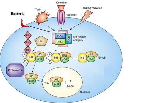
1.3.4.2 Cell signaling pathway modulated by the transcription factor NF- κB
pag.37
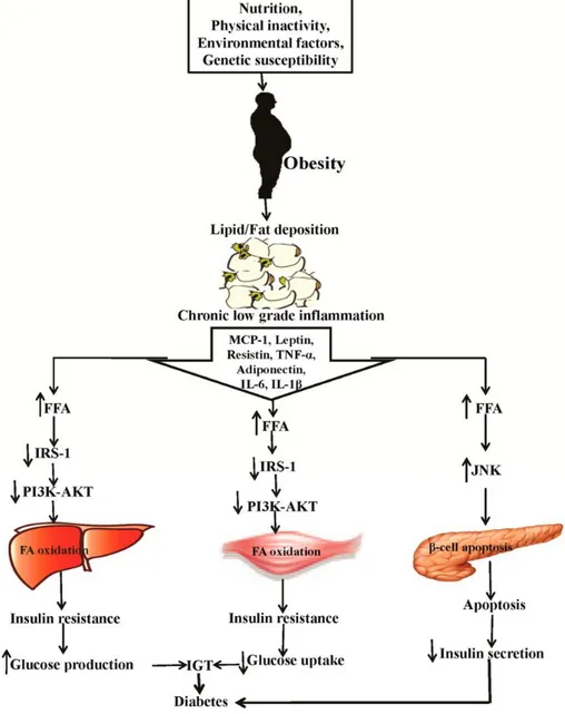
1.3.5 Obesity, inflammation and reduced insulin sensitivity pag.39
1.4 INSULIN RESISTANCE IN ADIPOSE TISSUE pag.42
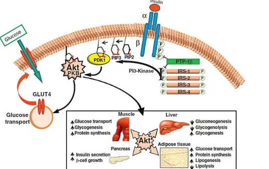
1.4.1 Metabolic effects of insulin pag.42
1.4.2 Insulin resistance and signalling pathways pag.45
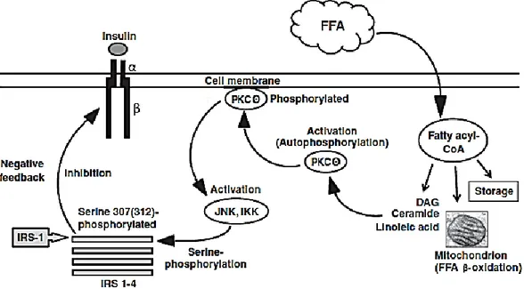
1.4.3 FFA and insulin receptor inactivation pag.48
1.4.4 Metabolic consequences of insulin resistance pag.50
1.4.5 Role of macrophages in the genesis of insulin resistance pag.52
CHAPTER 2. Anthocyanins
pag.552.1 INTRODUCTION pag.56
2.2 CHEMICAL STRUCTURE OF ANTHOCYANINS pag.56
2.3
ANTHOCYANINS CONSUMPTION THROUGH DIET
pag.592.4 ANTHOCYANIN BIOAVAILABILITY pag.60
2.5BIOLOGICAL ANTHOCYANINS ACTIVITIES pag.62
2.6ROLE OF ANTHOCYANINS IN OBESITY pag.64
2.6.1 Modulation of oxidative stress pag.64
2.6.2 Inflammatory response regulation pag.65
2.6.3 Insulin signal regulation pag.68
2.7RESULTS OBTAINED WITH ANTHOCYANINS ACCORDING TO MY RESEARCH EXPERIENCE
pag.70
2.7.1 Effects of AC on insulin resistance in endothelial cells pag.70 2.7.2 Anti-inflammatory effects of C3G on intestinal epithelial cells pag.72
2.8 CONCLUSION AND PROSPECTS pag.75
Part 2 :EXPERIMENTAL
pag.773.1 INTRODUCTION pag.78
3.2 MATERIALS AND METHODS pag.82
3.2.1 Reagents pag.82
3.2.3 Cell culture and treatment pag.83
3.2.3.1 3T3-L1 cells pag.83
3.2.3.2 SGBS cells pag.86
3.2.4 Oil Red Assay pag.88
3.2.5 Evaluation of gene expression pag.89
3.2.5.1 RNA extraction pag.89
3.2.5.2 RNA integrity pag.89
3.2.5.3 Preparation of cDNA pag.89
3.2.6 Quantitative RT-PCR pag.90
3.2.6.1 3T3-L1 experiments pag.90
3.2.6.2 SGBS experiments pag.92
3.2.6.3 Post-analysis Elaboration pag.92
3.2.7 Western blot analysis pag.93
3.2.7.1 Extraction of cellular protein pag.93
3.2.7.1.1 Nuclear and cytoplasmic proteins pag.93
3.2.7.1.2 Total proteins pag.93
3.2.7.2 Determination of protein content pag.93
3.2.7.3 Immunoblotting pag.94
3.2.8 Statistical analysis pag.94
3.3 RESULTS AND DISCUSSION pag.95
3.3.1 STUDIES ON 3T3-L1 ADIPOCYTES EXPOSED TO PALMITIC ACID
pag.95
3.3.1.1 Protective effect of C3G against PA-induced hypertrophy pag.95
3.3.1.2 Effect of C3G on PPAR-γ expression pag.96
3.3.1.3 Effect of C3G on FABP4 gene expression pag.98 3.3.1.4 Protective effect of C3G on PA-induced NF-κB pathway pag.99 3.3.1.5 Effect of C3G on PA-induced insulin resistance pag.103 3.3.1.6 Effect of C3G on Adiponectin gene expression pag.108
3.3.2 STUDIES ON HUMAN SGBS ADIPOCYTES EXPOSED TO PALMITIC ACID
pag.109
3.3.2.1 Effects of C3G on the overexpression of proinflammatory genes induced by PA
pag.109
3.3.2.2 Effect of C3G on insulin resistance induced by PA pag.112 3.3.2.3 Effect of C3G on the Adiponectin gene expression pag.114
Abbreviations
AC Anthocyanins
ACC Acetyl-coenzyme A carboxylase
ACS Acyl-CoA synthetase
AGAT 1-acyl-glycerol-3-phosphate acyltransferase AMPK AMP-activated protein kinase
AP-1 Activator protein 1
ATP Adenosine triphosphate
BAT Brown adipose tissue
BMI Body mass index
C/EBPs CAAT/Enhancer binding proteins
C3G Cyanidin-3-O-glucoside
cAMP Cyclic adenosine monophosphate
CART Cocaine- and Amphetamine-regulated transcript CCR-2 C-C chemokine receptor type 2
CCR5 C-C chemokine receptor type 5 CD-1 Cluster of differentiation-1 protein CIDE-A Cell death activator
CK2 Cytokine receptor kinase 2
CNS Central nervous system
COX Cyclooxygenase
CPT-1 Carnitine palmitoyl transferase 1
CRP C-reactive protein
CSF-1 Colony stimulating factor-1
DAG Diacylglycerols
ECM Extracellular matrix
eNOS Endothelial nitric oxide synthase
ER Endoplasmic reticulum
ET-1 Endothelin-1
FABP4 Fatty acid-binding protein 4 FAS Fatty acid synthase
FATP1 Fatty acid transport protein 1
FFA Free fatty acids
FOXO1 Forkhead Box O1
FSP27 Fat-specific protein 27
GH Growth hormone
GLUT-4 Glucose transporter type 4
HFD High-fat diet
HO-1 Hemeoxygenase
HSL Hormone-sensitive lipase
ICAM-1 Intercellular Adhesion Molecule-1
IFN-γ Interferon gamma
IGF Insulin-like growth factor
IKK IkB kinase
IL-6 Interleukin-6
INOS Inducible nitric oxide synthase
IRE-1 Inositol-requiring enzyme 1 IRS-1 Insulin receptor substrate-1
JNK C-Jun NH2-terminal kinase
JNK c-Jun N-terminal kinase
LD Lipid droplets
LPL Lipoprotein lipase
LPS Lipopolysaccharide
MAPK Mitogen activated protein kinase MCP-1/CCL2 Monocyte chemoattractant protein-1
NF-κb Nuclear factor-κb
NO Nitric oxide
NQO-1 Quinone reductase-oxide 1
NRF2 NF-E2-related factor-2
NST Nonshivering thermogenesis
PA Palmitic acid
PAI-1 Plasminogen activator inhibitor-1 PEPCK Phosphoenolpyruvate carboxykinase PI3K Phosphatidylinositol-3-phosphate
PKA Protein kinase A
PKC Protein kinase C
PKR Protein kinase RNA-activated
PNS Peripheral nervous system
PPAR-γ Peroxisome proliferator-activated receptor γ RBP4 Retinol-binding protein-4
ROS Reactive oxygen species
SFRPs Secreted frizzled-related proteins
SH2 Src homology 2 domain
SNS Sympathetic nervous system
SOCS Suppressor of cytokine signalling
SREBP-1c Sterol regulatory element binding protein-1c T2DM Type 2 diabetes mellitus
TLR Toll-Like Receptor
TNF-α Tumor necrosis factor-α
TSH Thyrostatic hormone
UCP-1 Uncoupling protein-1
UPR Unfolded protein response
VCAM-1 Vascular cell adhesion molecule-1 VEGF Vascular endothelial growth factors WAT White adipose tissue
XBP-1 X-box binding protein 1 ZFP36 Zinc finger protein 36
Abstract
Obesity is a metabolic disorder of multifactorial origin correlated with an elevated morbidity and mortality rates. It predisposes to the metabolic syndrome and is characterized by excess adipose tissue, altered levels of circulating proinflammatory adipokines, imbalances of the adaptive immune system and local and systemic chronic inflammation. In particular, obesity is associated with a state of "chronic low-grade inflammation", which plays an important pathophysiological role in the development and progression of many chronic pathologies, such as insulin resistance, endothelial dysfunction and dyslipidemia. Furthermore lipotoxicity, common in the adipose tissue, contributes to exacerbate the problems associated with these pathological conditions. FFA, including palmitic acid (PA), are in fact considered among the main causes of the onset of inflammation and insulin resistance in the adipose tissue.
In recent years, epidemiological evidences have shown that anthocyanins, natural phenols commonly present in food and vegetables from Mediterranean Diet possesses not only a high antioxidant and inflammatory activity, but also a marked anti-obesity and insulin sensitizing effect. Therefore, the aim of this work was to evaluate the potential beneficial effects of cyanidin-3-O-glucoside (C3G) in counteracting the inflammatory condition and the insulin resistance induced by high concentrations of PA at the adipose tissue level, through the use of an in vitro experimental models on murine (3T3-L1) and human (SGBS) adipocytes.
In all experiments fully differentiated 3T3-L1 and SGBS adipocytes were pretreated with different concentrations of C3G for 24 h and then exposed to high concentrations of PA for further 24 h in order to induce cellular hypertrophy. To evaluate the insulin resistance condition, cells were subsequently treated with insulin.
In particular, to characterize the effect of PA on the inflammatory process and the insulin resistance at molecular level and to demonstrate the protective effect of C3G in such conditions, for 3T3-L1 cells, we evaluated cellular signal pathways involved in adipogenesis (PPAR-γ pathway), inflammatory process (NF-kB pathway) and insulin resistance (IRS-1/PI3K/Akt pathway).
Instead, in order to confirm the effects on human SGBS cells we assessed the mRNA levels of the main cytokines modulated by NF-kB (TNF-α, IL-6, IL-8, and MCP-1), and GLUT-1, GLUT-4, hexokinase and adiponectin as markers of insulin sensitivity.
Data reported in this thesis demonstrate that C3G ameliorates inflammation and insulin resistance conditions induced by PA, thus suggesting new potential roles for this natural compound in the prevention and treatment of pathological conditions linked to obesity.
Keywords: Lipotoxicity, Free fatty acids, Anthocyanin, Cyanidin-3-O-glucoside, Inflammation, Insulin resistance.
1
2
CHAPTER 1
Obesity, Inflammation and
Insulin resistance
3
1.1 Introduction
Obesity is a chronic multifactorial disease, characterized by an excessive accumulation of body fat, which can lead to adverse health effects, resulting in a reduction of life expectancy. It is determined in most cases by improper life styles: among the predisposing conditions there are hypercaloric feeding and reduced energy expenditure, due to inadequate physical activity (Makki et al., 2013).
In the last decades, obesity has become one of the major public health problems worldwide, mainly because its prevalence is in constant and worrying increase both in western countries and in low-middle income classes. It is turned to be a major risk factor for many chronic diseases, such as type 2 diabetes mellitus (T2DM), cardiovascular and tumoral diseases, resulting in a reduced quality of life, high morbidity and mortality rates. It has been estimated that 44% of cases of T2DM, 23% of ischemic heart disease and up to 41% of some cancers, are due to obesity and overweight conditions (Muenning et al., 2006).
In recent years the increase of this condition,with all its correlated effects, has been more and more increased especially among young people (Knight et al., 2011). It is therefore essential to apply important strategies to cope with the increase in weight in the population at risk.
1.2 The adipose tissue
1.2.1 Morphogenesis
In human organism there are two types of adipose tissue: white adipose tissue (WAT) and brown adipose tissue (BAT). However, despite the common origin, the function of the two types of tissue is totally different.
WAT is the main tissue associated with energy storage at the body level; to WAT it is also attributed the function of isolation and mechanical protection of some vital organs. It consists of very large cells, with a diameter of about 50-100 µm, able to accumulate inside large amounts of triglycerides, which gather to form a large oily drop. Due to the considerable size of this drop, the cytoplasm and the nucleus are relegated to the periphery. White adipocytes are not simple fat
4
reservoirs, but metabolically active cells, able to synthesize and store triglycerides. The WAT mature adipocytes show the expression profile required for the synthesis of triacylglycerols, for glucose uptake and lipogenesis, as well as for lipolysis (Fantuzzi et al., 2005). This phenotype makes it possible that, when the body's energy supply is excessive, or when the expenditure is decreased, the excess energy is efficiently deposited at the WAT level as triacylglycerols. On the other hand, in cases of caloric need (when the energy intake is inadequate and/or the energy consumption is increased) WAT mobilizes lipid deposits resulting in the release of fatty acids and glycerol which are carried through the blood into the tissue, where they are oxidized to obtain energy (Large et al., 2004). Adipocytes have also the ability to convert excess glucose into reserve triglycerides.
BAT, on the other hand, has significant differences. It consists of smaller cells, whose brownish-red colour (hence the name of ‘‘Brown’’ fat) is due to rich vascularization and to the presence of cytochromes contained in the numerous mitochondria present at the cellular level (Cannon et al., 2004; Cinti et al., 2005). Unlike white adipocyte, the brown adipocytes do not contain a single large adipose mass but many small drops of triglycerides, known as lipid vacuoles. Therefore the nucleus and the cytoplasm are not located in periphery but well distinguishable within the cell. Besides a morphological there is also a diverse functional nature. Whereas in white adipocytes the triglyceride hydrolysis occurs according to the body's energy requirements, in the brown adipocytes fat degradation occurs in response to a lowering of body temperature. The role of BAT is to metabolize fatty acids to produce heat (Harms et al., 2013). BAT is mainly specialized in “nonshivering thermogenesis” (NST). This thermogenesis, also called chemical thermogenesis, is physiologically stimulated by noradrenaline, released by the nerve fibers of the sympathetic system that innervates it, and provides for the production of heat through exothermic biochemical reactions. The transduction of thermogenesis signal occurs mainly through the β3-adrenergic receptors on the brown adipocytes membrane and it is coupled to the activation of adenylyl cyclase leading to an increase in cytosolic levels of cyclic adenosine monophosphate (cAMP) (Schulz et al., 2011).
5
The cAMP increase also induces hydrolysis of triglyceride reserves through activation of protein kinase A (PKA). The small lipid droplets, typical of brown adipocytes, make this process faster: they are more easily accessible for hydrolysis and rapid fatty acids oxidation. This BAT-specific role is supported by the high presence in mitocondria of uncoupling protein-1 (UCP1), which determines the coupling of the respiratory chain to adenosine triphosphate (ATP) synthetase, thus allowing the use of energy derived from oxidation of fatty acids for heat production (Dulloo et al., 2010). Significant BAT deposits are found in rodents and other animals throughout their life. In humans, however, they are mainly present in newborn, although recent studies have shown that brown adipocytes may persist even in adults (Nedeergaard et al., 2007).
Recently, along with these two types of adipose tissue, the "brite" ("Brown-in-white") or beige adipose tissue has emerged. This tissue is mainly located at the supraclavicular level with intermediate characteristics between the WAT and BAT (Fig. 1). This type of adipose tissue is dispersed among the white adipocytes, to which it is similar for the low mitochondrial protein UCP1 concentration; however, like BAT, it responds to cAMP stimulation, thus increasing the activity of UCP1 and mitochondrial respiration. Similarly to brown adipocytes, the activity of beige adipocytes is stimulated by cold, sympathetic stimulation, and natriuretic peptides. The main differences between the two types of adipose tissue (brown and beige), is that the first is rich in UCP1 in basal conditions, while the latter is enriched with this protein only in response to certain stimuli. It is therefore an adipose tissue easily adaptable to energy dissipation (Wu et al., 2012; Cui et al., 2017; Harms et al., 2013).
6
Fig. 1: Different types of adipose tissue and their characteristics (Rui et al., 2017).
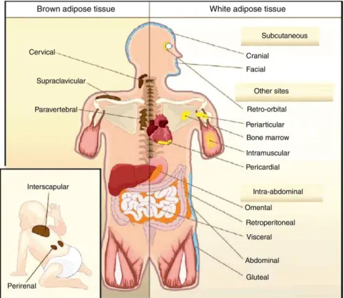
Most adipose tissue in the adult is composed of WAT subcutaneous and visceral deposits. Subcutaneous adipose tissue affects the entire body surface; in women it is particularly developed in the gluteal-femoral and mammary region, but in men it is mainly present in the abdominal area. A significant portion, about 10% of the total, is deposited around the muscular bundles of the limbs and at the visceral level, while smaller deposits are found in the epicardial region and in the mediastinum (Fig. 2).
7
Fig. 2: Body distribution of WAT and BAT in humans (adapted from Gesta et al., 2007).
WAT has also a very heterogeneous composition according to body localization (Sanchez et al., 2005). If the cell that distinguishes the adipose tissue is the adipocyte, this is not the only type of cell present in the adipose tissue, and not even the most abundant one. Pre-adipocytes, macrophages, neutrophils, lymphocytes, and endothelial cells, are among the different cell types of adipose tissue. The balance between these different cellular forms, as well as their expression profile, is closely related to the maintenance of energy homeostasis. The increase in the form of adipocytes, in the number, and in the type of lymphocytes and infiltrated macrophages, is in fact strictly related to the problems of the metabolic syndrome (Esposito et al., 2006; Lumeng et al., 2011).
Therefore, the study of preadipocytes regulation of proliferation and differentiation, as well as the understanding of the interaction between the different cells types present in the adipose tissue, gives us new targets which allow coping with these pathologies.
8 1.2.2 Adipogenesis
Adipogenesis is a multi-step process whose cellular and molecular events have been extensively studied in recent years, mainly due to some cell lines generation, such as the murine preadipocyte 3T3-L1 line (Green et al., 1975). They have allowed the understanding of both the differentiation of preadipocytes in mature adipocytes and the mechanisms underlying the main metabolic functions of the cell, such as lipolysis, incorporation of insulin-mediated glucose, and lipogenesis. During this process, an undifferentiated mesenchymal cell becomes a pre-adipocyte, which in turn differentiates into a mature pre-adipocyte, a cell used for fat storage. Until few years ago, adipogenesis was considered a function which ended in the first years of life with the presence of a fixed number of adipocytes predestined at birth. On the contrary, today it is widely accepted that the body fat is the place of a continuous cell turn-over through which the mesenchymal stem cells are engaged in the processes of proliferation in preadipocytes, stopping their growth and differentiating into mature adipocytes. The number of adipocytes thus depends on a balance between adipogenesis and apoptosis (Lefterova et al., 2009). Adipogenesis occurs therefore in response both to normal cell renewal and to the need of increasing the fat reserves that happens when the nutritional needs exceed (Hausman et al., 2001). Two phases of adipose tissue growth can be distinguished: a first one is characterized by a marked hyperplasia, which increases the number of cells, and a second phase in which the number of adipocytes is apparently stable and instead shows a hypertrophic growth.
Hyperplasia is caused by the differentiation of precursors to mature adipocytes and it is an irreversible process, in contrast to hypertrophy (Avram et al., 2007). Some authors have developed a theory according to which adipose tissue initially grows thanks to the combination of hyperplasia and hypertrophy; then, as the number of cells rapidly reaches a plateau, cell hypertrophy continues until reaching a "maximum" of cell size (Otto and Lane, 2005).
When the "maximum" is reached, according to the "critical fat cell size hypothesis", adipocytes produce and release a series of paracrine factors that control the proliferation of pre-adipocytes and they are therefore involved in the development of obesity (Hausman et al., 2001). This activation is mainly induced by insulin-like growth factor (IGF) released by hypertrophic adipocytes and free
9
insulin, unable to bind to the specific receptor on adipocyte. There is therefore an initial differentiation of stem cells towards adipoblasts and pre-adipocytes and a subsequent activation of stem cell mitosis to restore the basal number of them in the stroma (Arner et al., 2009).
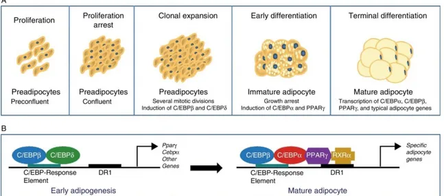
Once the differentiative stimulus has been induced, the cells are subjected to profound changes at transcriptional and morphological levels (Avram et al., 2007). One of the first events of adipogenesis is growth arrest, which normally occurs through contact inhibition. Following this, preadipocytes must receive an appropriate combination of mitogenic and adipogenic signals to continue through the next stages of differentiation, which will lead to the progressive acquisition of morphological and biochemical characteristics typical of mature adipocytes (Tang
et al., 2012; Ali et al., 2013) (Fig. 3).
In particular, the adipogenic process is initialized by the drastic transition from the elongated fibroblast to the spherical one, typical of adipocytes. The morphological modifications are then accompanied by changes in extracellular matrix (ECM) and cytoskeleton components. The terminal stage of differentiation is instead characterized by the activation of a transcriptional cascade that leads to an increase in the expression of key proteins involved in the synthesis of triglycerides, an increase in the number of glucose transporters and increased sensitivity of insulin receptors. In this last phase, the newly formed adipocyte becomes in fact a highly specialised endocrine cell, able to secrete important hormones related to the regulation of energy homeostasis (Arner et al., 2009).
10
Fig. 3: Differentiation of preadipocytes into adipocytes (Ràfols et al., 2014). (A) Scheme of the transition process from preadipocyte to mature adipocyte including the different stages. (B) Sequential model of transcriptional control during adipogenesis.
During adipogenesis the increase in insulin sensitivity is observed through the increased synthesis of its receptor and of the insulin-dependent glucose transporter type 4 (GLUT-4); it is also observed a high transcription of the genes coding for fatty acid-binding protein 4 (FABP4), fatty acid transport protein 1 (FATP1), encoding for a fatty acid transporter, and for lipoprotein lipase (LPL), involved in lipid "storage" and metabolism control (Furuhashi et al., 2014; Wu et al., 2006; Mead et al., 2002). However, the different steps involved in the transition from pre-adipocytes to adipocytes are mainly regulated by the activation of a transcriptional cascade involving the nuclear receptor PPAR-γ (peroxisome proliferator-activated receptor γ) and some members of the C/EBPs family (CAAT/Enhancer binding proteins) (Nerlov 2007; Wang et al., 2017).
1.2.2.1 PPAR-γ
Transcription factors PPARs are members of the nuclear receptor superfamily. Up to now three different subtypes of PPARs (α, β/δ, and γ) have been identified; their actions result in a wide and diverse range of biological effects based on tissue localization and on the chemical profile of the ligand involved in the activation (D’amore et al., 2013; Wang et al., 2010).
11
In particular, PPAR-α is found in a variety of tissues including liver, heart and skeletal muscles, in which it has the function of fatty acids oxidation (Semple et al., 2006; Kiec-Wilk et al., 2005; Ferrè et al., 2004), and anti-inflammatory activity (Kiec-Wilk et al., 2005).
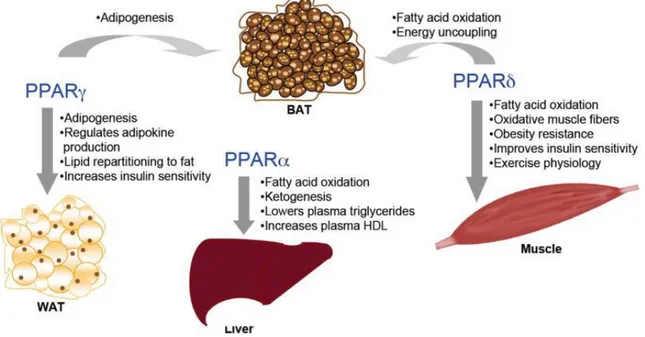
PPAR β/δ is ubiquitous, but mostly expressed in skeletal muscles (Semple et al., 2006; Gurnell, 2005). During prenatal development, it promotes the formation of organs through the regulation of cytotrophoblast migration, and influences stem cell differentiation (Kiec-Wilk et al., 2005). PPAR-γ instead has been identified as one of the most important regulators of adipogenesis (Fig. 4).
Fig. 4: Physiological and/or pharmacological PPARs roles in energy metabolism (Wang et al., 2010).
There are two isoforms of PPAR-γ (PPAR-γ1 e PPAR-γ2), originating from an alternative splicing, and they are both expressed in the adipose tissue, but only the isoform 2 is a specific marker of adipose tissue (Imai et al., 2004). The gene coding for the protein PPAR-γ not only plays a key role in the development of fat cells, but also for the maintenance of differentiation. Its silencing in the already differentiated 3T3-L1 adipocytes, in fact, causes de-differentiation with lipid loss and decreased expression of adipocytes typical markers (Feve et al., 2005; Semple et al., 2006; Kiec-Wilk et al., 2005). Up to now in fact, no factor capable of
12
promoting adipogenesis in absence of PPAR-γ has been identified (Agostini et al., 2006).
Some studies have recently shown that the downregulation of PPAR-γ by stressors agents such as tumor necrosis factor-α (TNF-α) leads to triglyceride storage alteration in the fat cells (Fig. 5).
Fig. 5: Adipocyte dysfunction linking obesity to Insulin resistance (Guilherme
et al., 2008).
PPAR-γ expression can be regulated at the transcriptional level by TNF-α through the activation of Nuclear Factor-κB (NF-κB) and Activator protein 1 (AP1) protein, that negatively regulates PPAR expression (Ye et al., 2008; Li et al., 2018). In the adipocytes there is a rapid turnover of PPAR gene expression (Tang et al., 2006) and the treatment of cultivated adipocytes with TNF-α could determine gene degradation. Translational control of PPAR is mediated by mitogen activated protein kinase-4 (MAPK4), a kinase upregulated by TNF-α (arrow 3). In addition, caspase activation through TNF-α could trigger the PPAR protein degradation in adipocytes (dashed arrow 4). The regulation of PPAR activity is also negatively regulated by kinase-mediated phosphorylation (Diradourian et al., 2005) and ubiquitylation (Hauser et al., 2000), which promotes PPAR degradation through a proteasome-dependent pathway (arrow 5). The multiple-level action of TNF-α could therefore lead to a decrease in PPAR activity. A precise regulation of PPAR expression and function can contribute to the control of triglycerides biosynthesis and lipid droplets hydrolysis and deposition. This can take place thanks to the regulation of triglyceride metabolism enzyme expression as phosphoenolpyruvate carboxykinase (PEPCK), fatty acid synthase (FAS), acyl-CoA synthetase (ACS), LPL, and lipid-droplet proteins, including cell death activator (CIDE-A), fat-specific protein 27 (FSP27), and perilipin (Langhi et al., 2015) (arrow 6).
13 1.2.2.2 C/EBP
The C/EBPs transcription factor family includes 3 members: C/EBP-α, c/EBP-β, c/EBP-δ. These are the first transcription factors involved in adipocyte differentiation (Otto et al., 2005) and they are expressed at specific times during adipogenesis. The earliest are β and δ, which, in turn, promote the expression of c/EBP-α and PPAR-γ, essential for adipocytes total differentiation. The expression of C/EBP-β appears to be essential in the early stages of differentiation and this shows how animals deficient in C/EBP-β display a reduction of adipose tissue (Lefterova et al., 2014).
Murine models with suppression of c/EBP-β and c/EBP-δ, maintain the ability to generate adipose tissue, although the efficiency with which this phenomenon occurs is reduced. Surprisingly, they express normal levels of PPAR-γ, c/EBP-α, and FABP4, in spite of a reduced total fat mass, indicating, therefore, that there may be compensatory mechanisms involved in the activation of adipocyte genes (Lee et al., 2004). Differently, the expression of C/EBP-α is required for adipogenesis with PPAR-γ, althoughthe latter appears to be the dominant process (Zuo et al., 2006) (Fig. 6).
Fig. 6: Diagram of the transcriptional activity of PPAR-γ and C/EBPs during adipogenesis (adapted from Lefterova et al., 2014).
14 1.2.3 Physiology of adipose tissue
For a long time, adipose tissue has been considered an organ with a poorly active role in global energy homeostasis. It was believed that its function, in addition to providing a thermal and mechanical isolation, was only to store excess energy in the form of triglycerides with high caloric density, and to return it, according to needs, as free fatty acids. In recent years the concept of adipose tissue has radically changed, and nowadays it is considered as an endocrine organ that secretes many factors with autocrine, paracrine, and endocrine function (Sikaris et al., 2004; Waki et al., 2007).
The adipose tissue is involved in the regulation of the fat mass and nutrient homeostasis, as well as in the immune response, in blood pressure control, in haemostasis, and in thyroid and reproductive system functions control (Trayhurn et al., 2005; Grant et al., 2015).
Among the various substances secretes by WAT, fatty acids represent the most important molecules. These are esterified and stored in dynamic organelles called lipid droplets (LD) that during periods of excessive food availability act as a deposit for fatty acids and excess cholesterol which would otherwise be harmful to cells (Farese et al., 2009). In addition to the adipogenesis process, two other important metabolic activities occur in the adipose tissue: the synthesis of fatty acids and their storage (lipogenesis), and mobilization or triglyceride hydrolysis (lipolysis). The first one occurs during periods in which there is a situation of excess energy, the second one during periods of caloric deficit and energy demand by other organs or tissues. Both lipogenesis and lipolysis are regulated by the integration of endocrine and neural mechanisms that cooperate in order to maintain the level of balanced and constant body fat under normal conditions (Ameer et al., 2014; Duncan et al., 2007).
Lipogenesis is a process regulated by LPL action. Synthesized and secreted by adipocytes, this enzyme is transported to the endothelium where it acts by hydrolyzing triglycerides from lipoproteins, thus releasing fatty acids and monoacylglycerol. LPL glycosylation is an important adjustment step both for its secretion and its enzymatic activity. LPL activity increases after meals, probably due to the stimulation by insulin itself, allowing the clearance of lipids from the
15
bloodstream and their storage in adipose tissue. LPL action is also affected by other factors, such as adenosine, which stimulates it, and cortisol, which acts by promoting insulin action; LPL activity is therefore reduced in the absence of insulin. Among the hormones that also inhibit LPL action catecholamines are included.
Glucose metabolism is essential within the adipocyte: it provides energy and allows to maintain a normal degree of free fatty acids esterification; in fact, during glycolysis, α-glycerophosphate is formed, and with it free fatty acids are esterified to form triglycerides (Herman et al., 2006). Adipocytes are also able, through the lipolysis, to release in the circulation fatty acids that are used by most tissues as an energy source when glucose is not present in sufficient concentration, feeding mitochondrial systems of fatty acid oxidation and creating metabolic intermediates that act as substrates or signalling molecules (Zechner et al., 2012). Lipolysis is largely determined by the action of hormone-sensitive lipase (HSL), an enzyme that hydrolyzes triglycerides by releasing fatty acids and glycerol (Jaworski et al., 2007). The enzyme activity depends on its phosphorylation, and the hormones that affect lipolysis regulate this state. The main regulatory mechanism occurs through the action of PKA, an enzyme whose function is activated by the increase in cAMP resulting from stimulation of adenylate cyclase. There are also other protein kinases that mediate HSL phosphorylation, such as the mitogen-activated protein kinase (MAPK) and the AMP-activated protein kinase (AMPK). The lipolytic stimuli also include catecholamines, glucagon, growth hormone (GH), cortisol, and thyrostatic hormone (TSH).
The most important anti-lipolytic hormone is insulin, whose action negatively regulates HSL phosphorylation. The action of the HSL is also increased by the perilipin protein, which is present on the membrane of the lipid-intracellular droplets and acts as a protective layer to prevent the action of the enzyme (Brasaemle et al., 2007). A local antilipolitic action is also carried out by molecules directly produced by adipocytes, including adenosine and prostaglandins. The correct functioning of the lipogenesis/lipolysis balance is necessary to monitor all other metabolic processes in the organism. In presence of nutrients and energy excess or obesity, the adipose tissue ability can be overwhelmed, causing stress, injury and malfunctioning. For example, insulin
16
resistance under these conditions leads to higher levels of basal lipolysis and a decreased ability to synthesize and esterify fatty acids for storage or neutralization by downregulation of synthetics mechanisms (Morigny et al., 2016; Ranganathan et al., 2006).
This dysfunctional state contributes to systemic lipotoxicity, because the excess of fatty acids released or absorbed from diet, are moved into circulation and deposited in organs that are not predisposed to store lipids. The esterification purpose is to prevent the harmful effects of fatty acids (Listenberger et al., 2003) by increasing the storage capacity of LD in adipocytes and by determining protection against the development of diabetes and insulin resistance (Schaffer et al., 2016; Sezer et al., 2017).
The increase of lipolysis is also linked to the secretion of FABP4 (Cao et al., 2013; Ertunc et al., 2015), which is an important mediator of immunometabolic local responses in adipose tissue and links the lipolytic state to glucose metabolism into the liver and other sites (Cao et al., 2006). Although adipocytes represent a specialized site for neutralizing fatty acids, this ability is not infinite and these cells are not entirely insensitive to the accumulation of excess lipids that can lead to various inflammatory responses (Sezer et al., 2017). Among the other lipid molecules secretes by WAT are prostanoids, cholesterol, and retinol, which are stored to be subsequently released (Trayhurn et al., 2005), and steroid hormones (sexual steroids and glucocorticoids), which at the level of the WAT can move from inactive to active forms and vice versa, with significant autocrine and paracrine roles (Li et al., 2017). In addition to these lipid substances, adipocytes and associated cells also produce and release, in systemic circulation, a series of hormones, factors and signals protein, called adipokines or adipocytokines, important for the maintenance of energy homeostasis, and their alterations contribute to the onset of complications associated with obesity (Fernández-Sánchez et al., 2011).
17 1.2.4 Adipose tissue as an endocrine organ
All molecules produced and secreted by adipose tissue with autocrine, paracrine, or endocrine functions are called adipokines. The term “adipokine” should be used to designate proteins synthesized and secreted directly from adipocytes (Trayhurn et al., 2004). However, this term is used generically for all proteins secreted by WAT, although they are mainly synthesized by other cell types, such as infiltrated macrophages, present in the adipose tissue (Weisberg et al., 2003). Adipokines have highly diversified chemical structures and important physiological roles. They include proteins involved in the regulation of energy intake and energy balance (leptin), in the regulation of arterial blood pressure (angiotensinogen), in vascular hemostasis (PAI-1), in lipid metabolism (RBP-4, CETP), in carbohydrate homeostasis (adiponectin, resistin, visfatine), and in angiogenesis (VEGF), as well as growth factors (TGF) and acute phase and oxidative stress proteins (hepatoglobulin, and A1-acid glycoprotein) (Antuna-Puente et al., 2008). It is also interesting to note how, many of them, are linked to the immune system, such as the classical cytokines TNF-α, 1, 6, 8, IL-10, IL-4, IL-13, and MCP-1. This can therefore underline the close relationship between inflammation and obesity (Ouchi et al., 2011; Gregor et al., 2011).
This wide range of factors and protein signals suggests that adipose tissue is a complex organ, highly integrated in the physiology and metabolism of mammals, capable of establishing communication links with other tissues and organs, including the central nervous system (CNS), the liver, the skeletal muscles, and the surrenal cortex, through effects on the neuroendocrine pathways (Mathieu et al., 2009). It is important to emphasize, however, that endocrine function is not only typical of the WAT, because many of these factors are also synthesized by BAT (Cannon et al., 2004; Villarroya et al., 2013). According to their effect in the adipose tissue, these adipokines can be classified as pro- or anti-inflammatory agents (Fig. 7).
18
Fig.7: Physiological and metabolic processes regulated by WAT through adipokine secretion (Ràfols et al., 2014). CETP: cholesterol ester transfer protein; IL1: interleukin 1; IL1Ra: interleukin receptor antagonist-1; IL4: interleukin 4; IL6: interleukin 6: IL8: interleukin 8; IL10: interleucin-10; IL18: interleukin-18; MCP-1: monocyte chemoattractant protein-1; NGF: nerve growth factor; NPY: neuropeptide Y; RBP-4: retinolbinding protein-4; TGFβ: transforming growth factor β; TNFα: tumor necrosis factor alpha: VEGF: vascular endothelial growth factor.
1.2.5 Pro-inflammatory Adipokines 1.2.5.1 Leptin
The identification in 1994 of the Leptin gene (OB) and its receptor has started the endocrine era of adipocyte. It is a 16 kDa hormone formed by 167 amino acids, located on chromosome 6 in mice and chromosome 7 in humans. It is mainly secreted by adipocytes proportionally to the mass of adipose tissue and to nutritional conditions. Leptin is mainly produced in WAT and it is strongly involved in the regulation of lipid metabolism and energy consumption (Lastra et al., 2006; Kelesidis et al., 2011; Moon et al., 2013).
In obesity conditions it has shown an influence on dietary behaviour through hypothalamic regulation in CNS. Once secreted by adipose tissue, it circulates in the blood linked to the plasma proteins, and diffuses in the CNS through the binding to the capillaries in the median eminence and, through saturable transport,
19
through the choroid plexus receptor. In the ventromedial nucleus of the hypothalamus, leptin stimulates the cytokine receptor kinase 2 (CK2), the synthesis of the melanocyte stimulating hormone and cocaine- and amphetamine-regulated transcript (CART) which, via paracrine mechanisms, stimulates receptors 3 and 4 of the melanocortin lateral nucleus, causing satiety (Sikaris et al., 2004; Fonseca-Alaniz et al., 2007).
The functions of leptin are not limited in the hypothalamus and are not exclusively concerned with the maintenance of energy homeostasis (Baratta et al., 2002), but involve the entire CNS and peripheral nervous system (PNS) (Bjørbæck et al., 2004). Leptin, in fact, is involved in several physiological processes such as: regulation of metabolism, growth, development, and regulation of certain endocrinological and immunologic processes, reproduction, cardiovascular physiopathology and maintenance of respiratory function (La Cava et al., 2004). The discovery of leptin has thus confirmed the existence of a channel of communication between adipose tissue and brain, which aims to regulate the accumulation of fat in the adipocytes (Fig. 8).
Fig. 8: Effects of leptin (Kahn et al., 2000).
Leptin exerts multiple actions to regulate glucose homeostasis through autocrine, paracrine, endocrine, and neural pathways.
20
Leptin inhibits lipogenesis and stimulates lipolysis, reducing intracellular lipid levels in skeletal muscles, liver and pancreatic β cells, thus improving insulin sensitivity. The limbic system stimulates dopamine reuptake, decrease appetite, and, through the nucleus locus coeruleus, activates the sympathetic nervous system (SNS) that increases resting energy expenditure (Fonseca-Alaniz et al., 2007; Minokoshi et al., 2012). Catecholamines influence the leptin secretion while other regulators of its synthesis are glucocorticoids (Sikaris et al., 2004); however, it has been shown that the main determinant in leptin secretion is glucose metabolism, because leptin concentration in bloodstream decreases in fasting conditions or caloric restriction and increases in response to food intake (Dulloo et al., 2010). Obesity is associated with increased leptin levels and it has been shown that the apparent decrease in anorectic effects and weight loss are the result of a mechanism of resistance to it (Fonseca-Alaniz et al., 2007). The obese subjects therefore present leptin resistance (Zhou et al., 2013). This condition also causes altered leptin transport through the brain barrier, hyperleptinemia (Kievit et al., 2006), autophagy (Quan et al., 2012), and ER stress (Ozcan et al., 2009). Besides intervening in the body weight regulation, leptin regulates puberty and reproduction, placental and fetal functions, immune response, muscular and hepatic insulin sensitivity (Kelesidis et al., 2010). In patients with lipoatrophy the lack of adipose tissue causes severe hypolepinemia, which is associated with severe insulin resistance, hepatic steatosis and dyslipidemia. In these patients, in addition, leptin treatment results in a marked improvement of glucose metabolism, dyslipidemia and hepatosteosis. Furthermore, hyperleptinemia, which belongs to the majority of the obese subjects, seems to have a proatherogenic role contributing to insulin resistance, altering endothelial function, and promoting platelet aggregation and arterial thrombosis (Martens et al., 2006). In inflammation, leptin acts directly on macrophages by increasing the phagocytic activity and the production of proinflammatory cytokines; when leptin is administered, higher levels of C-reactive protein are produced, thus proving its inflammatory effect (Steffes et al., 2006). In fact, leptin activates monocytes and macrophages, leading to the production of MCP-1 and VEGF in the hepatic stellate cells (Aleffi et al., 2005). Other inflammatory signals such as TNF-α and lipopolysaccharide (LPS) also stimulate the expression of leptin and its receptor
21
(Gan et al., 2012). Leptin also increases the production of pro-inflammatory Th1 cytokines and suppresses the production of anti-inflammatory Th2 cytokines, such as IL-4 (Iikuni et al., 2008). When there is weight loss the circulating hormone levels decrease and, in turn, also those of the inflammatory markers associated with obesity (Hukshorn et al., 2004). Leptin, besides promoting oxidative stress and vascular inflammation, stimulates the proliferation and migration of endothelial cells and smooth muscle ones, favouring the development of atherosclerosis (Cachofeiro et al., 2006). It is also important to emphasize that leptin can also be produced in the placenta, in the spinal cord, in the muscle, and perhaps in the brain (Sikaris et al., 2004).
1.2.5.2 Interleukin-6
Interleukin-6 (IL-6) is a cytokine with pro- and anti-inflammatory action (Fonseca-Alaniz et al., 2007). It is produced by macrophages, adipocytes (Cachofeiro et al., 2006), immune system cells, fibroblasts, endothelial cells, and skeletal muscles (Sanchez et al., 2005). IL-6 is highly expressed in adipose tissue and it is positively related to obesity in humans. Circulating cytokine levels are related to body mass index (BMI), insulin resistance, and carbohydrate intolerance (Lastra et al., 2006). Elevated plasma levels of IL-6 are considered as a predisposing factor for type 2 diabetes and myocardial infarction (Qu et al., 2014). IL-6 also influences glucose tolerance through the negative regulation of visfatin; it also inhibits the secretion of adiponectin (Fonseca-Alaniz et al., 2007) and in animal models increases triglycerides levels by increasing gluconeogenesis and glycogenolysis and inhibiting glycogenesis. IL-6 also induces hepatic C-reactive protein (CRP) production, which is known to be a risk factor for cardiovascular complications (Sacheck et al., 2008). High levels of IL-6 have been detected in atherosclerotic plaques in humans. In addition, IL-6 alters endothelium-dependent dilatation in veins, for this reason it appears to be an important aggravating factor of coronary heart disease (Neal et al., 2008). IL-6 activity is however controversial. The peripheral administration of IL-6 leads to an interruption of insulin signal due to the increase of suppressor of cytokine signaling-3 (SOCS3) expression in hepatocytes, suggesting that the expression of IL-6 in obesity determines insulin resistance (Kim et al., 2009). In contrast, in
22
vivo studies, have found that IL-6 deficient mice show obesity and hepatic inflammation, and IL-6 intake reverses insulin resistance (Matthews et al., 2010). IL-6 intake therefore would improve energy expenditure by reducing obesity. Thus the role of IL-6 in obesity and insulin resistance depends on specific expression sites integrated with other adipokines and cytokines.
1.2.5.3 Tumor necrosis factor-α
Tumor necrosis factor-α (TNF-α) is a pro-inflammatory cytokine produced mainly by monocytes, lymphocytes, adipose tissue, and muscles (Ouchi et al., 2011). It appears involved in several systemic inflammatory responses and its irregular production contributes to the pathogenesis of the metabolic syndrome resulting in a higher incidence of obesity, diabetes, and cardiovascular diseases (Sanchez et al., 2005). The high levels of TNF-α are related to an increase in adipose tissue metabolism. In fact, it is also secreted by preadipocytes and adipocytes and its production is increased in obese subjects. TNF-α is also capable of suppressing the differentiation of preadipocytes and inducing delipidation and dedifferentiation of adipocytes (Kurebayashi et al., 2001). Therefore it appears to be closely related to insulin resistance and obesity (Tzanavari et al., 2010).
In several in vitro and in vivo studies, TNF-α has been shown to induce insulin resistance (Borst et al., 2004; Swaroop et al., 2012). In this condition, in particular, there is an increase in the release of free fatty acids in adipocytes, blocking of adiponectin synthesis, and reduction of tyrosine phosphorylation in insulin receptor, which is essential for progression of intracellular hormone signalling (Lastra et al., 2006). Thus, TNF-α appears positively related to insulin resistance and its neutralization should improve insulin sensitivity. However, all these aspects are still controversial. Recent studies have shown that a short-term intake of TNF-α significantly suppresses inflammation in obese subjects with type 2 diabetes, but does not show improvement in insulin sensitivity (Swaroop et al., 2012).
In contrast, long-term treatment with TNF-α causes remission of inflammatory diseases such as rheumatoid arthritis, greatly improving insulin sensitivity (Gonzales-Gay et al., 2006; Stanley et al., 2011). TNF-α also plays an important role in inflammation by determining activation of NF-κB, resulting in an increase
23
in adhesion molecules expression on the surface of muscle cells, inflammatory state of adipose tissue, endothelial dysfunction, and atherogenesis (Lastra et al., 2006).
1.2.5.4 Monocyte chemoattractant protein-1 and CC-Chemokine receptor type 5
Chemokines and their receptors play an essential role in mediating immune cell infiltration in adipose tissue. Monocyte chemoattractant protein-1 (MCP-1) (also called CCL2) and CC-Chemokine receptor type 5 (CCR5) are respectively a typical chemokine and a chemokine receptor that mediate inflammatory responses and are significantly increased in obese subjects. In vivo studies have shown how deficient mice in MCP-1 receptor exhibit reduced macrophagic infiltration, inflammation and insulin resistance (Weisberg et al., 2006). In addition, the deletion of the CCR5 receptor in obese rats show improved inflammation, insulin sensitivity and hepatic steatosis resulting in reduced macrophagic infiltration and differentiation of macrophages into the anti-inflammatory M2 isoform (Kitade et al., 2012; Kanda et al., 2006). However, MCP-1 role in inflammation and insulin resistance is still unclear. In another study, obese mice deficient in MCP-1 do not show differences in the accumulation of macrophages and inflammation in the adipose tissue (Kirk et al., 2008). However, the reason for this difference is still unclear: it is possible that a deficiency of MCP-1 could be compensated by other chemokines to it related.
1.2.5.5 Other Pro-inflammatory Adipokines
In addition to Leptin, IL-6, TNF-α and MCP-1, WAT expresses and releases a variety of other pro-inflammatory cytokines and chemokines including Retinol-binding protein 4, Resistin, Angiotensinogen and Plasminogen Activator Inhibitor–1.
The Retinol-binding protein 4 (RBP4) is a specific protein for circulating transport of retinol (vitamin A). It is expressed in the liver, in adipocytes, and in macrophages, and shows elevated plasma levels in different animal models of obesity and insulin resistance. Recently, it has been shown that retinol-binding protein constitutes an adipocitary signal that can contribute to the pathogenesis of
24
T2DM in obesity. The increase of its plasma concentration seems to induce the hepatic expression of neoglucogenetic enzymes and contributes to the insulin resistance in skeletal muscles (Yang et al., 2005). Recent in vivo studies have shown that the intake of recombinant RBP4 in healthy rats determines insulin resistance (Yang et al., 2005). The expression of RBP4 seems, in fact, to be inversely related to GLUT-4 in adipocytes, thus determining induction of insulin resistance condition by inhibiting insulin-induced insulin receptor substrate-1 (IRS-1) tyrosine phosphorylation. Clinical studies have also shown that increased levels of RBP4 are closely associated with high blood pressure, high levels of triacylglycerol, high BMI (Graham et al., 2006), subclinical inflammation, and renal diseases (Akbay et al., 2010). RBP4 stimulates primary human endothelial cells to produce pro-inflammatory molecules such as vascular cell adhesion molecule-1 (VCAM-1), MCP-1, and IL-6, determining endothelial inflammation progression in cardiovascular disorders and microvascular complications in diabetes (Farjo et al., 2012).
The Resistin (ADSF/FIZZ3/XCP1), a 10 kDa polypeptide with 114 amino acids, has been identified as a pulmonary inflammation and insulin resistance inducer (Mojiminiyi et al., 2007). It belongs to the rich cysteine family and circulates as hexamer or trimer. The hexameric form is the most abundant but the less active, while the trimeric instead determines insulin resistance. Resistin is involved in the activation of the SOCS3 protein which triggers suppression of the insulin mediated signal in adipocytes (Steppan et al., 2005). Studies in mice have shown that, as a result of the deficiency of this protein, there is also a better glucose tolerance and insulin sensitivity (Qi et al., 2006). Instead the resistin function in humans is still controversial. Monocytes and macrophages are the main source of resistin in humans; its expression in adipocytes is however restricted only to rodents. Inflammatory cytokines such as IL-1, IL-6, TNF-α, and LPS induce resistin expression in human macrophages. Resistin also stimulates human peripheral mononuclear cells to produce IL-6 and TNF-α through the NF-κB pathway. Rosiglitazone and PPAR-γ agonists instead suppress its expression in adipose tissue, determining attenuation of inflammatory responses (Bokarewa et al., 2005). Resistin also activates JNK and p38 MAPK determining induction of insulin resistance through the bond in hypothalamus with the Toll-like receptor-4
25
(TLR4) (Benomar et al., 2013). Induction of resistin synthesis can be attenuated by PPAR-γ agonists (Kusminski et al., 2005; Lehrke et al., 2004). It has been shown that the treatment of patients with T2DM with pioglitazone, a PPAR-γ agonist, results in a decrease in serum resistin levels (Bajaj et al., 2004). In addition, several factors, such as pituitary, steroid and thyroid hormones, adrenalin, β3-adrenergic receptor activation, endothelin-1 and insulin, modulate resistin expression (Kusminski et al., 2005; Lehrke et al., 2004).
Adipose tissue is, in addition, an important site for the production of angiotensinogen and angiotensin II. Higher levels of Angiotensinogen can be found in adipose tissue of obese individuals compared to normal weight subjects, and a positive relation between plasma levels of angiotensinogen and adiposity can be detected (Goossens et al., 2003). It is clear, therefore, that the increased synthesis of angiotensinogen and angiotensin II can contribute to hypertension that is frequently associated with obesity. In addition, angiotensin II appears to exert disparate proinflammatory effects in adipocyte and stimulates oxidative stress, effects that can be inhibited by blocking angiotensin receptor 1. Angiotensinogen can play an important role in regulating blood and fatty acids flow into adipose tissue (Dulloo et al., 2010). It is expressed in multiple cell types within the adipose tissue; its expression and secretion are higher in visceral tissue compared to the subcutaneous one and its high levels are related to the metabolic syndrome (Fonseca-Alaniz et al., 2007). The plasminogen activator inhibitor-1 (PAI-1) is the first physiological inhibitor of plasminogen activators in blood and contributes to thrombosis and to the development of chronic cardiovascular diseases. PAI-1 can play an important role in regulating blood and fatty acids flow of adipose tissue. Plasma levels of PAI-1 are regulated by the accumulation of visceral fat and its high concentration is associated with insulin resistance and proinflammatory cytokines (Sikaris et al., 2004).
26 1.2.6 Anti-inflammatory Adipokines 1.2.6.1 Adiponectin
Adiponectin, also called Acrp30, ApM1, AdipoQ and GBP28 (Ruan et al., 2016), is a protein of 247 amino acids isolated for the first time in 1995 into adipose tissue (Scherer et al., 1995). It is highly expressed in adipocytes with potent anti-inflammatory activities. It presents a collagen-like N-terminal and a globular C-terminal domain, and circulates as a trimer, hexamer and other high molecular weight structures. Under physiological conditions, it is present in blood at elevated concentrations (5-10 μg/ml); in obese subjects, in those with T2DM and in those suffering from cardiovascular disease, the circulating levels of adiponectin are reduced. Proinflammatory factors such as TNF-α, IL-6, reactive oxygen species (ROS) and hypoxia suppress the expression in adipocytes (Li et al., 2009). Recently, it has been shown that not only inflammatory signals, but also iron overload in adipocytes, suppress the expression of adiponectin in obese subjects through Forkhead Box O1 gene (FOXO1) (Gabrielsen et al., 2012). Conversely, PPAR-γ antagonists stimulate adiponectin expression in adipocytes (Maeda et al., 2001). Adiponectin activates AMPK through its receptors AdipoR1 and AdipoR2, resulting in increased fatty acid oxidation and glucose uptake in muscles and suppressing hepatic gluconeogenesis (Yamauchi et al., 2002). Exogenous adiponectin intake also leads to improvement in insulin sensitivity (Maeda et al., 2002; Kim et al., 2007). Moreover, Adiponectin has a direct effect on glucose uptake in skeletal muscles and adipose tissue through an increased translocation of GLUT-4 on the plasma membrane (Ceddia et al., 2005; Fu et al., 2005).
Adiponectin inhibits, in macrophages, the production LPS-induced of TNF-α by inhibiting the activation of NF-κB pathway and stimulating the production of IL-10 which presents an anti-inflammatory activity (Kumada et al., 2004). It also promotes the differentiation of anti-inflammatory macrophages M2 and modulates T-cells activation and inflammatory NK-cells function. Adiponectin receptors are upregulated at the surface of T-cells after antigenic stimulation and mediate apoptosis of specific T-cells by suppressing the antigen-specific expansion (Wilk et al., 2011). Moreover, adiponectin suppresses the production of interferon
27
gamma (IFN-γ) mediated by Toll-Like Receptor (TLR) in NK-cells without determining cytotoxicity (Wilk et al., 2013). Thus adiponectin shows to possess important antiatherogenic, anti-diabetic and anti-inflammatory properties. Subjects with elevated plasma levels of adiponectin therefore have a significantly reduced risk of major cardiovascular events with consequent reduction of atherosclerosis and hepatic diseases (Hui et al., 2012).
1.2.6.2 SFRPs (Secreted frizzled-related proteins)
The secreted frizzled-related proteins (SFRPs) are a family of soluble proteins that consists of five secreted glycoproteins (SFRP1, SFRP2, SFRP3, SFRP4, SFRP5) that act as extracellular signalling ligands. Each SFRP is composed of around 300 amino acids and contains a cysteine-rich domain (CRD) that presents a 30-50% sequence homology with the CRD of Frizzled (Fz) receptors. SFRPs are the main proteins responsible for binding to 'Wingless/Integrated' (WNT) proteins on the plasma membrane (You et al., 2013; Garcia Tobilla et al., 2016).
SFRPs are responsible for the control of several biological phenomena and human abnormalities development. Aberrant gene expression and abnormal secreted protein levels are releated to a wide range of pathologies in humans (Liu et al., 2018). In particular, the isoform SFRP5 has been identified as an anti-inflammatory adipokine which may be implicated in obesity adipocyte disfunction (Anunciado-Koza et al., 2016). Lower SFRP5 levels have been observed in obese rather than in lean subjects. This confirms therefore the relation between this protein and BMI (Hu et al., 2013). The expression of SFRP5 is low during the white and brown adipocytes differentiation while it is elevated in mature adipocytes (Wang et al., 2014). SFRP5 acts by WNT signalling pathway inhibition in white adipose tissues and recent studies have shown that this protein can play a key role in lipid metabolism, inflammation, and T2DM (Chen et al., 2017).
SFRP5-deficient mouse models exhibit marked glucose intolerance, insulin resistance, as well as adipose tissue inflammation (Carstensen-Kirberg et al., 2016; Ehrlund et al., 2013). Recent studies have shown that SFRP5 modulates adipogenesis by increasing adypocite differentiation and lipid accumulation and storage (Van Camp et al., 2014). This protein represses the WNT protein action
28
that reduces the expression levels of pre-adipogenic transcription factors, such as the members of the C/EBP family and PPAR-γ (Ehrlund et al., 2013). Therefore, the anti-inflammatory and insulin-sensitizing properties of SFRP5 have been determined by studies that have shown increased insulin sensitivity and decreased macrophage infiltration and pro-inflammatory protein production (Carstensen-Kirberg et al., 2017). SFRP5 acts by inhibition of c-Jun N-terminal kinase (JNK) activation and antagonism of serine phosphorylation on IRS-1.
All this confirms that SFRP5 could represent a promising therapeutic target in the treatment of obesity, T2DM, and other metabolic diseases (Ouchi et al., 2010; Lu et al., 2013).
1.2.6.3 Other Anti-inflammatory Adipokines
In addition to Adiponectin and Secreted frizzled-related proteins, adipose tissue expresses and releases a variety of other anti-inflammatory cytokines including Omentin–1 and Apelin.
The omental adipose tissue secretes omentin-1, which is mainly expressed by the cells of the omental stromal vascular fraction, but not by adipocytes (Schaffler et al., 2005). Omentin-1 increases insulin-induced glucose uptake at the level of visceral and subcutaneous human adipocytes through the increase of AKT/PKB phosphorylation (Yang et al., 2006). Interestingly, Omentin-1 attenuates CRP- and TNF-α-induced NF-κB activation in human endothelial cells. Some studies have also demonstrated how Omentin-1 promotes macrophage differentiation in the anti-inflammatory M2 phenotype, thus suppressing inflammatory response and formation of LDL-induced foamy cells. This therefore suggests that Omentin-1 could be an anti-inflammatory adipokin in humans (Tan et al., 20Omentin-10).
Apelin is a protein expressed in many tissues such as lung, mammary gland, and testicles, that has been identified as an endogenous ligand of the G protein coupled to the apelin receptor (APJ) (Lv et al., 2017). It has several physiological functions to regulate multiple metabolic functions (Carpene et al., 2007). Adipocytes produce apelin, and its plasma levels are increased in obese subjects. Apelin improves glucose uptake in an AMPK dependent way and suppresses lipolysis. Apelin deficient mice show insulin resistance suggesting that this
29
protein can improve glucose homeostasis and insulin sensitivity (Yue et al., 2010-2011). Apelin is also involved in the inflammatory responses of obese individuals. Its expression is positively associated with TNF-α, some studies have shown, in fact, that the treatment with TNF-α induces its expression in adipose tissue. Furthermore, apelin activates JNK and NF-κB thus inducing the adhesion of molecules such as Intercellular Adhesion Molecule-1 (ICAM-1) in HUVEC cells (Lu et al., 2012). However, apelin intake reduces renal inflammation to improve diabetic nephropathy by suppressing the expression of MCP-1, monocytes infiltration, and NF-κB activation (Day et al., 2013). Thus the precise role of apelin in the regulation of inflammatory responses remains undefined.
1.3 Lipotoxicity and inflammation in adipose tissue
1.3.1 Lipotoxicity
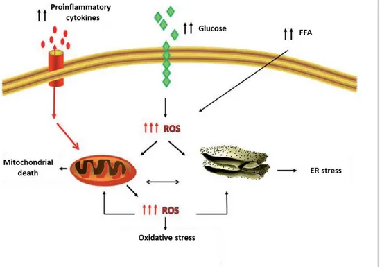
The condition of lipotoxicity, typical of obese subjects, causes alteration of the body homeostasis following prolonged exposure to high concentrations of free fatty acids (FFA), sterols and phospholipids. These molecules are important cellular constituents and provide energy for the body metabolic activities, regulating different homeostatic processes inside and outside the cell, including organelles homeostasis, organs communication, immune function, energy metabolism, and cell survival. Although they perform important physiological roles, when in excess or altered they can be highly harmful, causing cell death, chronic inflammation and alterations of the energetic metabolism with consequent diffuse homeostatic alterations. The condition of lipotoxicity plays an essential role in different metabolic diseases such as inflammation and insulin resistance (Hotamisligil et al., 2006).
Chronic low-grade metabolic inflammation, termed "metainflammation", is considered one of the main features of metabolic diseases such as obesity and diabetes, and it is diffused in several metabolic tissues including adipose tissue, liver, muscles, brain, and intestine (Hotamisligil et al., 2006). Just as lipotoxicity gives rise to metainflammation, alterations in lipid metabolism and typical cellular signalling pathways can lead to an increase in the onset of many diseases (Fu et