The effect of moderate treadmill exercise on the resistive
index of the medial long posterior ciliary artery in dogs
Michela Pugliese
1*, Monica Ragusa
2, Vito Biondi
1, Annamaria
Passantino
1, Kai Zhang
3, Francesco Macri
11Department of Veterinary Science, University of Messina, Polo Universitario Annunziata,
Messina, Italy
2Department of Experimental and Clinical Medicine, University of Catanzaro, Viale Europa,
Catanzaro, Italy
3Department of Traditional Chinese Veterinary Medicine. Lanzhou Institute of Husbandry
and Pharmaceutical Sciences of CAAS, Xiaoxihu, Qilihe District, Lanzhou city, Gansu province, People’s Republic of China
*Corresponding author: [email protected]
Citation: Pugliese M, Ragusa M, Biondi V, Passantino A, Zhang K, Macri F (2019): The effect of moderate treadmill
exercise on the resistive index of the medial long posterior ciliary artery in dogs. Veterinarni Medicina 64, 400–406. Abstract: The resistive index (RI) is an indirect measurement of arterial resistance by means of a ratio between the peak systolic and end diastolic velocities recorded with a spectral Doppler device, especially used to evaluate the vascular damage in ocular diseases such as glaucoma. Some ocular variables such as the intraocular pressure (IOP), the choroidal thickness, the axial length and the ocular blood flow may be influenced by physical exercise. The purpose of this study was to evaluate the influence of the exercise on the RI of the medial long posterior ciliary artery in dogs, and correlate the data obtained with the IOP values. Ten clinically healthy dogs were sub-jected to moderate physical exercise on a canine motorised treadmill at different speeds for 45 minutes. A colour Doppler examination was performed and the RI values were calculated for the medial long posterior ciliary artery at rest, immediately after the exercise, and after 60 minutes at the end of the exercise. At the same times, the IOP was recorded by applanation tonometry. The data were analysed by a two-way repeated ANOVA measurement in order to compare the RI and the IOP. Wilcoxon’s test was applied for the post hoc comparison. Spearman’s rank correlation for non-normal distribution was used to determine a relationship between the RI and the IOP. The at rest RI was 0.722 +/–0.022, IOP 12.38 +/3.21 mm Hg. A significant decrease in the RI was observed imme-diately after the exercise (0.697 +/–0.035) and during the passive recovery phase (0.682 +/–0.042). A significant decrease in the IOP (11+/3.39 mmHg) was recorded after 60 min of the passive recovery phase; at the end of the exercise, a slight decrease (12.29+/4.26 mm Hg) mm Hg was detected. During the test, a linear correlation between the RI and the IOP was observed. Our results suggest that exercise induces the modification of the ophthalmic blood flow in dogs, presumably related to the compensatory neuro-hormonal mechanisms.
Keywords: physical exercise, ocular variables, ocular blood flow
Colour Doppler Imaging (CDI) is a valid method to study the ocular vascular flow velocity and char-acteristics. It permits one to have dynamic real-time anatomical vascular information; moreover,
it allows one to evaluate the presence, the direc-tion and the type of blood into a vessel (Szatmari et al. 2001). The internal and external ophthalmic artery, anterior ciliary artery, the short and long
posterior ciliary arteries, primary retinal arteries are the ocular and orbital vessels most frequent-ly imaged (Gelatt-Nicholson et al. 1999a). Using CDI, impedance indices such as the resistive in-dex (RI), pulsatility inin-dex and systolic-to-diastolic peak velocity ratio characterising the resistance to flow in the vascular system may be calculated (Ferrandis et al. 2013).
The resistive index (RI) or Pourcelot ratio allows for the non-invasive assessment of the peripheral vascular resistance (Bude and Rubin 1999), vascu-lar compliance, conductance and transmural pres-sure (Ostrowska et al. 2016). It is meapres-sured from the interpretation of the shape calculated by the spectral analysis of the vascular signals (Pozniak et al. 1988), as the ratio of the peak systolic veloc-ity and the end diastolic velocveloc-ity of the blood flow. An increase in the ophthalmic blood flow causes a decrease in the intraocular pressure (IOP) – ac-cording to the Goldman equation (Nassr et al. 2009). A high RI value is related to an increase in the distal vascular resistance and a decrease in the perfusion (Liu et al. 1997). In veterinary oph-thalmology, normal RI values on the ocular and orbital vessels have been reported in healthy dogs (Gelatt-Nicholson et al. 1999a; Novellas et al. 2007); its use is especially interesting in animals suffering from diseases such as glaucoma, which helps deter-mine a significant alteration of the ocular vascular pattern (Gelatt-Nicholson et al. 1999b; Lee et al. 2002). An increase in the RI on the medial long posterior ciliary artery was described in glauco-matous dogs treated with antihypertensive drugs, as a direct consequence of the intraocular pressure (IOP) decrease (Choi et al. 2011).
It has been reported that some ocular variables such as IOP (Gale et al. 2009), choroidal thick-ness (Sayin et al. 2015), axial length (Hong et al. 2014) and ocular blood flow (Beck et al. 1995; Kiss et al. 2001; Nemeth et al. 2002; Lovasik et al. 2003; Pournaras et al. 2004) may be influenced by physi-cal exercise in humans. Data describing the effect of the physical exercise on the orbital and ocular blood velocity indicators, determined by Doppler imaging, have, to the best of author’s knowledge, not been reported in the veterinary literature for dogs.
The aim of this study was to evaluate the effect of some moderate exercise on a treadmill on the RI of the medial long posterior ciliary artery (mLPCA) in dogs, correlating the variations in the RI with the changes in the IOP.
MATERIAL AND METHODS
The study was carried with the approval of the Ethical Committee of the Department of Veteri-nary Sciences, the University of Messina (Italy) (Protocol No.2017-11) in accordance with Italian and European regulations on animal welfare. Ten privately-owned dogs of both sexes (seven males and three females), belonging to different breeds (five cross-breed, three Hounds, one Beagle, one Cocker Spaniel), aged two to six years old (a mean of 3.8 ± 2.1 SD) whose body weight ranged between 12–20 kg (a mean of 14 ± 4.3 SD) were included.
Written consent from each owner was obtained before the dog’s enrolment in the study. All sub-jects were determined as healthy and normotensive on the basis of a thorough physical examination and an arterial blood pressure measurement. Furthermore, an ophthalmological examination, including a Schirmer tear test, fluorescein stain-ing, slit-lamp biomicroscope, applanation tonom-etry, gonioscopy and a dilated fundus examination was performed in each dog. The dogs were not pre-trained to run on a treadmill, not familiar with the procedures and the equipment for RI and IOP evaluation. In order to acclimatise them to the procedures, the RI and IOP were evaluated once a day for five days in each dog. All dogs underwent a moderate exercise test on a motorised treadmill (Professional canine treadmill, Grillo®, Modena, Italy) for 45 min at different velocities: 2.5 km/h for 15 min, 5.0 km/h for 20 min, and 2.5 km/h for 10 min, once per day (at 08.00 h). The treadmill presented the following technical features: length 2.40 m (running surface 0.410 m), width 2.50 m (run-ning surface 0.640 m), height 0.600 m, total weight 120 kg, speed 2.5–1.4 km/h. The environmental
tem-perature was continuously recorded and maintained between 18–21 °C (64.40–69.80 °F), the relative
hu-midity ranged between 50–60%. The RI and IOP were recorded at rest (T0), immediately after the exercise (T1) and after 16 min of passive recovery (T2). One drop of a topical anaesthetic (Ossibuprocaine hy-drochloride 0.4%, Novartis; Italy) was instilled in the conjunctival fornix of the left eye before being test-ed. The IOP was recorded by applanation tonometry (Tonopen-Vet, Medtronic, Solan, USA). CDI ultra-sonography (MyLab40, Esaote, Italy) was performed using a 12–18 MHz probe. A coupling gel
was ap-plied from the dorsal region of the left zygomatic arch, positioning the transducer in a horizontal
SPSS, Inc., Chicago, USA).The data were expressed as mean ± standard deviation (SD) and analysed by two-way repeated ANOVA (analysis of variance) measurement in order to compare the RI and IOP before (T0) and after the exercise (T1, T2). Where the ANOVA was showing an acceptable level of sig-nificance (P ≤ 0.05), Wilcoxon’s test was applied for the post hoc comparison. Spearman’s correla-tion for non-normal distribucorrela-tion was used to de-termine the relationship between the RI and IOP. The statistical significance was defined as P < 0.05. RESULTS
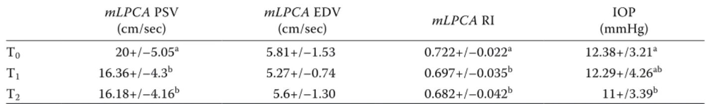
The effects of moderate physical exercise on the RI and IOP are summarised in Table 1. The val-ues of the IOP and RI are expressed as a mean ± SD with their conventional units. The exercise induced plane. The identification of ocular vessel
was ob-tained visualising the retro bulbar fat and the lateral wall of each eye (Lee et al. 2002). Once the mLPCA was localised, the Peak Systolic Velocity (PSV) and End Diastolic Velocity (EDV) were evaluated. The RI was calculated by the formula (PSV-EDV)/PSV (Macri et al. 2015) (Figure 1). At the end of the pro-cedure, each eye was washed with a sterile physi-ological solution, in order to remove the ultrasound gel. The procedures were performed by the same examiners (respectively, M.R. for the IOP and F.M. for the RI) with the dogs restrained gently and placed in sternal recumbence. A mean value of the five measurements at each time point was used in the calculations for the IOP and RI. The mean values of the first measurement on each day were considered the baseline values.
Statistical analysis. The statistical analyses were
performed using a software package (Version 17.0,
Figure 1. Pulsed Doppler ul-trasonogram of the mLPCA in a dog. RI was calculated by the formula (PSV-EDV)/PSV EDV = end diastolic velocity; mLPCA = medial long posterior ciliary artery; PSV = peak systolic velocity; RI = resistive index
Table 1. The effects of the exercise on the PSV, ESV, RI of the medial long posterior ciliary artery and the IOP mLPCA PSV
(cm/sec) mLPCA EDV (cm/sec) mLPCA RI (mmHg)IOP T0 20+/–5.05a 5.81+/–1.53 0.722+/–0.022a 12.38+/3.21a T1 16.36+/–4.3b 5.27+/–0.74 0.697+/–0.035b 12.29+/4.26ab T2 16.18+/–4.16b 5.6+/–1.30 0.682+/–0.042b 11+/3.39b
EDV = end diastolic velocity; IOP = intraocular pressure; mLPCA = medial long posterior ciliary artery; PSV = peak systolic velocity; RI = resistive index
a decrease in the RI and IOP immediately after the exercise, which was also maintained during the rest phase. Our results evidenced a significant vari-ation in the RI values on the mLPCA between T1 and T0 (P < 0.025), and between T2 and T1 (P < 0.021). The IOP decreased significantly between T2 and T0 (P < 0.001). No significant differences be-tween T1 and T2 were recorded. The IOP and RI appeared highly positively correlated (P ≤ 0.001; R 0.425) (Table 2).
DISCUSSION
CDI is a useful method to investigate ocular and orbital blood flow characteristics (Aburnand Sergott 1993; Munk et al. 1993; Rojanapongpun and Drance 1993; Baxter et al. 1995; Dudea 2011) that has been successfully applied in clinical settings for the evalu-ation of the circulatory status in retinal vascular disorders and glaucoma (Williamson and Harris 1996; Rankin 1999; MacKinnon et al. 2000; Akal et al. 2014). The topography and direction of the blood flow in many ocular and retro-orbital vessels, such as the external and internal ophthalmic artery, anterior ciliary artery, short and long ciliary arter-ies and primary retinal artery, have been described clearly in dogs (Gelatt-Nicholson et al. 1999a; Lee et al. 2002; Novellas et al. 2007). Switching to the spectral Doppler mode, quantitative velocity meas-urements and the calculation of resistive indices may be evaluated. The resistive index has been used to assess the ocular vascular resistance in canine glaucoma and to monitor the hypotensive effect
of a drug’s administration (Gelatt-Nicholson et al. 1999b; Gelatt et al. 2003; Kallberg et al. 2003; Vrbovska et al. 2017). The present study has fo-cused on assessing the effect of moderate exercise on the RI of the medial long posterior ciliary artery (mLPCA) using CDI, and to determine if the moder-ate exercise is effective in reducing the IOP, through adjustments of vasculature resistance. The mean values of the RI recorded in the dogs at T0 differed from the data reported by Gelatt and Brooks (1999) in healthy non-conscious dogs (0.51 +/–0.08). In our
opinion, this difference was attributable to the effect of the chemical sedation on the orbital tension and ophthalmic blood flow.
In humans, many investigators have demonstrated that physical exercise induces reductions in the arte-rial vascular resistance, apparently associated with an increase in ocular perfusion, suggesting that the ocular blood flow or microvascular pressures may be autoregulated by the vascular adjustment prox-imal to the orbit itself (Beck et al. 1995). The RI provides an indirect measurement of the arterial resistance by means of a ratio between the peak sys-tolic, end diastolic and mean velocity of the spectral doppler waveform (Novellas et al. 2007). Our results showed that moderate physical exercise produces a decrease in the RI following on to a PSV and EDV decrease. Both a decrease in the blood flow and the vessel’s narrowing may be involved in the indices of the blood flow velocity and resistance measured with the CDI (Roff et al. 1999). The constant level of blood circulation in the eyeball during the exer-cise is maintained by the local myogenic constric-tion of the smooth muscle cells in the arterial wall
Table 2. Spearman´s rank correlation along a two-tailed P for each parameter observed
PSV EDV RI IOP
PSV correlation coefficientsignificance –– .033.852 .158.505 .267.008 EDV correlation coefficientsignificance .033.852 –– .219.516 .286*.004 RI correlation coefficientsignificance .158.505 .219.516 –– < 0.001.425** IOP correlation coefficientsignificance .267.008 .286*.004 < 0.001.425** –– EDV = end diastolic velocity; IOP = intraocular pressure; PSV = peak systolic velocity; RI = resistive index *a significant difference between the measurements pre-exercise vs. post-exercise at the 5% significance level **a significant difference between the measurements pre-exercise vs. post-exercise at the 1% significance level
(Bayliss effect) and by the vasoconstriction induced by an increase in the sympathetic drive (Birch et al. 1995; Movaffaghy et al. 1998; Blum et al. 1999). Although changes in the dimensions of the arteries are considered an indirect indicator of the volume flow (Nemeth et al. 2002), the small vessel’s diam-eter of the mLPCA was below the spatial resolution of the ultrasound biometric system so it was not possible to measure the vessels and the changes in the arterial dimension induced by the exercise.
A decreased blood flow in the mLPCA, extraocu-lar branch of the ophthalmic artery, may be inter-preted as a sign of autoregulation of the retinal circulation. The autoregulation represents the in-trinsic ability of the vascular bed to maintain con-stant organ blood flow, through the increase of the vessel’s tone despite changes in the perfusion pres-sure (Polska et al. 2007). In order to enpres-sure an ad-equate supply of oxygen and nutrients to the retinal tissue, choroidal circulation is able to keep the per-fusion relatively constant in spite of the changing intraocular pressure (Findl et al. 1997). Indeed, it has been reported a reduction in the blood flow in the ophthalmic artery and its branches after the exercise is associated with the unchanged blood flow velocities in the central retinal artery (Nemeth et al. 2002). Thus, our data suggested that the long posterior ciliary arteries may receive less blood during and after the dynamic exercise than in the rest condition (Nemeth et al. 2002). The decrease in the IOP after the moderate exercise observed in the present study, is consistent with the results of many earlier studies (Kiuchi et al. 1994; Martin et al. 1999; Albaugh et al. 2014; Wylegała 2016). Other authors have suggested that the decrease in the IOP during and after dynamic exercising avail it to maintain the constant blood flow in the central retinal artery by increasing the ocular per-fusion pressure and moderating the changes in the resistivity indices caused by the exercise (Zhu et al. 2018). The highly significant positive correlation between the IOP and RI variations suggests that the IOP is also influenced by the cardiovascular and haematological adaptations necessary to guaran-tee the correct oxygen and blood-borne substrate supply to activate the muscles during exercise and the release of metabolites (Giudice et al. 2010).
In conclusion, as described in human patients, aerobic exercise could be the cause of significant changes in the IOP and in the RI, presumably relat-ed to compensative neuro-hormonal mechanisms.
REFERENCES
Aburn NS, Sergott RC (1993): Orbital colour Doppler imag-ing. Eye 7, 639–647.
Albaugh RA, Keil SM, Ou Z, Bello NM (2014): Intraocular pressure changes in equine athletes during endurance competitions. Veterinary Ophthalmology 17, 154–159. Akal A, Ulas T, Goncu T, Karakas E, Karakas O, Kurnaz F,
Boyaci FN, Yilmaz OF, Bata A, Yildiz S (2014): Evaluation of resistive index using Color Doppler Imaging of orbital arteries in geriatric patients with hypertension. Indian Journal of Ophthalmology 62, 671–674.
Baxter GM, Williamson TH (1995): Color Doppler imaging of the eye: normal ranges, reproducibility, and observer variation. Journal of Ultrasound in Medicine 14, 91–96. Beck D, Harris A, Evans D, Martin B (1995): Ophthalmic
arterial hemodynamics during isometric exercise. Journal of Glaucoma 4, 317–321.
Birch AA, Dirnhuber MJ, Hartley Davies R, Iannotti F, Neil-Dwyer G (1995): Assessment of autoregulation by means of periodic changes in blood pressure. Stroke 26, 834–837. Blum M, Bachmann K, Wintzer D, Riemer T, Vilser W,
Stro-bel J (1999): Non-invasive measurement of the Bayliss effect in retinal autoregulation. Graefe´s Archives for Clinical and Experimental Ophthalmology 237, 296–300. Bude RO, Rubin JM (1999): Relationship between the resis-tive index and vascular compliance and resistance. Radi-ology 211, 411–417.
Choi H, Lee Y, Yeon S, Lee H, Lee H (2011): Effects of anti-glaucoma drugs on resistive index of the medial long posterior ciliary artery using color Doppler imaging in Beagle dogs. Journal of Veterinary Science 12, 99–101. Dudea SM (2011): Ultrasonography of the eye and orbit.
Medical Ultrasonography 13, 171–174.
Ferrandis I, Jakovljevic S, Aprea F, Corletto F (2013): Effect of two sedative protocols and hepatosplenic disease on Doppler indices of splenic arteries in dogs: a preliminary study. Veterinary Journal 197, 712–716.
Findl O, Strenn K, Wolzt M (1997): Effects of changes in in-traocular pressure on human ocular haemodynamics. Current Eye Research 16, 1024–1029.
Gale MB, Wells AP, Wilson GW (2009): Effects of exercise on ocular physiology and disease. Survey of Ophthalmol-ogy 54, 349–355.
Gelatt KN, Brooks DE (1999): The canine glaucomas. In: Gelatt KN (ed): Veterinary Ophthalmology. 3rd edn. Lip-pincott Williams and Wilkins, Philadelphia. 701–754 p. Gelatt KN, Miyabayashi T, Gelatt-Nicholson KJ, MacKay
EO (2003): Progressive changes in ophthalmic blood ve-locities in Beagles with primary open angle glaucoma. Veterinary Ophthalmology 6, 77–84.
Gelatt-Nicholson KJ, Gelatt KN, Mackay EO, Brooks DE, Newell SM (1999a): Doppler imaging of the ophthalmic vasculature of the normal dog: blood velocity measurements and reproducibility. Veterinary Ophthalmology 2, 87–96. Gelatt-Nicholson KJ, Gelatt KN, Mackay EO, Brooks DE,
Newell SM (1999b): Comparative Doppler imaging of the ophthalmic vasculature in normal beagles and beagles with inherited primary open angle glaucoma. Veterinary Ophthalmology 2, 97–105.
Giudice E, Giannetto C, Casella S, Piccione G (2010): The effect of aerobic exercise on intraocular pressure in horse. Acta Veterinaria Brno 79, 409–413.
Hong J, Zhang H, Kuo DS, Wang H, Huo Y, Yang D, Wang N (2014): The short-term effects of exercise on intraocu-lar pressure, choroidal thickness and axial length. PLoS One 29. doi: 10.1371/journal.pone.0104294.
Kallberg ME, Brooks DE, Komaromy AM, Miyabayashi T, Bradshaw PT (2003): The effect of an L-type calcium channel blocker on the hemodynamics of orbital arteries in dogs. Veterinary Ophthalmology 6, 141–146. Kiss B, Dallinger S, Polak K, Findl O, Eichler HG,
Schmet-terer L (2001): Ocular hemodynamics during isometric exercise. Microvascular Research 61, 1–13.
Kiuchi Y, Mishima HK, Hotehama Y, Furumoto A, Hirota A, Onari K (1994): Exercise intensity determines the mag-nitude of IOP decrease after running. Japanese Journal of Ophthalmology 38, 191–195.
Lee H, Chang D, Eom K, Choi H, Seo K, Choi M, Yoon J (2002): Use of color Doppler imaging for determining the resistive index of the medial long posterior ciliary artery in clinical normal conscious dogs. American Jour-nal of Veterinary Research 63, 211–214.
Liu CJ, Chou Y, Chou JC, Chiou HJ, Chiang SC, Liu JH (1997): Retrobulbar haemodynamic changes studied by colour Doppler imaging in glaucoma. Eye 11, 818–826. Lovasik JV, Kergoat H, Riva CE, Petrig BL, Geiser M (2003):
Choroidal blood flow during exercise-induced changes in the ocular perfusion pressure. Investigative Ophthal-mology & Visual Science 44, 2126–2132.
MacKinnon JR, McKillop G, O’Brien C, Swa K, Butt Z, Nel-son P (2000): Colour Doppler imaging of the ocular cir-culation in diabetic retinopathy. Acta Ophthalmologica Scandinavia 78, 386–389.
Macri F, Pugliese M, Di Petro S, Coco MA, Liotta L, Niutta PP, Nardi S, Quartuccio M, Lanteri G, Piccionello AP (2015): Doppler ultrasonographic estimation of renal resis-tive index in horse: Comparison between left and right kidneys. Journal of Equine Veterinary Science 35, 111–115. Martin B, Harris A, Hammel T, Malinovsky V (1999): Mech-anism of exercise-induced ocular hypotension. Investiga-tive Ophthalmology & Visual Science 40, 1011–1015.
Movaffaghy A, Chamot SR, Petrig BL, Riva CE (1998): Blood flow in the human optic nerve head during isometric ex-ercise. Experimental Eye Research 67, 561– 568. Munk P, Downey D, Nicolle D, Vellet AD, Rankin R, Lin DT
(1993): The role of colour flow Doppler ultrasonography in the investigation of disease in the eye and orbit. Ca-nadian Journal of Ophthalmology 28, 171–176.
Nassr MA, Morris CL, Netland PA, Karcioglu ZA (2009): Intraocular pressure change in orbital disease. Survey of Ophthalmology 54, 519–544.
Nemeth J, Knezy K, Tapaszto B, Kovacs R, Harkanyi Z (2002): Different autoregulation response to dynamic exercise in ophthalmic and central retinal arteries: a color Doppler study in healthy subjects. Graefes Archives for Clinical and Experimental Ophthalmology 240, 835–840. Novellas R, Espada Y, Ruiz de Gopegui R (2007): Doppler ultrasonographic estimation of renal and ocular resistive and pulsatility indices in normal dogs and cats. Veterinary Radiology and Ultrasound 48, 69–73.
Ostrowska J, Kielbowicz Z, Zaleska-Dorobisz U, Atamaniuk W, Pietsch-Fulbiszewska A, Kinda W (2016): Resistive index (RI) obtained in renal interlobar arteries of normal dogs and cats by means of Doppler ultrasonography. Pa-kistan Veterinary Journal 36, 45–48.
Polska E, Simader C, Weigert G, Doelemeyer A, Kolod-jaschna J, Scharmann O, Schmetterer L (2007): Regulation of choroidal blood flow during combined changes in in-traocular pressure and arterial blood pressure. Investiga-tive Ophthalmology & Visual Science 48, 3768–3774. Pournaras CJ, Riva CE, Bresson-Dumont H, De Gottrau P,
Bechetoille A (2004): Regulation of optic nerve head blood flow in normal tension glaucoma patients. Euro-pean Journal Ophthalmology 14, 226–235.
Pozniak MA, Kelcz F, Stratta RJ, Oberley TD (1988): Extra-neous factors affecting resistive index. Investigative Ra-diology 23, 899–904.
Rankin SJ (1999): Color Doppler imaging of the retrobulbar circulation in glaucoma. Survey of Ophthalmology 43, S176–S182.
Roff EJ, Harris A, Chung HS, Hosking SL, Morrison AM, Halter PJ, Kagemann L (1999): Comprehensive assess-ment of retinal, choroidal and retrobulbar haemodynam-ics during blood gas perturbation. Graefe’s Archives for Clinical Experimental Ophthalmology 237, 984–990. Rojanapongpun P, Drance SM (1993): Velocity of ophthalmic
arterial flow recorded by Doppler ultrasound in normal subjects. American Journal of Ophthalmology 115, 174–180. Sayin N, Kara N, Pekel G, Altinkaynak H (2015): Choroi-dal thickness changes after dynamic exercise as measured by spectral-domain optical coherence tomography. Indian Journal Ophthalmology 63, 445–450.
Szatmari V, Sotonyi P, Voros K (2001): Normal duplex Dop-pler waveforms of major abdominal blood vessels in dogs: a review. Veterinary Radiology and Ultrasound 42, 93–107. Vrbovska T, Hornakova L, Drahovska Z, Kozar M, Trbolova
A (2017): The use of colour Doppler imaging to determine the effects of administration of butorphanol, medetomi-dine and ketamine on indices of feline ocular impedance. Veterinarni Medicina 62, 35–40.
Williamson TH, Harris A (1996): Color Doppler ultrasound imaging of the eye and orbit. Survey of Ophthalmology 40, 255–256.
Wylegała A (2016): The effects of physical exercises on ocu-lar physiology: A review. Journal of Glaucoma 25, 843–849. Zhu MM, Lai JSM, Choy BNK, Shum JWH, Lo ACY, Ng
ALK, Chan JCH, So KF (2018): Physical exercise and glau-coma: a review on the roles of physical exercise on in-traocular pressure control, ocular blood flow regulation, neuroprotection and glaucoma-related mental health. Acta Ophthalmologica 96, e676–e691.
Received: December 5, 2018 Accepted after corrections: August 13, 2019