UNIVERSITÀ DEGLI STUDI DI SIENA
DIPARTIMENTO DI BIOTECNOLOGIE, CHIMICA E FARMACIA
DOTTORATO DI RICERCA IN
SCIENZE CHIMICHE E FARMACEUTICHE - CICLO XXXIII COORDINATORE: Prof. Maurizio Taddei
IDENTIFICATION AND DEVELOPMENT OF ACTIVE
PRINCIPLES FROM PLANT SOURCES
SETTORE SCIENTIFICO-DISCIPLINARE: CHIM/08
DOTTORANDO: Paolo Governa
TUTOR: Prof. Fabrizio Manetti
i
TABLE OF CONTENTS
TABLE OF CONTENTS ... i
ABSTRACT ... 1
CHAPTER 1. Introduction ... 3
1.1. The use of natural products as pharmaceutical tools ... 3
1.2. The concept of phytocomplex ... 4
1.3. The problem of pharmacokinetics ... 5
1.4. Aim of the study ... 6
1.5. References ... 7
PART 1 ... 11
CHAPTER 2. Comparing the effects of in vitro simulated gastrointestinal digestion on isolated natural products and complex herbal extracts ... 12
2.1. Introduction ... 12
2.2. Materials and methods ... 13
2.2.1. Chemicals ... 13
2.2.2. Plant material and extraction ... 14
2.2.3. In vitro simulated gastrointestinal digestion ... 14
2.2.4. Chromatographic conditions ... 14
2.2.5. Statistical analysis ... 15
2.3. Results and discussion ... 15
2.3.1. Gastrointestinal stability of different classes of isolated phenolic compounds ... 15
2.3.2. Chemical characterization of the herbal extracts ... 17
2.3.3. The mixture of constituents in herbal extracts protects single compounds from in vitro simulated gastrointestinal digestion ... 20
2.4. Conclusions ... 23
2.5. References ... 24
CHAPTER 3. Stability and bioaccessibility of Cannabis sativa L. extracts under in vitro simulated gastrointestinal digestion ... 28
TABLE OF CONTENTS
ii
3.1. Introduction ... 28
3.2. Materials and methods ... 30
3.2.1. Chemicals ... 30
3.2.2. Plant material and extraction ... 30
3.2.3. In vitro simulated gastrointestinal digestion ... 31
3.2.4. Chromatographic conditions ... 31
3.2.5. Statistical analysis ... 32
3.3. Results and discussion ... 32
3.3.1. Quantification of cannabinoids ... 32
3.3.2. In vitro simulated gastrointestinal digestion and bioaccessibility ... 34
3.4. Conclusions ... 37
3.5. References ... 38
CHAPTER 4. Effect of in vitro simulated digestion on the anti-Helicobacter pylori activity of different propolis extracts ... 41
4.1. Introduction ... 41
4.2. Materials and methods ... 44
4.2.1. Sample preparation and chemical analysis ... 44
4.2.2. In vitro simulated gastric digestion ... 44
4.2.3. Anti-HP activity ... 45
4.2.4. Urease inhibition assay ... 45
4.2.5. Computational details ... 46
4.2.6. Statistical analysis ... 47
4.3. Results and discussion ... 47
4.3.1. Chemical analysis ... 47
4.3.2. Stability of propolis constituents under in vitro simulated gastric digestion ... 49
4.3.3. Anti-HP activity of DPE and its main constituents ... 50
4.3.4. Urease inhibition ... 51
4.4. Conclusions ... 53
4.5. References ... 54
CHAPTER 5. Effect of in vitro simulated digestion on the antioxidant activity of different Camellia sinensis (L.) Kuntze leaves extracts ... 63
iii
5.2. Materials and methods ... 64
5.2.1. Sample preparation and chemical analysis ... 64
5.2.2. In vitro simulated digestion ... 65
5.2.3. DPPH test ... 65
5.2.4. Cell culture ... 66
5.2.5. Evaluation of the antioxidant activity ... 66
5.2.6. Measurement of trans-epithelial electric resistance ... 66
5.2.7. DPPH-HPLC-DAD analysis ... 67
5.2.8. Statistical analysis ... 67
5.3. Results and discussion ... 67
5.3.1. Chemical analysis ... 67
5.3.2. Stability of tea constituents under in vitro simulated digestion ... 68
5.3.3. The antioxidant activity of teas is reduced by digestion ... 70
5.4. Conclusion ... 74
5.5. References ... 75
PART 2 ... 80
CHAPTER 6. Exploiting the Caco-2 permeability model to study the bioavailability of complex mixtures of compounds ... 81
6.1. Introduction ... 81
6.2. Materials and methods ... 83
6.2.1. Computational details ... 83
6.2.2. Chemicals ... 84
6.2.3. Cell culture ... 84
6.2.4. Caco-2 permeability assay ... 85
6.2.5. Chromatographic conditions... 86
6.2.6. Statistical analysis ... 87
6.3. Results and discussion ... 87
6.3.1. In silico selection and coupling of compounds ... 87
6.3.2. Caco-2 permeability assay ... 95
6.4. Conclusions ... 103
TABLE OF CONTENTS
iv
CHAPTER 7. Structure-based identification of P-glycoprotein inhibitors
from natural sources ... 108
7.1. Introduction ... 108
7.2. Materials and methods ... 113
7.2.1. Computational details ... 113
7.2.1.1. Protein and ligands preparation ... 113
7.2.1.2. Molecular docking ... 114
7.2.1.3. 3D common pharmacophore generation ... 115
7.2.2. Statistical analysis ... 117
7.3. Results and discussion ... 117
7.3.1. Validation of the docking protocol ... 117
7.3.1.1. Redocking of zosuquidar... 117
7.3.1.2. Docking of Validation set 1 ... 119
7.3.1.3. Docking of Validation set 2 and 3D common pharmacophore generation ... 121
7.3.2. Virtual screening results ... 124
7.4. Conclusions ... 128
7.5. References ... 129
CHAPTER 8. Conclusions ... 134
ACKNOWLEDGEMENTS ... 136
APPENDIX I. List of publications ... 139
1 ABSTRACT
Plants are the source of wide variety of molecules with potential therapeutic application. More than 80 percent of the world population still relies on traditional medicine systems, which are mostly based on herbal remedies. Furthermore, about 35 percent of the currently available medicines have been developed using natural products as a lead compound. One of the major limitations for the clinical use of natural products, both as isolated compounds and as complex herbal extracts, is represented by the limited knowledge of their pharmacokinetic properties. Indeed, several experimental models, including in vivo, cell-free and cell-based, together with in silico methods have been developed for the evaluation of pharmacokinetic parameters, such as gastrointestinal stability, intestinal absorption, and hepatic metabolism. However, these methods are barely applied to natural products, particularly to herbal extracts. The main difficulty in the study of pharmacokinetic parameters of herbal products originates from their complex nature: a herbal product, in fact, is composed of hundreds of molecules. As a consequence, the choice of a marker compound for pharmacokinetic studies is crucial but sometimes difficult. Moreover, the mixture of constituents in an herbal extract could potentially modulate the biological properties of single natural products. On this basis, extrapolation of pharmacokinetic parameters of herbal extracts by only relying on data acquired for single compounds could be a challenging issue.
Obtaining pharmacokinetic data on different natural products and herbal extracts is, hence, an urgent task. Particularly, ability to predict at least several of these parameters by using fast and cost-effective computational tools will be of great benefit to the pharmaceutical and food supplement research and industry.
In this study, we used a simple cell-free method to evaluate the effect of in vitro simulated gastrointestinal digestion on the stability, bioaccessibility, and biological effects of several natural products, belonging to different classes of polyphenols, and herbal extracts containing them. We also used the Caco-2 cell permeability assay to
2
evaluate the intestinal absorption of mixtures of compounds administered at the same time.
In most cases, we found that administration of herbal extracts protects its constituents from degradation induced by the digestive processes, while intestinal bioavailability is often reduced when using mixtures of compounds.
Moreover, an in silico virtual screening protocol based on molecular docking and pharmacophore modeling was used to identify new putative P-glycoprotein ligands from natural sources. The computational tool was first checked for its ability to discern between known P-gp binders and non-binders, and then used to prioritize compounds from commercial sources of natural compounds. Assays on selected compounds will provide full validation of the virtual screening approach.
The overall aim of this work is to enlarge the knowledge on pharmacokinetic parameters of natural products and herbal extracts to be used for setting up a computational model able to predict the pharmacokinetics parameters of complex mixtures of compounds.
3
CHAPTER 1 Introduction
1.1. The use of natural products as pharmaceutical tools
Natural products have been used in the traditional medicine systems for millennia. Indeed, traditional Chinese medicine has a history of over 3000 years [1], while ancient Egyptian left traces of natural products use in the Ebers Papyrus, dated 2900 BC [2,3]. In that times, a combination of prayers, magic practices and herbal extracts were used for treating diseases. It was during the Greek civilization, first, and during the Roman Empire, later, that divine intervention in medicine was refused and a new scientific approach to diseases and their management was developed [4]. This revolution opened the way for a more modern medicinal use of natural products.
Until the establishment of synthetic medicinal chemistry, crude plant extracts were the most important sources of therapeutic products. It was in 1816, with the isolation of morphine from Papaver somniferum L., that natural products started to be used as the basis for the development of more potent or selective drugs. Other natural products that deeply contributed to the history of drug discovery includes salicin, digitoxin, quinine and pilocarpine [5].
Although synthetic drug discovery contributed to overcome several intrinsic limit of the older plant-based medicine, the World Health Organization estimated that more than 80% of the world population still rely on traditional medicine systems [6]. Moreover, since less than 10% of the world’s biodiversity has been investigated for the possible therapeutic application, natural products still represent an invaluable source of chemical entities, which are used for the discovery of novel drugs [3,5].
Currently, the use of herbal extracts and isolated natural products is emerging not only in the pharmaceutical field, but also in the food industry and in cosmetics, making natural products an interesting study topic not only for research purposes, but also from an economical point of view [7].
4 1.2. The concept of phytocomplex
Plant extracts contains a mixture of several constituents. The ensemble of this constituents is called “phytocomplex”. Not all the constituents of a phytocomplex can exert physiologic or therapeutic activities. Indeed, along with the active principles, a number of other inactive compounds are usually extracted. While these molecules do not have a biological effect themselves, they can modulate the effect of the active principles, by modifying their pharmacodynamics and pharmacokinetics [8].
The interaction between the constituents of a phytocomplex can positively or negatively affect the biological activity of the active principle. When the use of the phytocomplex leads to clinical advantages, i.e. it enhances the pharmacological profile of the active principle alone, herbal extracts are to be preferred, compared to isolated compounds [9]. This effect is sometimes defined as the “entourage effect” [10]. Different scenarios can explain this phenomenon, including synergism, increase of bioavailability, or mechanism of action involving more than one target simultaneously. Also, sometimes, natural products can be chemically instable when purified, while they are more stable in the phytocomplex. A well-known example of synergism is represented by ginkgolides from Gingko biloba L. leaves extract, which are platelet-activating factor antagonists. Their activity is strongly enhanced by the presence of ginkgoflavones [11]. An example of the multi-target enhancement of the biological activity is represented by Matricaria recutita L.: at the acidic pH of the stomach, matricine is converted to camazulen carboxylate, which inhibits cyclooxygenase 2. Flavonoids in the M. recutita extract also have anti-inflammatory activity, which potentiate the effect of camazulen carboxylate [12]. Moreover, bisabolol, another constituent of M. recutita extract, exerts gastroprotective activity by activating ATP-sensitive potassium channels and by lowering the degradation of reduced glutathione [13,14]. A similar example is represented by Glycyrrhiza glabra L., which exert anti-inflammatory activity through the inhibition of cyclooxygenase-2 and 5-lipoxygenase [15] but is also gastroprotective by increasing the levels of prostaglandin E2, through the inhibition of 15-hydroxyprostaglandin dehydrogenase and delta-13-prostaglandin reductase [16]. Finally, several examples exist of instable purified natural products,
CHAPTER 1
5
which are instead stabilized by the phytocomplex, including Valeriana officinalis L. [17], Humulus lupulus L. [18], and Hypericum perforatum L. [19].
There are also cases in which a specific therapeutic condition can only be treated by using plant phytocomplexes, as synthetic monomolecular drugs do not exist yet for that particular indication. This is the case of herbal adaptogens, which are drugs able to preserve the homeostasis in response to fatigue and stress stimuli, through the modulation of the hypothalamic-pituitary-adrenal axis and of stress-related mediators, such as heat-shock proteins and mitogen-activated protein kinases [20]. Some examples of herbal drugs with adaptogeinc activity include: Rhodiola rosea L, Eleutherococcus senticosus (Rupr. & Maxim.) Maxim., Schisandra chinensis (Turcz.) Baill., and Panax ginseng C.A.Mey. [21,22].
The phytocomplex not always positively affects the pharmacological profile of its constituents. Namdar defined this phenomenon as “parasitage effect”, which is the opposite of the entourage effect [23]. There are many examples in which the single natural compounds demonstrated to be more active than the whole phytocomplex, including the anticancer constituents of Taxus brevifolia Nutt., the central nervous system stimulants contained in Camellia sinensis (L.) Kuntze and Coffea arabica L., cocaine from Erythroxylum coca Lam., morphine from Papaver somniferum L., and the antimalaric constituents of Artemisia annua L.
1.3. The problem of pharmacokinetics
Even though their increasing popularity, the pharmaceutical applications of herbal products are limited by the shortage of clinical trials and by the inadequate knowledge of their pharmacokinetics [24]. Some medicinal plants are enlisted in the pharmacopoeia of different countries and in other official documents, such as the European Medicine Agency (EMA) and the World Health Organization monographs. In Europe, the herbal species which are enlisted in the EMA monographs as “well-established use” have been evaluated for their safety and clinical effectiveness, and information on their pharmacokinetics are adequate that they are authorized to be registered as drug [7]. However, among the vast amount of herbal species with potential therapeutic use, only a few are enlisted in these monographs, or have been
6
investigated for their pharmacokinetic properties. This is mainly due to the difficulties of evaluating the absorption, distribution, metabolism, and excretion (ADME) properties of mixture of compounds administered at one time.
It is known that natural compounds suffer from several pharmacokinetic issues, including poor solubility, metabolism in the gastrointestinal tract, effect of efflux mechanisms, and high first-pass metabolism [25]. In particular, polyphenols, one of the major class of natural compounds found in plants, are characterized by a largely variable bioavailability, which depend on their physico-chemical properties [26,27].
Today, a large number of experimental models is available for investigating the pharmacokinetic properties of a drug candidate [28,29]. ADME parameters can be deeply evaluated with the support of in vivo studies [28]. Moreover, together with the development of more efficient analytical techniques (i.e., HPLC, MS, NMR), in vitro and in silico systems are emerging tools to investigate the bioavailability of novel drug and to support and guide the identification of metabolites with an extremely accurate level of detail [30].
Although being largely utilized for single molecule studies, pharmacokinetic models are barely applied to more complex mixtures of molecules, such as herbal products, due to the necessity of choosing a small number of chemical markers [24]. Indeed, most pharmacokinetic principles of synthetic single molecule drugs are frequently applied to herbal products, even if this has not been validated through experimental studies [31].
1.4. Aim of the study
The main aim of this study was to acquire data on some parameter involved in the bioavailability of natural products, such as stability to gastrointestinal digestion, bioaccessibility, intestinal permeability and effect of efflux protein, evaluating the differences between isolated compounds and herbal products.
The part 1 of the thesis, is focused on the effect of in vitro simulated gastrointestinal digestion on the stability of selected polyphenols and herbal extracts containing them. Some practical examples of how digestive process can affect the
CHAPTER 1
7
bioaccessibility and the biological activity of natural products of interest is also provided.
Part 2 is focused on bioavailability parameters, such as intestinal permeability and efflux mechanisms. In particular, the cell-based Caco-2 permeability model was used for evaluating the changes in intestinal permeability using isolated compounds and couples of molecules. Moreover, a computational protocol was set up for the evaluation of P-glycoprotein binders from natural sources, with the aim of discerning among possible substrates and inhibitors.
This work represents the first step of a broader project aimed at setting up an experimental model which, by combining in vitro cell-free and cell-based models to computational techniques, will be able to predict the ADME properties of complex mixture of molecules, focusing on natural products.
1.5. References
1. Gu, S.; Pei, J. Innovating Chinese Herbal Medicine: from traditional health practice to scientific drug discovery. Front. Pharmacol. 2017, 8, 381.
2. Lemonnier, N.; Zhou, G.-B.; Prasher, B.; Mukerji, M.; Chen, Z.; Brahmachari, S. K.; Noble, D.; Auffray, C.; Sagner, M. Traditional knowledge-based medicine: a review of history, principles, and relevance in the present context of p4 systems medicine. Prog. Prev. Med. 2017, 2.
3. Cragg, G. M.; Newman, D. J. Biodiversity: a continuing source of novel drug leads. Pure Appl. Chem. 77, 7–24.
4. Tipton, C. M. The history of “exercise is medicine” in ancient civilizations. Adv. Physiol. Educ. 2014, 38, 109–117.
5. Dias, D. A.; Urban, S.; Roessner, U. A historical overview of natural products in drug discovery. Metabolites 2012, 2, 303–336.
6. World Health Organization. WHO global report on traditional and complementary medicine; 2019.
8
7. Biagi, M.; Pecorari, R.; Appendino, G.; Miraldi, E.; Magnano, A. R.; Governa, P.; Cettolin, G.; Giachetti, D. Herbal products in Italy: the thin line between phytotherapy, nutrition and parapharmaceuticals; a normative overview of the fastest growing market in Europe. Pharmaceuticals 2016, 9, E65.
8. Williamson, E. M. Synergy and other interactions in phytomedicines. Phytomedicine 2001, 8, 401–409.
9. Gilbert, B.; Alves, L. F. Synergy in plant medicines. Curr. Med. Chem. 2003, 10, 13– 20.
10. Ribeiro, S. Whole organisms or pure compounds? entourage effect versus drug specificity. In Plant medicines, healing and psychedelic science: cultural perspectives; labate, B. C.; Cavnar, C., Eds.; Springer International Publishing: Cham, 2018; pp. 133–149.
11. European Medicine Agency. Assessment report on Ginkgo biloba L., folium
https://www.ema.europa.eu/en/documents/herbal-report/final-assessment-report-ginkgo-biloba-l-folium_en.pdf.
12. European Medicine Agency. Assessment report on Matricaria recutita L. flos and
Matricaria recutita L. aetheroleum
http://www.ema.europa.eu/docs/en_GB/document_library/Herbal_-_HMPC_assessment_report/2014/07/WC500170079.pdf.
13. Moura Rocha, N. F.; Venâncio, E. T.; Moura, B. A.; Gomes Silva, M. I.; Aquino Neto, M. R.; Vasconcelos Rios, E. R.; de Sousa, D. P.; Mendes Vasconcelos, S. M.; de França Fonteles, M. M.; de Sousa, F. C. F. Gastroprotection of (-)-alpha-bisabolol on acute gastric mucosal lesions in mice: the possible involved pharmacological mechanisms. Fundam. Clin. Pharmacol. 2010, 24, 63–71.
14. Bezerra, S. B.; Leal, L. K. A. M.; Nogueira, N. A. P.; Campos, A. R. Bisabolol-induced gastroprotection against acute gastric lesions: role of prostaglandins, nitric oxide, and KATP+ channels. J. Med. Food 2009, 12, 1403–1406.
15. Chandrasekaran, C. V; Deepak, H. B.; Thiyagarajan, P.; Kathiresan, S.; Sangli, G. K.; Deepak, M.; Agarwal, A. Dual inhibitory effect of Glycyrrhiza glabra (GutGardTM)
CHAPTER 1
9
on COX and LOX products. Phytomedicine 2011, 18, 278–284.
16. Baker, M. E. Licorice and enzymes other than 11 beta-hydroxysteroid dehydrogenase: an evolutionary perspective. Steroids 1994, 59, 136–141.
17. European Medicine Agency. Assessment report on Valeriana officinalis L., radix
and Valeriana officinalis L. aetheroleum
https://www.ema.europa.eu/en/documents/herbal-report/final-assessment-report-valeriana-officinalis-l-radix-valeriana-officinalis-l-aetheroleum_en.pdf.
18. European Madicine Agency. Assessment report on Humulus lupulus L., flos
https://www.ema.europa.eu/en/documents/herbal-report/final-assessment-report-humulus-lupulus-l-flos_en.pdf.
19. European Medicine Agency. Assessment report on Hypericum perforatum L., herba https://www.ema.europa.eu/en/documents/herbal-report/assessment-report-hypericum-perforatum-l-herba_en.pdf.
20. Panossian, A.; Wikman, G. Effects of Adaptogens on the central nervous system and the molecular mechanisms associated with their stress-protective activity. Pharmaceuticals (Basel). 2010, 3, 188–224.
21. Borgonetti, V.; Governa, P.; Biagi, M.; Dalia, P.; Corsi, L. Rhodiola rosea L. modulates inflammatory processes in a CRH-activated BV2 cell model. Phytomedicine 2020, 68, 153143.
22. Panossian, A. G. Adaptogens: tonic herbs for fatigue and stress. Altern. Complement. Ther. 2003, 9, 327–331.
23. Namdar, D.; Anis, O.; Poulin, P.; Koltai, H. Chronological review and rational and future prospects of cannabis-based drug development. Molecules 2020, 25, 4821.
24. He, S.; Chan, E.; Zhou, S. ADME properties of herbal medicines in humans: evidence , challenges and strategies. Curr. Pharm. Des. 2011, 17, 357–407.
25. Mukherjee, P. K.; Harwansh, R. K.; Bhattacharyya, S. Bioavailability of herbal products: approach toward improved pharmacokinetics. In Evidence-based validation of herbal medicine; Mukherjee, P. K., Ed.; Elsevier: Boston, 2015; pp. 217–245.
10
26. Manach, C.; Williamson, G.; Morand, C.; Scalbert, A.; Rémésy, C. Bioavailability and bioefficacy of polyphenols in humans. I. Review of 97 bioavailability studies. Am. J. Clin. Nutr. 2005, 81, 230S-242S.
27. Manach, C.; Scalbert, A.; Morand, C.; Rémésy, C.; Jiménez, L. Polyphenols: food sources and bioavailability. Am. J. Clin. Nutr. 2004, 79, 727–747.
28. Pelkonen, O.; Turpeinen, M.; Raunio, H. In vivo-in vitro-in silico pharmacokinetic modelling in drug development - current status and future directions. Clin. Pharmacokinet. 2011, 50, 483–491.
29. Sager, J. E.; Yu, J.; Ragueneau-majlessi, I.; Isoherranen, N. Physiologically based pharmacokinetic (PBPK) modeling and simulation approaches: a systematic review of published models, applications, and model verification. Drug Metab. Dispos. 2015, 43, 1823–1837.
30. Kirchmair, J.; Göller, A. H.; Lang, D.; Kunze, J.; Testa, B.; Wilson, I. D.; Glen, R. C.; Schneider, G. Predicting drug metabolism: experiment and/or computation? Nat. Rev. Drug Discov. 2015, 14, 387–404.
31. Kirchmair, J.; Williamson, M. J.; Tyzack, J. D.; Tan, L.; Bond, P. J.; Bender, A.; Glen, R. C. Computational prediction of metabolism: sites, products, SAR, P450 enzyme dynamics, and mechanisms. 2012, 52, 617-648.
11 PART 1
12 CHAPTER 2
Comparing the effects of in vitro simulated gastrointestinal digestion on isolated natural products and complex herbal extracts
2.1. Introduction
To exert their nutritional and/or pharmacological effect, orally administered natural products have to overcome the possible degradation occurring during gastrointestinal digestion and to be absorbed by the intestine. The bioavailability differs critically from one polyphenol to another [1]. Several factors, including polarity, molecular weight, different association with the plant matrix, and the effect of transport protein, can influence their bioavailability. Particularly, structural changes of natural compounds during digestion can lead to the maintenance or modification of their biological activity, by influencing their bioavailability [2].
The experimental evaluation of natural products digestion can be achieved using different models. Human and animal studies are expensive and limited by ethical issues, thus, in vitro and in silico methods are emerging [3]. In particular, cell-free methods can be used for simulating gastrointestinal digestion [4], while cell-based methods are more useful to evaluate the intestinal absorption [5].
Several models have been developed for simulating gastrointestinal digestion in vitro, and they can be classified into two main categories: static and dynamic models. Static models define fixed parameters, such (pH, temperature, and the period of time) to independently simulate the oral, gastric and intestinal environment. They are generally simpler and are useful for screening of several samples. Differently, dynamic models take into account the continuous changes of the physicochemical conditions and include mechanical simulation of peristaltic forces [3]. These are more complex models, which are less likely to be used for evaluating large number of samples but can provide experimental conditions more similar to physiological conditions, which
CHAPTER 2
13
better resemble in vivo digestion. Moreover, they are usually automated and paired with computational management of different digestive phases [6,7].
These methods have been successfully used for the analysis of the stability to digestion and bioaccessibility of plant extracts, particularly in the field of food science. Nevertheless, when developing novel drugs from natural sources, information about the bioavailability of the active principle is often derived from data obtained using a single molecule. If the active principle consists of a complex mixture of molecules, such as in the case of herbal extracts, translation of the data obtained on single molecules are not always straightforward. Indeed, the presence of other constituents in the extract can alter the digestion process of the active principle, leading to increased or reduced stability, thus, influencing the final bioavailability.
In this work, we set up a simple method for the analysis of the stability to in vitro simulated gastrointestinal digestion of 10 natural compounds, belonging to different classes of polyphenols. We focused on these classes of compounds because they are among the most studied natural products, being the chemical marker of several medicinal plants, and have been reported to possess controversial bioavailability [8]. The same method was also applied to different herbal products containing mixtures of selected phenolic constituents from the previous 10 compounds, to verify whether the compound stability may differ in complex mixtures.
2.2. Materials and methods
2.2.1. Chemicals
All solvents used in this work were purchased from Sigma-Aldrich (Milan, Italy). Apigenin, apigenin-7-glucoside, chlorogenic acid, bisdemethoxycurcumin (BMC), curcumin (CUR), demethoxycurcumin (DMC), resveratrol and resveratrol-3-O-glucoside were purchased from Sigma-Aldrich. Cyanidin chloride and cyanidin-3-O-glucoside were purchased from Extrasynthese (Genay, France). NaCl, pepsin from porcine gastric mucosa, pancreatin from porcine pancreas, and bile salts mixture were
14
purchased from Sigma Aldrich. Na2CO3 was purchased from Sodalco S.p.A. (Corsico, Italy).
2.2.2. Plant material and extraction
Curcuma longa L. dried rhizomes, Matricaria recutita L. flowers, and Cynara scolimus L. leaves were purchased from Erbamea (San Giustino, Perugia, Italy). Powdered C. longa rhizomes were extracted in methanol. Comminuted M. recutita flowers were extracted for 24 h using 70% v/v ethanol, 70% v/v methanol and pure methanol, with a 1:6 drug-extract ratio (DER). Comminuted C. scolimus leaves were extracted for 24 h using 80% v/v methanol and double distilled water (ddH20), with a 1:6 DER.
2.2.3. In vitro simulated gastrointestinal digestion
In vitro simulated digestion was carried out as previously described [3], with slight modifications. Briefly, extracts and reference standards were suspended in 20 mL simulated gastric juice that contained pepsin from porcine gastric mucosa (300 UI/mL) and NaCl (10 mg/mL), obtaining a final 1:20 dilution for the extracts and a concentration of 1 mg/mL for the reference standards. The pH of the solution was adjusted to 1.7 using HCl. Samples were incubated for 2 h at 37 °C with shaking. Then, pancreatin from porcine pancreas (10 mg/mL) and bile salts mixture (20 mg/mL) were added and the pH was increased by adding Na2CO3 (15 mg/mL) to simulate the intestinal environment. Intestinal digestion was carried out for 2 h at 37 °C with shaking. Samples were then centrifuged, filtered and immediately used for further analysis.
2.2.4. Chromatographic conditions
Samples (10 µL) were injected into a HPLC-DAD system, consisting of a Shimadzu Prominence LC 2030 3D instrument, equipped with a Bondpak® C18 column (10 µm, 125 Å, 3.9 mm, Waters Corporation, Milford, MA). The mobile phase consisted of ddH2O + 0.1% v/v formic acid (A) and acetonitrile + 0.1% v/v formic acid (B). The following method was applied: B from 10% at 0 min to 25% at 15 min and
CHAPTER 2
15
then from 25% to 35% at 18 min and from 35% to 50% at 25 min. Flow rate was set to 0.8 mL/min and column temperature to 28 °C. Chromatograms were recorded at 320 nm for resveratrol and resveratrol-3-O-glucoside, 330 nm for chlorogenic acid, 366 nm for apigenin and apigenin-7-glucoside, and 520 nm for cyanidin and cyanidin-3-O-glucoside. A different method was used for C. longa and curcuminoids [9]: A from 50% at 0 min to 45% at 8 min, then isocratic until 10 min. Flow rate was set to 0.9 mL/min and column temperature to 28 °C. Calibration curves were set up using reference standards at concentration ranging from 0.008 to 0.500 mg/mL, with correlation coefficients (R2) > 0.99.
2.2.5. Statistical analysis
The statistical differences between the results were determined by the analysis of the variance (ANOVA) or by using the Student’s t-test. Values are expressed in the range of +/- standard deviation and p<0.05 was considered statistically significant. Graphs and calculations were performed using GraphPad Prism.
2.3. Results and discussion
2.3.1. Gastrointestinal stability of different classes of isolated phenolic compounds
The analyzed phenolic compounds were found to be differently affected by in vitro simulated gastrointestinal digestion. Indeed, stilbenes and flavones showed higher stability, compared to the other classes of polyphenols, with antocyanins being the less stable (table 2.1).
Simulated gastric digestion has been reported to have little effect on flavones recovery, which is, however, deeply affected by intestinal digestion [10]. Interestingly, we observed that glycosylation doubled the stability of apigenin to gastrointestinal digestion, thus suggesting a protective role of the sugar moiety. This result is similar to the increased recovery of quercetin-3-glucoside, compared to quercetin, observed by
16
Boyer and co-workers [11] and is consistent with the higher bioavailability of glycosides, compared to that of aglycones, observed in humans [12–14].
Table 2.1. relative gastrointestinal stability of reference standards. Values (%) are expressed as mean ± standard deviation.
Chemical classification Sample Gastrointestinal
stability
Flavones apigenin 21.80 ± 10.54
apigenin-7-glucoside 43.95 ± 5.67 Caffeoylquinic
derivarives chlorogenic acid 1.48 ± 0.86
Anthocyanins cyanidin chloride < 0.10
cyanidin-3-O-glucoside 2.48 ± 1.28 Curcuminoids BMC 17.50 ± 6.43 CUR 0.25 ± 0.09 DMC 0.90 ± 0.33 Stilbenes resveratrol 26.98 ± 0.13 resveratrol-3-O-glucoside 59.88 ± 13.26
Caffeoylquinic acids, such as chlorogenic acid, are known to be instable in aqueous solution [15], especially at non-acidic pH. The degradation of chlorogenic acid after simulated digestion is consistent with previously published studies [16,17] and may be due to the precipitation of insoluble bile salts complex.
The very low recovery of anthocyanins after simulated digestion has been already reported in the past, using raspberry [18], pomegranate [19], and strawberry [20] extracts, as well as red wine [21]. Indeed, in aqueous solutions, antocyanins exist in a dynamic equilibrium between a variety of different molecular forms. These forms are highly unstable to oxygen, temperature, light, enzymes and, particularly pH changes. Although being mostly stable at low pH, such as in the stomach, the increase
CHAPTER 2
17
of pH in the intestine causes the conversion of the red flavylium cation to the blue quinonoidal structure, through a rapid loss of proton, or to the colorless hemiketal form, through a slower hydration process. This last form can further tautomerize to generate the cis- and trans chalcones. Moreover, anthocyanins can bind to components of the pancreatine/bile salts mixtures, generating insoluble complexes [18]. Thus, digestive processes have a huge impact on the bioavailability of anthocyanins. In a similar way to flavones, glycosylation increased the recovery of cyanidin.
Due to its high lipophilicity, CUR is not soluble in water, thus, its recovery in simulated gastrointestinal fluids was very low. This is consistent with the work of Ubeyitogullari, who reported the bioaccessibility of crude CUR, physically mixed with empty nanoporous starch aerogels, to be 0.4% [22]. Similar results were also obtained by Park, who studied nanostructured lipid carrier to improve the bioaccessibility of CUR [23]. Intriguingly, even if CUR represents the most abundant curcuminoid in turmeric, demethoxylated forms have been reported to have a better intestinal absorption in humans, compared to CUR [24].
Stilbenes were found to be the most stable group tested. Also in this case, glycosilation caused a marked increase in the stability of resveratrol to simulated in vitro digestion, suggesting that the hydroxyl groups of resveratrol can undergo metabolic conversion during digestion. This was confirmed by Hu, who synthetized resveratrol esters with caprylic acid, to improve its stability during simulated gastrointestinal digestion [25].
2.3.2. Chemical characterization of the herbal extracts
Based on these results, we decided to repeat the experiments by using herbal extracts containing selected constituents from table 2.1. In particular, we selected constituents belonging to the classes of flavones, caffeoylquinic derivatives and curcuminoids. We decided to exclude stilbenes as they resulted the most stable among the tested compounds and anthocyanins as their gastrointestinal stability as single constituents, as well as herbal extracts, is already detailed in literature.
18
The HPLC-DAD chromatograms of M. recutita, C. scolimus, and C. longa extracts are shown in figure 2.1.
Figure 2.1. HPLC-DAD chromatograms of the selected herbal extracts. (A) M.
recutita, recorded at 366 nm. Apigenin-7-glucoside RT = 17.8 min; apigenin RT =
23.8 min. (B) C. scolimus, recrded at 330 nm. Chlorogenic acid RT = 7.9 min. (C) C.
CHAPTER 2
19
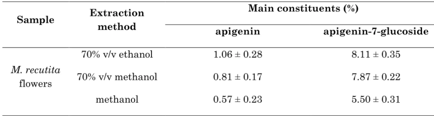
M. recutita flowers were used as a source of apigenin and apigenin-7-glucoside [26]. We used three different solvents to maximize the flavones extraction. The chemical characterization of each extract is reported in table 2.2. Consistently with the work by Haghi, we obtained higher amount of apigenin and apigenin-7-glucoside in the 70% v/v ethanol extract, with apigenin-7-glucoside being more abundant than apigenin [27]. Thus, we chose this extract to perform the digestion experiments.
Table 2.2. Chemical characterization of M. recutita extracts.
Sample Extraction method Main constituents (%) apigenin apigenin-7-glucoside M. recutita flowers 70% v/v ethanol 1.06 ± 0.28 8.11 ± 0.35 70% v/v methanol 0.81 ± 0.17 7.87 ± 0.22 methanol 0.57 ± 0.23 5.50 ± 0.31
Artichoke leave extracts are usually standardized for their content in caffeoylquinic acids, expressed as chlorogenic acid. Song reported that methanol has the highest extraction efficiency on polyphenols and chlorogenic acid, compared to ethanol or water [28]. Indeed, we confirmed that 80% v/v methanol is much more effective in extracting chlorogenic acid, compared to ddH2O (table 2.3), hence, we performed in vitro simulated digestion experiments on the 80% v/v methanol extract.
Table 2.3. Chemical characterization of C. scolimus extracts.
Sample Extraction method Chlorogenic acid (%)
C. scolimus leaves 80% v/v methanol 5.04 ± 0.21
ddH20 <0.1
Finally, a C. longa rhizome extract containing 48.07% total curcuminoids was obtained using methanol as a solvent. CUR was the most abundant constituent, representing 67.63% of total curcuminoids, followed by DMC and BMC, representing
20
22.95% and 9.42% of total curcuminoids, respectively (table 2.4). This phytochemical profile is coherent with that of commonly available C. longa extract.
Table 2.4. Chemical characterization of C. longa extract.
Sample Extraction method Main constituents (%) BMC CUR DMC C. longa rhizomes Methanol 4.53 ± 0.54 32.51 ± 0.32 11.03 ± 0.28
2.3.3. The mixture of constituents in herbal extracts protects single compounds from in vitro simulated gastrointestinal digestion
The effect of food matrix on the stability of food constituents has been widely studied [29]. A plethora of examples can be found in literature regarding the effects of food matrix on polyphenols digestion and bioaccessibility [17,20,30–32]. Herbal extracts contain a mixture of several constituents, which can influence the bioavailability of each other. One of the mechanisms by which this may occur is the modulation of the stability to gastrointestinal digestion. In the examples reported in this chapter, it is evident that the gastrointestinal stability of single constituents is different when they are used as isolated compounds, compared to their stability when they are enclosed in the complex mixture of the herbal extract. Indeed, figure 2.2 shows that both apigenin and apigenin-7-glucoside recovery after simulated digestion are significantly increased (+68% and +120%, respectively), compared to the reference standards alone.
CHAPTER 2
21
Figure 2.2. Stability of M. recutita flavones after in vitro simulated gastrointestinal digestion. * p<0.05 vs reference standard; *** p<0.001 vs reference standard,
two-way ANOVA followed by Tuckey’s post-hoc.
Noticeably, also in the case of M. recutita extract, glycosylation improved the stability of apigenin. Indeed, apigenin-7-glucoside was completely recovered, suggesting that other constituents of the extract may act by protecting this compound from degradation during digestion.
Similarly, chlorogenic acid stability upon simulated digestion was significantly affected by C. scolymus phytocomplex. Indeed, its recovery increased from 1.48%, using the reference standard alone, to 55.08%, when using the herbal extract (figure 2.3).
22
Figure 2.3. Stability of chlorogenic acid in C. scolymus extract after in vitro simulated gastrointestinal digestion. **p<0.01 vs reference standard, paired t test.
In 2015, D’Antuono and colleagues investigated the in vitro bioaccessibility, intestinal uptake and bioavailability of Cynara cardunculus (L.) subsp. scolymus Hayek polyphenols. Consistently with our results, they observed a much higher stability of chlorogenic acid in the herbal extract, compared to the reference standard, after simulated gastrointestinal digestion [33]. One possible explanation for this marked difference may be related to the ability of artichoke extracts to inhibit digestive enzymes [34].
Regarding curcuminoids, CUR stability to gastrointestinal digestion remained very low when using the herbal extract, even if a not statistically significant increase from 0.25% to 3.79% was observed. A much more evident effect was observed for DMC, whose stability increased from 0.90% when tested as single compound to 17.79% when in herbal matrix, and for BMC from 17.5% when tested as single compound to 104.78% in turmeric extract (figure 2.4).
CHAPTER 2
23
Figure 2.4. Stability of C. longa curcuminoids after in vitro simulated
gastrointestinal digestion. * p<0.05 vs reference standard; *** p<0.001 vs reference standard, two-way ANOVA followed by Tuckey’s post-hoc.
Despite being present in lower concentration in the extract, BMC can potentially reach the intestine at a concentration three-fold higher than that of CUR and DMC, which partially explains the higher bioavailability of BMC observed in humans, compared to CUR [24].
2.4. Conclusions
Oral bioavailability is one of the main factors that limit the clinical use of natural products. To reach the site of absorption, an orally-administered drug has to overcome the digestion process without being extensively metabolized. In this work we evaluated the effect of in vitro simulated gastrointestinal digestion on different classes of polyphenols. Moreover, we investigated the changes in the gastrointestinal stability of these compounds when used as a complex mixture of molecules, such as in the case of herbal extracts. Using M. recutita, C. scolymus, and C. longa as simple examples, we found that the phytocomplex significantly influenced the recovery of single constituents. In particular, we found a protective role of the phytocomplex, resulting in higher stability of the compounds to the digestive process. There might also be cases in which the phytocomplex may reduce, or leave unaltered, the stability of single constituents. These results provide evidence that it is extremely important to take into
24
account the effect of gastrointestinal digestion when evaluating the biological effectiveness of an herbal extract, as this cannot be extrapolated from data obtained using single compounds.
2.5. References
1. Manach, C.; Williamson, G.; Morand, C.; Scalbert, A.; Rémésy, C. Bioavailability and bioefficacy of polyphenols in humans. I. Review of 97 bioavailability studies. Am. J. Clin. Nutr. 2005, 81, 230S-242S.
2. Wojtunik-Kulesza, K.; Oniszczuk, A.; Oniszczuk, T.; Combrzyński, M.; Nowakowska, D.; Matwijczuk, A. Influence of in vitro digestion on composition, bioaccessibility and antioxidant activity of food polyphenols - a non-systematic review. Nutrients 2020, 12, 1401.
3. Alminger, M.; Aura, A. M.; Bohn, T.; Dufour, C.; El, S. N.; Gomes, A.; Karakaya, S.; Martínez-Cuesta, M. C.; Mcdougall, G. J.; Requena, T.; Santos, C. N. In vitro models for studying secondary plant metabolite digestion and bioaccessibility. Compr. Rev. Food Sci. Food Saf. 2014, 13, 413–436.
4. Stewart, R. J. C.; Morton, H.; Coad, J.; Pedley, K. C. In vitro digestion for assessing micronutrient bioavailability: the importance of digestion duration. Int. J. Food Sci. Nutr. 2019, 70, 71–77.
5. Hubatsch, I.; Ragnarsson, E. G. E.; Artursson, P. Determination of drug permeability and prediction of drug absorption in Caco-2 monolayers. Nat. Protoc. 2007, 2, 2111–2119.
6. Kong, F.; Singh, R. P. A human gastric simulator (HGS) to study food digestion in human stomach. J. Food Sci. 2010, 75, E627-35.
7. Minekus, M.; Marteau, P.; Havenaar, R.; Veld, J. H. J. H. in’t A multicompartmental dynamic computer-controlled model simulating the stomach and small intestine. Altern. to Lab. Anim. 1995, 23, 197–209.
CHAPTER 2
25
8. Manach, C.; Scalbert, A.; Morand, C.; Rémésy, C.; Jiménez, L. Polyphenols: food sources and bioavailability. Am. J. Clin. Nutr. 2004, 79, 727–747.
9. Governa, P.; Marchi, M.; Cocetta, V.; Leo, B. De; Saunders, P. T. K.; Catanzaro, D.; Miraldi, E.; Montopoli, M.; Biagi, M. Effects of Boswellia serrata Roxb. and Curcuma longa L. in an in vitro intestinal inflammation model using immune cells and Caco-2. Pharmaceuticals 2018, 11, 126.
10. Vallejo, F.; Gil-Izquierdo, A.; Pérez-Vicente, A.; García-Viguera, C. In Vitro Gastrointestinal Digestion Study of broccoli inflorescence phenolic compounds, glucosinolates, and vitamin C. J. Agric. Food Chem. 2004, 52, 135–138.
11. Boyer, J.; Brown, D.; Liu, R. H. In vitro digestion and lactase treatment influence uptake of quercetin and quercetin glucoside by the Caco-2 cell monolayer. Nutr. J. 2005, 4, 1.
12. Ross, J. A.; Kasum, C. M. Dietary flavonoids: bioavailability, metabolic effects, and safety. Annu. Rev. Nutr. 2002, 22, 19–34.
13. Hollman, P. C.; Katan, M. B. Dietary flavonoids: intake, health effects and bioavailability. Food Chem. Toxicol. 1999, 37, 937–942.
14. Hollman, P. C.; Katan, M. B. Absorption, metabolism and health effects of dietary flavonoids in man. Biomed. Pharmacother. 1997, 51, 305–310.
15. Clifford, M. N. Chlorogenic acids and other cinnamates - nature, occurrence and dietary burden. J. Sci. Food Agric. 1999, 79, 362—372.
16. Liu, G.; Ying, D.; Guo, B.; Cheng, L. J.; May, B.; Bird, T.; Sanguansri, L.; Cao, Y.; Augustin, M. Extrusion of apple pomace increases antioxidant activity upon in vitro digestion. Food Funct. 2019, 10, 951–963.
17. Kamiloglu, S.; Ozkan, G.; Isik, H.; Horoz, O.; Van Camp, J.; Capanoglu, E. Black carrot pomace as a source of polyphenols for enhancing the nutritional value of cake: an in vitro digestion study with a standardized static model. LWT 2017, 77, 475–481. 18. McDougall, G. J.; Dobson, P.; Smith, P.; Blake, A.; Stewart, D. Assessing potential bioavailability of raspberry anthocyanins using an in vitro digestion system. J. Agric.
26 Food Chem. 2005, 53, 5896–5904.
19. Pérez-Vicente, A.; Gil-Izquierdo, A.; García-Viguera, C. In vitro gastrointestinal digestion study of pomegranate juice phenolic compounds, anthocyanins, and vitamin C. J. Agric. Food Chem. 2002, 50, 2308–2312.
20. Gil-Izquierdo, A.; Zafrilla, P.; Tomás-Barberán, F. A. An in vitro method to simulate phenolic compound release from the food matrix in the gastrointestinal tract. Eur. Food Res. Technol. 2002, 214, 155–159.
21. McDougall, G. J.; Fyffe, S.; Dobson, P.; Stewart, D. Anthocyanins from red wine - their stability under simulated gastrointestinal digestion. Phytochemistry 2005, 66, 2540–2548.
22. Ubeyitogullari, A.; Ciftci, O. N. A novel and green nanoparticle formation approach to forming low-crystallinity curcumin nanoparticles to improve curcumin’s bioaccessibility. Sci. Rep. 2019, 9, 19112.
23. Park, S. J.; Garcia, C. V; Shin, G. H.; Kim, J. T. Improvement of curcuminoid bioaccessibility from turmeric by a nanostructured lipid carrier system. Food Chem. 2018, 251, 51–57.
24. Cuomo, J.; Appendino, G.; Dern, A. S.; Schneider, E.; McKinnon, T. P.; Brown, M. J.; Togni, S.; Dixon, B. M. Comparative absorption of a standardized curcuminoid mixture and its lecithin formulation. J. Nat. Prod. 2011, 74, 664–669.
25. Hu, X.-P.; Yin, F.-W.; Zhou, D.-Y.; Xie, H.-K.; Zhu, B.-W.; Ma, X.-C.; Tian, X.-G.; Wang, C.; Shahidi, F. Stability of resveratrol esters with caprylic acid during simulated in vitro gastrointestinal digestion. Food Chem. 2019, 276, 675–679.
26. European Medicine Agency. Assessment report on Matricaria recutita L. flos and
Matricaria recutita L. aetheroleum
http://www.ema.europa.eu/docs/en_GB/document_library/Herbal_-_HMPC_assessment_report/2014/07/WC500170079.pdf.
27. Haghi, G.; Hatami, A.; Safaei, A.; Mehran, M. Analysis of phenolic compounds in Matricaria chamomilla and its extracts by UPLC-UV. Res. Pharm. Sci. 2014, 9, 31–37.
CHAPTER 2
27
28. Shuhui Song; Hongju He; Xiaowei Tang; Wenqi Wang Determination of polyphenols and chlorogenic acid in artichoke (Cynara scolymus L.). In Acta horticulturae; International Society for Horticultural Science (ISHS), Leuven, Belgium, 2010; pp. 167–172.
29. Wojtunik-Kulesza, K.; Oniszczuk, A.; Oniszczuk, T.; Combrzyński, M.; Nowakowska, D.; Matwijczuk, A. Influence of in vitro digestion on composition, bioaccessibility and antioxidant activity of food polyphenols - a non-systematic review. Nutrients 2020, 12, 1401.
30. Tarko, T.; Duda-Chodak, A. Influence of food matrix on the bioaccessibility of fruit polyphenolic compounds. J. Agric. Food Chem. 2020, 68, 1315–1325.
31. Pineda-Vadillo, C.; Nau, F.; Dubiard, C. G.; Cheynier, V.; Meudec, E.; Sanz-Buenhombre, M.; Guadarrama, A.; Tóth, T.; Csavajda, É.; Hingyi, H.; Karakaya, S.; Sibakov, J.; Capozzi, F.; Bordoni, A.; Dupont, D. In vitro digestion of dairy and egg products enriched with grape extracts: effect of the food matrix on polyphenol bioaccessibility and antioxidant activity. Food Res. Int. 2016, 88, 284–292.
32. Mandalari, G.; Vardakou, M.; Faulks, R.; Bisignano, C.; Martorana, M.; Smeriglio, A.; Trombetta, D. Food matrix effects of polyphenol bioaccessibility from almond skin during simulated human digestion. Nutrients 2016, 8, 568.
33. D’Antuono, I.; Garbetta, A.; Linsalata, V.; Minervini, F.; Cardinali, A. Polyphenols from artichoke heads (Cynara cardunculus (L.) subsp. scolymus Hayek): in vitro bio-accessibility, intestinal uptake and bioavailability. Food Funct. 2015, 6, 1268–1277. 34. Mahboubi, M. Cynara scolymus (artichoke) and its efficacy in management of obesity. Bull. Fac. Pharmacy, Cairo Univ. 2018, 56, 115–120.
28 CHAPTER 3
Stability and bioaccessibility of Cannabis sativa L. extracts under in vitro simulated gastrointestinal digestion
3.1. Introduction
Cannabis sativa L. (Cannabaceae) has been used since ancient times as a medicinal tool and as a source of textile fiber and, more recently, as a psychoactive drug for recreational uses [1,2]. Thanks to the discovery of the endocannabinoid system as a regulator of several physiologic functions, C. sativa is attracting the interest of the pharmaceutical industry, because of the intriguing therapeutic potential of its constituents [3].
Indeed, a wide variety of molecules are produced by C. sativa, the more characteristic being represented by a class of terpenophenolic compounds, known as cannabinoids [4]. Among the over 400 compounds found in C. sativa [5], more than 90 different cannabinoids, including their breakdown products, have been reported [6]. Two other main classes of constituents, terpenes and flavonoids, are present in the C. sativa phytocomplex, even if their pharmacological role is still under evaluation [5].
Cannabinoids in C. sativa are typically present as acidic precursors [5]. However, their decarboxylated derivatives are responsible for the most therapeutic effects, thus, several decarboxylation methods have been proposed [7].
Δ9-tetrahydrocannabinol (THC) is the major psychotropic constituent of C. sativa. It is a partial agonist at cannabinoid receptor 1 (CB1) and 2 (CB2) [8]. The higher affinity for CB1 is responsible for the insurgence of psychotropic effects [4]. THC, and its synthetic version dronabinol, have been investigated for their analgesic, antispasmodic and antiemetic activity [9].
Cannabidiol (CBD), on the other hand, is the major non-psychotropic cannabinoid in C. sativa. It is a weak inverse agonist at CB2 and it has been approved
CHAPTER 3
29
by the FDA for the treatment of intractable childhood-onset seizures [10]. Other than its anti-epileptic effect, CBD has been studied for its potential therapeutic role in mood disorders [11], multiple sclerosis [12], and in a number of diseases associated with oxidative stress, and inflammation [3,13].
THC levels in cannabis dramatically increased from 1970 to 2017, increasing the risk of addiction and mental health disorders [14]. However, CBD seems to be able to moderate the toxic effects of THC [15,16].
Apart from the pharmacological properties of isolated cannabinoids, in the last decade, many efforts have been made to investigate the clinical effectiveness of the whole C. sativa extract [12,17–19]. Despite the growing scientific interest, however, only one standardized extract with a fixed THC:CBD ratio (1:1) has been registered as a medicinal product so far [20]. In Italy, eight variety of medical cannabis with different content of THC and CBD and cultivated indoor in The Netherlands, Canada and Italy, are authorized. These can be dispensed as herbal substance, used by patients for self-medication by vaporization, as an herbal tea or as galenic herbal preparations, such as oil or other standardized extracts[21]. However, information on the pharmacodynamics and pharmacokinetics of these preparations are lacking.
Moreover, the controversial, unregulated growing market of the so-called “cannabis light” products, i.e. C. sativa herbal material or derived preparations with THC content lower than 0.2%, complicated the situation by increasing the availability of products which have not been clinically evaluated for their pharmaco-toxicological profile.
Thus, there is the need for the development of efficient analysis methods to characterize the chemical and biological properties of different C. sativa products. In particular, information about the stability of these products after oral ingestion would be of great use for guiding the development of novel cannabis-based drugs.
Recently, we analyzed the chemical composition, the microbial contamination, and the levels of heavy metals of 12 non-psychotropic C. sativa products, finding that most of the cannabis light products do not comply with the Italian food legislation, or have different amount of THC and CBD than the quantity declared on the label [22].
30
In this study, we aimed at evaluating the stability to digestion of different non-psychotropic C. sativa products. We used three different extraction methods, namely decoction, oleolite and hydroalcoholic extraction, to extract two different C. sativa varieties with THC level lower than 0.2%, before and after heat-induced decarboxylation of cannabinoids. CBD, THC, and their relative acidic forms (CBDA and THCA) were quantified by HPLC-DAD and their stability and bioaccessibility under in vitro simulated gastrointestinal digestion were evaluated.
3.2. Materials and methods
3.2.1. Chemicals
All solvents used in this work were purchased from Sigma-Aldrich. NaCl, pepsin from porcine gastric mucosa, pancreatin from porcine pancreas, and bile salts mixture were from Sigma Aldrich. Na2CO3 was purchased from Sodalco S.p.A. CBD was purchased from Linnea SA (Riazzino, TI, Switzerland).
3.2.2. Plant material and extraction
The dried inflorescences of a commercially available product from indoor cultivation named “Mary-light” and of a field-grown C. sativa var. carmagnole, with different CBD content, were extracted for 4 h, using ddH2O, 96% v/v ethanol or extravirgin olive oil (EVO), with (d) or without heat decarboxylation of cannabinoids. We obtained four different extracts, namely non-decarboxylated Mary-light (M), decarboxylated Mary-light (dM), non-decarboxylated C. sativa var. carmagnole (C) and decarboxylated C. sativa var. carmagnole (dC) extracts. The drug-extract ratio (DER) of each extract was 1:10. Heat decarboxylation of cannabinoids was performed following the method of Romano and Hazekamp [23], by heating plant material in an oven at 145 °C for 30 min.
CHAPTER 3
31
3.2.3. In vitro simulated gastrointestinal digestion
In vitro simulated digestion was carried out as previously described [24], with slight modifications. Briefly, extracts (1 mL) and CBD were suspended in 20 mL of simulated gastric juice that contained pepsin from porcine gastric mucosa (300 UI/mL) and NaCl (10 mg/mL), obtaining a final 1:20 dilution for the extracts and a concentration of 1 mg/mL for CBD. The pH of the solution was adjusted to 1.7 using HCl. Samples were incubated for 2 h at 37 °C with shaking. One mL gastric solution was sampled and stored at -80 °C for further analysis. Then, pancreatin from porcine pancreas (10 mg/mL) and bile salts mixture (20 mg/mL) were added to the solutions to simulate the intestinal environment. To evaluate the bioaccessibility of the samples, semi-permeable cellulose dialysis tubes (Sigma Aldrich), with a molecular cut-off of 12 kDa, containing 19 mL of NaHCO3 (15 mg/mL) in ddH2O, were inserted into the solutions. Intestinal digestion was carried out for 2 h at 37 °C with shaking. The fraction of the samples able to permeate into the dialysis tubes was considered “bioaccessibile” or “serum available”, while the fraction which remained out of the dialysis tubes was considered “not bioaccessible” or “colon available”. Samples were then centrifuged, filtered, and immediately used for further analysis.
3.2.4. Chromatographic conditions
Samples (20 µL) were injected into a HPLC-DAD system, consisting of a Shimadzu Prominence LC 2030 3D instrument, equipped with a Bondpak® C18 column (10 µm, 125 Å, 3.9 mm, Waters Corporation). The mobile phase consisted of ddH2O + 0.1% v/v formic acid (A) and acetonitrile + 0.1% v/v formic acid (B). The following method was applied: A 35% at 0 min for 3 min, then from 35% to 10% at 10 min, A 10% for 2 min and from 10% to 35% at 14 min, then A 35% for 1 min. Flow rate was set to 1.2 mL/min and column temperature to 28 °C. Chromatograms were recorded at 225 nm. The calibration curve of CBD was set up using concentrations ranging from 0.008 to 0.500 mg/mL, with R2 > 0.99. The quantification of CBDA, THC and THCA was performed using CBD as a reference standard, by conversion according to the Dutch office for Medicinal Cannabis analytic monograph for Cannabis Flos Version 7.1 (November 28, 2014).
32
3.2.5. Statistical analysis
The statistical differences between the results were determined by ANOVA or by using the Student’s t-test. Values are expressed in the range of +/- standard deviation and p<0.05 was considered statistically significant. Graphs and calculations were performed using GraphPad Prism.
3.3. Results and discussion
3.3.1. Quantification of cannabinoids
The HPLC-DAD chromatograms of the extracts (figure 3.1) show that, despite being suggested by several therapeutic systems, water extraction by decoction is not a suitable method for the extraction of cannabinoids, while ethanol and EVO gave higher yields, with ethanol being significantly better. In nature, cannabinoids occur mostly as acidic precursors, while their decarboxylated derivatives are usually produced by manufacturing processes or smoking [25].
CHAPTER 3
33
Figure 3.1. HPLC-DAD chromatograms recorded at 225 nm of the (A) ethanol, (B) olive oil and (C) water extracts. M (red), dM (green), C (blue) and dC (black). CBDA
34
Consistently, CBD and THC were not detected neither in M ethanol and EVO extracts, nor in C EVO extract. Small amounts of CBD were found in C ethanol extract. On the contrary, the acidic form of cannabinoids was much more present in the non-decarboxylated extracts, with CBDA being 63% higher in M, compared to C. THC content was always below the Italian legal limit of 0.2 %. The quantification of the cannabinoids in the extracts is reported in table 3.1.
Table 1. Cannabinoids content of the extracts (% w/v). Data are expressed as mean ± standard deviation. n.d. = not determined.
Solvent Sample CBDA CBD THCA THC
E tO H M dM 0.60 ± 0.03 0.06 ± 0.07 0.53 ± 0.09 n.d. 0.05 ± 0.04 n.d. 0.05 ± 0.02 n.d. C 0.38 ± 0.06 0.08 ± 0.01 0.01 ± 0.01 n.d. dC n.d. 0.36 ± 0.02 n.d. 0.04 ± 0.03 EVO M 0.42 ± 0.04 n.d. 0.01 ± 0.01 n.d. dM 0.07 ± 0.02 0.34 ± 0.02 n.d. n.d. C 0.26 ± 0.03 n.d. n.d. n.d. dC 0.06 ± 0.02 0.25 ± 0.07 n.d. n.d. ddH 2 O M dM n.d. n.d. n.d. n.d. n.d. n.d. n.d. n.d. C n.d. n.d. n.d. n.d. dC n.d. n.d. n.d. n.d.
3.3.2. In vitro simulated gastrointestinal digestion and bioaccessibility
The stability of isolated CBD after gastrointestinal digestion resulted to be approximately 75%, and a large part (60%) of the initial amount of CBD was able to permeate across the dialysis tube, reaching the serum available fraction, thus showing a good bioaccessibility (figure 3.2).
CHAPTER 3
35
Figure 3.2. CBD bioaccessibility after in vitro simulated gastrointestinal digestion. * p<0.001 vs colon available, unpaired t test.
Differences in cannabinoids recovery and bioaccessibility were found when comparing the different C. sativa preparations (figure 3.3).
Indeed, the total recovery of CBDA was approximately 30% and 60% in M and C ethanol extracts, respectively, with the colon available to serum available ratio (CSR) being almost identical. Interestingly, the stability of CBDA to in vitro simulated digestion resulted to be significantly higher when using the EVO extracts. The total recovery of CBDA was almost 100% for M and C, respectively. 87% of CBDA was found in the colon available fraction for both M and C, while a small, yet significant, difference was found in the serum available, with a CSR of 6.4 and 8.9 for M and C, respectively. Small amount of CBDA were also present in the decarboxylated EVO extracts, with its stability to simulated digestion being lower (approximately 42% and 37% in dM and dC, respectively) and only colon available, compared to the non-decarboxylated one.
CBD recovery was comparable in each ethanol extract, with an average total recovery of 42%. A similar value was obtained for dC EVO extract, while dM EVO extract showed a protective effect on CBD stability, with the colon available recovery being approximately 47% and 90%, respectively. Similarly to CBDA, the CBD colon available fraction was higher than the serum available fraction, with an average CSR of 10 in the ethanol extracts, while no CBD was detected in the serum available
36
fractions of EVO extracts. While the absence of CBD in the serum available fraction of the EVO extracts may be due to a limitation of the experimental procedure, as the formation of micelles may interfere with the permeation through the dialysis tube, a lower bioaccessibility of CBD when using C. sativa extracts, compared to the reference standard alone, can be speculated.
Figure 3.3. (A) Colon and (B) serum available fractions of different C. sativa extracts after in vitro simulated gastrointestinal digestion. *** p<0.001 EtOH vs EVO; ###
p<0.001 field-grown vs indoor cultivation; °°° p<0.001 non-decarboxylated vs decarboxylated
The THCA and THC content of the analyzed extracts was very low, thus, the measurement of their stability and bioaccessibility after in vitro simulated gastrointestinal digestion was not the aim of this work. However, it is interesting to note that, the presence of THCA and THC was not detected in the serum available