Presta
Paola Chiodelli, Stefania Mitola, Cosetta Ravelli, Pasqua Oreste, Marco Rusnati and Marco
Endothelial Growth Factor Receptor-2 Agonist Gremlin
Print ISSN: 1079-5642. Online ISSN: 1524-4636
Copyright © 2011 American Heart Association, Inc. All rights reserved.
Greenville Avenue, Dallas, TX 75231
is published by the American Heart Association, 7272
Arteriosclerosis, Thrombosis, and Vascular Biology
doi: 10.1161/ATVBAHA.111.235184
2011;
2011;31:e116-e127; originally published online September 15,
Arterioscler Thromb Vasc Biol.
http://atvb.ahajournals.org/content/31/12/e116
World Wide Web at:
The online version of this article, along with updated information and services, is located on the
http://atvb.ahajournals.org//subscriptions/
at:
is online
Arteriosclerosis, Thrombosis, and Vascular Biology
Information about subscribing to
Subscriptions:
http://www.lww.com/reprints
Information about reprints can be found online at:
Reprints:
document.
Question and Answer
Permissions and Rights
page under Services. Further information about this process is available in the
which permission is being requested is located, click Request Permissions in the middle column of the Web
Copyright Clearance Center, not the Editorial Office. Once the online version of the published article for
can be obtained via RightsLink, a service of the
Arteriosclerosis, Thrombosis, and Vascular Biology
in
Requests for permissions to reproduce figures, tables, or portions of articles originally published
Permissions:
by guest on October 6, 2013 http://atvb.ahajournals.org/
Downloaded from http://atvb.ahajournals.org/ by guest on October 6, 2013 Downloaded from http://atvb.ahajournals.org/ by guest on October 6, 2013 Downloaded from http://atvb.ahajournals.org/ by guest on October 6, 2013 Downloaded from http://atvb.ahajournals.org/ by guest on October 6, 2013 Downloaded from http://atvb.ahajournals.org/ by guest on October 6, 2013 Downloaded from http://atvb.ahajournals.org/ by guest on October 6, 2013 Downloaded from http://atvb.ahajournals.org/ by guest on October 6, 2013 Downloaded from http://atvb.ahajournals.org/ by guest on October 6, 2013 Downloaded from http://atvb.ahajournals.org/ by guest on October 6, 2013 Downloaded from http://atvb.ahajournals.org/ by guest on October 6, 2013 Downloaded from http://atvb.ahajournals.org/ by guest on October 6, 2013 Downloaded from http://atvb.ahajournals.org/ by guest on October 6, 2013 Downloaded from
Activity of the Vascular Endothelial Growth Factor
Receptor-2 Agonist Gremlin
Paola Chiodelli, Stefania Mitola, Cosetta Ravelli, Pasqua Oreste, Marco Rusnati, Marco Presta
Objective—Heparan sulfate proteoglycans (HSPGs) modulate the interaction of proangiogenic heparin-binding vascular
endothelial growth factors (VEGFs) with signaling VEGF receptor-2 (VEGFR2) and neuropilin coreceptors in
endothelial cells (ECs). The bone morphogenic protein antagonist gremlin is a proangiogenic ligand of VEGFR2,
distinct from canonical VEGFs. Here we investigated the role of HSPGs in VEGFR2 interaction, signaling, and
proangiogenic capacity of gremlin in ECs.
Methods and Results—Surface plasmon resonance demonstrated that gremlin binds heparin and heparan sulfate, but not
other glycosaminoglycans, via N-, 2-O, and 6-O-sulfated groups of the polysaccharide. Accordingly, gremlin binds
HSPGs of the EC surface and extracellular matrix. Gremlin/HSPG interaction is prevented by free heparin and heparan
sulfate digestion or undersulfation following EC treatment with heparinase II or sodium chlorate. However, at variance
with canonical heparin-binding VEGFs, gremlin does not interact with neuropilin-1 coreceptor. On the other hand,
HSPGs mediate VEGFR2 engagement and autophosphorylation, extracellular signaling-regulated kinase
1/2and p38
mitogen-activated protein kinase activation, and consequent proangiogenic responses of ECs to gremlin. On this basis,
we evaluated the gremlin-antagonist activity of a panel of chemically sulfated derivatives of the Escherichia coli K5
polysaccharide. The results demonstrate that the highly N,O-sulfated derivative K5-N,OS(H) binds gremlin with high
potency, thus inhibiting VEGFR2 interaction and angiogenic activity in vitro and in vivo.
Conclusion—HSPGs act as functional gremlin coreceptors in ECs, affecting its productive interaction with VEGFR2 and
angiogenic activity. This has allowed the identification of the biotechnological K5-N,OS(H) as a novel angiostatic
gremlin antagonist. (Arterioscler Thromb Vasc Biol. 2011;31:e116-e127.)
Key Words: angiogenesis
䡲 endothelium 䡲 gremlin 䡲 heparan sufate proteoglycans
A
ngiogenesis plays a key role in various physiological
and pathological conditions, including embryonic
devel-opment, wound repair, inflammation, and tumor growth.
1Nu-merous inducers of angiogenesis have been identified, including
the members of the vascular endothelial growth factor (VEGF)
family.
2Different isoforms of mammalian VEGFs interact with
tyrosine kinase VEGF receptors (VEGFRs) expressed on the
surface of endothelial cells (ECs)
3and with heparan sulfate (HS)
proteoglycan (HSPG) and neuropilin (NRP) coreceptors,
4,5thus
activating a proangiogenic response.
HSPGs consist of a core protein and of glycosaminoglycan
(GAG) chains represented by unbranched heparin-like
poly-saccharides. They are found in free forms, in the extracellular
matrix (ECM), or associated with the plasma membrane
where they regulate the function of a wide range of ligands.
6,7In particular, endothelial HSPGs modulate angiogenesis by
affecting bioavailability and interaction of heparin-binding
VEGFs with signaling VEGFRs and NRP coreceptors.
8,9Heparin/HS interaction with angiogenic growth factors
de-pends on the degree/distribution of sulfate groups and length
of the GAG chain, distinct oligosaccharide sequences
medi-ating its binding activity.
10 –12Moreover, the study of the
biochemical bases of this interaction has been exploited to
design angiostatic heparin-like compounds, chemically
mod-ified heparins, and biotechnological heparins.
13–16Gremlin belongs to the CAN (Cerberus and Dan) family of
cystine-knot–secreted proteins.
17,18It binds various bone
morphogenic proteins (BMPs), thus preventing their
interac-tion with cell surface receptors.
19The capacity of gremlin to
bind BMPs and to inhibit their interaction with the cognate
transforming growth factor-
 (TGF-) family receptors is
thought to play a role in embryonic development of various
organ systems, such as bone, kidney, and lung.
20 –22Also,
gremlin has been implicated in the pathogenesis of human
diseases, including pulmonary hypertension and idiopathic
pulmonary fibrosis
23and diabetic nephropathy.
24Moreover,
Received on: April 15, 2011; final version accepted on: August 30, 2011.
From the Unit of General Pathology and Immunology, Department of Biomedical Sciences and Biotechnology, School of Medicine, University of Brescia, Brescia, Italy (P.C., S.M., C.R., M.R., M.P.); Glycores 2000, Milan, Italy (P.O.).
Correspondence to Marco Presta, General Pathology, Department of Biomedical Sciences and Biotechnology, Viale Europa 11, 25123 Brescia, Italy. E-mail [email protected]
© 2011 American Heart Association, Inc.
Arterioscler Thromb Vasc Biol is available at http://atvb.ahajournals.org DOI: 10.1161/ATVBAHA.111.235184
gremlin is produced by human tumors
25,26and is expressed
by proangiogenic ECs in vitro and tumor endothelium in
vivo.
27Gremlin stimulates EC intracellular signaling and
migration in vitro, leading to a potent angiogenic response
in vivo.
27,28This is due to its capacity to bind and activate
VEGFR2, the main transducer of VEGF-mediated
angio-genic signals, in a BMP-independent manner.
29Thus,
gremlin acts as a VEGFR2 agonist distinct from VEGF
family members that may play paracrine/autocrine
func-tions in tumor neovascularization.
Scattered experimental evidence indicates that gremlin
may act as a heparin-binding angiogenic factor: gremlin binds
to heparin-Sepharose beads
27; it associates to the surface of
producing cells
30; and a heparin-binding domain has been
identified in sclerostin, a BMP antagonist structurally related
with gremlin.
31,32Here, we investigated the capacity of
gremlin to interact with heparin and cell-associated HSPGs
and the role of endothelial HSPGs in mediating
gremlin-dependent VEGFR2 activation and angiogenic activity. The
results demonstrate that gremlin binds heparin/HS. However,
at variance with canonical heparin-binding VEGFR2 ligands,
gremlin interacts with HSPGs but not with NRP-1 in ECs.
Nevertheless, cell surface HSPGs play a nonredundant role in
mediating the ability of gremlin to bind and activate VEGFR2
in endothelium, thus triggering an NRP-1-independent
proan-giogenic response. Accordingly, we have identified a
chem-ically modified, heparin-like derivative of the capsular
Esch-erichia coli K5 polysaccharide
16as a potent angiostatic
gremlin antagonist.
Methods
Chemicals
Recombinant gremlin, anti-gremlin antibody, and human recombi-nant VEGF-A165 were from R&D Systems (Minneapolis, MN); anti-phospho (P)-VEGFR2 antibody was from Cell Signaling Tech-nology (Boston, MA); anti-tubulin antibody, heparinase II, phorbol myristate acetate (PMA), and protease inhibitors were from Sigma-Aldrich (St Louis, MO); anti-P-extracellular signaling–regulated kinase (anti-P-ERK1/2), anti-VEGFR2, anti-ERK2, anti-p38, and anti-P-p38 mitogen-activated protein kinase antibodies were from Santa Cruz Biotechnology (Santa Cruz, CA); anti-actin antibody was from Chemicon, Millipore (Billerica, MA); anti-focal adhesion kinase (clone 2A7) was from Upstate, Millipore; sodium chlorate was from BDH Laboratory Supplies (Pole, United Kingdom); type I HS was from Opocrin (Corlo, Italy); and unmodified, fractionated very-low–molecular weight (MW) heparins, chemically modified heparins, and K5 polysaccharide derivatives were from Glycores 2000 (Milan, Italy). K5 derivatives were obtained by N-deacetyla-tion/N-sulfation, O-sulfation, or both of a single batch of E. coli K5 polysaccharide (see Leali et al33 for a description of these
com-pounds). Dermatan sulfate, chondroitin-4-sulfate, chondroitin-6-sulfate, and hyaluronic acid were obtained from Dr M. Del Rosso, University of Florence, Italy (see Rusnati et al34for a description of
these GAGs). Tinzaparin sodium was from LEO Pharmaceutical Products, Ltd (Ballerup, Denmark). The HS-specific monoclonal antibody 10E435was kindly provided by G. David (Leuven
Univer-sity, Leuven, Belgium).
Surface Plasmon Resonance Analysis
Surface plasmon resonance (SPR) measurements were performed on a BIAcore X instrument (GE Healthcare). For the study of gremlin/ heparin interaction, heparin was immobilized onto a CM3 sensor chip (GE Healthcare) as described.36 A streptavidin-coated sensor
chip was used as reference and for blank subtraction. Increasing
concentrations of gremlin in 10 mmol/L HEPES buffer (HBS-EP), pH 7.4, containing 150 mmol/L NaCl, 3 mmol/L EDTA, and 0.005% surfactant P20 (HBS-EP) (GE Healthcare) were injected over the heparin-coated or streptavidin surfaces for 4 minutes and then washed until dissociation was observed. After each run, the sensor chip was regenerated by injection of HBS-EP containing 2.0 mol/L NaCl.
In all experiments, association and dissociation rates (konand koff,
respectively) were obtained by fitting the raw sensorgrams with the 1:1 Langmuir binding model using the Biaevaluation software (BIAcore). Equilibrium affinity constants (expressed as dissociation constant, kd) were either derived from the kinetic parameters
(kd⫽koff/kon) or determined from Scatchard plot analysis of the
equilibrium binding data.
Cell Cultures
Human umbilical vein endothelial (HUVE) cells were used at early passages (passages I–IV) and grown on plastic surface coated with porcine gelatin (Sigma-Aldrich) in M199 medium (Invitrogen, Carls-bad, CA) supplemented with 20% fetal calf serum (FCS) (Invitro-gen), EC growth factor (100g/mL) (Sigma-Aldrich), and porcine heparin (100g/mL, Sigma-Aldrich). Fetal bovine aortic endothelial GM 7373 cells37were grown in Dulbecco’s modified Eagle medium
(Invitrogen) containing 10% FCS, vitamins, and essential and nonessential amino acids. These cells were transfected according to standard protocols with a pcDNA3.1 expression vector harboring the mouse VEGFR2 cDNA (provided by G. Breier, Max Planck Insti-tute, Bad Nauhein, Germany) to generate stable GM7373-VEGFR2 transfectants. Wild-type Chinese hamster ovary cells (CHO)-K1 cells and GAG-deficient A745 CHO cells38were kindly provided by
J.D. Esko (University of California, La Jolla, CA) and grown in Ham’s F-12 medium supplemented with 10% FCS.
To inhibit the sulfation of cell-associated HS chains, GM7373 and HUVE cells were grown for 48 hours in presence of sodium chlorate (50 and 30 mmol/L, respectively).39Alternatively, cells
were incubated for 2 hours at 37°C with heparinase II (15 mU/mL in phosphate-buffered saline [PBS], Sigma-Aldrich) before experimentation.
Human pancreatic carcinoma (PANC-1) cells were grown in Dulbecco’s modified Eagle medium containing 10% FCS, vitamins, and essential and nonessential amino acids. Gremlin-overexpressing PANC-1 cells (PANC-1-gremlin cells) were generated by infection with the pRRLsin.cPPT.PGK lentiviral vector (kindly provided by L. Naldini, San Raffaele Institute, Milan, Italy) harboring the rat gremlin cDNA.40Green fluorescent protein (GFP)– overexpressing
PANC-1 cells (PANC-1-GFP cells) were generated with the same procedures and used as a negative control. PANC-1-GFP and PANC-1-gremlin cells were grown for 48 hours in serum-free Dulbecco’s modified Eagle medium and their conditioned media were analyzed for gremlin content by Western blotting and for the capacity to induce VEGFR2 phosphorylation in HUVE cells.
Gremlin/Cell-Associated HSPG Binding Assays
CHO-K1 cells; GAG-deficient A745 CHO-K1 cells; and untreated, chlorate-treated, or heparinase II–treated GM7373 cells were incu-bated with gremlin (100 ng/mL) for 2 hours at 4°C in serum-free medium in the absence or in the presence of the indicated competitor. Then, cells were washed twice with PBS or with PBS plus 1.5 mol/L NaCl to remove gremlin bound to HSPGs.41Next,immunofluores-cence analysis was performed using an anti-gremlin antibody fol-lowed by Alexa Fluor 488 anti-goat IgG (Invitrogen), and cells were photographed using a Zeiss Axiovert 200M epifluorescence micro-scope equipped with a Plan-Apochromat⫻63/1.4 NA oil objective. Alternatively, HSPG-bound gremlin was collected in SDS-PAGE sample buffer and analyzed with anti-gremlin antibody by Western blotting.
In a parallel set of experiments, cell-free subendothelial ECM preparations were obtained by incubating GM7373 cells for 3 minutes at room temperature with 0.5% Triton X plus 0.00375% NH3 in PBS.42 Next, cell-free ECM preparations were incubated
with gremlin and analyzed for HSPG-bound gremlin as described above.
VEGFR2 Cross-Linking Assay
Confluent GM7373-VEGFR2 cells were incubated for 2 hours at 4°C with gremlin dissolved in PBS containing the bis(sulfosucci-namide) suberate cross-linking reagent (BS3, Pierce). Then, cells were lysed in 50 mmol/L Tris-HCl buffer (pH 7.4) containing 150 mmol/L NaCl, 1% Triton X-100, 1.0 mmol/L Na3VO4, and protease inhibitors. Lysates were immunoprecipitated and separated by SDS-PAGE followed by Western blotting with anti-gremlin or anti-VEGFR2 antibodies.
ELISA
Ninety-six-well plates were coated for 16 hours at room temperature with the extracellular domain of soluble VEGFR2D1–7 (soluble VEGFR2, Calbiochem) or rat NRP-1 receptor (R&D Systems) in PBS followed by a 3-hour blocking step with 1% bovine serum albumin. Next, gremlin or VEGF-A165 (in PBS containing 0.1% bovine serum albumin, 5 mmol/L EDTA, 0.004% Tween 20 [PBET]) were sequentially incubated for 1 hour at 37°C and for 1 hour at room temperature. For competitive ELISA, gremlin was added in presence of K5 derivates. Then, wells were further incubated for 1 hour at 37°C and for an additional 1 hour at room temperature with an anti-gremlin antibody or anti-VEGF antibody (R&D Systems), both at 100 ng/mL in PBET. Finally, wells were incubated for 1 hour at room temperature with a secondary anti-goat or anti-mouse horseradish peroxidase– conjugated antibody (Santa Cruz Biotechnology).
VEGFR2/NRP-1 Coimmunoprecipitation Analysis
Confluent HUVE cells were made quiescent by a 16-hour starvation in M199 medium containing 5% FCS. Cells were then stimulated with gremlin (50 ng/mL) or VEGF-A165 (30 ng/mL) and lysed in 50 mmol/L Tris-HCl buffer, pH 7.4, containing 150 mmol/L NaCl, 1% Triton X-100, 10% glycerol, and a protease/phosphatase inhib-itors mix (Sigma-Aldrich). After centrifugation (15 minutes, 10 000g), supernatants were precleared by 1 hour of incubation with protein A–Sepharose (Sigma-Aldrich). Samples (1 mg of proteins) were incubated overnight at 4°C with rabbit polyclonal anti-VEGFR2 antibody (Santa Cruz Biotechnology), and immune com-plexes were recovered on protein A–Sepharose. Immunoprecipitates were washed 4 times with lysis buffer, twice with the same buffer without Triton X-100 and once with TBS. Proteins were solubilized under reducing conditions, separated by SDS-PAGE (8%), and analyzed by Western blotting with anti-NRP-1 and anti-P-VEGFR2 (Tyr1175) antibodies. Uniform immunoprecipitation was confirmed using an anti-VEGFR2 antibody.Immunofluorescence Analysis
Cells were seeded on glass coverslips in M199 medium containing 2% FCS. Cells were treated with gremlin (100 ng/mL) for 10 minutes at 37°C, fixed in 3% paraformaldehyde/2% sucrose in PBS, permeabilized with 0.5% Triton X-100, and saturated with goat serum in PBS. Then, cells were incubated with an anti-P-VEGFR2 antibody followed by Alexa Fluor 488 anti-rabbit IgG (Invitrogen). Cells were analyzed using a Zeiss Axiovert 200M epifluorescence microscope or a LSM510 Meta confocal microscope equipped with a Plan-Apochromat⫻63/1.4 NA oil objective.
Western Blot Analysis
Confluent HUVE cells were made quiescent by a 16 hours-starvation in M199 medium containing 5% FCS. After stimulation with gremlin (100 ng/mL), cells were lysed in 50 mmol/L Tris–HCl buffer, pH 7.4, containing 150 mmol/L NaCl, 1% Triton X-100, 0.1% Brij and a protease/phosphatase inhibitors mix (Sigma-Aldrich) and 50 to 100 g aliquots of the cell extracts were analyzed by 6% to 8% SDS-PAGE followed by Western blotting with anti-P-VEGFR2, P-ERK1/2or P-p38 mitogen-activated protein kinase antibodies. In
all the experiments, uniform loading of the gels was confirmed using anti-focal adhesion kinase, VEGFR2, ERK2or p38 antibodies.
Chemotaxis Assay
HUVE cells (1.0⫻106 cells/mL) were seeded in M199 medium containing 2% FCS in the upper compartment of a Boyden chamber containing gelatin-coated polyvinylpyrrolidone-free polycarbonate filters (8 m pore size, Costar, Cambridge, MA). Gremlin (10 ng/mL) or PMA (100 ng/mL) in M199 containing 2% FCS was placed in the lower compartment. A 2% FCS medium was used as negative control. After 4 hours of incubation at 37°C, cells that had migrated to the lower side of the filter were stained with Diff-Quik (Dade-Behring, Milan, Italy). Five random fields were counted for each triplicate sample.
Cell Motility Assay
Cell motility was assessed by time-lapse videomicroscopy. Cells were seeded at 150 cells/cm2in 24-well plates for 2 hours and then stimulated with gremlin (100 ng/mL). Constant temperature (37°C) and pCO2(5%) were maintained throughout the experimental period by means of an heatable stage and climate chamber. Cells were observed under an inverted photomicroscope (Zeiss Axiovert 200M), and phase-contrast snap photographs (1 frame every 8 minutes) were digitally recorded for 360 minutes. Cell paths (25–30 cells per experimental point) were generated from centroid positions and migration parameters were analyzed with the Chemotaxis and Migration tool of ImageJ software (http://rsbweb.nih.gov/ij).
EC Sprouting Assay
Sprouting of HUVE cell aggregates embedded in fibrin gel was analyzed as described.27Briefly, spheroids were prepared in 20%
methylcellulose medium, embedded in fibrin gel, and stimulated with gremlin (50 ng/mL) in the absence or in the presence of K5 derivatives. Formation of radially growing cell sprouts was observed during the next 24 hours and photographed at a⫻200 magnification using an Axiovert 200M microscope equipped with a⫻20 objective (LD A Plan 20⫻/0.30PH1, Zeiss).
Chick Embryo Chorioallantoic Membrane Assay
Alginate beads (5L) containing vehicle or gremlin (100 ng/bead) were placed on the chorioallantoic membrane of fertilized white Leghorn chicken eggs at day 11 of incubation28in the absence or inthe presence of K5 or K5-N,OS(H) (both at 1g/bead). After 72 hours, newly formed blood vessels converging toward the implant were counted at a⫻5 magnification using a STEMI SR stereomi-croscope equipped with an objective f of 100 mm with adapter ring 475070 (Zeiss).
Human Artery Ring Assay
One-millimeter human umbilical artery rings were embedded in fibrin gel28 in the presence of the conditioned medium from
PANC-1-GFP or PANC-1-gremlin cells in the absence or in the presence of 100 ng/mL K5 derivatives. After 6 days, EC sprouts, morphologically distinguishable from scattering fibroblasts/smooth muscle cells, were counted. Then, rings were photographed at a ⫻100 magnification using an Axiovert 200M microscope equipped with a⫻20 objective (LD A PLAN 20X/0.30PH1, Zeiss).
Statistical Analysis
Data were expressed as mean⫾SD. The analysis was performed using ANOVA models with probability values correction for multi-ple comparisons.43
Results
Characterization of Gremlin/Heparin Interaction
Real-time SPR analysis was exploited to characterize
grem-lin/heparin interaction. Increasing concentrations of gremlin
were injected over a streptavidin-activated BIAcore sensor
chip coated with biotinylated heparin (Figure 1A). Analysis
of kinetic parameters (k
onand k
off⫽1.86⫻10
5
[mol/L]
⫺1per
second
⫺1and 3.66
⫻10
⫺3per second
⫺1, respectively), and
Scatchard plot analysis of steady-state SPR (data not shown)
demonstrates that gremlin/heparin interaction occurs with
high affinity (K
d⫽20 nmol/L).
The capacity of different GAGs to inhibit the binding of
gremlin to immobilized heparin was then evaluated in a SPR
competition assay (Figure 1B). Free heparin and HS
pre-vented the binding of gremlin to immobilized heparin with
ID
50values of 0.12 and 0.7
g/mL, respectively (data not
shown), whereas dermatan sulfate, chondroitin-4-sulfate,
chondroitin-6-sulfate, and hyaluronic acid were ineffective.
Thus, GAG/gremlin interaction depends, at least in part, on
differences in GAG structure and degree of sulfation. On this
basis, selectively desulfated heparins were evaluated in the
SPR competition assay (Figure 1C). No competition was
exerted by totally O-desulfated heparin, whereas
2-O-desulfated, 6-O-2-O-desulfated, and N-desulfated/N-acetylated
heparins show a significant reduction in the capacity to
inhibit the binding of gremlin to the heparin-coated sensor
chip (ID
50⫽0.6, 0.6, and 0.9
g/mL, respectively) compared
with unmodified heparin (ID
50⫽0.12
g/mL). Taken
to-gether, the data indicate that at least some 2-O-, 6-O-, and
N-sulfate groups must be organized on the same heparin
chain to allow an optimal interaction with gremlin.
Also, the influence of the length of the heparin chain on its
binding activity was investigated by using tinzaparin, a
thera-peutic low-MW heparin whose MW ranges between 5.5 and 7.5
kDa,
44and 3 different very-low-MW heparins (average MWs of
4.0, 3.0, and 2.1 kDa, respectively). On a molar basis, tinzaparin
binds gremlin with an affinity that is similar to unmodified
heparin, whereas the capacity of very-low-MW heparins to bind
gremlin is progressively reduced as a function of size, with
2.1-kDa heparin showing the weakest activity (Figure 1D).
Gremlin Binds EC-Associated HSPGs
We then evaluated the capacity of gremlin to interact with
cell-associated HSPGs. In a first set of experiments,
HSPG-bearing CHO-K1 cells and GAG-deficient A745 CHO-K1 cell
mutants
45were incubated with gremlin at 4°C to prevent the
internalization of the protein and decorated with anti-gremlin
antibody. Confocal microscopy (Figure 2) and
immunofluores-cence analysis (Figure 3A) demonstrated the capacity of gremlin
Figure 1. Surface plasmon resonance (SPR) analysis of gremlin/
heparin interaction. A, Sensorgram overlay showing the binding of increasing amounts of gremlin (1.25, 2.5, 5.0, 10.0, 20.0, 40.0, and 80.0 nmol/L) to a heparin-coated BIAcore sensor chip. The response (in resonance units [RU]) was recorded as a func-tion of time. B to D, SPR competitive studies performed by injecting gremlin (20 nmol/L) on the heparin surface in the pres-ence of different glycosaminoglycans (all at 1.0g/mL) (B); increasing concentrations of unmodified heparin (Œ),
N-desulfated/N-acetylated heparin (E), 6-O-desulfated heparin (f), 2-O-desulfated heparin (䡺), and totally O-desulfated heparin (F) (C); or increasing concentrations of unmodified heparin (Œ), very-low–molecular weight (MW) 4.0-kDa heparin (f), 3.0-kDa heparin (䉬), 2.1-kDa heparin (F), or tinzaparin (E) (D). The response (in RU) was recorded at the end of the injection. Each point is the mean⫾SD of 2 or 3 independent experiments. HS indicates heparan sulfate; DS, dermatan sulfate; C-4-S, chondroitin-4-sulfate; C-6-S, chondroitin-6-sulfate; HA, hyal-uronic acid.
Figure 2. Confocal analysis of gremlin binding to cell-surface
heparan sulfate proteoglycans (HSPGs). CHO-K1 cells (A) and glycosaminoglycan-deficient A745 CHO-K1 cells (B) were incu-bated with gremlin (100 ng/mL) for 2 hours at 4°C. At the end of incubation, nonpermeabilized cells were decorated with anti-gremlin antibody (in green) and 4⬘,6-diamidino-2-phenylindole (in blue). z-Stack sections and orthogonal z reconstitution were an-alyzed by confocal microscopy.
to bind the surface of CHO-K1 cells but not of GAG-deficient
A745 CHO-K1 cells. The binding of gremlin to CHO-K1 cell
surface was inhibited by free heparin or HS, whereas hyaluronic
acid was ineffective. Also, in keeping with a postulated HSPG
interaction, a 1.5 mol/L NaCl wash detached gremlin bound to
the CHO-K1 cell surface. Similar results were obtained when
cell-surface bound gremlin was analyzed by Western blotting of
the cell monolayers (Figure 3B).
We then evaluated the capacity of gremlin to bind
EC-associated HSPGs. In keeping with the data obtained with
CHO-K1 cells, immunofluorescence analysis revealed that
gremlin binds the surface of bovine aortic endothelial
GM7373 cells and that the binding is competed by heparin or
abrogated by a 1.5 mol/L NaCl wash (data not shown).
Accordingly, digestion of cell-associated HSPGs by
hepari-nase II or inhibition of the sulfation of the HS chains by
pretreatment of ECs with sodium chlorate both prevented the
binding of gremlin to the EC surface (Figure 3C). Similar
results were obtained with HUVE cells (see below).
Finally, we analyzed the capacity of gremlin to bind
HSPGs associated with the cell-free subendothelial ECM
deposited by GM7373 cells (Figure 3C). Also in this case,
binding of gremlin to subendothelial ECM was prevented by
digestion with heparinase II, pretreatment of ECs with
chlo-rate before ECM isolation, or coincubation with heparin.
Again, a 1.5 mol/L NaCl wash was able to remove gremlin
from subendothelial ECM-associated HSPGs.
HSPGs Are Required for VEGFR2 Binding and
Activation by Gremlin
Previous observations had implicated HSPGs in the
interac-tion of heparin-binding VEGF isoforms with tyrosine
kinase-Figure 3. Binding of gremlin to cell-associated heparan sulfate proteoglycans (HSPGs). A, CHO-K1 cells and
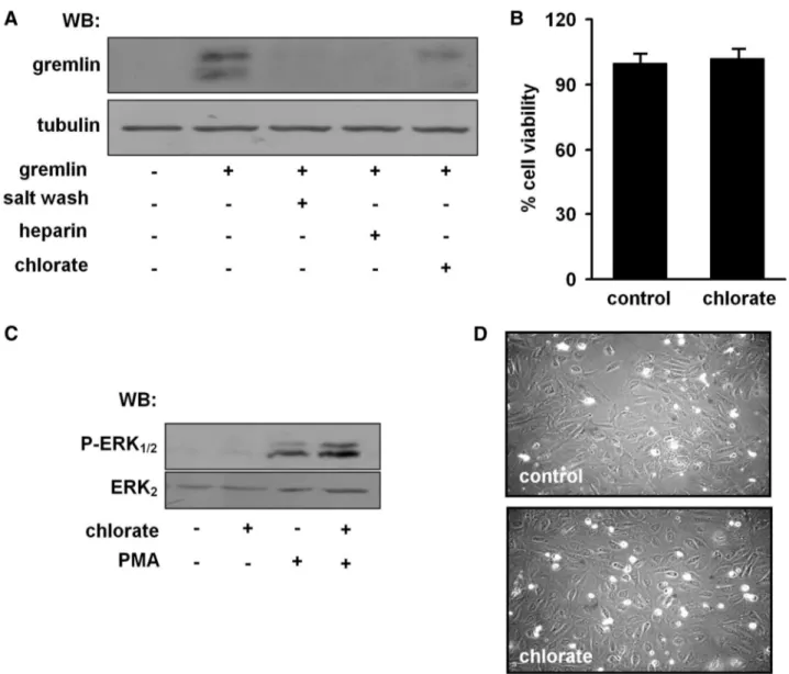
glycosaminoglycan-deficient A745 CHO-K1 cell mutants were incubated for 2 hours at 4°C with gremlin alone (100 ng/mL) or in the presence of heparin, heparan sulfate (HS), or hyaluronic acid (HA) (all at 1.0g/mL). Then, nonpermeabilized cells were washed with PBS or with PBS plus 1.5 mol/L NaCl (salt wash) and decorated with an anti-gremlin antibody (in green) followed by nuclear counterstaining with 4 ⬘,6-diamidino-2-phenylindole (in blue). B, CHO-K1 and A745 CHO-K1 cells were incubated with gremlin as in A, and cell lysates were ana-lyzed by Western blot (WB) with an anti-gremlin antibody. C, Subendothelial extracellular matrices (ECMs) were prepared from untreated or chlorate- or heparinase II–treated GM7373 cell monolayers. Then, living cells or their ECMs were incubated for 2 hours at 4°C with gremlin (100 ng/mL) in the absence or in the presence of heparin (1.0g/mL), washed with PBS or with PBS plus 1.5 mol/L NaCl (salt wash) and analyzed by Western blotting with an anti-gremlin antibody. Uniform loading of the samples was confirmed by Western blotting with an anti-actin antibody. The data are representative of 2 or 3 independent experiments that gave similar results.
VEGFR2 and NRP-1 coreceptor in ECs.
4,46However, at
variance with the prototypic heparin-binding VEGFR2 ligand
VEGF-A165 and similar to VEGF-A
121that lacks the
heparin/NRP-1 binding domain,
4,47gremlin does not induce
VEGFR2/NRP-1 complex formation when administered to
HUVE cells (Figure 4A), thus indicating that HSPG
interac-tion is not per se sufficient to drive NRP-1 engagement. Also,
gremlin does not directly bind recombinant NRP-1 produced
in mammalian cells in ELISA (Figure 4B) or NRP-1
ex-pressed on HUVE cells in cross-linking experiments (data not
shown). Thus, gremlin appears to differ from canonical
heparin-binding VEGFs in the ability to co-opt VEGFR2
coreceptors.
On this basis, we investigated whether HSPG
corecep-tors play any role in mediating a productive interaction of
gremlin with VEGFR2.
29In a first set of experiments, we
exploited VEGFR2-overexpressing endothelial GM7373
cells (GM7373-VEGFR2 cells) that express HSPGs and high
levels of VEGFR2 on their cell surface (Figure 5). These cells
were incubated at 4°C with gremlin in the presence of the
bis(sulfosuccinamide)suberate cross-linking reagent BS3.
Af-ter incubation, cell lysates were immunoprecipitated with
anti-gremlin antibodies, and immunocomplexes were
ana-lyzed by Western blotting with anti-VEGFR2 antibodies.
When cross-linked to the GM7373-VEGFR2 cell surface,
gremlin forms a 250-kDa VEGFR2 complex whose
forma-tion is abrogated when the cross-linking reacforma-tion was
per-formed in chlorate-treated cells (Figure 6A). Accordingly,
chlorate pretreatment abolished the capacity of gremlin to
induce VEGFR2 autophosphorylation in HUVE cells, as
shown by immunostaining of intact cells and Western
blot-ting of the cell extracts using an anti-P-VEGFR2 antibody
(Figure 6B and 6C). Also, the capacity of gremlin to induce
ERK
1/2and p38 mitogen-activated protein kinase
phosphor-ylation was hampered in chlorate-pretreated HUVE cells
(Figure 6C). Similar results were obtained when HUVE cells
were incubated with heparinase II before gremlin stimulation
(data not shown). Also, addition of increasing concentrations
of unmodified heparin to the cell culture medium restored the
capacity of gremlin to induce VEGFR2 autophosphorylation
in chlorate-pretreated HUVE cells (Figure 6D). It must be
pointed out that even though chlorate pretreatment prevented
the binding of gremlin to the HUVE cell surface (Figure 7A),
it did not affect cell viability and the capacity of the protein
kinase C activator PMA to induce ERK
1/2phosphorylation in
these cells (Figure 7B and 7C), confirming the specificity of
the effect. Taken together, these observations demonstrate
that HSPGs are required for a productive interaction of
gremlin with VEGFR2.
HSPGs Mediate the Angiogenic Activity
of Gremlin
Stimulation of EC motility is part of the VEGFR2-dependent
angiogenic program activated by gremlin.
27,29To assess the
Figure 4. Gremlin does not interact with neuropilin-1 (NRP-1). A,
Quiescent human umbilical vein endothelial (HUVE) cells were incubated for 15 minutes at room temperature in the absence (control) or in the presence of vascular endothelial growth factor (VEGF)–A165 (30 ng/mL) or gremlin (50 ng/mL), lysed, and immunoprecipitated (IP) with anti-VEGF receptor-2 (VEGFR2) antibody, and immunocomplexes were probed in a Western blot (WB) with anti-P-VEGFR2 or anti-NRP-1 antibodies. Uniform loading of the gel was confirmed by WB with VEGFR2 anti-body. The P-VEGFR2/VEGFR2 and NRP-1/VEGFR2 ratios are shown at the bottom of the corresponding blots, as calculated by densitometric measurement of the bands. B, 96-well plates coated with 2g/mL NRP-1 were incubated with increasing concentrations of VEGF-A165 or gremlin. Then, the capacity of VEGF-A165 or gremlin to bind to the immobilized receptor was assessed by incubation with VEGF-Ala or gremlin anti-bodies, respectively.
Figure 5. Heparan sulfate proteoglycan (HSPG) and vascular
endothelial growth factor receptor-2 (VEGFR2) expression by endothelial GM7373-VEGFR2 cells. Parental and VEGFR2-overexpressing endothelial GM7373 cells were analyzed for VEGFR2 expression by Western blotting (WB) of the cell lysates (A) and by immunofluorescence analysis with an anti-VEGFR2 antibody (B, a and b). Cells were also decorated with the anti-heparan sulfate monoclonal antibody 10E4 (B, c and d). In B, cells were also counterstained with 4 ⬘,6-diamidino-2-phenylindole (in cyan).
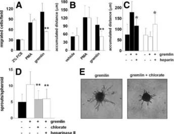
role of HSPGs in mediating the angiogenic activity of
gremlin, chlorate-pretreated HUVE cells were tested in a
Boyden chamber assay for their capacity to migrate through
a gelatin-coated filter in response to gremlin. Despite a
similar capacity to adhere to immobilized gelatin (Figure 7D)
and to migrate in response to PMA (Figure 8A),
chlorate-pretreated HUVE cells displayed a dramatic reduction in their
chemotactic response to gremlin compared with control cells
(Figure 8A). Again, HSPG digestion following heparinase II
incubation mimicked the effect of chlorate, thus abolishing
the chemotactic activity of gremlin in HUVE cells (data not
shown). In keeping with these observations, chlorate
pretreat-ment abolished the motogenic response of HUVE cells to
gremlin as assessed by time-lapse microscopy, without
af-fecting EC motility induced by PMA (Figure 8B). Moreover,
as observed for gremlin-induced VEGFR2
autophosphoryla-tion (see Figure 6D), addiautophosphoryla-tion of heparin to chlorate-treated
cells restored their motogenic response to gremlin stimulation
(Figure 8C).
To confirm the role of HSPGs in mediating EC activation
by gremlin, spheroids of chlorate or heparinase II–treated
HUVE cells were embedded in a 3-dimensional fibrin gel and
stimulated with gremlin.
29As shown in Figure 8D and 8E,
HSPG digestion or undersulfation caused a significant
reduc-tion in the number and length of EC sprouts induced by
gremlin.
Heparin-Like K5 Derivatives as
Gremlin Antagonists
The capsular E. coli K5 polysaccharide has the same structure
as the heparin precursor N-acetyl heparosan.
16K5 derivatives
can be generated by chemical sulfation in the N- or
O-position, leading to heparin-like molecules with defined
sulfation patterns
48and able to hamper the angiogenic activity
of the heparin-binding fibroblast growth factor-2.
33To assess their ability to act as gremlin antagonists, a panel
of selectively sulfated K5 derivatives (including the
N-sulfated derivative K5-NS; the O-sulfated derivatives with
low and high degrees of sulfation, K5-OS(L) and K5-OS(H);
and the N,O-sulfated derivatives with low and high degrees of
sulfation, K5-N,OS(L) and K5-N,OS(H))
33were evaluated
for their capacity to prevent gremlin/heparin interaction in a
SPR competition assay. As shown in Figure 9A, K5-N,OS(H)
and K5-N,OS(L) competed for the binding of gremlin to
immobilized heparin with high potency (ID
50of 0.05 and 0.2
Figure 6. Endothelial heparan sulfate proteoglycans (HSPGs)
mediate vascular endothelial growth factor receptor-2 (VEGFR2)/ gremlin interaction and signaling. A, Control and chlorate-treated GM7373-VEGFR2 cells were incubated for 2 hours at 4°C with gremlin (100 ng/mL). After a further incubation with the cross-linking reagent BS3, cell lysates (1.0 mg) were immuno-precipitated with anti-gremlin antibody and probed with the same antibody or with an anti-VEGFR2 antibody in a Western blot. B, Serum-starved control and chlorate-treated human umbilical vein endothelial (HUVE) cells were stimulated for 10 minutes at 37°C with gremlin (100 ng/mL), decorated with
anti-VEGFR2 or anti-P-VEGFR2 antibodies (in green) followed by nuclear counterstaining with 4⬘,6-diamidino-2-phenylindole (in blue), and observed under a confocal microscope. C, Serum-starved control and chlorate-treated HUVE cells were stimulated for 10 minutes at 37°C with gremlin (100 ng/mL) and cell lysates (50g) were analyzed by Western blotting with anti-P-VEGFR2, anti-P-extracellular signaling–regulated kinase (P-ERK1/2) or
anti-P-p38 mitogen-activated protein kinase (MAPK) antibodies (p38). D, Serum-starved chlorate-treated HUVE cells were incu-bated for 10 minutes at 37°C with gremlin (100 ng/mL) and increasing concentrations of heparin. Then, cell lysates (50g) were analyzed by Western blotting with P-VEGFR2 anti-body. Uniform loading of the gels was confirmed by incubation of the membranes with antibodies directed against the unphos-phorylated form of the antigen. The data are representative of 2 or 3 independent experiments that gave similar results.
g/mL, respectively), whereas K5-OS(H) and K5-OS(L)
were less effective (ID
50of 0.5 and 1.0
g/mL, respectively).
K5-NS and unmodified K5 did not exert a significant
competition.
Then, the most active derivative K5-N,OS(H) was tested
for the capacity to affect the angiogenic activity exerted by
gremlin in vitro and in vivo. As shown in Figure 9B and 9C,
K5-N,OS(H) inhibits the binding of gremlin to the
extracel-lular domain of VEGFR2 in a competitive ELISA and its
ability to stimulate the sprouting of HUVE cell aggregates in
fibrin gel. Also, K5-N,OS(H) exerts a potent inhibitory
activity on blood vessel formation triggered by gremlin
delivered on the top of the chick embryo chorioallantoic
membrane via an alginate pellet implant (Figure 9D and 9E).
Accordingly, K5-N,OS(H) inhibits the angiogenic activity
exerted by gremlin in a tumor-driven ex vivo angiogenesis
assay in which fibrin-embedded human umbilical cord artery
rings
28are grown in the presence of the conditioned medium
from gremlin-overexpressing human pancreatic carcinoma
PANC-1 cells (Figure 10A and 10B). As shown in Figure
10C and 10D, 1-gremlin cells, but not control
PANC-1-GFP cells, induce a dramatic increase in the formation of
EC sprouts from human artery rings that is significantly
inhibited by K5-N,OS(H). When tested under the same
experimental conditions, unmodified K5 was ineffective in all
these assays.
Figure 7. Characterization of chlorate-treated human umbilical vein endothelial (HUVE) cells. A, HUVE cell monolayers were left
untreated or treated with chlorate. Then, cells were incubated for 2 hours at 4°C with gremlin (100 ng/mL) in the absence or in the presence of heparin (1g/mL), washed with PBS or with PBS plus 1.5 mol/L NaCl (salt wash). Then, cell lysates were analyzed by Western blotting with an anti-gremlin antibody. Uniform loading of the samples was confirmed by incubation of the membrane with an anti-tubulin antibody. B, Control and chlorate-treated HUVE cells were assessed for cell viability by the tetrazolium salt (3-[4,5-dimethylthiazol-2-yl]-2,5-diphenyltetrazolium bromide) colorimetric assay according to standard procedures. C, Control and chlorate-treated HUVE cells were stimulated for 10 minutes with phorbol myristate acetate (PMA) (10 ng/mL). Then, 50g-aliquots of cell lysates were analyzed by Western blotting with an anti-P-extracellular signaling–regulated kinase (P-ERK1/2) antibody. Uniform loading
of the gel was confirmed by incubation of the membrane with an anti-ERK2antibody. D, Control and chlorate-treated HUVE cells were
allowed to adhere onto substrate-immobilized gelatin for 2 hours at room temperature, and attached cells were photographed under an inverted microscope.
Discussion
This is the first characterization of the interaction of gremlin
with heparin/HSPGs and of its role in mediating the
angio-genic response triggered by this noncanonical VEGFR2
ligand in ECs. Compared with other sulfated GAGs, heparin
(SO
3⫺/COO
⫺⫽2.14)
34binds gremlin with high affinity
(K
d⫽20 nmol/L). In HS (SO
3⫺/COO
⫺⬵1.0),
34sulfate groups
are arranged in high-charge-density clusters intercalated with
low-charge-density regions,
49thus resulting in a lower
gremlin-binding activity. On the other hand, HS is a much
more effective gremlin interactor than dermatan sulfate,
chondroitin-4-sulfate, or chondroitin-6-sulfate, even though
they all share a similar charge, no significant binding being
observed with the nonsulfated hyaluronic acid and K5
poly-saccharide. Also, similar to other heparin-binding angiogenic
proteins
10,14the affinity for gremlin is affected by the size of
the polysaccharide chain. Indeed, whereas low-MW heparin
tinzaparin binds gremlin with an affinity similar to
unfrac-tionated heparin, very-low-MW heparins showed a reduced
interaction, and a negligible binding was observed for
2.1-kDa heparin. Interestingly, when compared with different
low-MW heparin preparations, tinzaparin has demonstrated
significant antiangiogenic and anticancer properties.
44Sev-eral sets of experimental evidence demonstrate that
physico-chemical and biological properties of different low-MW
heparins are not alike.
44Even though additional studies are
required to fully characterize the interaction of gremlin with
different heparin preparations, our data clearly indicate that
the degree of sulfation, charge distribution, disaccharide
composition, and size are all important in determining the
capacity of sulfated GAGs to bind gremlin.
Previous observations had shown that fibroblast growth
factor-2 binds avidly to a pentasaccharide region in which
N-sulfate groups and a single 2-O-sulfate group are essential
for interaction. In contrast, N-sulfation and 6-O-sulfation are
crucial for VEGF-A165 binding, whereas 2-O-sulfate groups
are less essential.
50Our data demonstrate that selective 2-O-,
6-O-, or N-desulfation significantly reduces gremlin/heparin
interaction, indicating that they are all required for high
affinity binding. These findings support the hypothesis that
heparin-binding cytokines may bind sulfated GAGs in a
distinct manner. Even though specific factor binding
se-quences may be hidden in heparin because of its high degree
Figure 8. Heparan sulfate proteoglycans (HSPGs) mediate the
angiogenic activity of gremlin in endothelial cells (ECs). A and B, Control (black bars) and chlorate-treated (open bars) human umbilical vein endothelial (HUVE) cells were assessed for their capacity to migrate in response to gremlin or phorbol myristate acetate (PMA) in a modified Boyden chamber assay (A) or by time-lapse videomicroscopy (B). C, Control (black bars) and chlorate-treated (open bars) HUVE cells were incubated with gremlin in the absence or in the presence of heparin (0.5g/mL) and assessed for their motility by time-lapse microscopy. D, HUVE cell spheroids obtained from control, chlorate-treated, or heparinase II–treated cells were embedded in fibrin gel and incubated with gremlin. Formation of radially growing cell sprouts was evaluated after 24 hours of incubation. E, Repre-sentative microphotographs of control and chlorate-treated HUVE cell spheroids stimulated with gremlin. In A to D, each data are the mean⫾SD of 2 or 3 independent experiments. Sig-nificantly different from gremlin alone under the same experi-mental conditions: *P⫽0.004, **P⬍0.001. FCS, fetal calf serum.
Figure 9. Sulfated K5 derivatives inhibit the angiogenic activity
of gremlin. A, Gremlin (20 nmol/L) was injected over a heparin-coated BIAcore sensor chip in the presence of increasing con-centrations of K5 (F), K5-NS (E), K5-OS(L) (f), K5-N,OS(L) (Œ), K5-OS(H) (䡺), or K5-N,OS(H) (‚), and the response (in reso-nance units [RU]) was recorded at the end of the injection. B, 96-well plates coated with the extracellular domain of soluble-vascular endothelial growth factor receptor-2 (sVEGFR2)D1–7
(200 ng/mL) were incubated with gremlin (50 ng/mL) in presence of increasing concentrations of K5 (F) or K5-N,OS(H) (E). Then, the formation of VEGFR2/gremlin complex was assessed by incubation with an anti-gremlin antibody. C, Human umbilical vein endothelial (HUVE) cell spheroids embedded in fibrin gel were incubated with vehicle or gremlin in the absence (black bars) or in the presence of K5 (open bars) or K5-N,OS(H) (gray bars) (both at 10 ng/mL). Formation of radially growing cell sprouts was evaluated after 24 hours of incubation. D, Alginate pellets containing gremlin (100 ng/pellet) in the absence or in the presence of K5 or K5-N,OS(H) (both at 1g/pellet) were implanted on chick embryo chorioallantoic membranes (10 eggs per group). After 3 days, newly formed microvessels converging vs the implant were counted. E, Representative images of cho-rioallantoic membranes implanted with alginate pellets contain-ing gremlin alone or gremlin added with K5-N,OS(H). Note the numerous newly formed thin microvessels converging in a spoke-wheel pattern vs the gremlin implant, significantly reduced in the presence of K5-N,OS(H). Significantly different from controls: **P⬍0.001.
of sulfation, the high heterogeneity in HS structure allows a
more refined tailoring of selective binding regions that may
influence the biological activity and bioavailability of
heparin/HS-binding growth factors.
In keeping with its heparin/HS binding capacity, gremlin
binds the surface of CHO-K1 cells but not of GAG-deficient
A745 CHO-K1 cells. Accordingly, gremlin binds HSPGs
exposed on the surface of ECs and deposited in the
suben-dothelial ECM, the binding being prevented by HS chain
digestion with heparinase II or by inhibition of HS chain
sulfation following EC treatment with chlorate. Similar to the
canonical heparin-binding VEGFR2 ligand VEGF-A165,
HSPG interaction appears to be essential for the binding of
gremlin to VEGFR2 and receptor activation in ECs. Indeed,
chlorate treatment or heparinase II digestion both hamper
gremlin/VEGFR2 interaction in HUVE cells. Accordingly,
VEGFR2 autophosphorylation triggered by gremlin is
abol-ished following HS digestion or undersulfation, yet is
re-stored by the addition of exogenous heparin. Similarly,
inhibition of HSPG interaction suppresses the ability of
gremlin to induce the activation of intracellular signaling
pathways in ECs, including ERK
1/2and p38
mitogen-acti-vated protein kinase phosphorylation. Also, it inhibits EC
chemotactic migration, sprouting, and motility, all activities
that are mediated by a productive interaction of gremlin with
VEGFR2.
29NRP-1 is a transmembrane glycoprotein involved in the
interaction with a plethora of binding partners, including
class-3 semaphorin receptors, heparin-binding growth
fac-tors, and HSPGs.
51Also, NRP-1 core protein is modified with
either a single HS or condroitin sulfate chain depending on
the expressing cell type, thus increasing the complexity of
NRP-1 extracellular interactions.
52Several sets of
experimen-tal evidence indicate that the binding of mammalian
heparin-binding VEGF isoforms to HS and NRP-1 is interdependent.
4Gremlin does not contain the amino acid sequence CDKPRR
present in the VEGF-A165 carboxyl terminus and involved in
the interaction with b1b2 ectodomains of the NRP-1 core
protein.
53Even though we cannot rule out the possibility that
gremlin may bind the HS chain of NRP-1 in target cells, our
data indicate that, at variance with VEGF-A165, gremlin does
not interact with recombinant NRP-1 and does not trigger the
formation of a VEGFR2/NRP-1 complex in ECs. Also, the
structural bases for heparin/HS interaction may differ
be-tween VEGF isoforms and gremlin. Indeed, VEGFs bind HS
via a highly basic carboxyl-terminal region distinct from the
cystine-knot domain of the protein and encoded by
alterna-tively spliced exons.
4At variance, gremlin may interact with
heparin/HS via a linear basic patch along the third loop of its
cystine-knot domain, as suggested by homology modeling
prediction studies based on the cystine-knot BMP antagonist
sclerostin.
31,32Thus, gremlin differs from canonical
heparin-binding VEGFs in the ability to co-opt VEGFR2 coreceptors
and appears to exert a VEGFR2-dependent proangiogenic
activation of ECs with mechanisms that are, at least in part,
different from VEGFs. For instance, HSPGs contribute to
VEGF-triggered angiogenesis by a mechanism dependent on
the modulation of
␣
6
1integrin expression on the EC
surface.
54At variance with VEGF-A, gremlin does not
upregulate the expression of
␣
6integrin in ECs (data not
shown), further underscoring the different mechanism of
action of the 2 angiogenic factors.
Chemical or enzymatic modifications of the E. coli capsular
K5 polysaccharide allow the production of “biotechnological”
semisynthetic heparin/HS-like compounds with selected
chem-ical features, with potential implications in the therapy of various
human pathologies, including thromboembolic diseases, viral
infection, and neoplasia.
16,55Here we evaluated the
gremlin-binding capacity of a series of specific N-, O-, or N,O-sulfated
nonanticoagulant K5 derivatives.
33Among the compounds
tested, highly sulfated K5-N,OS(H) binds gremlin with higher
affinity compared with the corresponding derivative K5-OS(H)
devoid of sulfate group in the N position but bearing an identical
total negative charge (SO
3⫺/COO
⫺of 3.8 for both
com-pounds
33). K5-OS(H) consists of the virtually homogeneous
repeat of GlcA2,3SO
3⫺-GlcNAc3,6SO
3⫺disaccharide units,
whereas most of K5-N,OS(H) sequences are represented by
GlcA2,3SO
3⫺-GlcNSO
3⫺,6SO
3⫺disaccharide units.
33Our
data indicate that the requirement for N-sulfation present in
K5-N,OS(H) cannot be fully overcome by the high density
charge present in the highly O-sulfated K5-OS(H). Similarly,
low sulfated K5-N,OS(L) binds gremlin more efficiently than
Figure 10. K5-N,OS(H) inhibits the angiogenic activity of
gremlin-overexpressing human pancreatic carcinoma-1 (PANC-1) cells. A, Conditioned media from PANC-1-green fluo-rescent protein (GFP) and PANC-1-gremlin cells grown for 48 hours in serum-free Dulbecco’s modified Eagle medium (DMEM) were analyzed by Western blotting with an gremlin anti-body. B, Human umbilical vein endothelial (HUVE) cells were stimulated for 15 minutes with the conditioned medium from PANC-1-GFP or PANC-1-gremlin cells. Then, 50g-aliquots of cell extracts were analyzed by Western blotting with an P-vascular endothelial growth factor receptor-2 (VEGFR2) anti-body. Uniform loading of the gel was confirmed by incubation of the membrane with an anti-VEGFR2 antibody. PANC-1-gremlin cells released gremlin in a bioactive form, as demonstrated by the capacity of their conditioned medium to induce VEGFR2 phosphorylation in HUVE cells. C and D, Human umbilical artery rings (4 rings per group) were embedded in fibrin gel and stimu-lated with the conditioned medium from 1-GFP or PANC-1-gremlin cells in the absence (black bars) or in the presence of K5 (open bars) or K5-N,OS(H) (gray bars) (both at 100 ng/mL). After 6 days, endothelial cell (EC) sprouts were counted (C) and photographed under an inverted microscope at a⫻200 magnifi-cation (D). Significantly different from controls: *P⬍0.03.
K5-OS(L), even though both compounds have a SO
3⫺/COO
⫺value of 1.7.
33On the other hand, K5-NS devoid of sulfate
groups in O positions (SO
3⫺/COO
⫺of 1.0) does not have a
significant interaction with gremlin. Thus, in agreement with
the results obtained with selectively desulfated heparins,
N-and O-sulfate groups are required for gremlin interaction with
sulfated K5 derivatives.
Gremlin exerts both dependent and
BMP-independent functions in different physiopathological
condi-tions by inhibiting BMP-mediated TGF-
 receptor activation
or by a direct activation of VEGFR2 signaling, respectively.
In this respect, it is interesting to note that a complex, not yet
fully elucidated cross-talk may exist among TGF-
/BMP,
VEGFR2, and canonical and noncanonical Wingless (Wnt)
signaling pathways during angiogenesis,
56making
unpredict-able the impact of the modulation of a single
agonist/antag-onist on the neovascularization process in different
patholog-ical conditions. Here, we demonstrate that K5-N,OS(H) is a
potent antagonist of gremlin, inhibiting VEGFR2 interaction
and angiogenic activity in vitro and in vivo. Also,
K5-N,OS(H) may prevent gremlin-driven tumor
neovasculariza-tion, as indicated by its capacity to suppress the angiogenic
potential of gremlin-overexpressing human tumor PANC-1
cells in an ex vivo human artery ring assay. Previous
observations had shown the ability of K5-N,OS(H) to inhibit
the angiogenic activity of the heparin-binding molecules
fibroblast growth factor-2 and HIV-1 Tat protein
16and to
reduce tumor invasion by hampering heparanase activity
16and tumor metastasis.
57Thus, K5-N,OS(H) may represent the
basis for the design of novel nonanticoagulant
biotechnolog-ical heparins endowed with a multitarget antineoplastic action
by interacting with different angiogenic growth factors and
limiting tumor cell invasion and metastasis. Also, preclinical
and clinical studies identify gremlin as a potential therapeutic
target in human fibrotic diseases characterized by alterations
of TGF-
/BMP signaling and vascular defects in different
organs as liver,
58kidney,
24and lung.
23Given the role of
gremlin in physiological development and different
patholog-ical conditions, additional preclinpatholog-ical in vivo studies will be
required to define the therapeutic capability and possible side
effects of gremlin antagonists, including K5-N,OS(H) and its
derivatives.
Acknowledgments
We thank Dr Sara Lovecchio for contributions to the experiments and Dr Stefano Calza for statistical analysis.
Sources of Funding
This work was supported by grants from Ministero dell’Istruzione, dell’Università e della Ricerca (MIUR), Associazione Italiana per la Ricerca sul Cancro (AIRC) (Grant 10396), Fondazione Berlucchi, CARIPLO (Grant 2008-2264 and the NOBEL Project) (to M.P.); from MIUR, Istituto Superiore di Sanita (AIDS Project Grant 40H.51), and CARIPLO (Grant 2008-2198) (to M.R.); and from MIUR and My First AIRC Grant 9161 (to S.M.). Dr Ravelli was supported by a Fondazione Italiana per la Ricerca sul Cancro fellowship.
Disclosures
None.References
1. Carmeliet P, Jain RK. Angiogenesis in cancer and other diseases.
Nature. 2000;407:249 –257.
2. Yancopoulos GD, Davis S, Gale NW, Rudge JS, Wiegand SJ, Holash J. Vascular-specific growth factors and blood vessel formation. Nature. 2000;407:242–248.
3. Neufeld G, Cohen T, Gengrinovitch S, Poltorak Z. Vascular endothelial growth factor (VEGF) and its receptors. FASEB J. 1999;13:9 –22. 4. Grunewald FS, Prota AE, Giese A, Ballmer-Hofer K. Structure-function
analysis of VEGF receptor activation and the role of coreceptors in angiogenic signaling. Biochim Biophys Acta. 2010;1804:567–580. 5. Krilleke D, DeErkenez A, Schubert W, Giri I, Robinson GS, Ng YS,
Shima DT. Molecular mapping and functional characterization of the VEGF164 heparin-binding domain. J Biol Chem. 2007;282: 28045–28056.
6. Lindahl U. Heparan sulfate-protein interactions: a concept for drug design? Thromb Haemost. 2007;98:109 –115.
7. Iozzo RV, San Antonio JD. Heparan sulfate proteoglycans: heavy hitters in the angiogenesis arena. J Clin Invest. 2001;108:349 –355.
8. Houck KA, Leung DW, Rowland AM, Winer J, Ferrara N. Dual regu-lation of vascular endothelial growth factor bioavailability by genetic and proteolytic mechanisms. J Biol Chem. 1992;267:26031–26037. 9. Kawamura H, Li X, Goishi K, van Meeteren LA, Jakobsson L,
Cebe-Suarez S, Shimizu A, Edholm D, Ballmer-Hofer K, Kjellen L, Klagsbrun M, Claesson-Welsh L. Neuropilin-1 in regulation of VEGF-induced acti-vation of p38MAPK and endothelial cell organization. Blood. 2008;112: 3638 –3649.
10. Robinson CJ, Mulloy B, Gallagher JT, Stringer SE. VEGF165-binding sites within heparan sulfate encompass two highly sulfated domains and can be liberated by K5 lyase. J Biol Chem. 2006;281:1731–1740. 11. Maccarana M, Casu B, Lindahl U. Minimal sequence in heparin/heparan
sulfate required for binding of basic fibroblast growth factor. J Biol
Chem. 1994;269:3903–3903.
12. Guimond S, Maccarana M, Olwin BB, Lindahl U, Rapraeger AC. Acti-vating and inhibitory heparin sequences for FGF-2 (basic FGF). Distinct requirements for FGF-1, FGF-2, and FGF-4. J Biol Chem. 1993;268: 23906 –23914.
13. Rusnati M, Urbinati C. Polysulfated/sulfonated compounds for the devel-opment of drugs at the crossroad of viral infection and oncogenesis. Curr
Pharm Des. 2009;15:2946 –2957.
14. Presta M, Leali D, Stabile H, Ronca R, Camozzi M, Coco L, Moroni E, Liekens S, Rusnati M. Heparin derivatives as angiogenesis inhibitors.
Curr Pharm Des. 2003;9:553–566.
15. Presta M, Oreste P, Zoppetti G, Belleri M, Tanghetti E, Leali D, Urbinati C, Bugatti A, Ronca R, Nicoli S, Moroni E, Stabile H, Camozzi M, Hernandez GA, Mitola S, Dell’Era P, Rusnati M, Ribatti D. Antian-giogenic activity of semisynthetic biotechnological heparins: low-molecular-weight-sulfated Escherichia coli K5 polysaccharide deriv-atives as fibroblast growth factor antagonists. Arterioscler Thromb Vasc
Biol. 2005;25:71–76.
16. Rusnati M, Oreste P, Zoppetti G, Presta M. Biotechnological engineering of heparin/heparan sulphate: a novel area of multi-target drug discovery.
Curr Pharm Des. 2005;11:2489 –2499.
17. Pearce JJ, Penny G, Rossant J. A mouse cerberus/Dan-related gene family. Dev Biol. 1999;209:98 –110.
18. Vitt UA, Hsu SY, Hsueh AJ. Evolution and classification of cystine knot-containing hormones and related extracellular signaling molecules.
Mol Endocrinol. 2001;15:681– 694.
19. Balemans W, Van Hul W. Extracellular regulation of BMP signaling in vertebrates: a cocktail of modulators. Dev Biol. 2002;250:231–250. 20. Khokha MK, Hsu D, Brunet LJ, Dionne MS, Harland RM. Gremlin is the
bmp antagonist required for maintenance of SHH and FGF signals during limb patterning. Nat Genet. 2003;34:303–307.
21. Lu MM, Yang H, Zhang L, Shu W, Blair DG, Morrisey EE. The bone morphogenic protein antagonist gremlin regulates proximal-distal pat-terning of the lung. Dev Dyn. 2001;222:667– 680.
22. Michos O, Panman L, Vintersten K, Beier K, Zeller R, Zuniga A. Gremlin-mediated BMP antagonism induces the epithelial-mesenchymal feedback signaling controlling metanephric kidney and limb organo-genesis. Development. 2004;131:3401–3410.
23. Costello CM, Cahill E, Martin F, Gaine S, McLoughlin P. Role of gremlin in the lung: development and disease. Am J Respir Cell Mol Biol;42: 517–523.
24. Lappin DW, McMahon R, Murphy M, Brady HR. Gremlin: an example of the re-emergence of developmental programmes in diabetic nephrop-athy. Nephrol Dial Transplant. 2002;17(suppl 9):65– 67.
25. Namkoong H, Shin SM, Kim HK, Ha SA, Cho GW, Hur SY, Kim TE, Kim JW. The bone morphogenetic protein antagonist gremlin 1 is over-expressed in human cancers and interacts with YWHAH protein. BMC
Cancer. 2006;6:74.
26. Sneddon JB, Zhen HH, Montgomery K, van de Rijn M, Tward AD, West R, Gladstone H, Chang HY, Morganroth GS, Oro AE, Brown PO. Bone morphogenetic protein antagonist gremlin 1 is widely expressed by cancer-associated stromal cells and can promote tumor cell proliferation.
Proc Natl Acad Sci U S A. 2006;103:14842–14847.
27. Stabile H, Mitola S, Moroni E, Belleri M, Nicoli S, Coltrini D, Peri F, Pessi A, Orsatti L, Talamo F, Castronovo V, Waltregny D, Cotelli F, Ribatti D, Presta M. Bone morphogenic protein antagonist Drm/gremlin is a novel proangiogenic factor. Blood. 2007;109:1834 –1840. 28. Mitola S, Moroni E, Ravelli C, Andres G, Belleri M, Presta M.
Angiopoietin-1 mediates the pro-angiogenic activity of the bone mor-phogenic protein antagonist Drm. Blood. 2008;112:1154 –1157. 29. Mitola S, Ravelli C, Moroni E, Salvi V, Leali D, Ballmer-Hofer K,
Zammataro L, Presta M. Gremlin is a novel agonist of the major proan-giogenic receptor VEGFR2. Blood. 2010;116:3677–3680.
30. Topol LZ, Bardot B, Zhang Q, Resau J, Huillard E, Marx M, Calothy G, Blair DG. Biosynthesis, post-translation modification, and functional characterization of Drm/gremlin. J Biol Chem. 2000;275:8785– 8793. 31. Veverka V, Henry AJ, Slocombe PM, Ventom A, Mulloy B, Muskett FW,
Muzylak M, Greenslade K, Moore A, Zhang L, Gong J, Qian X, Paszty C, Taylor RJ, Robinson MK, Carr MD. Characterization of the structural features and interactions of sclerostin: molecular insight into a key reg-ulator of Wnt-mediated bone formation. J Biol Chem. 2009;284: 10890 –10900.
32. Rider CC, Mulloy B. Bone morphogenetic protein and growth differen-tiation factor cytokine families and their protein antagonists. Biochem J. 2010;429:1–12.
33. Leali D, Belleri M, Urbinati C, Coltrini D, Oreste P, Zoppetti G, Ribatti D, Rusnati M, Presta M. Fibroblast growth factor-2 antagonist activity and angiostatic capacity of sulfated Escherichia coli K5 polysaccharide derivatives. J Biol Chem. 2001;276:37900 –37908.
34. Rusnati M, Coltrini D, Oreste P, Zoppetti G, Albini A, Noonan D, d’Adda di Fagagna F, Giacca M, Presta M. Interaction of HIV-1 Tat protein with heparin: role of the backbone structure, sulfation, and size. J Biol Chem. 1997;272:11313–11320.
35. Zhang Z, Coomans C, David G. Membrane heparan sulfate proteoglycan-supported FGF2-FGFR1 signaling: evidence in support of the “coop-erative end structures” model. J Biol Chem. 2001;276:41921– 41929. 36. Rusnati M, Urbinati C, Caputo A, Possati L, Lortat-Jacob H, Giacca M,
Ribatti D, Presta M. Pentosan polysulfate as an inhibitor of extracellular HIV-1 Tat. J Biol Chem. 2001;276:22420 –22425.
37. Grinspan JB, Mueller SN, Levine EM. Bovine endothelial cells trans-formed in vitro by benzo(a)pyrene. J Cell Physiol. 1983;114:328 –338. 38. Esko JD. Genetic analysis of proteoglycan structure, function and
me-tabolism. Curr Opin Cell Biol. 1991;3:805– 816.
39. Safaiyan F, Kolset SO, Prydz K, Gottfridsson E, Lindahl U, Salmivirta M. Selective effects of sodium chlorate treatment on the sulfation of heparan sulfate. J Biol Chem. 1999;274:36267–36273.
40. De Palma M, Naldini L. Transduction of a gene expression cassette using advanced generation lentiviral vectors. Methods Enzymol. 2002;346: 514 –529.
41. Rusnati M, Urbinati C, Presta M. Internalization of basic fibroblast growth factor (bFGF) in cultured endothelial cells: role of the low affinity heparin-like bFGF receptors. J Cell Physiol. 1993;154:152–161. 42. Margosio B, Marchetti D, Vergani V, Giavazzi R, Rusnati M, Presta M,
Taraboletti G. Thrombospondin 1 as a scavenger for matrix-associated fibroblast growth factor 2. Blood. 2003;102:4399 – 4406.
43. Hothorn T, Bretz F, Westfall P. Simultaneous inference in general para-metric models. Biom J. 2008;50:346 –363.
44. Mousa SA, Petersen LJ. Anti-cancer properties of low-molecular-weight heparin: preclinical evidence. Thromb Haemost. 2009;102:258 –267. 45. Rostand KS, Esko JD. Microbial adherence to and invasion through
proteoglycans. Infect Immun. 1997;65:1– 8.
46. Gitay-Goren H, Soker S, Vlodavsky I, Neufeld G. The binding of vascular endothelial growth factor to its receptors is dependent on cell surface-associated heparin-like molecules. J Biol Chem. 1992;267: 6093– 6098.
47. Cohen T, Gitay-Goren H, Sharon R, Shibuya M, Halaban R, Levi BZ, Neufeld G. VEGF121, a vascular endothelial growth factor (VEGF) isoform lacking heparin binding ability, requires cell-surface heparan sulfates for efficient binding to the VEGF receptors of human melanoma cells. J Biol Chem. 1995;270:11322–11326.
48. Casu B, Grazioli G, Razi N, Guerrini M, Naggi A, Torri G, Oreste P, Tursi F, Zoppetti G, Lindahl U. Heparin-like compounds prepared by chemical modification of capsular polysaccharide from E. coli K5.
Carbohydr Res. 1994;263:271–284.
49. Esko JD, Lindahl U. Molecular diversity of heparan sulfate. J Clin Invest. 2001;108:169 –173.
50. Ono K, Hattori H, Takeshita S, Kurita A, Ishihara M. Structural features in heparin that interact with VEGF165 and modulate its biological activity. Glycobiology. 1999;9:705–711.
51. Pellet-Many C, Frankel P, Jia H, Zachary I. Neuropilins: structure, function and role in disease. Biochem J. 2008;411:211–226.
52. Shintani Y, Takashima S, Asano Y, Kato H, Liao Y, Yamazaki S, Tsukamoto O, Seguchi O, Yamamoto H, Fukushima T, Sugahara K, Kitakaze M, Hori M. Glycosaminoglycan modification of neuropilin-1 modulates VEGFR2 signaling. EMBO J. 2006;25:3045–3055. 53. Geretti E, Klagsbrun M. Neuropilins: novel targets for anti-angiogenesis
therapies. Cell Adh Migr. 2007;1:56 – 61.
54. Lee TH, Seng S, Li H, Kennel SJ, Avraham HK, Avraham S. Integrin regulation by vascular endothelial growth factor in human brain micro-vascular endothelial cells: role of␣61 integrin in angiogenesis. J Biol
Chem. 2006;281:40450 – 40460.
55. Rusnati M, Vicenzi E, Donalisio M, Oreste P, Landolfo S, Lembo D. Sulfated K5 Escherichia coli polysaccharide derivatives: a novel class of candidate antiviral microbicides. Pharmacol Ther. 2009;123:310 –322. 56. Goodwin AM, D’Amore PA. Wnt signaling in the vasculature. Angiogenesis.
2002;5:1–9.
57. Poggi A, Rossi C, Casella N, Bruno C, Sturiale L, Dossi C, Naggi A. Inhibition of b16-bl6 melanoma lung colonies by semisynthetic sulfami-noheparosan sulfates from E. coli K5 polysaccharide. Semin Thromb
Hemost. 2002;28:383–392.
58. Boers W, Aarrass S, Linthorst C, Pinzani M, Elferink RO, Bosma P. Transcriptional profiling reveals novel markers of liver fibrogenesis: gremlin and insulin-like growth factor-binding proteins. J Biol Chem. 2006;281:16289 –16295.