Sialic Acid Associated with
␣
v

3
Integrin Mediates HIV-1 Tat
Protein Interaction and Endothelial Cell Proangiogenic
Activation
*
Received for publication, December 23, 2011, and in revised form, April 3, 2012Published, JBC Papers in Press, April 23, 2012, DOI 10.1074/jbc.M111.337139 Paola Chiodelli‡, Chiara Urbinati‡, Stefania Mitola‡, Elena Tanghetti§, and Marco Rusnati‡1
From the Units of‡General Pathology and Immunology and§Histology, Department of Biomedical Sciences and Biotechnology, School of Medicine, University of Brescia, 25123 Brescia, Italy
Background:HIV-1 Tat engages␣v3, leading to endothelial cell (EC) proangiogenic activation.
Results:Sialic acid (NeuAc)-binding lectins and neuraminidase partially inhibit Tat/␣v3 interaction and consequent EC proangiogenic activation.
Conclusion:Endothelial␣v3-associated NeuAc is involved in Tat interaction and consequent EC proangiogenic activation. Significance:Integrin-associated NeuAc can be considered a target for the development of new treatments for angiogenesis/ AIDS-associated pathologies.
Sialic acid (NeuAc) is a major anion on endothelial cells (ECs) that regulates different biological processes including angiogen-esis. NeuAc is present in the oligosaccharidic portion of integ-rins, receptors that interact with extracellular matrix compo-nents and growth factors regulating cell adhesion, migration, and proliferation. Tat is a cationic polypeptide that, once released by HIV-1ⴙ cells, accumulates in the extracellular matrix, promoting EC adhesion and proangiogenic activation by engaging ␣v3. By using two complementary approaches (NeuAc removal by neuraminidase or its masking by NeuAc-binding lectin from Maackia amurensis, MAA), we investigated the presence of NeuAc on endothelial␣v3and its role in Tat interaction, EC adhesion, and proangiogenic activation.␣v3 immunoprecipitation with biotinylated MAA or Western blot analysis of neuraminidase-treated ECs demonstrated that NeuAc is associated with both the␣vand the3subunits. Sur-face plasmon resonance analysis demonstrated that the masking of␣v3-associated NeuAc by MAA prevents Tat/␣v3 interac-tion. MAA and neuraminidase prevent ␣v3-dependent EC adhesion to Tat, the consequent FAK and ERK1/2 phosphory-lation, and EC proliferation, migration, and regeneration in a wound-healing assay. Finally, MAA inhibits Tat-induced neo-vascularization in the ex vivo human artery ring sprouting assay. The inhibitions are specific because the NeuAc-unrelated lectin from Ulex europaeus is ineffective on Tat. Also, MAA and neuraminidase affect only weakly integrin-dependent EC adhe-sion and proangiogenic activation by fibronectin. In concluadhe-sion, NeuAc is associated with endothelial␣v3and mediates Tat-de-pendent EC adhesion and proangiogenic activation. These data point to the possibility to target integrin glycosylation for the treatment of angiogenesis/AIDS-associated pathologies.
Polyanionic macromolecules are extremely abundant in the extracellular environment, readily accessible to many proteins for interactions implicated in various biological functions. Among polyanions, sialic acid (NeuAc)-bearing gangliosides and glycoproteins are widely distributed in biological fluids, extracellular matrix, and cell membrane, where they act as receptors for various physiological ligands and for many human viruses, bacteria, and protozoa (1–3).
The term NeuAc encompasses a large family of sugars char-acterized by a nine-carbon sugar acid common in higher ani-mals and some microorganisms (4). NeuAc is found mainly as a terminal component of glycoprotein and gangliosides, where it regulates various molecular and cellular interactions (5).
NeuAc is the major surface anion on the endothelial cell (EC)2surface. Accordingly, the lectin from Maackia amurensis
(MAA), which specifically binds NeuAc residues attached to galactose through an␣(233) linkage, binds to ECs of retina, brain, and myocardium (6). NeuAc expression on ECs is regu-lated during ontogenesis, inflammation (7–9), and possibly neovascularization, as suggested by the observation that the binding of the NeuAc-binding lectin from Limax flavus to ECs increases during angiogenesis in the chick embryo chorioallan-toic membrane (8).
NeuAc is involved in different physiological and pathological functions of the endothelium; in its ganglioside- or glycopro-tein-associated form, it mediates EC infection by different microorganisms (10) and the transport of HIV-1 or of its pro-teins across the blood-brain barrier (11, 12). In its ganglioside-associated form, NeuAc takes part in the regulation of neovas-cularization (13–15). When associated with integrin subunits (including␣E(16),␣2, (17),␣3(18),␣4(19),␣5and␣v(20),1
*This work was supported by grants from Ministero dell’Istruzione, dell’Universita` e della Ricerca, Istituto Superiore di Sanita` (AIDS Project), and Cassa di Risparmio delle Provincie Lombarde (to M. R.).
1To whom correspondence should be addressed: General Pathology and Immunology, Dept. of Biomedical Sciences and Biotechnology, viale Europa 11, 25123 Brescia, Italy. Tel: 39-30-3717315; Fax: 39-30-3717747; E-mail: [email protected].
2The abbreviations used are: EC, endothelial cell; FAK, focal adhesion kinase; FN, fibronectin; MAA, lectin from M. amurensis; UEA, lectin from U.
euro-paeus; PDMP,D -threo-1-phenyl-2-decanoylamino-3-morpholino-1-propa-nol; PPPP,D -1-threo-1-phenyl-2-hexadecanoylamino-3-pyrrolidino-1-pro-panol-HCl; SPR, surface plasmon resonance; VEGFR2, vascular endothelial growth factor receptor-2; WB, Western blot; TRITC, tetramethylrhodamine isothiocyanate; MTS, 3-(4,5-dimethylthiazol-2-yl)-5-(3-carboxymethoxy-phenyl)-2-(4-sulfophenyl)-2H-tetrazolium, inner salt.
at BIBL DELLA FAC DI MED, on June 11, 2012
www.jbc.org
(17, 18, 20),2(21), and 4 (16, 20)), NeuAc contributes to leukocyte and tumor cell extravasation during inflammation and metastasization, respectively.
Integrins are widely distributed receptors that interact with extracellular matrix components, growth factors, and microbial proteins regulating adhesion, migration, and proliferation of various normal and transformed cell types (22). Among the various integrins,␣v3expressed on the surface of ECs plays a
central role in neovascularization (23). Interestingly, NeuAc has been found associated with␣v3integrin from melanoma
metastatic cell surface (18), but no data are available for␣v3
from ECs.
HIV-1 Tat is a cationic protein that, once released by HIV-1-infected cells (24), targets ECs, causing a variety of pathological effects that, in turn, lead to different angiogenesis-related AIDS-associated diseases such as Kaposi sarcoma and ocular microangiopathies. Extracellular Tat accumulates in the extra-cellular matrix where, by binding to endothelial␣v3, it
pro-motes EC adhesion and proangiogenic activation (25–27). Tat/ ␣v3interaction occurs both via the RGD motif and the basic
domain (RKKRRQRRR) of Tat (25). On the basis of what is described above, in this study, we decided to evaluate the pres-ence of NeuAc on integrin␣v3expressed at the EC surface and
to investigate its role in Tat engagement and consequent bio-logical activities.
EXPERIMENTAL PROCEDURES
Chemicals—Synthetic 86-amino acid Tat was from Xeptagen (Venezia, Italy). The recombinant wild type 86-amino acid form of HIV-1 Tat and its mutants Tat 1e (characterized by the deletion of the amino acid sequence that contains the RGD sequence) and Tat R3 A (in which the arginine residues 49, 52, 53, 55, 56, and 57 within the basic domain were mutated to alanine residues) were purified from Escherichia coli as gluta-thione S-transferase (GST) fusion proteins (28). GST moiety does not interfere with Tat molecular interactions and biolog-ical activities (25). Anti-vascular endothelial growth factor receptor-2 (VEGFR2) antibody was gifted by Prof. H. A. Weich, National Research Centre for Biotechnology, Braunschweig, Germany. The heptapeptides GRGDSPK and GRADSPK were from Neosystems Laboratoires, Strasbourg, France, K5NOSH was from Glycores 2000, Milan, Italy, specific␣v3antagonist
SCH221153 and its inactive analog SCH21668 (27) were from Schering-Plough (Kenilworth, NJ), phospho-FAK anti-body was from Santa Cruz Biotechnology (Santa Cruz, CA), anti-phospho-ERK1/2 antibody and anti-phospho-VEGFR2 antibody were from Cell Signaling Technology (Danvers, MA), biotinylated MAA was from Vector Laboratories (Burlingame, CA), streptavidin-Sepharose, 1-ethyl-3-(3-dimethylaminopro-pyl)carbodiimide hydrochloride, and N-hydroxy-succinimide were from GE Healthcare, anti-paxillin antibody was from Upstate Biotech Millipore (Lake Placid, NY), purified human ␣v3 integrin, anti-␣v3 LM 609, anti-fascin, anti-␣v and
anti-3antibodies were from Chemicon, Millipore (Billerica, MA), glucosyl ceramide synthase inhibitorD
-threo-1-phenyl-2-decanoyl-amino-3-morpholino-1-propanol (PDMP) and
D
-1-threo-1-phenyl-2-hexadecanoylamino-3-pyrrolidino-1-propanol-HCl (PPPP) were from Matreya, LLC (Pleasant Gap,
PA), anti-␣-tubulin antibody, TRITC-phalloidin, FITC-conju-gated anti-mouse IgG, neuraminidase from Clostridium
per-fringens, MAA, lectin from Ulex europaeus (UEA), poly-L -ly-sine, fibrinogen, fibronectin (FN), phorbol myristate acetate, 4-6-diamidino-2-phenylindole (DAPI), phenylmethylsulfonyl fluoride (PMSF), amino-n-caproic acid, leupeptin, Na3VO4, and NaF were from Sigma.
Surface Plasmon Resonance (SPR) Analysis—A BIAcore X instrument (GE Healthcare) was used. Two different immobi-lizations were used to study the Tat/␣v3interaction. (i) As
described previously (25), synthetic Tat (40g/ml) was allowed to react with a CM5 sensor chip activated with 50l of a mix-ture of 0.2M1-ethyl-3-(3-dimethylaminopropyl)carbodiimide
hydrochloride and 0.5MN-hydroxy-succinimide, leading to the
immobilization of 6,470 resonance units (0.35 pmol/mm2) of
protein. Similar results were obtained for the immobilization of bovine serum albumin (BSA), used as a negative control and for blank subtraction. Increasing concentrations of integrin␣v3
in 10 mMTris, pH 7.8, containing 10 nMMn2⫹were injected over the Tat or BSA surfaces in the absence or in the presence of MAA (250 nM) for 4 min and then washed until dissociation. In
parallel experiments, increasing concentrations of integrin ␣v3in 10 mMTris, pH 7.8, containing 10 nMMn
2⫹were
incu-bated for 1 h with neuraminidase (500 milliunits/ml) before injection. Samples containing ␣v3 to which neuraminidase was added only before injection were used as controls, demon-strating that the presence of the enzyme does not interfere sig-nificantly with Tat/␣v3interaction. After every run, the sensor
chip was regenerated by injection of 2.0M NaCl in 10 mM
HEPES, 3 mMEDTA, 150 mMNaCl, 0.005% surfactant P20. The
dissociation constant (Kd) of the Tat/␣v3interaction was
cal-culated by the Scatchard plot analysis of the steady-state SPR data. (ii) Anti-GST antibody was immobilized on a CM5 surface using standard amine-coupling chemistry allowing the immo-bilization of 25,000 resonance units, equal to 0.98 pmol. Then, wild type GST-Tat, GST-Tat 1e, and GST-Tat R3 A (120 g/ml in Tris 10 mMpH 7.8 containing 10 nMMn2⫹) were
injected over the anti-GST surface at a flow rate of 10l/min, allowing the immobilization of about 800 resonance units (equal to about 0.023 pmol) for wild type Tat and GST-Tat R3 A, and 1,300 resonance units (equal to 0.037 pmol) for GST-Tat 1e and for the GST moiety alone, used for blank subtraction.
Cell Culture—Transformed fetal bovine aortic endothelial GM7373 cells (obtained from the Human Genetic Mutant Cell Repository, Institute for Medical Research, Camden, NJ) (29), were grown in Dulbecco’s modified minimum essential medium (DMEM), 10% fetal calf serum (FCS), and antibiotics (Invitrogen, Paisley, UK). Removal of NeuAc from the cell sur-face was obtained by a 1-h incubation at 37 °C of cells with phosphate-buffered saline (PBS) containing neuraminidase from C. perfringens (from 125 to 500 milliunits/ml) and used for the various assays described below.
Detection of NeuAc on Integrin␣v3—GM7373 ECs (1⫻ 106
cells/sample) were treated with neuraminidase (from 125 to 500 milliunits/ml), washed, scraped in 50l of 50 mMTris-HCl,
pH 7.4, containing 150 mM NaCl, 1% Nonidet P-40, 0.25%
sodium deoxycholate, 1 mMPMSF, 4 mMamino-n-caproic acid,
at BIBL DELLA FAC DI MED, on June 11, 2012
www.jbc.org
10g/ml leupeptin, 1 mMNa3VO4, 50 mMNaF
(radioimmuno-precipitation modified lysis buffer) and centrifuged (10 min at 12,000 rpm). Cell extracts (30-g aliquots) were analyzed on nonreducing SDS-6% PAGE followed by Western blot (WB) with anti-␣vor anti-3antibodies. Human purified␣v3(100
ng) was incubated for 1 h at 37 °C with neuraminidase (125 milliunits/ml in PBS) and used as control.
For immunoprecipitation analysis, GM7373 EC cultures were lysed in radioimmunoprecipitation modified lysis buffer and centrifuged (10 min at 12,000 rpm). Cell extracts (400g) were incubated for 1 h at 25 °C with biotinylated MAA (1 g/sample) and for an additional 16 h at 4 °C with streptavidin-Sepharose (30l/sample), centrifuged (1 min at 3,000 rpm), and analyzed on nonreducing SDS-6% PAGE followed by WB with anti-␣vor anti-3antibodies. Human purified␣v3
integ-rin (250 ng) incubated with biotinylated MAA was used as control.
Cell Adhesion Assay—Adhesion assay was performed with GM7373 ECs on polystyrene nontissue culture microtiter plates coated with Tat, with the␣51-ligand FN, or with the
␣v3ligand fibrinogen as described (30).
Immunocytochemistry—GM7373 ECs (40,000/cm2) were
allowed to adhere to glass coverslips coated with Tat or FN for 4 h in DMEM containing 1% FCS. Cells were then treated with neuraminidase (125 milliunits/ml), washed, fixed, permeabi-lized, saturated (27), stained for actin by a 30-min incubation at room temperature with 0.9 g/ml TRITC-phalloidin in PBS containing 3% BSA (PBS/BSA), co-stained for paxillin by a 1-h incubation at room temperature with anti-paxillin antibody (1:800 in PBS/BSA), further incubated for 45 min at room temperature with FITC-conjugated anti-mouse IgG (1:200 in PBS/BSA), and photographed under an Axioplan 2 micro-scope equipped for epifluorescence (Carl Zeiss, Gottingen, Germany).
FAK, ERK1/2, and VEGFR2 Phosphorylation Analysis —Con-fluent GM7373 EC cultures were maintained in serum-free DMEM for 16 h, detached, and resuspended in DMEM 1% FCS. Aliquots of 1,000,000 cells were treated with neuraminidase (125 milliunits/ml), maintained in suspension, or allowed to adhere for 30 min to Tat- or FN-coated 35-mm nontissue cul-ture plates. Alternatively, cells preincubated for 60 min with the lectins were allowed to adhere to Tat- or FN-coated 35-mm nontissue culture plates for a further 30 min. At the end of the incubations, cells were lysed in radioimmunoprecipitation modified lysis buffer and centrifuged (10 min at 13,000 rpm). Twentyg of protein/sample were analyzed on reducing 8% SDS-PAGE followed by WB with phospho-FAK, anti-phospho-ERK1/2, or anti-phospho-VEGFR2 antibodies. Equal loading of the different lanes was confirmed by WB with anti-␣-tubulin antibody. The extent of FAK, ERK1/2, or VEGFR2 phosphorylation was quantified by using the Image Pro-Plus analysis system (Media Cybernetics, Silver Spring, MD). Briefly, the autoradiographies for phosphorylated second messengers or tubulin were digitized on a high resolution monitor and stored within the memory of the Pro-Plus analysis system. The integrated densities of the bands were then calculated, and the values of those corresponding to phosphorylated second mes-sengers were normalized to tubulin levels.
EC Proliferation Assay—GM7373 ECs (25,000/well) were seeded onto Tat- or FN-coated polystyrene nontissue culture microtiter plates and incubated for 4 h in DMEM 1% FCS. Then, cells were treated with neuraminidase (125 milliunits/ ml) and further incubated for 24 or 48 h in DMEM, 0.4% FCS containing 5-bromo-2-deoxyuridine in the presence of MAA or UEA (62 nM). Incorporation of 5-bromo-2-deoxyuridine was
detected with the colorimetric cell proliferation ELISA kit (Roche Applied Science, Mannheim, Germany).
EC Membrane Ruffling and Wound-healing Assays —Conflu-ent cultures of GM7373 ECs were allowed to adhere to Tat- or FN-coated 35-mm polystyrene nontissue culture plates, treated with neuraminidase (125 milliunits/ml), wounded with a rub-ber policeman, and incubated with DMEM, 0.4% FCS for 3 or 48 h for membrane ruffling and wound-healing assay, respec-tively. Alternatively, neuraminidase-untreated ECs were incu-bated as described above in the presence of MAA or UEA (62 nM). For membrane ruffling assay, at the end of the 3-h
incuba-tion, ECs were stained for nuclei (DAPI), immunostained for fascin (by a 1-h incubation with anti-fascin antibody followed by a further 45-min incubation with TRITC-conjugated anti-mouse IgG antibody), and photographed under an Axioplan 2 microscope equipped for epifluorescence (Carl Zeiss). Then, the number of ruffling-positive cells was counted. For the wound-healing assay, at the end of the 48-h incubation, wounded monolayers were photographed under the inverted microscope. The extent of wound repair (due to both EC migra-tion and proliferamigra-tion (31)) was evaluated by measuring the area of the wound by computerized image analysis using the Image Pro-Plus analysis system (Media Cybernetics).
MTS Assay—Tat-adherent GM7373 ECs were treated with neuraminidase or lectins as described above and then incu-bated for 24 or 48 h in DMEM, 0.4% FCS. Then, cell viability was assessed by using the CellTiter 96 AQueouskit from Promega
(Madison, WI).
Human Artery Ring Sprouting Assay—This assay was per-formed as already described (32). Briefly, 1-mm-thick human umbilical artery rings were embedded in fibrin gel and cultured in human EC serum-free medium (Invitrogen) in the absence or in the presence of Tat (5 nM) and the lectins MAA or UEA (62
nM). After 6 days, rings were photographed at 100⫻
magnifica-tion using an AxioVert 200M microscope equipped with a 20⫻ objective (LD A PLAN 20⫻/0.30PH1, Zeiss), and EC sprouts were counted.
RESULTS
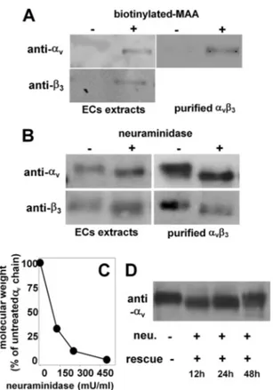
NeuAc Is Associated with Endothelial␣v3Integrin—To eval-uate the presence of NeuAc on glycan(s) linked to␣v3of the
endothelial surface, ECs were lysed, immunoprecipitated with the NeuAc-binding lectin MAA, and blotted with anti-␣vor
anti-3antibodies. As shown in Fig. 1A, MAA binds to both␣v
and3subunits, similarly to the␣vsubunit from human
pla-centa integrin. In a second set of experiments, ECs were treated with neuraminidase (500 milliunits/ml), lysed, and analyzed by WB with anti-␣vor anti-3antibodies. Neuraminidase
treat-ment causes a decrease of the molecular mass of both␣v(from
134 to 126 kDa) and3(from 70 to 67 kDa). Similar results were obtained with␣v3purified from human placenta (Fig. 1B). The
at BIBL DELLA FAC DI MED, on June 11, 2012
www.jbc.org
effect of neuraminidase is dose-dependent, with a partial removal of NeuAc residues obtained for concentrations of the enzyme lower than 500 milliunits/ml (Fig. 1C). After its enzy-matic removal, NeuAc is fully re-exposed on endothelial␣v3
only after 48 h (Fig. 1D).
NeuAc Is Involved in ␣v3/Tat Interaction—Tat binds to ␣v3(25). The contribution of the RGD motif and of the basic
domain to␣v3binding and cell-adhesive capacity was here
characterized by two experimental approaches. First, the mutants GST-Tat 1e (in which the RGD sequence has been deleted) and GST-Tat R3 A (in which the arginine residues of the basic domain have been substituted with alanine residues) were evaluated for their ␣v3 binding capacity in SPR. As
shown in Fig. 2A and Table 1, the two mutants retain the capac-ity to bind to␣v3, although decreased in respect to wild type
GST-Tat. Second, a cell adhesion assay was performed in the presence of the peptide GRGDSPK (which competes with the RGD motif of Tat for the binding to␣v3) or in the presence of
the K5 derivative K5NOSH (which inhibits EC adhesion to Tat (33) by binding to the basic domain of the transactivating fac-tor). As shown in Fig. 2B, when assayed at the doses of 12M
(GRGDSPK) and 75 nM (K5NOSH), the two compounds
slightly inhibit EC adhesion to Tat, but when assayed together,
FIGURE 1. Detection of NeuAc on integrin␣v3. A, GM7373 ECs or purified
human integrin␣v3were immunoprecipitated with biotinylated MAA and analyzed in WB with anti-␣vor anti-3antibodies. B and C, alternatively, they were incubated with 500 milliunits/ml (mU/ml) (B) or with the indicated con-centrations (C) of neuraminidase, lysed, and analyzed in WB with anti-␣vor anti-3antibodies. D, GM7373 ECs were treated with neuraminidase (neu., 500 milliunits/ml), washed, further incubated for the indicated periods of time in the absence of the enzyme (rescue), lysed, and analyzed in WB with anti-␣vantibodies. In panels A, B, and D, the data shown are representative of 2–3 other experiments that gave similar results. In C, data are expressed as the percentage of the molecular mass of the␣vsubunit from neuraminidase-treated ECs with respect to the intact protein from unneuraminidase-treated ECs.
FIGURE 2. Role of␣v3-associated NeuAc in Tat interaction. A, overlay of
blank-subtracted sensorgrams showing the binding of␣v3(10 nM) to sensor chip-immobilized wild type GST-Tat or its mutants GST-Tat 1e or GST-Tat R3 A. B, GM7373 ECs were allowed to adhere to Tat in the absence (ctrl) or in the presence of the indicated inhibitors. C, overlay of blank-subtracted sen-sorgrams showing the binding of native or neuraminidase-treated (neu.)␣v3 (12.5 nM) to sensor chip-immobilized synthetic Tat in the absence or in the presence of MAA (250 nM). The sensorgrams generated by injecting MAA (250 nM) or neuraminidase (500 milliunits/ml) on the Tat surface are also shown.
D, saturation curves obtained using the values of resonance units (RU) bound at
equilibrium from injection of increasing concentrations of native or neuramini-dase-treated␣v3onto immobilized synthetic Tat in the absence or in the pres-ence of MAA (250 nM). E,␣v3(50 nM) was injected onto the sensor chip contain-ing the indicated GST-Tat proteins in the absence or in the presence of MAA (250 nM). In A and C, the sensorgrams shown are representative of three others that gave similar results. In D, each point is the mean⫾ S.E. of 3 independent injec-tions. In B and E, each point is the mean⫾ S.E. of 3 independent experiments in duplicate and is expressed as the percentage of ECs adherent to Tat in the absence of any inhibitor or of␣v3bound to the sensor chip at the equilibrium in the absence of MAA, respectively (*⫽ p ⬍ 0.05, Student’s t test).
at BIBL DELLA FAC DI MED, on June 11, 2012
www.jbc.org
they completely inhibit the same process. Taken together, these data indicate that both the RGD and the basic domain of Tat contribute to its cell-adhesive capacity, the presence of one of the two domains being enough to ensure a partial␣v3binding
and cell-adhesive capacity to Tat.
Besides␣v3, the basic domain also mediates the binding of
Tat to the polyanionic heparin/heparan sulfate (28), the two interactions occurring with similar affinities (Table 1). These observations suggest that the basic domain of Tat, besides interacting with the negatively charged sulfated groups of hep-arin, may as well make contact with the negatively charged NeuAc residues of␣v3. To evaluate this possibility, two
dif-ferent experimental approaches were exploited. (i) We first evaluated the effect of neuraminidase and MAA on␣v3/Tat
interaction. Preliminary SPR analyses demonstrated that neuraminidase treatment of the integrin, as well as the presence of MAA, significantly inhibits its interaction with Tat. It is important to note that MAA and neuraminidase do not bind directly to Tat (Fig. 2C). In a second set of experiments, increas-ing concentrations of native or neuraminidase-treated ␣v3
were injected onto the Tat surface in the absence or in the presence of MAA. Then, the values of steady-state SPR data were used to generate the saturation curves shown in Fig. 2D. Scatchard plot analysis demonstrated that neuraminidase treatment, as well as MAA, decreases the affinity of the␣v3/ Tat interaction (Table 1). Interestingly, the reduction of the affinity observed for␣v3/Tat interaction in the absence or in
the presence of MAA (2.48 times) is in the same order of mag-nitude as the difference of the affinity measured for the inter-action of␣v3with wild type GST-Tat or with GST-Tat R3 A
(2.53 times) (Table 1), suggesting that MAA hampers the inter-action of NeuAc residues of␣v3to the basic domain of Tat. (ii)
The two Tat mutants GST-Tat 1e and GST-Tat R3 A where evaluated for their capacity to induce EC adhesion in the pres-ence of MAA. Similarly to what was observed with synthetic Tat, at 250 nM, MAA partially inhibits the binding of␣v3to
wild type GST-Tat (Fig. 2E). At the same concentration, MAA exerts a weak inhibition on␣v3/GST-Tat R3 A interaction
(possibly because this occurs only via the RGD sequence), whereas it efficiently inhibits ␣v3/GST-Tat 1e interaction
(possibly because this can occur only via the basic domain) (Fig. 2E). Taken together, these data indicate that at low concentra-tions, MAA binds to NeuAc residues of␣v3, inhibiting the
interaction of the integrin with the basic domain of Tat but leaving unaffected that with the RGD motif.
NeuAc Mediates␣v3-dependent EC Adhesion to Substrate-immobilized Tat—Substrate-immobilized Tat induces EC adhesion in an␣v3-dependent manner (25). Accordingly, by
using specific anti-␣v3or anti-VEGFR2 antibodies, here we
confirmed that EC adhesion to Tat specifically depends on the integrin but not on VEGFR2 (Fig. 3A). Also, our unpublished experiments with silencing RNAs directed against enzymes of the biosynthetic pathway of heparan sulfate demonstrated that these receptors are not involved in EC adhesion to Tat (data not shown).
TABLE 1
Affinity of the interaction of␣v3to the various Tat mutants immobi-lized to a BIAcore sensor chip
The values of dissociation constant (Kd) reported have been calculated by the
Scatchard plot analysis of the steady-state SPR data in the different experimental conditions adopted. For a comparison, the Kdof the Tat/heparin interaction
previ-ously calculated by Scatchard plot analysis of the steady-state SPR (33) is also reported.
Ligand Analyte Kd
nM
Synthetic Tat Native␣v3 19.9
␣v3after neuraminidase treatment 157.1
␣v3⫹ MAA 49.5
GST-Tat wild type Native␣v3 40.3
GST-Tat R3A 102.6 GST-Tat 1e 136.2 GST-Tat wild type Heparin 16.0
FIGURE 3. Role of NeuAc in␣v3-dependent EC adhesion to Tat and
cyto-skeleton organization. A, GM7373 ECs were treated with the indicated
con-centrations of neuraminidase and subjected to cell adhesion assay on Tat or FN. Alternatively, cells were subjected to adhesion assay in the presence of anti-VEGFR2 antibody (400g/ml, white arrow) or with anti-␣v3antibody (100g/ml, black arrow). B, GM7373 ECs were allowed to adhere to Tat or FN, incubated for 1 h with increasing concentrations of neuraminidase, and then further incubated in the absence of the enzyme for 24 h. C, GM7373 ECs were treated with PDMP (10Mfor 72 h), PPPP (1Mfor 48 h), or vehicle (ctrl) and subjected to cell adhesion assay on wells without coating (⫺) or coated with Tat or fibrinogen (FG). At the end of the incubations, adherent cells were counted. D, GM7373 ECs were treated with neuraminidase, allowed to adhere to Tat or FN, co-stained for nuclei (blue), paxillin (green), and actin (red), and photographed (630⫻). E, GM7373 ECs were incubated for 2 h at 37 °C with increasing concentrations of MAA (circles) or UEA (triangles) and then allowed to adhere to Tat (black symbols) or FN (white symbols). F, GM7373 ECs were incubated for 2 h at 37 °C with increasing concentrations of MAA and allowed to adhere onto the indicated GST-Tat proteins. In panels A–C, E, and F, each point is the mean⫾ S.E. of 3–4 independent experiments in duplicate (* ⫽
p⬍ 0.05, ** ⫽ p ⬍ 0.01, with respect to untreated controls, Student’s t test).
at BIBL DELLA FAC DI MED, on June 11, 2012
www.jbc.org
The removal of NeuAc from the EC surface by neuramini-dase prevents EC adhesion to Tat, only slightly affecting that to the␣51-ligand FN, here used as a control (Fig. 3A).
Neuramin-idase also causes the detachment of ECs already adhered to Tat over a 24-h period of incubation (Fig. 3B), without causing sig-nificant cell death (as assessed by MTS assay, data not shown). Besides integrins, NeuAc is also associated with gangliosides. To evaluate the possible involvement of these structures in EC adhesion to Tat, the cells were treated with PDMP or PPPP (which prevent ganglioside biosynthesis without affecting intracellular levels of ceramide (34)) and then evaluated for their capacity to adhere to Tat. When used at doses that effec-tively hamper the expression of NeuAc-bearing gangliosides (34), PDMP and PPPP do not affect ECs adhesion to Tat or to fibrinogen (another␣v3ligand here used as a control, Fig. 3C).
These results rule out the possibility that ganglioside-associ-ated NeuAc is responsible for the observed EC adhesion to Tat. EC adhesion to Tat induces cytoskeleton organization with the assembly of actin stress fibers and focal adhesion plaques containing integrins and paxillin (35). We then evaluated the involvement of NeuAc in cytoskeleton organization of Tat-ad-herent ECs. When tested at concentrations that do not hamper EC adhesion (125 milliunits/ml), neuraminidase prevents the proper organization of actin stress fibers and paxillin-contain-ing focal adhesion plaques. The effect is specific because neuraminidase did not alter focal adhesion plaque formation in FN-adherent ECs (Fig. 3D).
In a second set of experiments, we also evaluated whether MAA affects␣v3-dependent EC adhesion to Tat. As shown in Fig. 3E, MAA inhibits EC adhesion to Tat in a dose-dependent
way. The inhibition is specific because MAA does not affect EC adhesion to FN and UEA (a lectin that specifically binds to ␣-linked fucose) does not affect EC adhesion to Tat. In agree-ment with the results of the SPR analyses, MAA inhibits EC adhesion to GST-Tat 1e with a potency (ID50⬍ 125 nM) that is
higher than those with which it inhibits EC adhesion to GST-Tat R3 A and to wild type GST-GST-Tat (ID50 ⫽ 198 and 182,
respectively) (Fig. 3F).
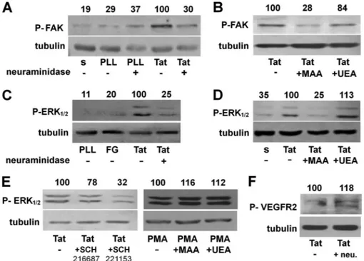
NeuAc Is Required for Signal Transduction Triggered by Tat/
␣v3Interaction in ECs—We evaluated the role of NeuAc in Tat/␣v3-dependent phosphorylation of FAK, a second
mes-senger involved in Tat/␣v3-dependent EC cytoskeleton
orga-nization and proangiogenic activation (25, 36). Preliminary experiment confirmed that FAK undergoes phosphorylation in ECs adherent to Tat but not in ECs maintained in suspension or adherent to poly-L-lysine (an integrin-independent adhesive
molecule). When used at 125 milliunits/ml (a dose that pre-vents cytoskeleton organization without hampering EC adhe-sion to Tat; Fig. 3), neuraminidase inhibits FAK phosphoryla-tion in Tat-adherent ECs (Fig. 4A). Accordingly, at 62 nM,
MAA, but not UEA, prevents FAK phosphorylation (Fig. 4B). It is relevant to note that at 62 nM, MAA does not hamper EC
adhesion to Tat (Fig. 3E). Besides FAK, EC adhesion to Tat induces the activation of ERK1/2, another second messenger involved in Tat-dependent EC proangiogenic activation (37). Neuraminidase pretreatment (Fig. 4C) and MAA, but not UEA (Fig. 4D), prevent ERK1/2 phosphorylation in Tat-adherent EC. In the same experimental conditions, ERK1/2 phosphoryla-tion is inhibited by the specific␣v3antagonist SCH221153 but not by its inactive analog SCH21668, indicating that ERK1/2
FIGURE 4. Role of NeuAc in Tat/␣v3-triggered signal transduction in ECs. A–E, serum-starved GM7373 ECs were allowed to adhere to the indicated proteins
in the presence of MAA or UEA (62 nM) or treated with neuraminidase (neu., 125 milliunits/ml) and then maintained in suspension (s) for 30 min or allowed to adhere for 1 h to the indicated proteins. Alternatively, cells were allowed to adhere to Tat and incubated in the presence of the␣v3antagonist SCH221153 or of its inactive analog SCH216687 (0.3M) or allowed to adhere to tissue culture plates and treated with free phorbol myristate acetate (PMA, 10 ng/ml) in the presence of MAA or UEA (62 nM) (E). At the end of the incubations, cells were lysed and analyzed for FAK (A and B), ERK1/2 (C–E), or VEGFR2 (F) phosphorylation (indicated by P). Tubulin was used as a loading control. The integrated densities of the bands were evaluated, normalized to tubulin, and expressed as the percentage with respect to the bands from EC adherent to Tat in the absence of any inhibitor. The data shown are representative of 2–3 additional experiments that gave similar results. PLL, poly-L-lysine.
at BIBL DELLA FAC DI MED, on June 11, 2012
www.jbc.org
activation is dependent, at least in part, on␣v3. Also, MAA does not affect ERK1/2 phosphorylation induced by phorbol myristate acetate in EC adherent to tissue culture plastic (Fig. 4E), suggesting that the specificity of the inhibitory effect exerted by MAA is likely due to its interaction with␣v3, which
prevents the binding of the integrin to Tat (Fig. 2).
Besides integrin ␣v3, the proangiogenic activity of Tat is
also mediated by VEGFR2 activation (38). Interestingly, this receptor bears NeuAc residues (39). On these bases, we evalu-ated the effect of the removal of NeuAc residues of VEGFR2 on its phosphorylation driven by Tat. As shown in Fig. 4F, neuramin-idase effectively removes NeuAc from VEGFR2, as assessed by the decrease of the molecular mass of the band corresponding to the receptor. However, neuraminidase treatment does not hamper VEGFR2 phosphorylation in response to Tat, which is instead even increased (Fig. 4F).
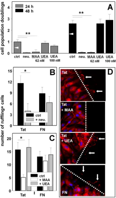
EC NeuAc Is Involved in ␣v3-dependent Proangiogenic Activity of Tat—EC adhesion, proliferation, and migration rep-resent essential steps of neovascularization (40). Accord-ingly, ECs adherent to substrate-immobilized Tat are induced to migrate and proliferate in an ␣v3-dependent
way (27). Preliminary experiments confirmed that Tat-ad-herent ECs proliferate more efficiently than those adTat-ad-herent to FN both at 24 h and at 48 h (Fig. 5A). Pretreatment with neuraminidase (125 milliunits/ml) inhibits the proliferation of Tat-adherent cells in a specific way because it leaves unaf-fected the basal proliferation of FN-adherent ECs or of ECs adherent on tissue culture plates (data not shown). Besides neuraminidase, MAA (62 nM), but not UEA (assayed at both
62 nMand 100 nM), inhibits the proliferation of Tat-adherent
ECs (Fig. 5A).
Cell membrane ruffling, which precedes the migration of EC body, is considered a morphological phenotype of motile cells (41) and has been already exploited to characterize the migra-tion of Tat-adherent ECs (27). As shown in Fig. 5D, when adherent to Tat or to FN, ECs rapidly form membrane ruffles at the edge of a wounded monolayer. Pretreatment with neuraminidase (125 milliunits/ml) inhibits membrane ruffling in Tat-adherent ECs and, to a lesser extent, in FN-adherent ECs (Fig. 5B). Accordingly, MAA (62 nM), but not UEA, inhibits
membrane ruffling only in Tat- but not FN-adherent ECs (Fig. 5C).
The ability of a substrate-immobilized protein to stimulate proliferation and motility in adherent ECs leads to an increased capacity of a mechanically wounded EC monolayer to cover the denuded area, a biological activity referred to as “motogenic activity” that has been used as a surrogate marker of angiogen-esis (31). Substrate-immobilized Tat induces motogenangiogen-esis of adherent ECs in an␣v3-dependent way (25).
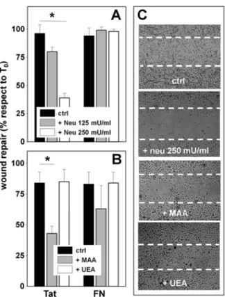
When allowed to adhere to Tat, treated with neuraminidase (250 milliunits/ml), and wounded, ECs show a decreased motogenic activity. The effect is specific because, in the same experimental conditions, neuraminidase does not affect the motogenic activity of FN-adherent ECs (Fig. 6A). In this regard, it is important to recall that when added after EC adhesion has occurred, neuraminidase at 250 nMdoes not cause the
detach-ment of Tat-adherent ECs (Fig. 3B) nor affect their viability over a 48-h period of time (data not shown). MAA (62 nM)
inhibits the motogenic activity of Tat. The effect is specific because MAA does not affect viability of Tat-adherent ECs (data not shown) nor the motogenic activity of FN-adherent ECs (Fig. 6, B and C). Also, UEA is ineffective on Tat-adherent ECs (Fig. 6, B and C).
Tat induces neovascularization via ␣v3 (27). We then decided to evaluate the involvement of NeuAc in Tat
proangio-FIGURE 5. Role of NeuAc in Tat/␣v3-dependent proliferation and
migration of ECs. GM7373 ECs adherent to the indicated proteins were
treated as follows. A, cells were treated with neuraminidase (neu., 125 milliunits/ml) and incubated in the absence of the enzyme for an addi-tional 24 or 48 h. Alternatively, cells were directly incubated for 24 or 48 h with the indicated concentrations of MAA or UEA. B, cells were incubated in the absence (ctrl) or in the presence of neuraminidase (125 milliunits/ ml), wounded, and further incubated for 30 min. C, cells were wounded and incubated for 30 min in the absence (ctrl) or in the presence of MAA or UEA (62 nM). At the end of the incubations, EC proliferation (A) or the number of ruffling-positive ECs (B and C) were evaluated. Each point is the mean ⫾ S.E. of 3– 4 independent experiments in duplicate (*⫽ p ⬍ 0.01, ** ⫽ p ⬍ 0.001, Student’s t test). In panel A, white arrowheads point to the proliferation measured in cells adherent to FN. D, represen-tative epifluorescence microphotographs (630⫻) of ECs at the edge of a wounded monolayer incubated in the absence (ctrl) or in the presence of MAA or UEA (62 nM) and stained with DAPI (blue) and with anti-fascin antibody (red). Arrows point to the most prominent ruffles.
at BIBL DELLA FAC DI MED, on June 11, 2012
www.jbc.org
genic activity by exploiting the human artery ring sprouting assay, an ex vivo model used to characterize the pro- or antian-giogenic properties of various anantian-giogenic growth factors and inhibitors (32). As shown in Fig. 7, Tat effectively induces an increase of the number of EC sprouts that generate from a human artery ring, and this activity can be inhibited by MAA, but not by UEA.
DISCUSSION
␣v3mediates EC adhesion and proangiogenic activation by
binding cationic angiogenic growth factors including FGF2 and HIV-1 Tat (27, 30). On the other hand, NeuAc has so far been identified on␣v3of melanoma metastatic cell surface, where it regulates cell adhesion (18, 20). We thus decided to investigate whether NeuAc is also associated with␣v3integrin of ECs and
whether it is involved in␣v3/Tat interaction and in the
conse-quent proangiogenic activation of ECs. These possibilities were investigated by two complementary approaches, consisting of the use of neuraminidase from C. perfringens (an enzyme that removes NeuAc from the cell surface) and of MAA (a lectin that specifically binds NeuAc residues).
In ECs, the removal of␣v3-associated NeuAc by
neuramin-idase is rapid and efficient, occurring in less than 2 h. The bio-chemical features of bacterial neuraminidase surely contribute to this (43), but so does the rapid recycling of integrins that are classically internalized to the early endosomes to be immedi-ately returned to the plasma membrane (44, 45). In this way, endogenous␣v3remains continuously exposed at the cell
sur-face, accessible to exogenous neuraminidase.
Fully sialylated␣v3integrins are re-exposed on ECs only
48 h after neuraminidase treatment. The long-lasting nature of ␣v3desialylation is likely due to the fact that after de novo
synthesis, integrins must undergo sialylation by sialyltrans-ferases in the Golgi apparatus (46). Whatever its cause, the long-lasting desialylation of␣v3by neuraminidase is in agree-ment with the capacity of the enzyme to inhibit processes, such as EC proliferation and motogenesis, that occur over 24 – 48-h periods.
MAA immunoprecipitates both ␣v and 3subunits from
ECs, indicating the presence of␣(233)-linked NeuAc on the two chains. We also obtained similar results with the lectin from Sambucus nigra that binds instead to ␣(236)-linked NeuAc (data not shown). Accordingly, neuraminidase from
C. perfringens, which hydrolyzes both ␣((233)-linked and ␣((236)-linked NeuAc, causes a decrease of the molecular mass of both␣vsubunit (⫺7 kDa, corresponding to about 25
NeuAc residues) and 3subunit (⫺3 kDa, corresponding to
about 10 NeuAc residues). These data are in agreement with the observations that␣vand3subunits possess 13 and 6 putative
N-glycosylation sites, respectively, with biantennary structures (UniProt accession number P05106) (18).
Neuraminidase cleaves ␣(233)-linked NeuAc residues faster than␣((236) linkages (47). Accordingly, neuraminidase operates a complete removal of ␣v3-associated NeuAc only
when used at high concentration (⬎250 milliunits/ml), whereas when used at suboptimal concentrations (125 milliunits/ml), desialylation of␣v3remains incomplete. Interestingly, when
used at higher concentrations, neuraminidase directly prevents (and even disrupts)␣v3-dependent EC adhesion to Tat (Fig. 3,
Aand B), whereas at lower concentrations, it leaves EC adhe-sion unaffected, although inhibiting Tat/␣v3-dependent
proangiogenic activation of Tat-adherent ECs. Taken together, these data suggest that the complete removal of␣v3 -associ-ated NeuAc prevents the binding of the integrin to Tat and the consequent EC adhesion. Instead, a partial removal of NeuAc
FIGURE 6. Role of NeuAc in Tat/␣v3-dependent motogenesis of ECs.
A, GM7373 ECs adherent to the indicated proteins were left untreated (ctrl) or
treated with neuraminidase (neu) at 125 or 250 milliunits/ml, wounded ,and further incubated for 48 h. B, GM7373 ECs adherent to the indicated proteins were wounded and incubated for 48 h in the absence (ctrl) or in the presence of MAA or UEA (62 nM). At the end of the incubations, the extension of the area of the wound repaired was evaluated. Each point is the mean⫾ S.E. of 3–5 fields measured in one experiment out of 2–3 that gave similar results (*⫽
p⬍ 0.05, Student’s t test). C, microphotographs (50⫻) of wounded
monolay-ers of Tat-adherent GM7373 ECs taken at the end of the 48-h period of incu-bation with the indicated treatments. Dashed lines mark the edge of the wound at the beginning of the experiment.
FIGURE 7. Effect of MAA on Tat-induced neovascularization. A, human umbilical artery rings were embedded in fibrin gel and incubated without (vehicle) or with Tat in the absence (ctrl) or in the presence of MAA or UEA. After 6 days, EC sprouts were counted. Each point is the mean⫾ S.E. of 10–29 artery rings randomly chosen (*⫽ p ⬍ 0.001, Student’s t test). B, representa-tive aorta rings in the different experimental conditions photographed under an inverted microscope at 200⫻ magnification.
at BIBL DELLA FAC DI MED, on June 11, 2012
www.jbc.org
residues allows an “unproductive” Tat/␣v3interaction enough for cell adhesion, but that does not mediate the signal transduc-tion cascade required for EC proangiogenic activatransduc-tion.
By removing NeuAc residues from ␣v3, neuraminidase
causes a decrease of the affinity of␣v3/Tat interaction and its
inhibition (Fig. 2). Accordingly, by binding NeuAc residues of ␣v3, MAA causes the same effects, also if at a lesser extent. The
inhibition exerted by neuraminidase and MAA on ␣v3/Tat
interaction is mirrored by their capacity to inhibit several␣v3 -dependent biological activities of Tat, connecting the two pro-cesses. In effect, MAA shares with neuraminidase the capacity to differently affect Tat/␣v3interaction and consequent
bio-logical activities in a concentration-dependent way. At higher concentrations (125–250 nM), it directly inhibits␣v3
-medi-ated EC adhesion to Tat, whereas at a lower concentration (less than 62 nM), it leaves cell adhesion unaffected, although
inhib-iting the ␣v3-dependent signal transduction and proangio-genic activation of Tat-adherent ECs. Interestingly, MAA scarcely affects ␣v3 interaction with (and EC adhesion to)
GST-Tat R3 A (in which the intact RGD motif mediates integ-rin interaction and EC adhesion), whereas it exerts a stronger inhibition on GST-Tat 1e that, lacking the RGD motif, binds to integrin and mediates EC adhesion via its basic domain. These findings, together with the notion that␣v3interaction occurs
via both the RGD motif and the basic domain of Tat (25) (Fig. 2B), suggest that at appropriate concentrations, MAA succeeds in inhibiting the interaction of the basic domain of Tat with NeuAc residues of ␣v3, leaving unaffected the
RGD-depen-dent␣v3/Tat interaction. This generates an unproductive Tat/
␣v3 interaction enough to promote cell adhesion but not
adequate to trigger signal transduction and hence EC proangio-genic activation.
Besides integrin␣v3, a wide variety of cell surface sialogly-coproteins can be affected by neuraminidase and MAA, possi-bly impacting the phenomena reported here. Although we can-not completely rule out this possibility, it is, however, relevant to point out that neuraminidase does not significantly affect (or even increase) Tat-driven phosphorylation of VEGFR2, a tyro-sine kinase receptor that bears NeuAc residues (39) and whose activation is required for Tat proangiogenic activity (38). Curi-ously, the proangiogenic fibroblast growth factor (FGF) recep-tor-1 also possesses NeuAc-bearing glycans whose removal leads to an increased capacity to bind its ligand FGF2 (48). Also, hypo- or desialylation of␣51integrin increases its affinity for
the natural ligand FN (49). Relevant to this point, FN acts as a proangiogenic factor (50), and␣51can act as a Tat receptor on ECs (26). Taken together, these results indicate that although neuraminidase and MAA can act on sialoglycoproteins differ-ent from␣v3, the overall inhibitory effect exerted on the Tat proangiogenic potential depends on their action on NeuAc linked to␣v3.
The results presented in this work open new fields of research, some of which have been totally unexplored so far. First, mammals have four types of endogenous sialidases, among which the plasma membrane-associated sialidase NEU3 and the secreted form of NEU2 (51) could be involved in the fine-tuning of the acidic sugar on the cell surface. On the other hand, sialidases are expressed by ECs (52), and
neutrophil-de-rived endogenous sialidase(s) have already been demonstrated to induce desialylation of the EC surface with implications in the process of inflammation (53). Taken together, these obser-vations point to a possible role of neuraminidases as regulators of the process of neovascularization. Second, besides Tat, VEGF (54) and FGF2 (14, 15) also bind to integrins and to NeuAc-bearing gangliosides (55), suggesting a broader involve-ment of integrin-associated NeuAc in angiogenesis. Third, sia-lyltransferases are a family of enzymes located in the Golgi apparatus that mediate sialylation of various glycoconjugates (56) including integrins (46). Also, they are expressed by ECs (57). Thus, besides neuraminidases, sialyltransferases may also be involved in the regulation of angiogenesis. Fourth, integrin ␣v3is a target for antitumor therapies based on antibodies or
RGD-based compounds that prevent the interaction of the integrin with its proangiogenic ligands (58). The␣v3
antago-nist activity of MAA points to NeuAc-binding lectins as tem-plates for the design of novel integrin antagonists endowed with antiangiogenic potential. Fifth, in the field of virology, integrins act as entry receptors for several viruses (59), suggesting the possibility of blocking viral infection by means of NeuAc-bind-ing lectin-like compounds or by NeuAc analogues. Relevant to this point, the NeuAc derivative NMSO3 has been demon-strated to exert a potent inhibition against HIV-1 (42).
In conclusion, the results presented in this study open up the possibility that modulation of integrin glycosylation could be a promising strategy for regulating angiogenesis and viral infection.
Acknowledgments—We thank Mauro Giacca (International Centre for Genetic Engineering and Biotechnology, Trieste, Italy) for the E. coli strain expressing the various GST-Tat mutants, Pasqua Oreste for the K5NOSH, Alessandra Armato for technical assistance, and Eugenio Monti and Marco Presta (University of Brescia) for helpful discussion.
REFERENCES
1. Urbinati, C., Chiodelli, P., and Rusnati, M. (2008) Polyanionic drugs and viral oncogenesis: a novel approach to control infection, tumor-associated inflammation, and angiogenesis. Molecules 13, 2758 –2785
2. dos Santos, W. L., Rahman, J., Klein, N., and Male, D. K. (1995) Distribu-tion and analysis of surface charge on brain endothelium in vitro and in
situ. Acta Neuropathol. 90, 305–311
3. Vorbrodt, A. W. (1989) Ultracytochemical characterization of anionic sites in the wall of brain capillaries. J. Neurocytol. 18, 359 –368
4. Traving, C., and Schauer, R. (1998) Structure, function, and metabolism of sialic acids. Cell. Mol. Life Sci. 54, 1330 –1349
5. Schauer, R. (2009) Sialic acids as regulators of molecular and cellular in-teractions. Curr. Opin. Struct. Biol. 19, 507–514
6. Murea¸n, V., and Simionescu, N. (1987) High and low molecular weight tracers for the electron microscopical detection of sialoglycoconjugates.
Histochem. J. 19, 170 –178
7. Welim, H. B., Thies, M., and Herken, R. (1989) Appearance of lectin-binding sites during vascularization of the primordium of the central ner-vous system in 10 –12-day-old mouse embryos. Cell Tissue Res. 255, 627– 630
8. Henry, C. B., and DeFouw, D. O. (1996) Distribution of anionic sites on microvascular endothelium of the chick chorioallantoic membrane.
Tis-sue Cell 28,449 – 454
9. Doiron, A. L., Kirkpatrick, A. P., and Rinker, K. D. (2004) TGF- and TNF-␣ affect cell surface proteoglycan and sialic acid expression on
at BIBL DELLA FAC DI MED, on June 11, 2012
www.jbc.org
cular endothelial cells. Biomed. Sci. Instrum. 40, 331–336
10. Karlsson, K. A. (1991) Glycobiology: a growing field for drug design.
Trends Pharmacol. Sci. 12,265–272
11. Banks, W. A., Robinson, S. M., Wolf, K. M., Bess, J. W., Jr., and Arthur, L. O. (2004) Binding, internalization, and membrane incorporation of human immunodeficiency virus-1 at the blood-brain barrier is differen-tially regulated. Neuroscience 128, 143–153
12. Banks, W. A., and Kastin, A. J. (1998) Characterization of lectin-mediated brain uptake of HIV-1 GP120. J. Neurosci. Res. 54, 522–529
13. Chung, T. W., Kim, S. J., Choi, H. J., Kim, K. J., Kim, M. J., Kim, S. H., Lee, H. J., Ko, J. H., Lee, Y. C., Suzuki, A., and Kim, C. H. (2009) Ganglioside GM3inhibits VEGF/VEGFR2-mediated angiogenesis: direct interaction of GM3with VEGFR2. Glycobiology 19, 229 –239
14. Rusnati, M., Tanghetti, E., Urbinati, C., Tulipano, G., Marchesini, S., Ziche, M., and Presta, M. (1999) Interaction of fibroblast growth factor-2 (FGF-2) with free gangliosides: biochemical characterization and biolog-ical consequences in endothelial cell cultures. Mol. Biol. Cell 10, 313–327 15. Rusnati, M., Urbinati, C., Tanghetti, E., Dell’Era, P., Lortat-Jacob, H., and Presta, M. (2002) Cell membrane GM1ganglioside is a functional core-ceptor for fibroblast growth factor 2. Proc. Natl. Acad. Sci. U.S.A. 99, 4367– 4372
16. Kajiji, S., Tamura, R. N., and Quaranta, V. (1989) A novel integrin (␣E4) from human epithelial cells suggests a fourth family of integrin adhesion receptors. EMBO J. 8, 673– 680
17. Graham, K. L., Halasz, P., Tan, Y., Hewish, M. J., Takada, Y., Mackow, E. R., Robinson, M. K., and Coulson, B. S. (2003) Integrin-using rotaviruses bind ␣21integrin␣2I domain via VP4 DGE sequence and recognize ␣X2and ␣V3by using VP7 during cell entry. J. Virol. 77, 9969 –9978
18. Kremser, M. E., Przybyło, M., Hoja-Łukowicz, D., Pochec´, E., Amoresano, A., Carpentieri, A., Bubka, M., and Lityńska, A. (2008) Characterization of ␣31and␣v3integrin N-oligosaccharides in metastatic melanoma WM9 and WM239 cell lines. Biochim. Biophys. Acta 1780, 1421–1431 19. Woodard-Grice, A. V., McBrayer, A. C., Wakefield, J. K., Zhuo, Y., and
Bellis, S. L. (2008) Proteolytic shedding of ST6Gal-I by BACE1 regulates the glycosylation and function of␣41 integrins. J. Biol. Chem. 283, 26364 –26373
20. Chiang, C. H., Wang, C. H., Chang, H. C., More, S. V., Li, W. S., and Hung, W. C. (2010) A novel sialyltransferase inhibitor AL10 suppresses invasion and metastasis of lung cancer cells by inhibiting integrin-mediated signal-ing. J. Cell Physiol. 223, 492– 499
21. Morova, J., Osicka, R., Masin, J., and Sebo, P. (2008) RTX cytotoxins rec-ognize 2 integrin receptors through N-linked oligosaccharides. Proc.
Natl. Acad. Sci. U.S.A. 105,5355–5360
22. Mousa, S. A. (2008) Cell adhesion molecules: potential therapeutic and diagnostic implications. Mol. Biotechnol. 38, 33– 40
23. Eliceiri, B. P., and Cheresh, D. A. (1998) The role of␣vintegrins during angiogenesis. Mol. Med. 4, 741–750
24. Noonan, D., and Albini, A. (2000) From the outside in: extracellular activ-ities of HIV Tat. Adv. Pharmacol. 48, 229 –250
25. Urbinati, C., Bugatti, A., Giacca, M., Schlaepfer, D., Presta, M., and Rus-nati, M. (2005)␣v3integrin-dependent activation of focal adhesion ki-nase mediates NF-B activation and motogenic activity by HIV-1 Tat in endothelial cells. J. Cell Sci. 118, 3949 –3958
26. Barillari, G., Sgadari, C., Fiorelli, V., Samaniego, F., Colombini, S., Manzari, V., Modesti, A., Nair, B. C., Cafaro, A., Stürzl, M., and Ensoli, B. (1999) The Tat protein of human immunodeficiency virus type-1 promotes vascular cell growth and locomotion by engaging the␣51and␣v3integrins and by mobilizing sequestered basic fibroblast growth factor. Blood 94, 663– 672
27. Urbinati, C., Mitola, S., Tanghetti, E., Kumar, C., Waltenberger, J., Ribatti, D., Presta, M., and Rusnati, M. (2005) Integrin␣v3as a target for blocking HIV-1 Tat-induced endothelial cell activation in vitro and angiogenesis in
vivo. Arterioscler. Thromb. Vasc. Biol. 25, 2315–2320
28. Rusnati, M., Tulipano, G., Urbinati, C., Tanghetti, E., Giuliani, R., Giacca, M., Ciomei, M., Corallini, A., and Presta, M. (1998) The basic domain in HIV-1 Tat protein as a target for polysulfonated heparin-mimicking ex-tracellular Tat antagonists. J. Biol. Chem. 273, 16027–16037
29. Grinspan, J. B., Mueller, S. N., and Levine, E. M. (1983) Bovine endothelial
cells transformed in vitro by benzo(a)pyrene. J. Cell Physiol. 114, 328 –338 30. Rusnati, M., Tanghetti, E., Dell’Era, P., Gualandris, A., and Presta, M. (1997)␣v3integrin mediates the cell-adhesive capacity and biological activity of basic fibroblast growth factor (FGF-2) in cultured endothelial cells. Mol. Biol. Cell 8, 2449 –2461
31. Lauder, H., Frost, E. E., Hiley, C. R., and Fan, T. P. (1998) Quantification of the repair process involved in the repair of a cell monolayer using an in
vitromodel of mechanical injury. Angiogenesis 2, 67– 80
32. Mitola, S., Moroni, E., Ravelli, C., Andres, G., Belleri, M., and Presta, M. (2008) Angiopoietin-1 mediates the proangiogenic activity of the bone morphogenic protein antagonist Drm. Blood 112, 1154 –1157
33. Urbinati, C., Bugatti, A., Oreste, P., Zoppetti, G., Waltenberger, J., Mitola, S., Ribatti, D., Presta, M., and Rusnati, M. (2004) Chemically sulfated
Esch-erichia coliK5 polysaccharide derivatives as extracellular HIV-1 Tat pro-tein antagonists. FEBS Lett. 568, 171–177
34. Kopitz, J., Bergmann, M., and Gabius, H. J. (2010) How adhesion/growth-regulatory galectins-1 and -3 attain cell specificity: case study defining their target on neuroblastoma cells (SK-N-MC) and marked affinity reg-ulation by affecting microdomain organization of the membrane. IUBMB
Life 62,624 – 628
35. Urbinati, C., Ravelli, C., Tanghetti, E., Belleri, M., Giacopuzzi, E., Monti, E., Presta, M., and Rusnati, M. (2012) Substrate-immobilized HIV-1 Tat drives VEGFR2/␣v3integrin complex formation and polarization in en-dothelial cells. Arterioscler. Thromb. Vasc. Biol. 32, e25– e34
36. Angelucci, A., and Bologna, M. (2007) Targeting vascular cell migration as a strategy for blocking angiogenesis: the central role of focal adhesion protein tyrosine kinase family. Curr. Pharm. Des. 13, 2129 –2145 37. Rusnati, M., Urbinati, C., Musulin, B., Ribatti, D., Albini, A., Noonan, D.,
Marchisone, C., Waltenberger, J., and Presta, M. (2001) Activation of en-dothelial cell mitogen-activated protein kinase ERK(1/2) by extracellular HIV-1 Tat protein. Endothelium 8, 65–74
38. Albini, A., Soldi, R., Giunciuglio, D., Giraudo, E., Benelli, R., Primo, L., Noonan, D., Salio, M., Camussi, G., Rockl, W., and Bussolino, F. (1996) The angiogenesis induced by HIV-1 tat protein is mediated by the Flk-1/ KDR receptor on vascular endothelial cells. Nat. Med. 2, 1371–1375 39. Nacev, B. A., Grassi, P., Dell, A., Haslam, S. M., and Liu, J. O. (2011) The
antifungal drug itraconazole inhibits vascular endothelial growth factor receptor 2 (VEGFR2) glycosylation, trafficking, and signaling in endothe-lial cells. J. Biol. Chem. 286, 44045– 44056
40. Folkman, J., and Klagsbrun, M. (1987) Angiogenic factors. Science 235, 442– 447
41. Ridley, A. J., Paterson, H. F., Johnston, C. L., Diekmann, D., and Hall, A. (1992) The small GTP-binding protein rac regulates growth factor-in-duced membrane ruffling. Cell 70, 401– 410
42. Terada, M., Fujita, S., Suda, I., and Mastico, R. (2005) Polysulfated sialic acid derivatives as anti-human immunodeficiency virus. Biomed.
Phar-macother. 59,423– 429
43. Bouwstra, J. B., Deyl, C. M., and Vliegenthart, J. F. (1987) Purification and kinetic properties of sialidase from Clostridium perfringens. Biol. Chem.
Hoppe Seyler 368,269 –275
44. Caswell, P. T., Vadrevu, S., and Norman, J. C. (2009) Integrins: masters and slaves of endocytic transport. Nat. Rev. Mol. Cell Biol. 10, 843– 853 45. Roberts, M., Barry, S., Woods, A., van der Sluijs, P., and Norman, J. (2001)
PDGF-regulated rab4-dependent recycling of␣v3 integrin from early endosomes is necessary for cell adhesion and spreading. Curr. Biol. 11, 1392–1402
46. Christie, D. R., Shaikh, F. M., Lucas, J. A., 4th, Lucas, J. A., 3rd, and Bellis, S. L. (2008) ST6Gal-I expression in ovarian cancer cells promotes an in-vasive phenotype by altering integrin glycosylation and function. J.
Ovar-ian Res. 1, 3
47. Corfield, A. P., Higa, H., Paulson, J. C., and Schauer, R. (1983) The speci-ficity of viral and bacterial sialidases for␣(233)- and ␣(236)-linked sialic acids in glycoproteins. Biochim. Biophys. Acta 744, 121–126
48. Duchesne, L., Tissot, B., Rudd, T. R., Dell, A., and Fernig, D. G. (2006)
N-Glycosylation of fibroblast growth factor receptor 1 regulates ligand and heparan sulfate co-receptor binding. J. Biol. Chem. 281, 27178 –27189
49. Semel, A. C., Seales, E. C., Singhal, A., Eklund, E. A., Colley, K. J., and Bellis,
at BIBL DELLA FAC DI MED, on June 11, 2012
www.jbc.org
S. L. (2002) Hyposialylation of integrins stimulates the activity of myeloid fibronectin receptors. J. Biol. Chem. 277, 32830 –32836
50. Scatena, M., Almeida, M., Chaisson, M. L., Fausto, N., Nicosia, R. F., and Giachelli, C. M. (1998) NF-B mediates ␣v3integrin-induced endothelial cell survival. J. Cell Biol. 141, 1083–1093
51. Monti, E., Bonten, E., D’Azzo, A., Bresciani, R., Venerando, B., Borsani, G., Schauer, R., and Tettamanti, G. (2010) Sialidases in vertebrates: a family of enzymes tailored for several cell functions. Adv. Carbohydr. Chem.
Biochem. 64,403– 479
52. Renkonen, R., Mattila, P., Majuri, M. L., Räbinä, J., Toppila, S., Renkonen, J., Hirvas, L., Niittymäki, J., Turunen, J. P., Renkonen, O., and Paavonen, T. (1997) In vitro experimental studies of sialyl Lewis x and sialyl Lewis a on endothelial and carcinoma cells: crucial glycans on selectin ligands.
Gly-coconj. J. 14, 593– 600
53. Sakarya, S., Rifat, S., Zhou, J., Bannerman, D. D., Stamatos, N. M., Cross, A. S., and Goldblum, S. E. (2004) Mobilization of neutrophil sialidase activity desialylates the pulmonary vascular endothelial surface and in-creases resting neutrophil adhesion to and migration across the endothe-lium. Glycobiology 14, 481– 494
54. Lang, Z., Guerrera, M., Li, R., and Ladisch, S. (2001) Ganglioside GD1a enhances VEGF-induced endothelial cell proliferation and migration.
Biochem. Biophys. Res. Commun. 282,1031–1037
55. Rusnati, M., and Presta, M. (2006) Extracellular angiogenic growth factor interactions: an angiogenesis interactome survey. Endothelium 13, 93–111
56. Harduin-Lepers, A., Mollicone, R., Delannoy, P., and Oriol, R. (2005) The animal sialyltransferases and sialyltransferase-related genes: a phyloge-netic approach. Glycobiology 15, 805– 817
57. Brockhausen, I., Lehotay, M., Yang, J. M., Qin, W., Young, D., Lucien, J., Coles, J., and Paulsen, H. (2002) Glycoprotein biosynthesis in porcine aortic endothelial cells and changes in the apoptotic cell population.
Gly-cobiology 12,33– 45
58. Auzzas, L., Zanardi, F., Battistini, L., Burreddu, P., Carta, P., Rassu, G., Curti, C., and Casiraghi, G. (2010) Targeting␣v3integrin: design and applications of mono- and multifunctional RGD-based peptides and semi-peptides. Curr. Med. Chem. 17, 1255–1299
59. Stewart, P. L., and Nemerow, G. R. (2007) Cell integrins: commonly used receptors for diverse viral pathogens. Trends Microbiol. 15, 500 –507
at BIBL DELLA FAC DI MED, on June 11, 2012
www.jbc.org