Renal Apolipoprotein A-I Amyloidosis: A Rare and Usually
Ignored Cause of Hereditary Tubulointerstitial Nephritis
Gina Gregorini,* Claudia Izzi,* Laura Obici,
†Regina Tardanico,
‡Christoph Ro¨cken,
§Battista Fabio Viola,* Mariano Capistrano,* Simona Donadei,
†Luciano Biasi,
储Tiziano Scalvini,
¶Giampaolo Merlini,
†and Francesco Scolari*
*Division of Nephrology,
‡Pathology Department,
储Division of Infectious Diseases, and
¶Department of Internal
Medicine, Spedali Civili, Brescia, Italy;
†Amyloid Center, Biotechnology Research Laboratories, IRCCS San Matteo,
Pavia, Italy; and
§Department Pathology, Otto von Guericke University of Magdeburg, Magdeburg, Germany
Apolipoprotein A-I amyloidosis is a rare, late-onset, autosomal dominant condition characterized by systemic deposition of amyloid in tissues, the major clinical problems being related to renal, hepatic, and cardiac involvement. Described is the clinical and histologic picture of renal involvement as a result of apolipoprotein A-I amyloidosis in five families of Italian ancestry. In all of the affected family members, the disease was caused by the Leu75Pro heterozygous mutation in exon 4 of apolipoprotein A-I gene, as demonstrated by direct sequencing and RFLP analysis. Immunohistochemistry confirmed that amyloid deposits were specifically stained with an anti-apolipoprotein A-I antibody. The clinical phenotype was mainly characterized by a variable combination of kidney and liver disturbance. The occurrence of renal involvement seemed to be almost universal, although its severity varied greatly ranging from subclinical organ damage to overt, slowly progressive renal dysfunction. The renal presentation was consistent with a tubulointerstitial disease, as suggested by the findings of defective urine-concentrating capacity, moderate polyuria, negative urinalysis, and mild tubular proteinuria. Histology confirmed tubulointerstitial nephritis. Surprising, amyloid was restricted to nonglomerular regions and limited to the renal medulla. This location of apolipoprotein A-I amyloid differs sharply from other systemic amyloidoses that are mainly characterized by glomerular and vascular deposits. The tubulointerstitial nephritis as a result of hereditary apolipoprotein A-I amyloidosis is a rare disease and a challenging diagnosis to recognize. Patients who present with familial tubulointerstitial nephritis associated with liver disease require a high index of suspicion for apolipoprotein A-I amyloidosis.
J Am Soc Nephrol 16: 3680 –3686, 2005. doi: 10.1681/ASN.2005040382
H
ereditary systemic amyloidoses are late-onset, auto-somal dominant disorders caused by mutations in the genes that encode a group of plasma proteins. Originally classified by clinical presentation, progress in bio-chemistry has made possible the classification of hereditary amyloidoses according to the specific protein involved (1–3). The most common form is transthyretin amyloidosis. Sequen-tially, six other proteins have been associated with clinically overt hereditary amyloidosis: Apolipoprotein A-I (apoA-I), cysta-tin C, lysozyme, fibrinogen A␣-chain, gelsolin, and apoA-II (2).ApoA-I is a 28-kd nonglycosylated protein that constitutes the major apolipoprotein of HDL (4). In a mutated form, apoA-I represents the amyloidogenic precursor in some cases of famil-ial amyloidosis. N-terminal fragments of apoA-I, correspond-ing to the first 83 to 93 residues, have been identified as the main components of apoA-I amyloid fibrils (2). To date, 12 amyloidogenic apoA-I mutations that are associated with
dep-osition of amyloid fibrils predominantly in the liver, kidney, and heart have been described; other tissues and organs that are less frequently involved include the skin, the testes, the larynx, and the peripheral nerves (5–17).
Recently, we reported 13 unrelated individuals from North-ern Italy with a predominant hepatic amyloidosis associated with a new apoA-I mutation indicated as Leu75Pro. In nine patients, chronic renal failure also developed, but no kidney biopsy was available at that time to elucidate the basis of this renal dysfunction. We now report a detailed characterization of the clinical and pathologic features of renal involvement ob-served in five families with apoA-I amyloidosis associated with the Leu75Pro substitution. Two of these kindreds were in-cluded in the previous study (15), and three are new families.
Materials and Methods
Patients
Five families (Figure 1) who had hereditary apoA-I amyloidosis and were native of the province of Brescia in Northern Italy were studied. Families 1 and 3 have already been reported (Figure 1, Family D and Family A [15]). Family trees were obtained by personal interview of the patients and their relatives. Cyrillic software, version 2.1 (Cherwell Scientific, Oxford, UK) was used for pedigree drawing and data
col-Received April 11, 2005. Accepted August 22, 2005.
Published online ahead of print. Publication date available at www.jasn.org. Address correspondence to:Dr. Francesco Scolari, Cattedra e Divisione di Ne-frologia, Universita` e Spedali Civili, Piazza le Spedali Civili 1, Brescia 25125, Italy. Phone:⫹39-030-3995628; Fax: ⫹39-030-3995012; E-mail: [email protected]
lection. All individuals who participated in the study gave informed consent according to the Helsinki declaration.
Diagnostic Criteria
Diagnosis of apoA-I amyloidosis was established on the basis of family history, disease-related organ dysfunction, histologic demon-stration of amyloid deposits on renal and/or hepatic tissue biopsy, and detection of the mutation in the apoA-I gene. At-risk adult family
members were offered a presymptomatic genetic testing. Carriers of the mutation underwent a clinical, laboratory, and instrumental assessment, including renal and hepatic function tests and abdominal ultrasonogra-phy.
The type of proteinuria was investigated using urinary protein elec-trophoresis. The tubular pattern of proteinuria was defined by the presence of a small albumin fraction (⬍20% of total urinary protein) and by the predominance of low molecular weight protein.
Histology and Immunohistochemistry
Renal biopsy specimens were processed according to standard tech-niques and examined by light microscopy and immunofluorescence study. Detection of amyloid deposits was carried out through Congo red staining, followed by microscopic examination under polarized light. Immunohistochemical characterization of amyloid deposits was performed on paraffin-embedded tissue sections using a panel of com-mercial primary antibodies against known amyloid fibril proteins, in-cluding and light chains, transthyretin, amyloid A, fibrinogen, and apoA-I (Dako, Milano, Italy). The avidin-biotin complex method was used to visualize the reaction.
Moreover, a peptide-antibody directed against apoA-I was generated using NH2-DEPPQSPWDRVKDLAC-CONH2 and NH2-CVLKDS-GRDYVSQFEG-CONH2 as immunogen as described elsewhere (18). IgG was obtained by using HiTrap protein G columns (Amersham Pharmacia Biotech, Freiburg, Germany). The specificity of the antibody was tested by Western blotting using apolipoproteins that were purified from human serum and by immunostaining of tissue sections from atherosclerotic arteries with intimal amyloid deposits. No immunostaining was found when the primary was omitted or replaced by preimmune serum. Immu-nostaining of tissue sections was performed with the anti–apoA-I antibody (dilution 1:1500). Immunoreaction was visualized with the avidin-biotin complex method applying a Vectastain ABC alkaline phosphatase kit (Biogene-Alexis GmbH, Gru¨nberg, Germany). Neufuchsin served as chro-mogen. The specimens were counterstained with hematoxylin.
Mutation Search in ApoA-I Gene
DNA was obtained, using standard procedures, from peripheral blood mononuclear cells of affected and healthy at-risk family mem-bers. Mutation detection was performed by direct sequencing of exons and exon-intron boundaries of the apoA-I gene, as reported elsewhere (15). RFLP analysis was performed to screen all at-risk family members as described previously (15). After digestion of a 392-bp amplicon with the restriction endonuclease HpaII, the occurrence of the mutation was revealed on agarose gel electrophoresis by the presence of two addi-tional fragments of 266 and 126 bp, respectively.
Results
Clinical Data
The pedigrees of the five families, including genetic testing results, are shown in Figure 1. The overall clinical and biochem-ical data are collected in Table 1.
Family 1
The proband (III-8) had a diagnosis of hepatitis C virus infection at age 60. Hepatic biopsy showed mild active viral hepatitis and amyloid deposits. Diagnosis of apoA-I amyloidosis was per-formed at age 61 (15). At age 65, renal function was slightly reduced, also when it was estimated using Cockcroft-Gault equa-tion (65 ml/min). Urinalysis showed a reduced concentrating ability and tubular proteinuria. Patient III-2, who had a history of hypertension, moderate polyuria, and nocturia, first was seen at age 53 with mild chronic renal failure; urinalysis was character-ized by trace proteinuria and specific urine gravity of 1.010. A renal biopsy, which contained only cortical tissue, revealed inter-stitial fibrosis, tubular atrophy, and glomerular sclerosis; congo-philic material was not detected. Immunofluorescence study was negative. At age 58, because of mild elevation of alkaline phos-phatase (ALP) and␥-glutamyl transpeptidase (GGT) during cho-lecystectomy, he underwent liver biopsy that showed amyloid
deposits. At age 68, transesophageal echocardiography disclosed increased myocardial thickness and speckled-appearing myocar-dium, suggesting cardiac amyloidosis. Renal function progres-sively declined, requiring peritoneal dialysis; urinalysis confirmed mild proteinuria with a tubular pattern. One year later, he died of liver failure. Patient III-4 underwent surgical excision of right renal carcinoma at age 68. Chronic renal failure was documented; urinalysis was negative except for the urine specific gravity of 1.010; an elevation of ALP and GGT was also observed. At age 76, hepatosplenomegaly, portal hypertension, and a severe derange-ment of liver enzymes were docuderange-mented. At age 80, he was referred for gastrointestinal hemorrhage. A sudden death oc-curred. Patient IV-3 was referred at age 48 for hypertension and chronic renal failure; urinalysis was negative; urine specific grav-ity was 1.012. Renal biopsy provided a small fragment of renal cortex showing interstitial fibrosis and tubular atrophy. The glo-meruli revealed mild mesangial hypercellularity and no amyloid. Immunofluorescence study was negative. At age 55, renal and hepatic function were unchanged. Patient IV-4 was found at age 42 to have hypertension and chronic renal failure; urinalysis was negative; urinary specific gravity was 1.010. In his history, at age 35, a gynecomastia required surgical treatment; endocrine inves-tigation revealed hypergonadotropic hypogonadism secondary to testicular failure. Renal biopsy that included only cortical tissue was performed, showing a severe degree of interstitial fibrosis, tubular atrophy, and glomerular sclerosis. Amyloid was not found. Immunofluorescence study was negative. At age 48, hep-atosplenomegaly and a significant increase of serum concentra-tions of transaminases, ALP, and GGT were found. A liver biopsy showed large nodular hepatic amyloidosis that specifically stained with anti–apoA-I antibody.
Family 2
The proband (II-5) first was seen at age 56; she had a history of hypertension, moderate polyuria, nocturia, and chronic renal failure. Urinalysis showed small amounts of proteinuria (270 mg/24 h) with tubular pattern and urinary specific gravity of 1.010. Renal ultrasound and computed tomography (CT) scan revealed reduced kidneys and med-ullary cysts. A clinical diagnosis of medmed-ullary cystic kidney disease was made. Renal biopsy, which contained only med-ullary tissue, showed a diffuse interstitial amyloid deposi-tion limited to the inner medulla and was associated with tubular atrophy. At age 66, liver function tests suggested cholestasis; liver biopsy confirmed portal amyloid deposits that specifically stained with anti–apoA-I antibody. The pa-tient died at age 70 yr of breast cancer. Papa-tient II-1 was first seen at age 58 with a history of hypertension, moderate polyuria, nocturia, and chronic renal failure; urinalysis showed trace proteinuria and a specific urinary gravity of 1.012. Renal ultrasound and CT scan showed moderately reduced kidneys and small medullary cysts. At last follow-up, at age 74, renal function was unchanged. Abdomen ultrasound revealed smaller kidneys and hepatomegaly; liver function tests revealed mild cholestasis. Patient II-6 presented at age 46 with a history of polyuria for many years; mild chronic renal failure was found; urinalysis was negative
except for urinary specific gravity of 1.012. Liver function tests were normal. By renal ultrasound, kidney size was moderately reduced.
Family 3
The proband (III-8) received a diagnosis at age 55 of apoA-I amyloidosis on a liver biopsy that was performed because of jaundice and persistent elevation of ALP and GGT (15). At age 63, renal function was slightly reduced and was confirmed (67 ml/min) using Cockroft-Gault equation. Urinalysis showed no proteinuria or sediment abnormalities but reduced concentrat-ing ability. Patient III-4 first was seen at age 51 for hyperten-sion, mild reduction of renal function, and negative urinalysis except for urinary specific gravity of 1.012. Three years later, as a result of worsening of renal function and tubular proteinuria (240 mg/24 h), she underwent renal biopsy. The renal cortex
showed several sclerotic glomeruli, interstitial fibrosis, tubular atrophy, and no amyloid. However, in the inner medulla, ex-tensive peritubular and interstitial amyloid deposition was found. Coexistence of two different patterns of medullary amy-loid deposition were observed: Peritubular ribbon-like deposits and interstitial nodular areas, in association with tubular atro-phy. Immunohistochemical staining of amyloid was specifi-cally reactive to anti–apoA-I antibodies (Figure 2). Patient III-1 had a history of type 2 diabetes and hypertension. At age 65, he underwent a liver biopsy, which was performed for chronic hepatitis C and revealed mild active viral hepatitis and hepatic amyloidosis. Renal function was slightly reduced; urinalysis showed glomerular proteinuria (0.7 g/24 h), glycosuria, and urinary specific gravity of 1.015. At last follow-up, at age 70, renal and hepatic functions were unchanged.
Table 1. Clinical and biochemical features of patients
aPat ID
Gender/ Onset Age (yr)
Hyp
Renal Involvement (Onset/Last Follow-up)
Hepatic Involvement Follow-Up (yr) SCr (mg/dl) Cr Clear (ml/min) Proteinuria (mg/d) Pattern of Prot Urinary Specific Gravity (Normal Range 1.015 to 1.025) Morning Urinary Osmol (mmol/kg) Renal Biopsy Kidney Size (US) Family 1
III-2 M/53 Yes 1.8/10 50/⬍10 Trace/400 Tub 1.010/1.008 300/290 Cortical tissue: IF, TA, GS
Reduced Yes 16
III-4 M/68 Yes 1.6/3.2 58/20 Trace/trace ND 1.010/1.010 ND/ND ND Reduced Yes 12
III-8 F/60 Yes 0.8/1.05 nd/63 No/250 Tub Nd/1.012 Nd/320 ND Normal Yes 5
IV-3 M/48 Yes 2/2.3 40/37 No ND 1.012/1.010 320/300 Cortical tissue:
IF, TA, GS
Reduced No 7
IV-4 M/42 Yes 1.9/2 60/58 No ND 1.010/1.009 310/300 Cortical tissue:
IF, TA, GS
Reduced Yes 7
Family 2
II-1 F/58 Yes 2/2.5 40/35 Trace/trace ND 1.012/1.008 310/300 ND Reduced Yes 16
II-5 F/56 Yes 2.3/2.4 38/37 270/trace Tub 1.010/1.009 310/320 Amyloid
medullary deposits
Reduced Yes 10
II-6 M/46 Yes 1.6/nd 56/nd No/ND — 1.012/ND 310/Nd ND Reduced No ND
Family 3
III-1 M/65 Yes 1.8/2.0 50/48 Trace/700 ND/glom 1.015/1.012 ND/ND ND Reduced Yes 5
III-4 F/51 Yes 1.3/1.7 nd/45 No/240 Tub 1.012/1.010 ND/310 Amyloid
medullary deposits
Reduced No 4
III-8 M/55 Yes 1.1/1.3 nd/68 Trace/230 ND ND/1.012 ND/315 ND Reduced Yes 12
Family 4
II-1 M/56 Yes 1.6/2.3 ND/50 No/trace ND 1.012/1.010 ND/300 Amyloid
medullary deposits
Reduced Yes 11
Family 5
II-1 M/54 Yes 1.8/1.9 45/42 Trace/240 Tub 1.015/1.014 400/390 Amyloid
medullary deposits
Reduced Yes 1
a
Pat ID, patient identification number on pedigree; Hyp, hypertension; sCr, serum creatinine; Cr Clear, creatinine clearance (normal values: menⱖ90 ml/min; women ⱖ80 ml/min); US, ultrasound; M, male; F, female; ND, not done; tub, tubular; glom, glomerular; IF, interstitial fibrosis; TA, tubular atrophy; GS, glomerular sclerosis.
Family 4
The proband (II-1) first was seen at age 56 for hypertension and mild chronic renal failure with negative urinalysis, except for the urine specific gravity of 1.012. At age 64, he underwent left nephrectomy for renal carcinoma. At age 67, for persistent cholestasis, liver biopsy was performed and showed portal amyloidosis. The re-evaluation of the nephrectomy specimens disclosed small foci of amyloid deposits in the medullary in-terstitium; moreover, in the inner medullary zones, the normal parenchyma was replaced by a large band of amyloid (Figure 3A). Intense staining of amyloid deposits identified apoA-I as the major amyloid fibril protein constituent. The renal cortex did not contain amyloid. The adrenal glands were enlarged and massively infiltrated with amyloid.
Family 5
The proband (II-1), who had a diagnosis of chronic hepatitis C, was referred at age 64 because of hypertension and impaired
renal function. Urinalysis showed mild tubular proteinuria (240 mg/24 h). Liver function tests revealed mild cholestasis. Renal biopsy showed a variable degree of glomerular sclerosis, inter-stitial fibrosis, and tubular atrophy in the cortex. Immunofluo-rescence study was negative. In the medulla, an isolated nod-ular area of amyloid deposition was found (Figure 3B). Liver biopsy revealed a chronic active hepatitis and apoA-I–immu-noreactive amyloid deposits.
Discussion
Systemic amyloidoses are a well-recognized cause of glomer-ular pathology and result in a progressive loss of urinary pro-teins leading to nephrotic syndrome and to ESRD. In this report, we detail the peculiar clinical, anatomic, and pathologic features of kidney involvement in systemic apoA-I amyloid-osis, in which the renal disease seems to differ sharply from typical glomerulopathic systemic amyloidoses. Our findings
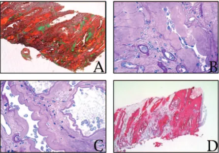
Figure 2. (A) Kidney biopsy specimen showing extensive peritubular and interstitial amyloid deposition in the inner medulla
(Congo red stain viewed under polarized light; areas of amyloid show apple green birefringence). (B and C) Kidney biopsy specimen showing peritubular ribbon-like amyloid deposits in the inner medulla (periodic acid-Schiff stain) (D) Kidney biopsy showing extended anti–apolipoprotein A-I (anti–apoA-I) immunoreactive amyloid deposits in the medulla. Magnification,⫻5 in A;⫻25 in B and C; ⫻40 in D.
Figure 3. (A) Nephrectomy specimen showing focal areas of amyloid deposition in the medullary interstitium; note in the inner
medulla a large band of amyloid material replacing normal parenchyma (hematoxylin-eosin stain). (B) Kidney biopsy specimen showing microscopic appearance of an isolated metachromatic nodular amyloidotic area located in the inner medulla (crystal violet stain). Magnification,⫻5.
also expand previous knowledge on the natural history of this autosomal dominant disorder, which recently was disclosed to be prevalent in Italy (15) but has been also recognized in people of various ethnic backgrounds (19).
The patients included in this study confirm our previous observation of the relatively mild phenotype of the amyloid disease associated with this apoA-I variant. Although the fibrils localize also into the testes, heart, and adrenal glands, the clinical picture seems to be dominated by either liver or kidney disease, with onset varying from the fourth decade to advanced age.
None of the families were known to be related to one an-other; tracing family trees to at least the great-grandparents of each index patient identified no ancestors shared by two or more kindreds. However, the finding of the same mutation in families who originated from different villages of the province of Brescia raises the possibility that a common mutation have been inherited from a shared ancestor (a founder mutation). The identification of additional families who carry the mutation and haplotype analysis will provide indication of whether the disease is due to inheritance of the same mutation from a common ancestor or to independent mutations.
Although the kidney involvement in hereditary apoA-I amy-loidosis long has been clinically recognized, very few reports have described the clinical and histologic picture (7,17). Thus, our families give us the opportunity to outline some distinctive aspects of the renal involvement that seem fragmentary in the literature, such as the nature and frequency of the renal disease. The renal disease invariably manifests as a tubulointerstitial disorder, characterized by the occurrence of moderate polyuria and nocturia as initial clinical signs, suggesting the presence of an acquired, mild form of nephrogenic diabetes insipidus. Uri-nalysis is frequently negative; proteinuria is a minor aspect of the disease and shows a tubular pattern.
The frequency of the renal involvement seems to be almost universal, although its severity varies greatly. The majority of the patients had a slowly progressive renal dysfunction, and only one reached end-stage renal failure. In a few patients with predominant hepatic involvement and no overt renal disease, a mild reduction in creatinine clearance was observed, associated with a defective urine-concentrating ability. This suggests that subclinical renal damage might be present in all f these patients, consistent with a latent tubulointerstitial disease.
A major finding of our study was the medullary location of amyloid. Histology confirmed that the renal disease was pri-marily tubulointerstitial nephritis, characterized by tubular at-rophy and interstitial fibrosis, with associated secondary focal glomerular sclerosis. However, amyloid deposition was re-stricted to nonglomerular regions and limited to the renal me-dulla. The renal cortex did not contain amyloid, and a striking absence of glomerular amyloid deposits was noted. The med-ullary location probably explains why renal biopsy specimens of the patients of family 1, which contained only cortical tissue, did not show congophilic material. This remarkable selectivity for the medullary compartment of apoA-I amyloid differs sharply from the glomerular and vascular involvement of other acquired and hereditary systemic amyloidoses (2). The only
exception is hereditary transthyretin amyloidosis, character-ized by predominant medullary distribution of the renal amy-loid deposits, which are mostly subclinical (20).
The medullary location of the apoA-I amyloid fibrils first was described ⬎30 yr ago, when the first variant of apoA-I (Arg26Gly) was reported in the original Iowa family (17). Sub-sequently, a few other reports confirmed that the renal involve-ment of apoA-I was interstitial rather than glomerular (7,9). To date, only one case of predominant glomerular involvement with nephrotic syndrome as a result of apoA-I amyloidosis has been reported (16), and this disparity in renal manifestations remains to be explained.
Many general questions about formation and deposition of any type of amyloidosis remain unanswered (1). In particular, the mechanisms by which some mutant forms of apoA-I de-posit as amyloid fibrils are still presently unknown and so are the possible factors that govern the anatomic distribution of apoA-I deposits, including this peculiar tropism for the renal medulla. The biochemical characterization of ex vivo apoA-I amyloid fibrils has invariably demonstrated that they are formed by N-terminal fragments of mature apoA-I with signif-icant C-terminal heterogeneity (21). This suggests that proteo-lytic remodeling of mutant apoA-I is likely to play a relevant role in the process of apoA-I fibrillogenesis. Although it is not known where this proteolytic cleavage might occur and by which proteases, mutations may enhance the amyloidogenic propensity of this protein through destabilization of its native structure (1). Such less stable conformation may be more sus-ceptible to proteolysis, increasing the amyloidogenic precursor protein pool.
A possible factor involved in the tissue localization of apoA-I amyloid deposits could be related to the physiologic function of apoA-I and to its metabolism. As the kidneys are the major organ of HDL catabolism, locally elevated concentrations of the amyloidogenic protein may result from specific interactions with putative local receptors involved in HDL metabolism. Moreover, peculiar local conditions, such as pH and osmolytes, may play an additional role in modulating fibril formation. Thus, the “renal medullary milieu,” characterized by high urea concentrations and acidic pH, might act as an additional amy-loidogenic hit, favoring amyloid formation (1,22). In the near future, work on animal and cellular models will be of consid-erable interest to understand the mechanism of preferential deposition of this apoA-I variant in the medullary interstitium. In summary, we have provided a detailed description of the peculiar tubulointerstitial involvement observed in renal apoA-I amyloidosis. Amyloid deposition is restricted to the medullary interstitium and makes this disease indistinguish-able on clinical grounds from other forms of hereditary tubu-lointerstitial nephritis with prevalent medullary involvement, such as medullary cystic kidney disease. The correct diagnosis can be missed even in patients who undergo renal biopsy, when the specimens contain only cortical tissue. Because renal urinary concentration capacity might be altered early in pa-tients with renal apoA-I amyloidosis, urinary concentration test might be helpful for the early identification of these patients.
apoA-I amyloidosis therefore represents a challenge. To meet this challenge, nephrologists and pathologists alike will have to maintain a high index of suspicion in patients who present with familial tubulointerstitial renal disease associated with liver involvement.
Acknowledgments
This study was supported by a grant from Cariplo Foundation, Milan, Italy.
We thank Anna Galletti (Pathology Department, Spedali Civili, Bres-cia, Italy) for the valuable technical assistance in immunohistochemis-try studies.
References
1. Merlini G, Bellotti V: Molecular mechanisms of amyloid-osis. N Engl J Med 349: 583–596, 2003
2. Benson MD: The hereditary amyloidosis. Best Pract Res Clin
Rheumatol 17: 909 –927, 2003
3. Lachmann H, Booth D, Booth S, Bybee A, Gilbertson A, Gilmore J, Pepys M, Hawkins P: Misdiagnosis of heredi-tary amyloidosis as AL (primary) amyloidosis. N Engl
J Med 346: 1786 –1791, 2002
4. Frank P, Marcel Y: Apolipoprotein A-I: Structure-function relationships. J Lipid Res 41: 853– 872, 2000
5. Nichols W, Gregg R, Brewer H, Benson M: A mutation in apolipoprotein A-I in Iowa type of familial amyloidotic polyneuropathy. Genomics 8: 318 –323, 1990
6. Soutar A, Hawkins P, Vigushin D, Tennent G, Hutton T, Nguyen O, Totty N, Feest T, Hsuan J, Pepys M: Apoli-poprotein AI mutation Arg-60 causes autosomal dominant amyloidosis. Proc Natl Acad Sci U S A 89: 7389 –7393, 1992 7. Vigushin D, Gough J, Allan D, Alguacil A, Penner B, Pettigrew N, Quinonez G, Bernstein K, Booth S, Booth D, Soutar A, Hawkins P, Pepys M: Familial nephropathic systemic amyloidosis caused by apolipoprotein AI variant Arg26. QJM 87: 149 –154, 1994
8. Booth D, Tan S, Booth S, Hsuan J, Totty N, Nguyen O, Hutton T, Vigushin D, Tennent G, Hutchinson W, Thom-son N, Soutar A, Hawkins P, Pepys M: A new apolipopro-tein AI variant, Trp50Arg, causes hereditary amyloidosis.
QJM 88: 695–702, 1995
9. Booth D, Tan S, Booth S, Tennent G, Hutchinson W, Hsuan J, Totty N, Truong O, Soutar A, Hawkins P, Brugurea M, Caballeria J, Sole M, Campistol J, Pepys M: Hereditary hepatic and systemic amyloidosis caused by a new dele-tion/insertion mutation in the apolipoprotein AI gene.
J Clin Invest 97: 2714 –2721, 1996
10. Persey M, Booth D, Booth S, van Zyl-Smit R, Adams B, Fattaar A, Tennent G, Hawkins P, Pepys M: Hereditary nephropathic systemic amyloidosis caused by a novel vari-ant apolipoprotein A-I. Kidney Int 53: 276 –281, 1998 11. Asl L, Liepnieks J, Asl K, Uemichi T, Moulin G, Desjoyaux
E, Loire R, Delpech M, Grateau G, Benson M: Hereditary amyloid cardiomyopathy caused by a variant apolipopro-tein AI. Am J Pathol 154: 221–227, 1999
12. Obici L, Bellotti V, Mangione P, Stoppini M, Arbustini E, Verga L, Zorzoli I, Anesi E, Zanotti G, Campana C, Vigano M, Merlini G: The new apolipoprotein A-I variant Leu(174)3 Ser causes hereditary cardiac amyloidosis, and the amyloid fibrils are constituted by the 93-residue N-terminal polypeptide. Am J Pathol 155: 695–702, 1999 13. Asl K, Liepnieks J, Nakamura M, Parker F, Benson M: A
novel apolipoprotein A-I variant, Arg173Pro, associated with cardiac and cutaneous amyloidosis. Biochem Biophys
Res Commun 257: 584 –588, 1999
14. de Sousa M, Vital C, Ostler D, Fernandes R, Pouget-Abadie J, Carles D, Saraiva M: Apolipoprotein AI and transthyre-tin as components of amyloid fibrils in a kindred with apoA-I Leu178His amyloidosis. Am J Pathol 156: 1911–1917, 2000
15. Obici L, Palladini G, Giorgetti S, Bellotti V, Gregorini G, Arbustini E, Verga L, Marciano S, Donadei S, Perfetti V, Calabresi L, Bergonzi C, Scolari F, Merlini GP: Liver biopsy discloses a new apolipoprotein A-I hereditary amyloidosis in several unrelated Italian families. Gastroenterology 128: 1418 –1422, 2004
16. Murphy C, Wang S, Weaver K, Gertz M, Weiss D, Solomon A: Renal apolipoprotein A-I amyloidosis associated with novel mutant Leu64Pro. Am J Kidney Dis 44: 1103–1109, 2004
17. Van Allen W, Frohlich JA, Davis JR: Inherited predisposi-tion to generalized amyloidosis. Clinical and pathological study of a family with neuropathy, nephropathy, and pep-tic ulcer. Neurology 19: 10 –25, 1969
18. Mucchiano GI, Haggqvist B, Sletten K, Westermark P: Apolipoprotein A-1-derived amyloid in atherosclerotic plaques of the human aorta. J Pathol 193: 270 –275, 2001 19. Coriu D, Dispenzieri A, Stevens FJ, Murphy CL, Shuching
W, Weiss DT, Solomon A: Hepatic amyloidosis resulting from deposition of the apolipoprotein A-I variant Leu75Pro. Amyloid 10: 215–223, 2003
20. Lobato L, Beirao I, Guimaraes SM, Droz D, Guimaraes S, Grunfeld JP, Noel LH: Familial amyloid polyneuropathy type I (Portuguese): Distribution and characterization of renal amyloid deposits. Am J Kidney Dis 31: 940 –946, 1998 21. Andreola A, Bellotti V, Giorgetti S, Mangione P, Obici L, Stoppini M, Torers J, Monzani E, Merlini G, Sunde M: Conformational switching and fibrillogenesis in the amy-loidogenic fragment of apolipoprotein A-I. J Biol Chem 278: 2444 –2451, 2003
22. Kim YS, Cape SP, Chi E, Raffen R, Wilkins-Stevens P, Stevens FJ, Manning MC, Randolph TW, Solomon A, Car-penter JF: Counteracting effects of renal solutes on amyloid fibril formation by immunoglobulin light chains. J Biol
Chem 276: 1626 –1633, 2001