Rassegna
Alterations of cerebral white matter structure in psychosis and their
clinical correlations: a systematic review of Diffusion Tensor
Imaging studies
Alterazioni strutturali della sostanza bianca cerebrale nella psicosi e le relative
correlazioni cliniche: una rassegna sistematica degli studi di diffusion
tensor imaging
SERENA PARNANZONE
1, DARIO SERRONE
1, MARIA CRISTINA ROSSETTI
1, SIMONA D’ONOFRIO
1,
ALESSANDRA SPLENDIANI
2, VALERIA MICELLI
2, ALESSANDRO ROSSI
1, FRANCESCA PACITTI
1**E-mail: [email protected]
1Department of Biotechnological and Applied Clinical Sciences (DISCAB), University of L’Aquila, Italy
2Department of Biothecnological and Applied Clinical Sciences (DISCAB), Neuroradiological Unit, University of L’Aquila, Italy
INTRODUCTON
Schizophrenia is a complex psychiatric syndrome compris-ing of psychiatric symptoms, includcompris-ing auditory hallucinations and delusions, cognitive deficits and social dysfunction1.
The majority of studies on structural brain changes in pa-tients at ultra-high risk for or affected by psychosis have been based on magnetic resonance imaging. Brain structural MRI is based on the differential behaviour of protons of wa-ter molecules in gray and white matwa-ter when exposed to a variable magnetic field. The contrast between structures varying in the response to magnetic field alterations allows delineating local groupings of neurons and fibers and deter-mining their sizes in absolute and relative terms2.
Most of MRI studies in schizophrenia suggest the involve-ment of white matter (WM) pathology in multiple cerebral re-gions in the neurobiology of this condition. As normal brain functions are served by macrostructural circuits of cortical and subcortical areas, disturbed communication between brain re-gions may be the core pathology of psychosis. WM consists of the axonal projections to other neurons and functional brain areas and is therefore key to neural communication. Myelina-tion is initiated prenatally and completed for most tracts with-in the first year birth but contwith-inues durwith-ing childhood, adoles-cence and adulthood and has a region-specific course where prefrontal regions myelinate the last3. Several lines of evi-dence point to myelin dysfunction, reduced oligodendrocyte number or integrity, or possibly hyperglutamatergic state4.
SUMMARY. Schizophrenia is a common, severe and chronically disabling mental illness. Most of MRI studies in schizophrenia suggest the involvement of white matter (WM) pathology in multiple cerebral regions in the neurobiology of this condition. White matter fiber tracts connecting numerous cortical regions have been the focus of a number of studies using a magnetic resonance technique called “Diffusion Tensor Imaging” (DTI). A literature search of published DTI studies was conducted using the major database National Centre for Biotech-nology information (NCBI) PubMed (MEDLINE). Our review covers 95 published papers. We summarise the main DTI findings involving the different brain regions in patients affected by or at high-risk for psychosis; we discuss clinical implications of these white matter disrup-tions and the limitadisrup-tions of current studies, listing the potential confounds and suggesting potential future research direcdisrup-tions.
KEY WORDS: DTI, psychosis, schizophrenia, white matter.
RIASSUNTO. La schizofrenia è una malattia mentale comune, grave e cronicamente invalidante. La maggior parte degli studi di risonanza magnetica in pazienti affetti da schizofrenia suggerisce il coinvolgimento della sostanza bianca di diverse regioni cerebrali nella patogenesi e nella neurobiologia di questa malattia. I fasci di sostanza bianca interposti tra le diverse regioni corticali sono stati oggetto di numerosi stu-di che utilizzano una tecnica stu-di risonanza magnetica chiamata “Diffusion Tensor Imaging” (DTI). Nel presente stustu-dio è stata condotta una revisione della letteratura sugli studi di DTI pubblicati utilizzando il database National Centre for Biotechnology (NCBI) PubMed (Medli-ne). Questa rassegna comprende 95 articoli pubblicati. Sono stati riportati i principali risultati degli studi di DTI in pazienti affetti da psico-si o ad alto rischio per lo sviluppo di ppsico-sicopsico-si; sono state discusse le implicazioni cliniche delle alterazioni della sostanza bianca e i limiti degli studi in corso elencando i potenziali fattori di confondimento e suggerendo possibili direzioni future per la ricerca.
Riv Psichiatr 2017; 52(2): 49-66
50
Abnormalities in WM structure and integrity have been correlated with psychotic symptoms, negative symptoms and cognitive deficits5.
WM is difficult to study in detail with conventional MRI be-cause of its high degree of homogeneity, moreover convention-al techniques do not convention-allow for the evconvention-aluation of its directionconvention-ali- directionali-ty and organization. WM fiber tracts connecting numerous cor-tical regions have been the focus of a number of studies using a magnetic resonance technique called Diffusion Tensor Imaging (DTI). It has become established in the last two decades as a valuable research tool. DTI assesses a non-invasive and in vivo quantification of the diffusion characteristics of water mole-cules: within a magnetic field these molecules tend to align into preferential directions according to their ability to diffuse across or along the arrangement of biological structures that surround them. In the brain water may diffuse freely in all directions (isotropic diffusion), or restricted along one particular direction of structured tissue such as WM tracts and fibers (anisotropy diffusion). Fractional anisotropy is a quantitative dimension and can take values between 0 and 1. If the anisotropy is high, then most of the diffusion occurs in the highly ordered directions, in-dicating a high level of orientation in the structure, therefore, decreased anisotropy may predict compromised white matter integrity6. Other measures used to compare different voxels in term of diffusion are mean diffusivity (MD), radial diffusivity (RD) and relative anisotropy (RA)7. Additionally different ap-proaches have been applied to study differences in regional brain anisotropy between subjects: some studies have used vox-el based approaches (VBA), where data sets have been processed with reference to FA normalized to a standard anatomical and averaged template, before being compared to similarly processed data sets; other studies have used a region of interest (ROI) approach in region of the brain thought to be im-plicated to psychosis. DTI is becoming increasingly important in the field of schizophrenia research8.
The aim of this study was to review the knowledge about the abnormalities of WM in patients at ultra-high risk for psychosis (UHR), patients with a first-episode psychosis (FEP) and chronic schizophrenia patients (SZ) compared with controls (HC), making clearer the role of WM integrity alterations in the etiopathogenesis, anatomical bases and clinical or neuro-cognitive correlates of the disorders.
In 25 studies the patient population included people con-sidered at ultra-high risk for psychosis. To be concon-sidered at high-risk for psychosis patients had to satisfy almost one of these criteria: 1) they had schizotypal personality features; 2) they had sub-threshold psychotic symptoms; 3) they had a first-degree relative with schizophrenia-like disorder; 4) they had brief psychotic moments with spontaneous remission in less than 1 week (Table 1).
We have decided to mention some of the studies exclud-ed because they can provide additional information.
In a study was examined the ability of DTI to differenti-ate between UHR, FEP and HC subjects: the results suggest that DTI allowed discrimination of UHR from HC sub-jects34.
Patients with only cannabis use disorder (CUD) have al-so been studied with DTI method: they had lower FA than HC in left inferior FOF27, and altered FA values in left ILF and left inferior FOF compared to HC; greater consumption of cannabis predicted a greater decrease in left ILF FA in CUD35.
In the study by Mittal et al.36youth at high-risk for psy-chosis presented neurological disfunction and abnormal neu-rodevelopment misured by the presence of neurological soft signs (NSS) and a decrease of FA in right/left superior CP at 12 months, controls showed a normative increase while there were no group differences at baseline. NSS predicted a lon-gitudinal decrease in cerebellar-thalamic FA and elevations in negative but not positive symptoms 12 months later.
According to Derosse et al.37 cumulative risk for psy-chosis (including low QI, low parental socioeconomic status, history of adolescent cannabis use and childhood trauma, high levels of subclinical psychotic-like experiences) was as-sociated with lower FA in left SLF.
In the study by Skranes et al.38very low birth weight dren had reduced FA values in CI, CE, CC, ILF, SLF; chil-dren with low QI had reduced FA in CE, SLF, ILF; fine mo-tor impairment was related to low FA in CI, CE and SLF; mild social deficits correlated with reduced FA in CE and SLF.
Prenatal and neonatal DTI were obtained in the offspring of mothers with schizophrenia or schizoaffective disorder and matched comparison mothers: there were no group dif-ferences in white matter diffusion tensor properties39.
In 41 studies the patient population included people ex-periencing a first episode of psychosis. (Table 2)
According to Peters et al.75FEP with cannabis use before age 17 showed increased directional coherence in the bilat-eral UF, anterior CI and FL while these abnormalities were absent in FEP without cannabis use before age 17: this is in contrast with most DTI studies which have produced evi-dence of WM hypoconnectivity.
In 46 studies the patient population included people with chronic schizophrenia (Table 3).
Tang et al.110obtained DTI and magnetic resonance spec-troscopy from 40 subjects with schizophrenia: N-Acetylas-partate and DTI anisotropy indices were reduced in medial temporal regions.
Patients with temporal lobe epilepsy and interictal psy-chosis were studied with DTI by Flügel et al.111; they showed lower FA values in both frontal and temporal regions and
METHODS
A literature search of published DTI studies was conducted using the major database National Centre for Biotechnology in-formation (NCBI) PubMed (Medline).
The key words used were: “schizophrenia” and “ DTI” or “dif-fusion tensor imaging”, “psychosis” and “DTI” or “dif“dif-fusion ten-sor imaging”. Studies were included if they satisfied the following criteria: the patient population had a diagnosis of psychosis or was considered at ultra-high risk for psychosis, diffusion tensor imag-ing was an imagimag-ing technique used, the article was published in English. Additionally, they were chosen if they were found to be relevant to the focus of this systematic review.
Our review covers 95 papers published between September 2005 and March 2015: 32 papers were excluded.
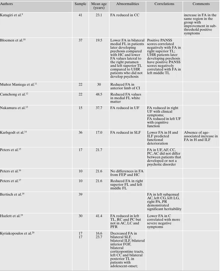
Table 1. Main findings of DTI studies in people considered at high-risk for psychosis.
Authors Sample Mean age
(years)
Abnormalities Correlations Comments
Katagiri et al.9 41 23.1 FA reduced in CC increase in FA in the
same region in the group with
improvement in sub-threshold positive symptoms
Bloemen et al.10 37 19.5 Lower FA in bilateral
medial FL in patients later developing psychosis compared with HC and lower FA values lateral to the right putamen and left superior TL compared to UHR patients who did not develop psychosis
Positive PANSS scores correlated negatively with FA in right superior TL; UHR patients later developing psychosis have positive PANSS scores negatively correlated with FA in left middle TL
Muñoz Maniega et al.11 22 30 Reduced FA in
anterior limb of CI
Camchong et al.12 22 48.5 Reduced FA values
in medial FL white matter
Nakamura et al.13 15 37.7 FA reduced in UF FA reduced in right
UF with clinical symptoms;
FA reduced in left UF with cognitive function
Karlsgodt et al.14 36 17.0 FA reduced in SLF Lower FA in H and
ILF predicted functional deterioration Absence of age-associated increase in FA in H and ILF
Peters et al.15 17 21.7 FA in UF, AF, CC,
PC, AC did not differ between patients that developed or not a psychotic disorder
Peters et al.16 10 21.6 No differences in FA
from FEP and HC
Peters et al.17 10 21.6 Reduced FA in right
superior FL and left middle FL
Bertisch et al.18 39 FA in left subgenual
AC, left CG, left LG, right PA, PR demonstrated significant heritability
Hazlett et al.19 30 41.4 FA reduced in left
TL, RC and PC but not in AC, LC and PFR
Lower FA in C correlated with more severe negative symptoms Kyriakopoulos et al.20 17 17 16.6 23.7 Decreased FA in bilateral SLF, bilateral ILF, bilateral inferior FOF, bilateral
corticopontine tracts, left CC and bilateral posterior TL in patients with adolescent-onset;
(years)
Decreased FA in bilateral ILF, bilateral inferior FOF, brain stem, cerebellum, right SLF, right CC, right UF, right C, right anterior CR, right posterior TL and corticopontine tract in patients with adult-onset
Carletti et al.21 32 23.4 FA, RD and AD
intermediate in UHR between controls and FEP
Progressive reduction in FA in subjects who developed psychosis
Benetti et al.22 46 24.3 Reduction of FA in
left long segment in patients without verbal auditory hallucinations
The sample is composed by both patients UHR and FEP
Hoptman et al.23 22 20 Reduced FA in left
posterior C, bilateral angular giry, left inferior frontal gyrus; increased FA in left subgenual anterior C and bilateral pontine tegmental WM and right middle/superior frontal gyri
Smallman et al.24 12 21.0 Increased FA in left
UF
Positive correlation between FA in right AF and hallucinatory experience
Domen et al.25 93 29.4 Did not differ from
HC
Boos el al.26 123 26.7 Higher mean FA in
left and right AF
Epstein et al.27 21 16.1 Lower FA in left ILF,
CST bilaterally, left inferior FOF
Derosse et al.28 67 36.1 Lower FA in inferior
FOF and greater asymmetry in UF
Lener et al.29 49 36.5 Lower FA in CC
compared with HC
This abnormality was more widespread in SZ
Lagopoulos et al.30 74 21.3 Lower FA in left
anterior CR, anterior TL
Jacobson et al.31 11 11-13 WM decrease in
inferior FOF, C, ILF
Goghari et al.32 24 40.2 Increase in FA in
right fimbria of the fornix
No significant association between FA and QI
Hohenberg et al.33 28 20.6 Increased MD in
SLF, posterior CR and CC, increased RD in posterior PL
CC=corpus callosum; UF=uncinate fasciculus; SLF=superior longitudinal fasciculus; H=hippocampus; ILF=inferior longitudinal fasciculus; AF=arcuate fasciculus; PC=posterior cingulate; AC=anterior cingulate; CG=cingulate gyrus; LG=lingual gyrus; PA=pericaudate area; PR=perilentiform region; TL=temporal lobe; PFR=prefrontal region; TR=thalamic radiations; RC=right cingulum; LC=left cingulum; C=cingulum; FMJ=forceps major; FMN= forceps minor; CE=external capsule; CR= corona radiate; CST=corticospinal tract; FOF=fronto-occipital fasciculus; PL=parietal lobe; CP=cerebellar peduncles; PO=parietal-FOF=fronto-occipital; LDL=low-density lipoprotein; CI=internal capsule; PANSS=positive and negative syndrome scale; WCST= Wisconsin card sorting test; GF=frontal gyrus; BPRS=brief psychiatric rating scale; FL=frontal lobe; OL=occipital lobe; T=thalamus; I=insula.
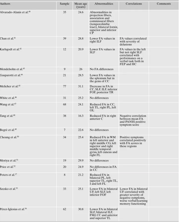
Table 2. Main findings of DTI studies in people experiencing a first episode of psychosis.
Authors Sample Mean age
(years)
Abnormalities Correlations Comments
Alvarado-Alanis et al.40 35 24.6 Abnormalities in
projection fibers, association and commissural fibers (temporolimbic tract), bilateral fornix, superior and inferior CP
Chan et al.41 39 28.8 Lower FA values in
right ILF
FA values correlated with severity of delusions
Karlsgodt et al.42 12 20.9 Lower FA values in
SLF
FA values in the left but not right SLF correlated with performance on a verbal task both in FEP and HC
Mendelsohn et al.43 9 26 No FA differences
Gasparotti et al.44 21 28.5 Lower FA values in
the splenium but in the genu of CC
Melicher et al.45 77 31.1 Decrease in FA in
CC, SLF, ILF, inferior FOF, posterior TR
White et al.46 31 25.2 No differences
Wang et al.47 68 24.1 Reduced FA in CC,
left TL, right PL, left OL
Tang et al.48 38 16.3 Reduced FA in right
anterior C
Negative correlation between mean FA and PANSS positive symptom score
Begrè et al.49 7 22.6 No differences
Cheung et al.50 34 25.4 Reduced FA in WM
in left anterior and right middle CG, left superior and right middle temporal gyrus, left cuneus and right FL
Positive symptoms correlated positively with FA scores in these regions
Moriya et al.51 19 29.9 No differences
Price et al.52 20 24.9 No differences in FA
in CC
Peters et al.17 8 21.2 Reduced FA in
bilateral PL, left superior TL, right TL, I and left FL
Szesko et al.53 33 25.1 Lower FA in bilateral
UF, left SLF, left inferior FOF Lower FA in bilateral UF correlated with greater severity of negative symptoms, worse verbal/learning memory functioning
Pérez-Iglesias et al.54 62 30.8 Lower FA in bilateral
SLF, bilateral ILF, FMJ, CC and anterior and superior TL
Riv Psichiatr 2017; 52(2): 49-66
54
(years)
Bijanki et al.55 31 23.1 Significant
correlation between global FA and negative symptoms (SANS) This correlation became non-significant with additioning age as a covariate
Price et al.52 19 23.8 FA reduced in left
UF
No correlations between FA and clinical ratings
Luck et al.56 32 23.6 FA reductions in the
fornix No significant correlation between FA and clinical or socio-demographic data
Kong et al.57 15 24.3 No significantly
decreased FA
Schneiderman et al.58 23 16.1 Decreased FA in TL
and SLF
Older age of onset tended to be associated with higher FA in ventral CI and ventral temporo-occipital WM
The study suggest that symptoms associated with the temporal lobe including auditory hallucinations would present before frontal associated symptoms including problem in executive functioning
Chen et al.59 20 46.9 Reduction in FA in
left PL, right PC
No significant correlations between FA value and PANSS and cognitive test scores, age and antipsychotic medication dosages
Late-onset schizophrenia
Friedman et al.60 40 26 Lower FA in ILF
Peters et al.61 30 22.7 Lower FA in CC,
bi-lateral PL, OL, FL, TL WM
FA correlated with polyunsaturated fatty acid concentration and negative correlat-ed with negative symptoms
Hao et al.62 21 Lower FA values in
cerebral peduncle, frontal regions, inferi-or tempinferi-oral gyrus, medial PL, hip-pocampal gyrus, I, right anterior C and right CR
Carletti et al.21 15 24.1 Widespread
reduc-tion in FA and in-creases in diffusivity compared to HC and UHR
Cheung et al.63 25 29 Lower FA values in
left FOF, left ILF, WM adjacent to right precuneus and right substantia nigra, CC, right posterior limb of CI and left cere-bral peduncle
(continued) - Table 2.
Authors Sample Mean age
(years)
Abnormalities Correlations Comments
Epstein et al.35 34 16.4 Lower FA in bilateral
CST, bilateral ILF, bilateral inferior FOF compared to HC
Epstein et al.27 55 16.9 Lower FA than HC
in bilateral CST, left ILF, left inferior FOF
Szesko et al.64 35 21.5 FA reductions within
PL and OL WM
Greater overall FA increases in patients with greater increases in LDL
No significant FA increases among patients following treatment
Lee et al.65 17 Lower FA in genu
and body of CC, UF, C, superior and inferior FOF, fornix, CE and CI, increased MD and RD in all WM regions; no difference for AD
FA in right inferior FOF had a positive correlation with negative, positive symptoms and all the items of WCST, FA of right CE showed positive correlation with category completed scores of WCST
Price et al.66 18 23.6 Reduced FA in CC
Qiu et al.67 32 28 No differences in FA Left thalamic FA
correlated with spatial working memory deficits
Dekker et al.68 26 21.1 Reduced FA in CC in
cannabis naïve FEP compared with FEP with early-onset cannabis use and with HC
Quan et al.69 16 21.1 FA reduced in
inferior GF-striatum tract, RD increased in bilateral rostral middle GF-striatum and bilateral inferior GF-striatum tracts
The number of WCST categories completed correlated positively with FA of right rostral middle GF-striatum tract and negatively with RD of right rostral middle GF-striatum tract, right inferior GF-striatum tract; BPRS score had no correlations
Szesko et al.70 10 26.9 Reduced FA in left
middle frontal gyrus, left posterior temporal gyrus, left CI
Kiriakopoulos et al.71 19 17.0 Lower FA in bilateral
parietal association and left middle CP, no areas with higher FA
Marques et al.72 63 27.7 Non responders to
treatment at baseline showed lower FA in UF, C, CC
After 12 weeks increase in FA in responders and non responders positively correlated to antipsychotic exposure
Riv Psichiatr 2017; 52(2): 49-66
56
higher MD in bilateral frontal regions, additionally the per-formance on some neuropsychological tests was related to frontotemporal FA reduction. Mao et al.112investigated in-terictal personality changes and white matter abnormalities in epilepsy patients: long disease duration and impairment of right AF integrity were independent risk factor of psychoti-cism.
Cocchi et al.113studied the relationship between structur-al and functionstructur-al deficits in schizophrenia patients: they showed decreased functional connectivity and impaired white matter integrity in a distributed network encompassing frontal, temporal, thalamic and striatal regions; in controls strong interregional coupling in neural activity was associat-ed with well-myelinatassociat-ed white matter pathways.
Compared with Parkinson’s disease patients without psy-chosis, those with psychosis had significantly lower FA in left frontal lobe, bilateral occipital lobe, left cingulated gyrus and left hippocampus114.
For an overview of the results see table 4.
DISCUSSION
The findings can be grouped into WM pathology affecting cortical regions, subcortical regions, inter-hemispheric fibers, association fibers and limbic system fibers. Corpus callosum consists of a commissural tract comprising the largest bundle of fibers connecting the two brain hemispheres.
Association fibers are: SLF which connects the frontal lobe with occipital and temporal areas, ILF, UF which are an-terior temporo-frontal fiber tracts connecting orbito-frontal with anterior and medial temporal lobes, FOF which extends backward from the frontal lobe and spreading into the tem-poral and occipital lobes, AF is a fiber tract that stems from the caudal part of the superior temporal gyrus and extends to the lateral prefrontal cortex, the superior and the middle frontal regions. Limbic system fibers are the cyngulum fibers that project both posteriorly from the cingulate gyrus to the
entorhinal cortex, temporal lobe, and anteriorly to the pre-motor, prefrontal regions and striatum. The fornix connects the hippocampus to the mamillary bodies, nucleus accum-bens, medial prefrontal cortex, and septal regions, thus this fiber serves as the main output and input pathway for hip-pocampus. Thalamic radiations are projection fibers that provides a functional loop between the cerebral cortex and the thalamus; they converged into the internal capsule, locat-ed between the putamen and the thalamus-caudate nucleus regions5.
Changes in WM integrity were found in chronic psychosis, first-episode psychosis and patients at ultra-high risk for psy-chosis, they may play a role in the primary pathophysiology, as opposed to being a result of secondary disease processes. These changes have been correlated with specific cognitive deficits as well as clinical symptoms, suggesting that biologi-cal changes may underlie these clinibiologi-cal factors in patients.
Previous DTI studies assessing the impact of WM disrup-tions on the disease process have had mixed results. Our study adds to a growing body of literature emphasizing the need for treatments targeting white matter function and structure in psychosis patients.
The main findings in patients at ultra-high risk for psy-chosis were a decreased FA in inferior FOF, temporal lobe WM, frontal lobe WM. They seem to have predictive value of onset of psychosis in high-risk individuals. Other studies in ultra-high risk patients showed lower FA in anterior CR, cor-ticospinal tracts, SLF, ILF, UF, CC and C. In addition, in-crease of FA values was seen in anterior C, left UF, AF, frontal lobe WM, right fornix and brain stem. The prediction of psychosis is a major topic in research and olds the hope for early intervention and prevention of full development of the illness, improving outcome and preserving WM integrity.
Decreases of FA in different tracts in patients at first-episode psychosis support notion of early disconnectivity be-tween brain regions: the most burned were CC, UF, ILF, SLF, inferior FOF, temporal lobe WM, parietal lobe WM and left frontal lobe WM. White matter abnormalities were also ob-served in C, occipital lobe, CI, corticospinal tracts, cerebral
(years)
Peters et al.16 10 21.2 No differences
Lu et al.73 21 22 Greater AD, RD, MD
in CC, C, CR, posterior TL, CI, ILF, inferior FOF
Luck et al.74 44 23.3 FA reduction in
bilateral UF and bilateral SLF but not in C
Greater WM changes in these tracts with poor outcome as compared to patients with good outcome
CC=corpus callosum; UF=uncinate fasciculus; SLF=superior longitudinal fasciculus; H=hippocampus; ILF=inferior longitudinal fasciculus; AF=arcuate fasciculus; PC=posterior cingulate; AC=anterior cingulate; CG=cingulate gyrus; LG=lingual gyrus; PA=pericaudate area; PR=perilentiform region; TL=temporal lobe; PFR=prefrontal region; TR=thalamic radiations; RC=right cingulum; LC=left cingulum; C=cingulum; FMJ=forceps major; FMN= forceps minor; CE=external capsule; CR= corona radiate; CST=corticospinal tract; FOF=fronto-occipital fasciculus; PL=parietal lobe; CP=cerebellar peduncles; PO=parietal-FOF=fronto-occipital; LDL=low-density lipoprotein; CI=internal capsule; PANSS=positive and negative syndrome scale; WCST= Wisconsin card sorting test; GF=frontal gyrus; BPRS=brief psychiatric rating scale; FL=frontal lobe; OL=occipital lobe; T=thalamus; I=insula.
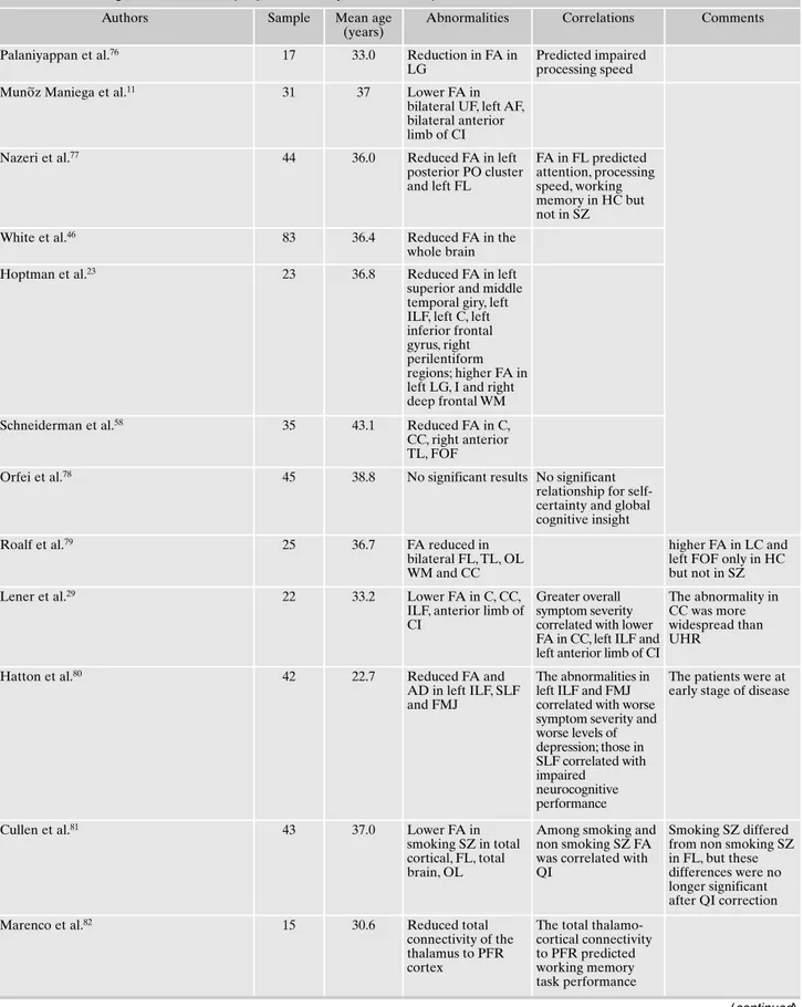
Table 3. Main findings of DTI studies in people affected by chronic schizophrenia.
Authors Sample Mean age
(years)
Abnormalities Correlations Comments
Palaniyappan et al.76 17 33.0 Reduction in FA in
LG
Predicted impaired processing speed
Mun~oz Maniega et al.11 31 37 Lower FA in
bilateral UF, left AF, bilateral anterior limb of CI
Nazeri et al.77 44 36.0 Reduced FA in left
posterior PO cluster and left FL FA in FL predicted attention, processing speed, working memory in HC but not in SZ
White et al.46 83 36.4 Reduced FA in the
whole brain
Hoptman et al.23 23 36.8 Reduced FA in left
superior and middle temporal giry, left ILF, left C, left inferior frontal gyrus, right perilentiform regions; higher FA in left LG, I and right deep frontal WM
Schneiderman et al.58 35 43.1 Reduced FA in C,
CC, right anterior TL, FOF
Orfei et al.78 45 38.8 No significant results No significant
relationship for self-certainty and global cognitive insight
Roalf et al.79 25 36.7 FA reduced in
bilateral FL, TL, OL WM and CC
higher FA in LC and left FOF only in HC but not in SZ
Lener et al.29 22 33.2 Lower FA in C, CC,
ILF, anterior limb of CI
Greater overall symptom severity correlated with lower FA in CC, left ILF and left anterior limb of CI
The abnormality in CC was more widespread than UHR
Hatton et al.80 42 22.7 Reduced FA and
AD in left ILF, SLF and FMJ
The abnormalities in left ILF and FMJ correlated with worse symptom severity and worse levels of depression; those in SLF correlated with impaired neurocognitive performance
The patients were at early stage of disease
Cullen et al.81 43 37.0 Lower FA in
smoking SZ in total cortical, FL, total brain, OL
Among smoking and non smoking SZ FA was correlated with QI
Smoking SZ differed from non smoking SZ in FL, but these differences were no longer significant after QI correction
Marenco et al.82 15 30.6 Reduced total
connectivity of the thalamus to PFR cortex
The total thalamo-cortical connectivity to PFR predicted working memory task performance
(years)
Yan et al.83 33 23.1 Decreased FA in
right AC
Correlated with stroop performance and symptom severity
Camchong et al.84 29 41.3 Connectivity
alteration in medial frontal e AC
Frontal connectivity is positively associated with symptoms and general cognitive ability measures
Lagopoulos et al.30 69 22.4 Lower FA in left
anterior CR, anterior TL
Weijer et al.85 44 36.9 Decreased FA in
CST, UF and C
Negative correlation between FA and age
The sample is composed by SZ with chronic severe hallucinations
Abdul-Rahman et al.86 33 39.4 Reduction in FA in
bilateral fornix and left AC, increase in RD in left AC and bilateral fornix, increase in AD in anterior left middle C
Decreased FA in left fornix and increased AD in RC correlated with greater severity of psychotic symptoms
Ardekani et al.87 50 30.3 FA and MD values
can be used to distinguish between SZ and HC
Choi et al.88 25 44.6 Decrease in mean
FA in anterior commissure
Anterior commissure integrity correlated negatively with age and decision making and correlated positively with total positive symptom score
Friedman et al.60 40 Lower FA in most
regions compared to HC
Antonius et al.89 36 37.4 Right superior GF,
left middle GF, bilateral parahippocampal gyrus, right T, left I, left fusiform gyrus, bilateral PC and left lentiform nucleus, left AC, RC, left LG, bilateral claustrum These abnormalities correlated with symptom unawareness; deficits of WM in right LG, left middle temporal gyrus and right precuneus related to misattribution of symptoms
No HC
Boos et al.26 126 26.6 No difference in
mean FA between SZ and HC; excessive decline in mean FA in genu, left UF, left inferior FOF, left ILF with increasing age
Negative correlation between FA in bilateral AF and symptom severity
SZ were young adult
Filippi et al.90 43 29.3 Decreased MD and
increased FA in right anterior and posterior limb of CI, bilateral
interhemispheric and corto-cortical connections, bilateral cerebellum and brain stem
These abnormalities related to a longer duration of the untreated psychosis and severity of positive symptoms (continued)
(continued) - Table 3.
Authors Sample Mean age
(years)
Abnormalities Correlations Comments
Domen et al.25 85 28.3 Lower mean FA in
CC, FMJ, FMN, bilateral CE, bilateral CR, bilateral posterior TR
Sungranyes et al.91 25 17.1 Reduced FA in
splenium and PC
Wagner et al.92 38 35.8 Lower FA in right
anterior limb of CI, right T, right CC
FA in right anterior limb of CI correlated with cognitive performance
Balevich et al.93 34 adults
17 adolescents 43.7 15.9 Adults most reduction in posterior region Adolescent most reduction in anterior region Negative correlation between negative symptoms and FA in right posterior lateral body in adults and left medial anterior body in adolescents
Bijanki et al.55 59 37.7 Global negative
symptoms correlated with global FA , upon addition of age as a covariate the relationship became non-significant
Goghari et al.32 25 41.3 No significant
relationship between FA and QI, symptoms or global functioning
Garver et al.94 13 33.7 Increase of
diffusivity in SZ considered drug-responders
This pathological increase in diffusivity was reduced following treatment-associated reduction of psychotic symptoms
Diffusivity of SZ considered poor responders did not differ from HC at baseline and following treatment
Rosenberger et al.95 27 39.1 Decline in FA with
age in SZ in C and UF but not in inferior FOF
Skelly et al.96 25 34.2 FA reduction in
multiple bilateral posterior limb of CI and bilateral CR (but stronger on the left hemisphere), in C (bilateral medial TL and right FL) , left ILF, left anterior TR, FMN and right inferior FOF
Inverse relationship of FA in left UF and left SLF with positive symptom score; positive correlation between negative symptoms and FA in right I
Nestor et al.97 18 39.1 FA in left C
correlated with orienting of attention
Liu et al.98 10 25.6 Lower FA in left
superior CP Caprihan et al.99 65 33.6 Abnormalities in TR, CST, FMJ, FMN, SLF, superior FOF (continued)
(years)
Cui et al.100 25 25.8 Reduced FA in left
posterior CR Negative correlation between FA in left frontoparietal lobe and positive symptom score; no correlation with duration of illness
Kong et al.57 15 24.3 Decreased FA in CC
in SZ
Levitt et al.101 16 39.4 No differences FA in anterior limb
of CI correlated positively with performance on measures of spatial and verbal declarative/episodic memory
Knochel et al.102 21 38.3 Changes in MD in
bilateral C and right UF; lower FA and higher MD in fornix in comparison with HC; lower FA in bilateral UF; higher MD in bilateral UF in HC No correlation with clinical parameters or with years of medication
Whitford et al.103 24 39.6 Subnormal levels of
FA in fibers connecting the rostral with the caudal anterior CG and the isthmus of C with
parahippocampal cortex
FA in fibers connecting the rostral with the caudal anterior CG correlated with positive symptoms, FA in fibers connecting the isthmus of C with parahippocampal cortex correlated with negative symptoms
McCarthy-Jones et al.104 113 39.1 Reduced FA and
increased RD in left AF in patients with Auditory Verbal Hallucinations (AVH) compared to HC and SZ without AVH
Sasamoto et al.105 35 36.6 Lower mean of FA
in CC, bilateral UF, CST, left SLF and superior FOF in SZ
Mean FA showed positive correlation with mean cortical thickness
Kawashima et al.106 15 24.5 Reduced FA in
bilateral UF but not in C
Early stage of illness
Hatton et al.107 42 22.7 Reduced FA in short
association fibres connecting the superior and the middle temporal gyri
Adolescent-onset psychosis subjects showed FA reductions in short association fibres connecting superior temporal gyrus and Heschl’s gyrus when compared to adult-onset subjects
peduncles and fornix. None of the studies included showed increased FA in patients with first-episode psychosis.
DTI abnormalities in first-episode patients are less robust than in chronic patients, suggesting that progression to more extensive abnormalities occurs after illness onset; there are also indications for accelerated aging effects in psychosis.
FA reductions were found in patients with chronic psy-chosis in CC, C, UF, left ILF, inferior FOF, SLF, FMN, FMJ, CR, corticospinal tracts, anterior CI, TR, temporal lobe WM, occipital lobe WM and frontal lobe WM. Changes in WM in-tegrity have been reported also in left AF, superior FOF, fornix and hippocampus.
White matter tracts that were reported to have increased FA in almost one study include brain stem, right frontal lobe WM, left occipital lobe WM, insula, CI, cerebellum, inter-hemispheric and cortico-cortical tracts.
Of the included studies, 13 did not report group differ-ences in anisotropy measures between patients and controls (3 in ultra-high risk patients, 8 in first-episode psychosis, 2 in chronic psychosis).
38 of the included studies (7 in UHR, 12 in FEP, 19 in SZ) found significant correlations between clinical or cognitive variables and FA values in some WM tracts. 3 studies showed a negative correlation between the severity of positive symp-toms and FA values in some WM tracts like temporal lobe WM, right anterior C, right frontal lobe WM, cingulated gyrus WM, left fornix, right anterior and posterior limb of CI, left UF, left SLF, fibers connecting the rostral with the caudal anterior CG, bilateral inter-hemispheric and cortico-cortical connections, cerebellum and brain stem. Regarding to hallu-cinatory experience a positive correlation was found with FA values in right AF, while severity of delusions was associated with FA values in right ILF.
In 3 studies negative symptoms were correlated negative-ly with FA values in some WM tracts including C, bilateral UF, CC, TL, OL, PL, FL and fibers connecting C with
parahippocampal cortex; in one paper a positive correlation was found between negative symptoms and WM integrity in right I.
FA values showed a relation with clinical symptoms in right UF, CC, left ILF, left anterior limb of CI, FMJ, right AC, frontal connectivity and bilateral AF.
Cognitive function was found to be related with WM deficits in left and right UF, right CE, SLF, right AC, frontal connectivity, right anterior limb of CI (this one was found to be proportional to performance on measures of spatial and verbal declarative/episodic memory). Left thalamic FA val-ues correlated with spatial working memory deficits. Frac-tional anisotropy in right rostral middle GF-striatum tract correlated positively with the number of WCST categories completed; FA reduction in LG predicted impaired process-ing speed while FA in left C correlated with orientprocess-ing of at-tention. According to Marenco et al.82the total thalamo-cor-tical connectivity to PFR predicted working memory task performance.
On the contrary, according to Lee et al.65FA in right infe-rior FOF had a positive relation with negative, positive symptoms and all the items of WCST; similarly, according to Choi et al.88anterior commissure integrity correlated nega-tively with decision making and posinega-tively with total positive symptoms score. In UHR patients increase in FA in CC was found to be correlated with improvement in subthreshold positive symptoms while, in other samples, patients later de-veloping psychosis had lower FA values in several tracts. In less numerous papers FA values did not differ between UHR patients that developed or not a psychotic disorder.
Functional deterioration in UHR was predicted by lower FA values in H and ILF, Goghari et al.32didn’t find signifi-cant relationship between FA and global functioning.
On the other side, no correlation with clinical/cognitive measures were found in 8 of the studies included (2 in UHR, 3 in FEP, 3 in SZ).
(continued) - Table 3.
Authors Sample Mean age
(years)
Abnormalities Correlations Comments
Zou et al.108 21 Reduced FA in
bilateral anterior limb of CI
Giezendanner et al.109 34 33.6 SZ born in summer
had lower FA in CC, bilateral inferior FOF, bilateral UF, right anterior CR, left posterior C, bilateral posterior CR, left posterior TR, bilateral CST, bilateral SLF, FMJ
Later age of onset was found in SZ born in winter months
CC=corpus callosum; UF=uncinate fasciculus; SLF=superior longitudinal fasciculus; H=hippocampus; ILF=inferior longitudinal fasciculus; AF=arcuate fasciculus; PC=posterior cingulate; AC=anterior cingulate; CG=cingulate gyrus; LG=lingual gyrus; PA=pericaudate area; PR=perilentiform region; TL=temporal lobe; PFR=prefrontal region; TR=thalamic radiations; RC=right cingulum; LC=left cingulum; C=cingulum; FMJ=forceps major; FMN= forceps minor; CE=external capsule; CR= corona radiate; CST=corticospinal tract; FOF=fronto-occipital fasciculus; PL=parietal lobe; CP=cerebellar peduncles; PO=parietal-FOF=fronto-occipital; LDL=low-density lipoprotein; CI=internal capsule; PANSS=positive and negative syndrome scale; WCST= Wisconsin card sorting test; GF=frontal gyrus; BPRS=brief psychiatric rating scale; FL=frontal lobe; OL=occipital lobe; T=thalamus; I=insula.
Riv Psichiatr 2017; 52(2): 49-66
62
White matter tract or area UHR FEP SZ
Decrease FA Increase FA Decrease FA Increase FA Decrease FA Increase FA
Corpus callosum Left Right 2 2 1 9 9 Cingulum Anterior Posterior 2 1 1 2 3 1 9 2 2 Uncinate fasciculus Right Left 2 1 4 1 8 Arcuate fasciculus Right Left 1 2 Inferior longitudinal fasciculus Right Left 2 1 4 1 2 1 4 Superior longitudinal fasciculus Right Left 2 5 3 Fronto-occipital fasciculus Superior Inferior 4 2 1 5 1 2 3 Temporal lobe Left Right 3 1 6 2 5 1 Parietal lobe Right Left 1 5 1 1 Occipital lobe Left Right 2 1 3 1 Frontal lobe Left Right 4 1 1 3 2 5 2 1 Hippocampus 1 2 Thalamic radiations Posterior Anterior 1 3 2 1 Internal capsule Anterior Posterior 1 2 1 5 1 1 External capsule Anterior Posterior 1 1 Corona Radiata Anterior Posterior 2 1 3 2 2 (continued)
Antonius et al.89studied the relation between symptoms unawareness and WM abnormalities, suggesting that misat-tribution of symptoms may be implied by loss of WM in-tegrity in right LG, TL and right precuneus.
The impact of medications on WM integrity is far from well understood. The vast majority of patients participating in DTI studies to date have been on antipsychotic medica-tion treatment. Although medicamedica-tion dose or cumulative ex-posure do not correlate with FA in most studies; some stud-ies reported positive findings: according to Marques et al.72 patients non-responders to treatment at baseline showed lower FA in UF, C, CC; additionally, in the same sample after 12 weeks increase in FA positively correlated to antipsychot-ic exposure.
Interestingly, in 2 studies FA values have been associated with metabolic measures like greater levels of LDL or polyunsaturated fatty acid concentration.
Several studies have shown age-related reduction in FA in schizophrenia, whereas other studies did not replicate this relationship. While some studies that examined correlations with age failed to identify a significant effect, 5 of the includ-ed papers showinclud-ed significant negative correlation between FA and age. Additionally, SZ adults showed most FA reduc-tion in SNC posterior region, while SZ adolescents had most FA reduction in SNC anterior region. Karlsgodt et al.14 found the absence of age-associated increase in FA in H and ILF in UHR patients.
Some studies pointed out the effect of some socio-de-mographic variables like gender, duration of untreated psychosis, duration of illness and age of onset on WM changes. Older age of onset tended to be associated with higher FA in ventral CI and ventral temporo-occipital WM, while adolescent-onset psychosis subjects showed
WM anomalies in short association fibers connecting supe-rior temporal gyrus and Heschl’s gyrus; suggesting that symptoms associated with TL WM anomalies including au-ditory hallucinations would present before FL WM symp-toms including problem in executive functioning. Later age of onset was found in SZ born in winter months, SZ born in summed had lower FA in CC, bilateral inferior FOF, bi-lateral UF, right anterior and bibi-lateral posterior CR, left posterior C, left posterior TR, bilateral SLF, bilateral CST and FMJ. Filippi et al.90found abnormalities in right ante-rior and posteante-rior limb of CI, bilateral inter-hemispheric and cortico-cortical connections, cerebellum and brain stem to be related with a longer duration of untreated psy-chosis. Cui et al.100showed no correlation of WM anom-alies with duration of illness. No significant associations were found between FA and QI in 2 papers, but in another one SZ patients had FA values proportional to QI and dif-ferences between smoking and non-smoking SZ were no longer significant after QI correction.
Focusing particularly on patients outcome, increase in FA values in affected tracts was predictive of improvement in symptoms and good outcome, while greater WM changes in some of these tracts, like bilateral UF and bilateral SLF, were associated with poor outcome.
There is a need to better understand the relationship be-tween neural changes with clinical manifestations, cognitive and social functioning and outcome. Understanding the pro-gression of these changes over the span of the illness is im-portant whilst taking into account the possible confounding effects of age, age of onset, duration of illness, sex, and treat-ment. This will potentially allow better staging of illness, identification of biomarkers for monitoring course of the ill-ness as well as response to treatment.
(continued) - Table 4.
White matter tract or area UHR FEP SZ
Decrease FA Increase FA Decrease FA Increase FA Decrease FA Increase FA
Corticospinal tracts 2 2 4 Cerebellar peduncules 1 1 Cerebral peduncles 2 Insula 1 1 1 Cerebellum 1 1 FMN 3 FMJ 1 4 Fornix Right Left 1 2 2 Anterior commissure 1
Inter-hemispheric and cortico-corti-cal tracts
1
Riv Psichiatr 2017; 52(2): 49-66
64
In conclusion, despite heterogeneity of DTI findings in psychosis, there is mounting evidence of disruptions of white matter integrity in cortical-subcortical brain regions, as well as associative and commissural tracts, highlighting neural changes in patients affected by or at high-risk for psychosis.
Future studies need to validate these findings in larger samples of subjects and in different populations as well as chart the progress of these cerebral WM changes over time so as to better appreciate the trajectory with illness course, treatment and chronicity.
Particularly, it can be useful combining DTI studies to functional RMN methods in order to investigate mediating factors that will enhance our knowledge about pathophysiol-ogy of psychosis.
REFERENCES
Picchioni M, Murray RM. Schizophrenia. BMJ 2007; 335: 91-5. 1.
Miguel-Hidalgo JJ. Brain structural and functional changes in 2.
adolescents with psychiatric disorders. Int J Adolesc Med He-alth 2013; 25: 245-56.
Benes F, Turtle M, Khan Y, Farol P. Myelination of a key relay 3.
zone in the hippocampal formation occurs in the human brain during childhood, adolescence and adulthood. Arch Gen Psi-chiatry 1994; 51: 477-84.
Lenroot RK, Giedd JN. Brain development in children and ado-4.
lescents: insights from anatomical magnetic resonance imaging. Neurosci Biobehav Rev 2006; 30: 718-29.
Kuswanto CN, Teh I, Lee TS, Sim K. Diffusion tensor imaging 5.
findings of white matter changes in first episode schizophrenia: a systematic review. Clin Psychopharmacol Neurosci 2012; 10: 13-24.
Peters BD, Blaas J, de Haan L. Diffusion tensor imaging in the 6.
early phase of schizophrenia: what have we learned? J Psychiatr Res 2010; 44: 993-1004.
Pierpaoli C, Jezzard P, Basser PJ, Barnett A, Di Chiro G. Diffu-7.
sion tensor MR imaging of the human brain. Radiology 1996; 201: 637-48.
Kyriakopoulos M, Bargiotas T, Barker GJ, Frangou S. Diffusion 8.
tensor imaging in schizophrenia. Eur Psychiatry 2008; 23: 255-73.
Katagiri N, Pantelis C, Nemoto T, et al. A longitudinal study in-9.
vestigating sub-threshold symptoms and white matter changes in individuals with an ‘at risk mental state’ (ARMS). Schizophr Res 2015; 162: 7-13.
Bloemen O, de Koning MB, Schmitz N, et al. White-matter mar-10.
kers for psychosis in a prospective ultra-high-risk cohort. Psy-chol Med 2010; 40: 1297-304.
Muñoz Maniega S, Lymer GK, Bastin ME, et al. A diffusion ten-11.
sor MRI study of white matter integrity in subjects at high ge-netic risk of schizophrenia. Schizophr Res 2008; 106: 132-9. Camchong J, Lim KO, Sponheim SR, Macdonald AW. Frontal 12.
white matter integrity as an endophenotype for schizophrenia: diffusion tensor imaging in monozygotic twins and patients’ nonpsychotic relatives. Front Hum Neurosci 2009; 3: 35. Nakamura M, McCarley RW, Kubicki M, et al. Fronto-temporal 13.
disconnectivity in schizotypal personality disorder: a diffusion tensor imaging study. Biol Psychiatry 2005; 58: 468-78. Karlsgodt KH, Niendam TA, Bearden CE, Cannon TD. White 14.
matter integrity and prediction of social and role functioning in subjects at ultra-high risk for psychosis. Biol Psychiatry 2009; 66: 562-9.
Peters BD, Dingemans PM, Dekker N, et al. White matter con-15.
king in first-episode schizophrenia, schizoaffective patients and subjects at ultra-high risk of psychosis. Neuropsychobiology 2008; 58: 19-28.
Peters BD, Schmitz N, Dingemans PM, et al. Preliminary evi-17.
dence for reduced frontal white matter integrity in subjects at ultra-high-risk for psychosis. Schizophr Res 2009; 111: 192-3. Bertisch H, Li D, Hoptman MJ, Delisi LE. Heritability estimates 18.
for cognitive factors and brain white matter integrity as markers of schizophrenia. Am J Med Genet B Neuropsychiatr Genet 2010; 153B: 885-94.
Hazlett EA, Goldstein KE, Tajima-Pozo K, et al. Cingulate and 19.
temporal lobe fractional anisotropy in schizotypal personality disorder. Neuroimage 2011; 55: 900-8.
Kyriakopoulos M, Perez-Iglesias R, Woolley JB, et al. Effect of 20.
age at onset of schizophrenia on white matter abnormalities. Br J Psychiatry 2009; 195: 346-53.
Carletti F, Woolley JB, Bhattacharyya S, et al. Alterations in whi-21.
te matter evident before the onset of psychosis. Schizophr Bull 2012; 38: 1170-9.
Benetti S, Pettersson-Yeo W, Allen P, et al. Auditory verbalm 22.
hallucinations and brain dysconnectivity in the perisylvian lan-guage network: a multimodal investigation. Schizophr Bull 2015; 41: 192-200.
Hoptman JM, Nierenberg J, Bertisch HC, et al. A DTI study of 23.
white matter microstructure in individuals at high genetic risk for schizophrenia. Schizophr Res 2008; 106: 115-24.
Smallman RP, Barkus E, Azadbakht H, et al. MRI diffusion trac-24.
tography study in individuals with schizotypal features: a pilot study. Psychiatry Res 2014; 221: 49-57.
Domen PA, Michielse S, Gronenschild E, et al. Microstructural 25.
white matter alterations in psychotic disorder: a family-based diffusion tensor imaging study. Schizophr Res 2013; 146: 291-300.
Boos HB, Mandl RC, van Haren NE, et al. Tract-based diffusion 26.
tensor imaging in patients with schizophrenia and their non-psy-chotic siblings. Eur Neuropsychopharmacol 2013; 23: 295-304. Epstein KA, Cullen KR, Mueller BA, Robinson P, Lee S, Kum-27.
ra S. White matter abnormalities and cognitive impairment in early-onset schizophrenia-spectrum disorders. J Am Acad Child Adolesc Psychiatry 2014; 53: 362-72. e1-2.
DeRosse P, Nitzburg GC, Ikuta T, Peters BD, Malhotra AK, Sze-28.
szko PR. Evidence from structural and diffusion tensor imaging for frontotemporal deficits in psychometric schizotypy. Schi-zophr Bull 2015; 41: 104-14.
Lener MS, Wong E, Tang CY, et al. White matter abnormalities 29.
in schizophrenia and schizotypal personality disorder. Schi-zophr Bull 2015; 41: 300-10.
Lagopoulos J, Hermens DF, Hatton SN, et al. Microstructural 30.
white matter changes are correlated with the stage of psychia-tric illness. Transl Psychiatry 2013; 3: e248.
Jacobson S, Kelleher I, Harley M, et al. Structural and functio-31.
nal brain correlates of subclinical psychotic symptoms in 11-13 year old schoolchildren. Neuroimage 2010; 49: 1875-85. Goghari VM, Billiet T, Sunaert S, Emsell L. A diffusion tensor 32.
imaging family study of the fornix in schizophrenia. Schizophr Res 2014; 159: 435-40.
von Hohenberg CC, Pasternak O, Kubicki M, et al. White mat-33.
ter microstructure in individuals at clinical high risk of psycho-sis: a whole-brain diffusion tensor imaging study. Schizophr Bull 2014; 40: 895-903.
Pettersson-Yeo W, Benetti S, Marquand AF, et al. Using genetic, 34.
cognitive and multimodal neuroimaging data to identify ultra-high risk and first-episode psychosis at the individual level. Psy-chol Med 2013; 43: 2547-62.
Epstein KA, Kumra S. White matter fractional anisotropy over 35.
cannabis use disorder: a naturalistic diffusion tensor imaging study. Psychiatry Res 2015; 232: 34-41.
Mittal VA, Dean DJ, Bernard JA, et al. Neurological soft signs 36.
predict abnormal cerebellar-thalamic tract development and negative symptoms in adolescents at high-risk for psychosis: a longitudinal perspective. Schizophr Bull 2014; 40: 1204-15. DeRosse P, Ikuta T, Peters BD, Karlsgodt KH, Szeszko PR, Mal-37.
hotra AK. Adding insult to injury: childhood and adolescent risk factors for psychosis predict lower fractional anisotropy in the superior longitudinal fasciculus in healthy adults. Psychiatry Res 2014; 224: 296-302.
Skranes J, Vangberg TR, Kulseng S, et al. Clinical findings and 38.
white matter abnormalities seen on diffusion tensor imaging in adolescents with very low birth weight. Brain 2007; 130 (Pt 3): 654-66.
Gilmore J, Kang C, Evans DD, et al. Prenatal and neonatal brain 39.
structure and white matter maturation in children at high risk for schizophrenia. Am J Psychiatry 2010; 167: 1083-91. Alvarado-Alanis P, León-Ortiz P, Reyes-Madrigal F, et al. Ab-40.
normal white matter integrity in antipsychotic-naive first-episo-de psychosis patients assessed by a DTI principal component analysis. Schizophr Res 2015; 162: 14-21.
Chan WY, Yang GL, Chia MY, et al. White matter abnormalities 41.
in first-episode schizophrenia: a combined structural MRI and DTI study. Schizophr Res 2010; 119: 52-60.
Karlsgodt KH, van Erp TG, Poldrack RA, Bearden CE, Nue-42.
chterlein KH, Cannon TD. Diffusion tensor imaging of the su-perior longitudinal fasciculus and working memory in recent-onset schizophrenia. Biol Psychiatry 2008; 63: 512-8.
Mendelsohn A, Strous RD, Bleich M, Assaf Y, Hendler T. Re-43.
gional axonal abnormalities in first episode schizophrenia: pre-liminary evidence based on high b-value diffusion-weighted imaging. Psychiatry Res 2006; 146: 223-9.
Gasparotti R, Valsecchi P, Carletti F, et al. Reduced fractional 44.
anisotropy of corpus callosum in first-contact, antipsychotic drug-naive patients with schizophrenia. Schizophr Res 2009; 108: 41-8.
Melicher T, Horacek J, Hlinka J, et al. White matter changes in 45.
first episode psychosis and their relation to the size of sample studied: a DTI study. Schizophr Res 2015; 162: 22-8.
White T, Magnotta VA, Bockholt HJ, et al. Global white matter 46.
abnormalities in schizophrenia: a multisite diffusion tensor ima-ging study. Schizophr Bull 2011; 37: 222-32.
Wang Q, Deng W, Huang C, et al. Abnormalities in connectivity 47.
of white matter tracts in patients with familial and non-familial schizophrenia. Psychol Med 2011; 41: 1691-700.
Tang J, Liao Y, Zhou B, et al. Abnormal anterior cingulum inte-48.
grity in first episode, early-onset schizophrenia: a diffusion ten-sor imaging study. Brain Res 2010; 1343: 199-205.
Begré S, Federspiel A, Kiefer C, Schroth G, Dierks T, Strik WK. 49.
Reduced hippocampal anisotropy related to anteriorization oh alpha EEG in schizophrenia. Neuroreport 2003; 14: 739-42. Cheung V, Chiu CP, Law CW, et al. Positive symptoms and whi-50.
te matter microstructure in never-medicated first episode schi-zophrenia. Psychol Med 2011; 41: 1709-19.
Moriya J, Kakeda S, Abe O, et al. Gray and white matter volu-51.
metric and diffusion tensor imaging (DTI) analyses in the early stage of first-episode schizophrenia. Schizophr Res 2010; 116: 196-203.
Price G, Cercignani M, Parker GJ, et al. White matter tracts in 52.
first-episode psychosis: a DTI tractography study of the uncina-te fasciculus. Neuroimage 2008; 39: 949-55.
Szeszko PR, Robinson DG, Ashtari M, et al. Clinical and neurop-53.
sychological correlates of white matter abnormalities in recent onset schizophrenia. Neuropsychopharmacology 2008; 33: 976-84. Pérez-Iglesias R, Tordesillas-Gutiérrez D, Barker GJ, et al. Whi-54.
te matter defects in first episode psychosis patients: a vowelwi-se analysis of diffusion tensor imaging. Neuroimage 2010; 49: 199-204.
Bijanki KR, Hodis B, Magnotta VA, Zeien E, Andreasen NC. 55.
Effects of age on white matter integrity and negative symptoms in schizophrenia. Schizophr Res 2015; 161: 29-35.
Luck D, Malla AK, Joober R, Lepage M Disrupted integrity of 56.
the fornix in first-episode schizophrenia. Schizophr Res 2010; 119: 61-4.
Kong X, Ouyang X, Tao H, et al. Complementary diffusion ten-57.
sor imaging study of the corpus callosum in patients with first-episode and chronic schizophrenia. J Psychiatry Neurosci 2011; 36: 120-5.
Schneiderman JS, Buchsbaum MS, Haznedar MM, et al. Age 58.
and diffusion tensor anisotropy in adolescent and adult patients with schizophrenia. Neuroimage 2009; 45: 662-71.
Chen L, Chen X, Liu W, et al. White matter microstructural ab-59.
normalities in patients with late-onset schizophrenia identified by a voxel-based diffusion tensor imaging. Psychiatry Res 2013; 212: 201-7.
Friedman J, Tang C, Carpenter D, et al. Diffusion tensor imaging 60.
findings in first-episode and chronic schizophrenia patients. Am J Psychiatry 2008; 165: 1024-32.
Peters BD, Machielsen MW, Hoen WP, et al. Polyunsaturated 61.
fatty acid concentration predicts myelin integrity in early-phase psychosis. Schizophr Bull 2013; 39: 830-8.
Hao Y, Liu Z, Jiang T, et al. White matter integrity of the whole 62.
brain is disrupted in first-episode schizophrenia. Neuroreport 2006; 17: 23-6.
Cheung C, Cheung C, McAlonan GM, et al. A diffusion tensor 63.
imaging study of structural dysconnectivity in never-medicated, first-episode schizophrenia. Psychol Med 2008; 38: 877-85. Szeszko PR, Robinson DG, Ikuta T, et al. White matter changes 64.
associated with antipsychotic treatment in first-episod psycho-sis. Neuropsychopharmacology 2014; 39: 1324-31.
Lee SH, Kubicki M, Asami T, et al. Extensive white matter abnor-65.
malities in patients with first-episode schizophrenia: a Diffusion Tensor Iimaging (DTI) study. Schizophr Res 2013; 143: 231-8. Price G, Cercignani M, Parker GJ, et al. Abnormal brain con-66.
nectivity in first-episode psychosis: a diffusion MRI tractogra-phy study of the corpus callosum. Neuroimage 2007; 35: 458-66. Qiu A, Zhong J, Graham S, Chia MY, Sim K. Combined analy-67.
ses of thalamic volume, shape and white matter integrity in first-episode schizophrenia. Neuroimage 2009; 47: 1163-71.
Dekker N, Schmitz N, Peters BD, van Amelsvoort TA, Linszen 68.
DH, de Haan L. Cannabis use and callosal white matter struc-ture and integrity in recent-onset schizophrenia. Psychiatry Res 2010; 181: 51-6.
Quan M, Lee SH, Kubicki M, et al. White matter tract abnor-69.
malities between rostral middle frontal gyrus, inferior frontal gyrus and striatum in first-episode schizophrenia. Schizophr Res 2013; 145: 1-10.
Szeszko P, Ardekani BA, Ashtari M, et al. White matter abnorma-70.
lities in first-episode schizophreniaor schizoaffective disorder: a diffusion tensor imaging study. Am J Psychiatry 2005; 162: 602-5. Kyriakopoulos M, Vyas NS, Barker GJ, Chitnis XA, Frangou S. 71.
A diffusion tensor imaging study of white matter in early-onset schizophrenia. Biol Psychiatry 2008; 63: 519-23.
Reis Marques T, Taylor H, Chaddock C, et al. White matter in-72.
tegrity as a predictor of response to treatment in first episode psychosis. Brain 2014; 137: 172-82.
Lu HL, Zhou XJ, Keedy SK, Reilly JL, Sweeney JA. White mat-73.
ter microstructure in untreated first episode bipolar disorder with psychosis: comparison with schizophrenia. Bipolar Disord 2011; 13: 604-13.
Luck D, Buchy L, Czechowska Y, et al. Fronto-temporal discon-74.
nectivity and clinical short-term outcome in first episode psycho-sis: a DTI-tractography study. J Psychiatr Res 2011; 45: 369-77. Peters BD, de Haan L, Vlieger EJ, Majoie CB, den Heeten GJ, 75.
Linszen DH. Recent-onset schizophrenia and adolescent canna-bis use: MRI evidence for structural hyperconnectivity? Psy-chopharmacol Bull 2009; 42: 75-88.
Riv Psichiatr 2017; 52(2): 49-66
66
chopharmacology 2013; 38: 1808-15.
Nazeri A, Chakravarty MM, Felsky D, et al. Alterations of super-77.
ficial white matter in schizophrenia and relationship to cognitive performance. Neuropsychopharmacology 2013; 38: 1954-62. Orfei MD, Piras F, Macci E, Caltagirone C, Spalletta G. The neu-78.
roanatomical correlates of cognitive insight in schizophrenia. Soc Cogn Affect Neurosci 2013; 8: 418-23.
Roalf DR, Ruparel K, Verma R, Elliott MA, Gur RE, Gur RC. 79.
White matter organization and neurocognitive performance va-riability in schizophrenia. Schizophr Res 2013; 143: 172-8. Hatton SN, Lagopoulos J, Hermens DF, Hickie IB, Scott E, Bennett 80.
MR. White matter tractography in early psychosis: clinical and neu-rocognitive associations. J Psychiatry Neurosci 2014; 39: 417-27. Cullen KR, Wallace S, Magnotta VA, et al. Cigarette smoking 81.
and white matter microstructure in schizophrenia. Psychiatry Res 2012; 201: 152-8.
Marenco S, Stein JL, Savostyanova AA, et al. Investigation of 82.
anatomical thalamo-cortical connectivity and FMRI activation in schizophrenia. Neuropsychopharmacology 2012; 37: 499-507. Yan H, Tian L, Yan J, et al. Functional and anatomical connecti-83.
vity abnormalities in cognitive division of anterior cingulate cor-tex in schizophrenia. PLoS One 2012; 7: e45659.
Camchong J, MacDonald AW 3rd, Bell C, Mueller BA, Lim KO. 84.
Altered functional and anatomical connectivity in schizophre-nia. Schizophr Bull 2011; 37: 640-50.
de Weijer AD, Mandl RC, Diederen KM, et al. Microstructural 85.
aterations of the arcuate fasciculus in schizophrenia patients with frequent auditory verbal hallucinations. Schizophr Res 2011; 130: 68-77.
Abdul-Rahman MF, Qiu A, Sim K. Regionally specific white 86.
matter disruptions of fornix and cingulum in schizophrenia. PLoS One 2011; 6: e18652.
Ardekani BA, Tabesh A, Sevy S, Robinson DG, Bilder RM, Sze-87.
szko PR. Diffusion tensor imaging reliably differentiates pa-tients with schizophrenia fron healthy volunteers. Hum Brain Mapp 2011; 32: 1-9.
Choi H, Kubicki M, Whitford TJ, et al. Diffusion tensor imaging 88.
of anterior commissural fibers in patients with schizophrenia. Schizophr Res 2011; 130: 78-85.
Antonius D, Prudent V, Rebani Y, et al. White matter integrity 89.
and lack of insight in schizophrenia and schizoaffective disor-der. Schizophr Res 2011; 128: 76-82.
Filippi M, Canu E, Gasparotti R, et al. Patterns of brain structu-90.
ral changes in first-contact, antipsychotic drug-naive patients with schizophrenia. AJNR Am J Neuroradiol 2014; 35: 30-7. Sugranyes G, Kyriakopoulos M, Dima D, et al. Multimodal ana-91.
lyses identify linked functional and white matter abnormalities within the working memory network in schizophrenia. Schi-zophr Res 2012; 138: 136-42.
Wagner G, De la Cruz F, Schachtzabel C, et al. Structural and 92.
functional dysconnectivity of the fronto-thalamic system in schi-zophrenia: a DCM-DTI study. Cortex 2015; 66: 35-45.
Balevich EC, Haznedar MM, Wang E, et al. Corpus callosum si-93.
ze and diffusion tensor anisotropy in adolescents and adults with schizophrenia. Psychiatry Res 2015; 231: 244-51.
Garver DL, Holcomb JA, Christensen JD. Compromised myelin 94.
integrity during psychosis with repair during remission in drug-responding schizophrenia. Int J Neuropsychopharmacol 2008; 11: 49-61.
Rosenberger G, Kubicki M, Nestor PG, et al. Age-related defi-95.
cits in fronto-temporal connections in schizophrenia: a diffusion tensor imaging study. Schizophr Res 2008; 102: 181-8.
Nestor PG, Kubicki M, Spencer KM, Niznikiewicz M, McCarley 97.
RW, Shenton ME. Attentional networks and cingulum bundle in chronic schizophrenia. Schizophr Res 2007; 90: 308-15. Liu H, Fan G, Xu K, Wang F. Changes in cerebellar functional 98.
connectivity and anatomical connectivity in schizophrenia: a combined resting-state functional MRI and diffusion tensor imaging study. J Magn Reson Imaging 2011; 34: 1430-8. Caprihan A, Abbott C, Yamamoto J, et al. Source-based mor-99.
phometry analysis of group differences in fractional anisotropy in schizophrenia. Brain Connect 2011; 1: 133-45.
Cui L, Chen Z, Deng W, et al. Assessment of white matter ab-100.
normalities in paranoid schizophrenia and bipolar mania pa-tients. Psychiatry Res 2011; 194: 347-53.
Levitt JJ, Kubicki M, Nestor PG, et al. A diffusion tensor ima-101.
ging study of the anterior limb of the internal capsule in schizo-phrenia. Psychiatry Res 2010; 184: 143-50.
Knöchel C, Stäblein M, Storchak H, et al. Multimodal asses-102.
sments of the hippocampal formation in schizophrenia and bi-polar disorder: evidences from neurobehavioral measures and functional and structural MRI. Neuroimage Clin 2014; 6: 134-44.
Whitford TJ, Lee SW, Oh JS, et al. Localized abnormalities in 103.
the cingulum bundle in patients with schizophrenia: a Diffusion Tensor tractography study. Neuroimage Clin 2014; 5: 93-9. Mc-Carthy Jones S, Oestreich LK, Australian Schizophrenia Re-104.
search Bank, Whitford TJ. Reduced integrity of the left arcuate fasciculus is specially associated with auditory verbal hallucina-tions in schizophrenia. Schizophr Res 2015; 162: 1-6.
Sasamoto A, Miyata J, Kubota M, et al. Global association bet-105.
ween cortical thinning and white matter integrity reduction in schizophrenia. Schizophr Bull 2014; 40: 420-7.
Kawashima T, Nakamura M, Bouix S, et al. Uncinate fasciculus 106.
abnormalities in recent onset schizophrenia and affective psy-chosis: a diffusion tensor imaging study. Schizophr Res 2009; 110: 119-26.
Hatton SN, Lagopoulos J, Hermens DF, Hickie IB, Scott E, Ben-107.
nett MR. Short association fibres of the insula-temporoparietal junction in early psychosis: a diffusion tensor imaging study. PLoS One 2014; 9: e112842.
Zou L, Xie JX, Yuan HS, Pei XL, Dong WT, Liu PC. Diffusion 108.
tensor imaging study of the anterior limb of internal capsules in neuroleptic-naive schizophrenia. Acad Radiol 2008; 15: 285-9. Giezendanner S, Walther S, Razavi N, et al. Alterations of white 109.
matter integrity related to the season of birth in schizophrenia: a DTI study. PLoS One 2013; 8: e75508.
Tang CY, Friedman J, Shungu D, et al. Correlations between 110.
Diffusion Tensor Imaging (DTI) and agnetic Resonance Spec-troscopy (1H MRS) in schizophrenic patients and normal con-trols. BMC Psychiatry 2007; 7: 25.
Flügel D, Cercignani M, Symms MR, et al. Diffusion tensor ima-111.
ging findings and their correlation with neuropsycological defi-cits in patients with temporal lobe epilepsy and interictal psy-chosis. Epilepsia 2006; 47: 941-4.
Mao LY, Ding J, Peng WF, et al. Disease duration and arcuate fa-112.
sciculus abnormalities correlate with psychoticism in patients with epilepsy. Seizure 2011; 20: 741-7.
Cocchi L, Harding IH, Lord A, Pantelis C, Yucel M, Zalesky A. 113.
Disruption of structure-function coupling in the schizophrenia connectome. Neuroimage Clin 2014; 4: 779-87.
Zhong J, Wu S, Zhao Y, et al. Why psychosis is frequently asso-114.
ciated with Parkinson’s disease? Neural Regen Res 2013; 8: 2548-56.