REVIEW ARTICLE
Advanced bioengineering technologies for preclinical
research
Elena Martineza,b,c*, Jean-Philippe St-Pierred*and Fabio Variolae,f,g
aInstitute for Bioengineering of Catalonia (IBEC), The Barcelona Institute of Science and Technology
(BIST), Barcelona, Spain;bCentro de Investigación Biomédica en Red (CIBER), Madrid, Spain; cDepartment of Electronics and Biomedical Engineering, University of Barcelona, Barcelona, Spain; dFaculty of Engineering, Department of Chemical and Biological Engineering, University of Ottawa,
Ottawa, Canada;eFaculty of Engineering, Department of Mechanical Engineering, University of
Ottawa, Ottawa, Canada;fFaculty of Medicine, Department of Cellular and Molecular Medicine,
University of Ottawa, Ottawa, Canada;gChildren’s Hospital of Eastern Ontario (CHEO), Ottawa,
Canada
ABSTRACT
Currentin vitro practices must overcome important challenges to compare favorably with human studies. The limited applic-ability of conventional in vitro assays and strategies can be explained by the fact that standard approaches do not enable recapitulation of the complexity of human tissues and physio-logical functions. To address this challenge, novel bioengineer-ing tools, techniques and technologies are rapidly emergbioengineer-ing to advance current fundamental knowledge and innovatein vitro practices. For example, organs-on-a-chip have recently appeared as a small-scale solution to overcome the transability, financial and ethical concerns associated with animal studies in drug discovery and development. In parallel, biomimetic inter-faces are increasingly recapitulating 3D structures with tissue-like dynamic properties to allow in-depth investigation of dis-ease mechanisms. This review aims at highlighting current bioengineering approaches poised to address the shortcomings of conventionalin vitro research practices towards the genera-tion of more effective solutions for improving human health.
ARTICLE HISTORY
Received 27 March 2019 Accepted 3 May 2019
KEYWORDS
In vitro assays; organoids; spheroids; microtissues; bioprinting; organs-on -a-chip; biomimetic models; biosensors
CONTACTFabio Variola [email protected] Faculty of Engineering, Department of Mechanical Engineering, University of Ottawa, Ottawa, Canada
*These authors contributed equally to this work https://doi.org/10.1080/23746149.2019.1622451
© 2019 The Author(s). Published by Informa UK Limited, trading as Taylor & Francis Group.
This is an Open Access article distributed under the terms of the Creative Commons Attribution License (http://creativecom mons.org/licenses/by/4.0/), which permits unrestricted use, distribution, and reproduction in any medium, provided the original work is properly cited.
1. Introduction
Significant efforts in bioengineering research are currently focused on the development of increasingly more effective strategies to enhance the rele-vance and accuracy of in vitro practices. Conventional in vitro methods such as standard two-dimensional (2D) cultures rely on rigid plastic/glass culture plates and/or transwell inserts to support the adhesion and pro-liferation of a 2D cellular monolayer. These traditional cultures are still almost exclusively employed because of their simplicity and low cost, the abundance of comparative literature, the precise environmental control
(e.g., temperature, CO2, exogenous nutrients and growth factors dissolved
in the medium) and the ease of observation and analysis [1,2]. However,
recent research has shifted toward more complex 3D systems in order to recreate a more realistic biochemical and biomechanical microenviron-ment that recapitulates the hierarchical architecture and dynamic nature
of native tissues [1]. In fact, although 2D assays have contributed to
significantly advance our understanding of numerous cell-mediated pro-cesses and are still an effective approach for preliminary screening (e.g., to
determine cytotoxic effects) [3], conventional cell monolayer cultures are
grown under simplified and physiologically unrealistic conditions, a factor
that limits their translation to in vivo systems [2]. In particular, in human
organ systems, cells interact with a complex 3D microenvironment (i.e., the extracellular matrix, ECM), continuously supplemented by nutrients and oxygen. These physiological aspects are not recapitulated in conven-tional 2D systems, causing significant deviations from the in vivo reality
[4]. For example, in the case of cultures of central nervous system neurons,
the 2D cellular microenvironment leads to aberrant cell–cell contacts and
network formation, unrealistically flattens soma and growth cones and
limits axon-dendrite outgrowth in all directions, ultimately causing
sig-nificant deviations from the in vivo response [2]. Similarly, conventional
2D models used in cancer biology modify cell morphology (e.g., forced
polarity,flattened cell shape), alter the mechanical/biochemical signals and
ultimately affect cell-to-cell communication [5,6]. Of note, such variations
in cell behavior observed in conventional 2D monolayers also translate into altered sensitivity and resistance to drugs, ultimately resulting in increased rejection rates during clinical trials due to low efficacy and unacceptable toxicity in vivo [7,8].
To validate in vitro results and bridge them with the in vivo reality, scientists have relied on animal testing, a long-standing practice employed
in many fields of biomedical research and medicine to understand a wide
range of physiological/pathological phenomena and assess novel therapies
prior to clinical trials with humans [9,10]. The main advantage of in vivo
physiological similarities with humans and replicate and organ-system level functions, factors that have permitted to achieve important
discoveries in a wide variety of research fields such as biology, physiology,
endocrinology, and pharmacology (e.g., pharmacokinetics and
pharmacody-namics) [10]. Despite these advantages, in vivo testing has been affected by
increasing costs and rising ethical issues, as well as challenges in the selection of the most informative models and in the extrapolation of data to humans due to interspecies differences [9,11]. In addition, animal models have shown significant differences with the human immune system in response to newly developed pharmaceutical drugs, a factor that has contributed to errant
pharmacokinetic predictions in drug discovery [8,12]. Taken together,
these limitations have spurred the search for alternative approaches to address fundamental aspects of biomedical problems.
In order to address the limitations of current in vitro practices and provide physiologically accurate alternatives to in vivo studies to lessen the current dependency on animal testing, recent efforts from the scientific community have focused on the development of in vitro systems capable of recapitulating key components of the architecture of human tissues and organ functions. In this context, bioengineering tools and methods, ranging from 3D tissue
cultures (section 2.1) to biomimetic models (section 2.5), are increasingly
demonstrating tremendous potential to overcome some of the limitations of conventional preclinical testing by combining the advantages of in vitro assays with the specificity of more complex in vivo models. The resulting synergy aims at replicating, in vitro, physiological processes and pathological condi-tions that are very challenging, and often impossible, to investigate directly in human patients, without the need of animal models. These bioengineering strategies are ultimately expected to generate accurate predictive and diagnos-tic data on a wide variety of biological processes, offering unique means to address specific health challenges (e.g., disease mechanisms) while developing real-world biomedical devices (e.g., drug screening assays).
This review brings attention on the latest in vitro bioengineering advances which promise to yield important long-term effects on human health. The goal of this work is not to report an exhaustive analysis of the existing literature on the subject but instead to provide the reader with a concise and critical overview of current bioengineering tools, methods and strategies for preclinical research.
2. Methods, strategies and technologies
2.1. Organoids, spheroids and 3D self-assembled tissue cultures
Pluripotent stem cells and organ-specific progenitor cells cultured as 3D entities have been shown to differentiate into a collection of cell types that
self-organize spatially into structures reminiscent of aspects of a specific
organ and exhibit some functionalities associated with that organ [13]. The
self-organization of these 3D cultures termed organoids occurs via two mechanisms: (i) cell sorting which is hypothesized to be driven by differ-ential intercellular adhesion between cell types, and (ii) spatially restricted cell fate decision, likely influenced by local differences in the signaling
environment resulting from diffusion limitations [13]. Organoids provide
an alternative to in vivo studies for the investigation of developmental, organ regenerative and homeostasis processes. This is because their for-mation implies the recapitulation of at least some of the mechanisms
involved in organogenesis, organ regeneration and/or maintenance [14].
The choice of cells used to generate organoids and particularly their maturation stage(s) dictate the mechanistic information that can be
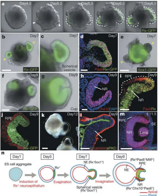
derived from the system. In a landmark study for the field, Sasai and
coworkers produced embryonic stem cell-derived optic cup-like organoids
[15]. The study revealed aspects of the morphogenesis process involved in
forming this complex structure, albeit painting an incomplete picture of
the in vivo process owing to the absence of supporting tissues (Figure 1). In
another important report that helped cement the emerging field,
tissue-specific adult stem cells were used to produce organoids with the
crypt-villus structures of the small intestine [16]. Their results highlighted
expression profile similarities with native adult tissue structures. This distinction may reflect the timescale of in vitro experiments compared to the duration of human developmental and regenerative or homeostatic processes. The replication of organ structures and functionalities within micron to millimeter scale constructs also provides the opportunity to study pathological processes, as well as drug safety and efficacy. Furthermore, the use of patient-specific adult stem cells and induced pluripotent stem cells make this technology particularly suited for
perso-nalized medicine applications [17].
A broad range of protocols have been devised to generate organoids, reflecting in part the diversity of organ-like structures that have already been produced using this approach and the variety of cell sources, differ-entiation stages and relevant co-cultures that can be selected to initiate the
process [13]. The choice of cells also impacts the need for supplementation
with exogenous factors to direct initial stem cell fate decisions. To ensure an adequate microenvironment for organoid formation, the cell masses are often encapsulated within hydrogels. Natural extracellular matrix such as Matrigel, a complex mixture of ECM molecules produced by
Engelbreth-Holm-Swarm mouse sarcoma, has been broadly used for this purpose [18].
While organoids exhibit organ-like structures and functionalities, they are not exact miniature recreations of the organs that they model. For example, organoids do not incorporate critical components of in vivo
organ system such as vascular networks, nor are they exposed to native physical conditioning (e.g., mechanical stresses and electrical stimulation). As such, caution must be used in extrapolating information collected with this approach in vitro to in vivo processes. Lack of reproducibility, which
Figure 1. Optic-cup-like organoid formed spontaneously from the 3D culture of mouse
embryonic stem cell aggregates in the presence of matrigel. Reprinted from reference [15] with permission. (a-l) show the temporal progression of the optic-cup-like structure formation along with the expression of key molecular markers for specific stages in the process. (m) shows the optic cup in a mouse embryo at time E11.5. (n) shows a schematic representation of key stages in the maturation of optic-cup-like. Copyright Nature Publishing Group (2011).
has been identified as differences in size, shape, cellular composition and 3D architecture, between organoids produced in a same experiment has
also limited the applicability [19]. Decreasing organoid-to-organoid
varia-bility is particularly important for the avaria-bility to identify statistically sig-nificant effects in drug screening experiments. A number of strategies have been proposed to address these issues. These range from the development of tailored bioreactors, to the replacement of Matrigel and other ECM hydrogels with synthetic hydrogel platforms that permit increased control over the presentation of spatiotemporal biochemical and biophysical cues for the 3D cell masses [13,19].
While the term spheroid has often been used interchangeably with organoid, it implies a number of important distinctions. Spheroids refer to the culture of tumor cells under non-adherent conditions that have included low-adhesion plates, hanging drop culture systems and
suspen-sion cultures in convective mixing bioreactors [20]. Spheroid cultures
develop heterogenous structures that are reminiscent of in vivo tumors in response to biomolecular gradients across the depth of the cell masses
[21]. This approach has thus been broadly used as in vitro investigative
tool to gain insight into tumor biology and for drug screening studies. The potential of dissociated somatic cells to re-aggregate into tissues with anisotropic architectures resembling native organization is also well established [22]. This self-organization capacity of somatic cells can also be exploited for improved in vitro culture models. For example, chondrocytes
cultured at high densities form 3D cartilage tissue [23], where the use of
a subpopulation of these cells found in the deeper aspect of cartilage allows the formation of tissues that can be induced to form stratified cartilage that incorporates a zone of calcified cartilage, thereby recreating the
cartilage-subchondral bone interface [24,25]. These tissue models have proved
useful to investigate the response of articular cartilage to bioactive
mole-cules [26], study the cellular responses to physiological mechanical signals,
which cannot be replicated with traditional 2D in vitro culture systems and for therapeutic applications [27,28].
2.2. Microtissues
Microengineered tissues, also termed microtissues, represent a new
para-digm in thefield of cell-based assays. They combine microfabrication and
tissue-engineering, to provide in vitro models with tissue-like character-istics such as three-dimensional (3D) architecture, multicellularity, cell-matrix interactions, and close-to-in vivo functionality. As minimal units representing the physiological behavior of tissues, microtissues are usually cultured on conventional well plates or trans-wells relying on nutrient passive diffusion to support cell growth. As a result, they can be used in
applications requiring high-throughput analysis such as drug development or toxicological assays.
Microtissues have been typically produced by adapting microfabrication technologies used in microelectronics to create miniaturized engineered constructs with precisely defined geometries. The main challenge has been, and still is, to successfully apply these engineering technologies,
conven-tionally used in hard materials, to soft materials and cells [29].
Microfabrication techniques such as replica molding, direct printing or photopatterning are used to precisely engineer hydrogels having tunable
cell response, mechanical properties and microarchitecture [30]. Hydrogels
are the preferred choice as engineered extracellular matrices, as they possess high-water content, mechanical properties similar to the soft native
ECM and allow diffusion-driven solute transport [31,32]. The benefits in
providing more physiologically-resembling, and thus predictive data of cell behavior are vastly reported and are related to the employment of micro-environments mimicking in vivo conditions. As such, microtissues repre-sent an enormous improvement of current reductionist cell culture monolayers, and open new research avenues by providing more reliable tools for basic research, disease modeling, and drug testing.
Currently, microtissues are successfully applied to predict drug-induced liver and cardiac toxicity, which is among the main causes of drug attrition
and market withdrawal [33]. While animal testing has shown severe predictive
limitations, in vitro testing has traditionally been hampered by the poor survival of both primary hepatocytes and cardiomyocytes as 2D monolayers. In this context, culturing cells in 3D microenvironments have proven to be beneficial to preserve the phenotype of primary cells for longer time periods and to help differentiation and maturation of cells derived from stem cells. For instance, primary hepatocytes cultured on top of micropatterned collagen
islands and surrounded by fibroblasts, in a configuration known as
micro-patterned co-cultures (MPCC) have high viability and functionality, and have been used in drug development applications, and drug-induced liver injury
predictions (Figure 2(a)) [34]. The technique can also be used with
hepato-cytes derived from induced-pluripotent stem cells, which show improved maturity markers [35,36]. In addition to liver, cardiac microtissues also con-stitute a prime example of the benefits of 3D cell culture in microenviron-ments that recapitulate the structural and electromechanical characteristics of
the native myocardium [37–39]. Microengineered human cardiac tissues,
usually fabricated using cell-laden natural-based hydrogels casted on posts [40–43], or around a wire template [44,45] are used in preclinical toxicology and drug screening assays (Figure 2(b-c)) [46,47]. Together with liver and heart, skin is also a major target of pharmaceutical research and, due to ethical-related issues restricting animal experimentation, of cosmetic research
mixed with collagen type I until contraction is observed. Then, keratinocytes are added and let proliferate. In a final step, the engineered tissue is cultured
in air-liquid interface conditions to promote tissue maturation (Figure 2(d))
[49]. While the use of liver and heart microtissues in routine cell-based assays is limited, several human epidermis tissue models have been approved by the Organization for Economic Cooperation and Development (OECD) and validated by the European Center for the Validation of Alternatives to animal
testing (ECVAM) for regulatory safety purposes [50]. Validated models to
measure skin corrosion are EpiDermTM (MatTek Corporation), EpiSkinTM (Episkin S.A.), SkinEthicTM RHE (Episkin S.A.) and epiCS® (CellSystems) models [OECD, July 2016. Test No. 431: In vitro skin corrosion: reconstructed human epidermis (RHE) test method. OECD Publishing]. Regarding skin irritation, the following models are validated: EpidermTM SIT, EpiSkinTM, SkinEthicTM RHE and LabCyte EPI-MODEL (Japan Tissue Engineering Co) Figure 2.(a) Micropatterned co-cultures (MPCC) withfibroblasts are employed to enhance the maturity of human hepatocytes derived from iPSCs. Reprinted from reference [36] with permission. (b) Cardiac microtissues casted on silicone posts: fabrication process and genera-tion of large microtissue arrays. Cross-secgenera-tional view of a single microtissue (scale bars: 800 μm (array), 100 μm (cross section). Reprinted from reference [41] with permission. (c) Biowire fabrication set-up: a suspension of cardiomyocytes in collagen type I gel is seeded in a PDMS channel around a suture wire. The wires can be easily incorporate in an electrostimulation chamber. Images of the biowire formed at low magnification and after Hematoxylin and Eosin (H&E) staining. Reprinted from reference [44] with permission. (d) Schematic representation of skin reconstruction in vitro. Histological sections of normal human skin and reconstructed skin a day 7 of air exposed culture conditions. Reprinted from reference [49] with permission.
[OECD, July 2015. Test No. 431: In vitro skin irritation: reconstructed human epidermis (RHE) test method. OECD Publishing].
Engineered microtissues also find relevant applications in modeling
human diseases. As an example, fibrosis, which is characterized by the
progressive stiffening of tissues limiting organ functionality and even producing its failure, lacks proper in vitro models that recapitulate the
changes in the mechanical properties of the affected tissues [51]. This is
a major drawback in the development of new therapies. Recently, arrays of
lung microtissues suspended over multiple flexible micropillars that
reca-pitulate the mechanical stiffening and contraction of alveolar tissue has
been reported (Figure 3(a)). The predictive capabilities of the model have
been addressed by analyzing the antifibrosis efficacy of two Food and Drug
Administration (FDA) approved drugs [52]. Also, lately, engineered
car-diac tissues fabricated onfilamentous polymer matrices using two-photon
polymerization have been used to reproduce the deficient contraction produced by cells deficient in the expression of sarcomere protein cardiac
myosin-binding protein C (MYBPC3) (Figure 3(b)) [53]. By employing
human induced pluripotent stem cells (iPSCs) bearing MYBPC3 mutation, this model aims to help in investigating the molecular mechanisms linking genetic alterations to the cardiomyopathy pathogenesis. Along the same line, human iPSC cells have also been successfully used to model skeletal
muscle dystrophies such as Duchenne dystrophy [54]. Muscle microtissues
faithfully reproducing skeletal muscle laminopathies are produced by dif-ferentiating cells within hydrogel matrices under tension to provide myo-fiber alignment. Complex multicellular microtissues including skeletal muscle, vascular endothelial cells, pericytes, and motor neurons are also proposed. Finally, microtissues of epithelial barriers are believed to be key in the study of host–pathogen interactions and the global fight against antibacterial resistance. By using replica molding, bioengineered intestinal tissues were produced on 3D porous silk scaffolds. These models mimicked the oxygen gradients existing in vivo in the gut lumen and allowed for the selective survival of bacteria in aerobic, microaerobic and anaerobic
con-ditions [55]. In a follow-up study, this model was used to study the effects
of long-term intestinal infection of Cryptosporidium parvum parasite [56].
Three-dimensional microtissues of lung mucosa bearing epithelial and stromal cellular components (fibroblasts) were produced on collagen gels and employed to investigate the role of Staphylococcus aureus exotoxins in
tissue pathology associated with pneumonia (Figure 3(c)) [57,58].
These examples aim at illustrating the relevance of microtissues in disease modeling and drug development processes. However, despite their proven improved predictive capabilities, microtissues are not used in routine pre-clinical assays. The access to representative cell sources, standardization issues and lack of high-throughput capabilities are still
challenging. While access to primary cells is limited and often expensive, cells derived from pluripotent stem cells are regarded as a promising alternative, especially as the differentiation process seems to be favored when cultured in 3D environments. Regarding standardization, there is still an open debate on the level of complexity required to obtain physio-logical representative data without compromising the reproducibility and manageability of the models. On the other hand, the reading output of a lot of the assays performed with microtissues relies on microscopy imaging. This prevents high throughput, intense labor and expensive microscopy set-ups for automation. In this sense, the use of biosensors
(reviewed in section 2.6) might be key to help increasing the microtissue
applications.
Figure 3. (a) Recapitulation of tissue fibrogenesis in lung microtissues. TGF-β1 treatment induced strong expressions fibrosis biomarkers (α-SMA stress fibers, cytosolic pro-collagen, and EDA-Fibronectin (Fn)), while SEM images of a time-lapse microscopy showed elevated contraction of the fibrotic tissue (scale bar: 200 µm). Reprinted from reference [52] with permission (http://creativecommons.org/licenses/by/4.0/) (b) Cardiac microtissues assembled onfiber matrices from healthy and MYBPC3 deficient cells (scale bar: 50 µm). Confocal images showed no structural disarray but calcium dynamics showed clear abnormalities. Reprinted from reference [53] with permission. (c) Lung tissue models to evaluate the severity of the damaged produced by several strains of Staphylococcus aureus found in patients with pneumonia. Reprinted from reference [58] with permission (http://creativecommons.org/ licenses/by/4.0/).
2.3. Tissue and organ 3D bioprinting
The promise of an eventual unlimited supply of functional engineered organs to ease the pressure on the transplant waiting lists has recently propelled 3D bioprinting into the public imagination. Three-dimensional bioprinting refers to a collection of technologies that rely on layer-by-layer assembly principles to precisely pattern mixtures of cells and biomolecules, often in combination with materials (termed bioinks) to create living anisotropic 3D structures reminiscent of our tissues and organs. These tools draw on 2D spatial data rendered by computer-aided design (CAD) software from an engineered tissue design to inform the spatial deposition of different bioinks within each layer of the structure. Despite growing research efforts towards the development of these techniques for regen-erative medicine, they have primarily been used to generate complex 3D microenvironments for in vitro experimentation. Indeed, these methods afford a level of spatial reproducibility that is essential to the generation of reliable and meaningful in vitro data, whether it pertains to gaining insight into physiological processes, understanding pathophysiological responses, or evaluating tissue responses to drugs. This feature combined with the ability to construct relatively large engineered tissues offer a unique oppor-tunity for in vitro preclinical studies that is not available with the other technologies and methodologies discussed in this review.
While methods of bioprinting share similarities with those of conventional additive manufacturing (AM) techniques used in rapid prototyping, they also have fundamental differences, made necessary by the incorporation of cells and biomolecules [59]. Indeed, the need to ensure that cell viability is main-tained during the process and that biomolecule denaturation is avoided imposes important constraints on printer design. For example, shear force generated during bioink delivery through capillaries can cause cell damage, while bioinks must avoid the use of cytotoxic compounds such that suitable biomaterials for 3D bioprinting remain limited. The most commonly used
bioprinter technologies are based on continuous extrusion of a bioink
fila-ment through a nozzle [60,61], or the delivery of discrete bioink droplets from an inkjet printer head onto a collector [62,63]. Laser-assisted bioprinting, which relies on the application of laser pulse onto the top face of a metallic ribbon to propel a bioink droplet from the layer bound to the opposite face of the ribbon onto a collector [64], and another technique that uses light to
cross-link the bioinks have also been employed [65]. These nozzle-free techniques
help address complications with orifice clogging of the first two techniques. Each of these approaches offer advantages and limitations, notably pertaining to spatial resolution, printing speed and cell viability, such that the choice of bioprinting device should be made with the intended application and the
Bioprinting has already been used to build a broad range of different 3D tissue models that incorporate aspects of the native spatial anisotropy. For example, a stratified pigmented skin model was developed that includes the dermal and epidermal layers, the latter of which is produced by the careful
placement of keratinocytes and melanocytes [66]. This in vitro tissue
model matured into a construct that more closely resembles native tissue structures than those produced by the traditional manual casting of the different cell types. Another study saw Ma et al. develop an in vitro liver model that recapitulates the spatial organization of hepatic progenitor cells (HPC) and support cells into hexagonal lobules that exhibit improved in vitro functionality compared to 2D cultures and 3D constructs consist-ing of a sconsist-ingle cell type, HPC [67].
The unique ability to precisely pattern cells and biomolecules in 3D structures has also provided an opportunity to tackle a paramount chal-lenge in regenerative medicine and with in vitro cultures, the incorporation of functional vascular networks within tissue constructs. With few excep-tions, cells typically exist in the body within 100–200 μm of blood vessels, beyond which nutrient and metabolite diffusion constraints can cause cell
necrosis [68]. Most approaches to produce 3D tissues models for in vitro
studies or in vivo implantation do not provide a direct route to the incorporation of such vascular networks. This has limited the size of 3D engineered tissues that can be studied for long periods in vitro. Perfusion
and convective flow bioreactors have been used to help address this
limitation, but these systems expose engineered tissues to non-physiological mechanical environments due to the stresses applied to
cells fromfluid flow [69]. Similarly, engineered tissues intended for
trans-plantation have often relied on the design of scaffolds with sufficiently large pores and interconnected porosity to accommodate new vasculariza-tion in vivo. This approach, even when complemented with the controlled release of proangiogenic factors, remains a slow process measured on the scale of days to weeks and can lead to substantial loss of viable resident
cells [70]. Bioprinting has become an essential tool in the development of
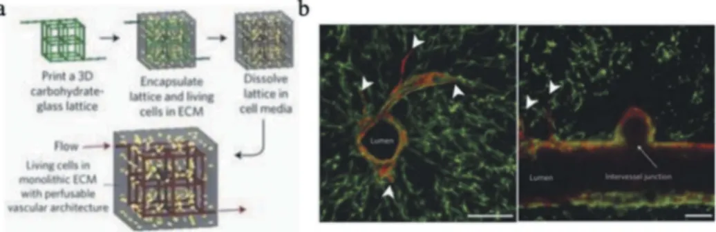
strategies to integrate functional vascular networks within engineered tissues. For example, a number of groups have used coaxial extrusion processes to produce tubular structures that can serve as vascular networks (Figure 4(a)) [71,72]. In this design, endothelial cells have been
incorpo-rated with the bioink used to form the tubular structure of vessels [72].
While this direct extrusion-based approach enables the formation of con-duits with appropriate lengths for clinically relevant constructs, it is also
not easily amenable to the fabrication of branched networks [73]. An
alternative approach that does support the incorporation of bifurcation relies on the deposition of sacrificial ink characterized by reversible phy-sical crosslinks (e.g., alginate, agarose, gelatin) within the 3D structure
during the printing process (Figure 4(b)) [74,75]. The temporary or fugi-tive ink patterns are subsequently liquefied and removed to reveal a network of channels that can be endothelialized and perfused with culture media, or sutured to blood vessels upon implantation. This strategy has been associated with limited long-term mechanical integrity of the channels, highlighting the need for the careful design of the bioinks to avoid rapid degradation. Similarly, efforts to reconstruct the supporting
structures of blood vessels may prove beneficial [73]. The resolution of
current bioprinting technologies also remains too low to support the
fabrication of multiscale vasculature down to sub-micron capillaries [76].
To address this issue, specifically developed bioinks that deliver proangio-genic factors and/or endothelial progenitors have been patterned to pro-mote the formation of capillary networks over time.
Despite the portrayal of 3D bioprinting in media coverage as a mature technology, a number of technological challenges need to be resolved before it can be used to supply functional engineered organs for clinical applications. Indeed, current printing technologies and bioink platforms fall short of enabling the recreation the structural, mechanical and bio-chemical complexity of tissues and organs at multiple length scales. While advances have been achieved in the presentation of temporal control mechanisms within bioprinted tissues and hydrogel systems in general, this critical component in the regulation of cellular behavior has yet to
match the level of complexity observed in native tissues [77]. The ability of
cells to self-organize into tissue-like structures in vitro may help bridge the gap between engineered and native structures; however, acceleration of the tissue maturation process will benefit from improved bioprinter hardware and increasingly modular bioinks that mimic more closely the native cell microenvironment. The current deposition speed of 3D bioprinters also remains insufficient for the fabrication of many human-scale tissues
Figure 4. (a) Schematic of the process to produce cell-laden hydrogels with perfusable
vascular networks using sacrificial glass fibers. (b) Cross-section of the construct showing vascular network lined with endothelial cells, vessel sprouts and intervessel junctions. Reprinted from reference [75] with permission. Copyright Nature Publishing Group (2012).
because the time required to print these structures and harsh conditions
during the bioprinting process can cause substantial cell death [59].
Further, while some tissue defects are amenable to in vivo tissue matura-tion upon implantamatura-tion, the ultimate and ambitious goal of producing full-scale functional engineered organs will require simultaneous innovations in bioreactor design. Such devices are essential to create a hospitable in vitro environment for the maturing tissues construct that includes the delivery of nutrients and other biochemical cues, the removal of metabo-lites and waste products and the simultaneous administration appropriate biochemical and biophysical cues, while accounting for mass transfer resistances across large engineered tissues. Nevertheless, bioprinting has already proved to be a powerful tool to support in vitro studies in disease
modeling as well as drug screening [78]. Bioprinting is still considered an
emerging technology, especially in comparison to the broader AM field;
however, it has demonstrated rapid growth in recent years. A report has predicted that the bioprinter market will reach $1.8 billion by 2027 and
that it could eventually reach the tens of billions of dollars [79].
Expectedly, a number of companies have proposed new products with a broad range of capabilities and price points ranging from $5,000 to
$250,000, making this technology affordable for most academic labs [80].
2.4. Organs-on-a-chip
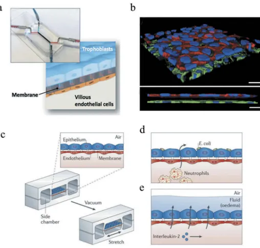
The fundamental design philosophy behind the organs-on-a-chip (OOC) technology is the reduction of whole human organs and/or organ systems to minimal functional units capable of maintaining key aspects of the native tissue architecture and of the in vivo physiology. In brief, OOC platforms consist of microchannels lined by multiple cell types spatially assembled in ways to replicate organ-specific interfaces (e.g., the fetal-maternal interface in
the placenta [81], Figure 5(a-b)). The resulting microengineered systems
through a system of pumps, valves andfilters, allow to precisely control the
perfusion of microliter amounts offluids through the microchannel network
in a regime of laminar flow (i.e., microfluidics). Together, these features
ultimately permit to mimic specific aspects of the whole-organ response, replicate the in vivo connections between different organs, investigate
phar-macokinetic mechanisms and drug toxicity, among others [82].
A variety of methods, ranging from etching techniques to laser
photo-ablation, have been used to fabricate microfluidic systems [83,84]. Among
these, the most common approach for microfabricating the channels within a solid chip relies on a combination of soft lithography and replicas, with polydimethylsiloxane (PDMS) as the material of choice because of its intrinsic advantages (i.e., low cost, ability to be poured and cross-linked into intricate features, transparency to allow microscopy observation)
[83,84]. To achieve the sought-after physiological relevance, the resulting microfluidic platforms are successively bioengineered in ways to integrate specific features that characterize living tissues and organs, such as the presence of an ECM-like microenvironment, temporal gradients of soluble factors (e.g., cytokines, growth factors, hormones) and the presence of biophysical forces. In this context, mechanical actuators that can generate different stress states (e.g., shear, compressive, tensile) to replicate the
Figure 5. (a) Placenta-on-a-chip, consisting of two microchannels separated by
a semipermeable membrane sandwiched between a trophoblast and an endothelial cell monolayer, mimicking the maternal-fetal interface. (b) Three-dimensional rendering (top) and cross-sectional view (bottom) of the bioengineered placental barrier consisting of tro-phoblast cells cultured on the apical side of the membrane and villous endothelial cells adhering on the basal side of the membrane. Scale bar: 30μm. Reprinted from reference [81] with permission. Copyright Royal Society of Chemistry (2017). (c) Schematic of a lung-on -a-chip, consisting of an epithelial and endothelial cell monolayer separated by a porous membrane to recapitulate the alveolar-capillary barrier of human lungs. In order to include biomechanical cues, a vacuum was applied to mimic the stretching of the tissue during breathing. The lung-on-a-chip permitted to reconstitute organ-level functions such as (d) the immune response to bacteria and (e) pulmonary edema. Reprinted from reference [89] with permission. Copyright Nature Publishing Group (2015).
mechanobiology of living tissues have been integrated in microfluidic
devices [85]. This aspect was particularly important in the development
of an accurate mechanobiological environment for epithelial cells in the
lung-on-a-chip device [86], since during normal breathing movements,
lungs undergo dynamic deformation ranging from 4% to 12% stretch
distension of the basement membrane (Figure 5(c-e)) [87].
Aside from these distinctive characteristics, the OOC technology has
made additional steps forward towards the biological fidelity required to
mimic their organ-level counterparts, while maintaining the ease of tradi-tional 2D cell cultures [88]. In particular, the direct use of human cells not only promises to address the translatability challenges and ethical issues
encountered with animal models [89], but is also expected to accelerate the
development of personalized platforms for precision medicine and
advanced drug screening [85], in parallel with progresses in iPSC biology
[90]: mature cells could be in fact harvested from individual patients,
reprogrammed and finally used in OOC platforms to build personalized
functional models for disease mechanisms and drug screening purposes
[91]. Other advantages of OOCs include [89]: (i) the ease of direct
real-time observation of cellular processes of interest with conventional optical microscopes; (ii) the ability of controlling environmental factors (e.g., temperature and gas concentration), precisely delivering soluble factors (e.g., nutrients, drugs, cytokines, hormones), collecting metabolites, waste and other secretory factors; (iii) the reduced cost associated with the smaller volumes of reagents required; (iv) reproducibility, integration with other technologies (e.g., sensors for electrical, optical and biochemical
readouts) [83] and possibility to link multiple OOCs together to investigate
interactions among different organs and recreate either normal or
patho-logical whole human physiology (i.e., body-on-a-chip) [92].
To date, all major organs have been reconstituted in OOC platforms: liver, skin, lung, and placenta are just few examples of the panoply of current applications of this technology. Regardless of the specific tissue type, OOCs
have been mainly employed for two principal purposes [93]: (i) Disease
modeling: a panoply of pathological states has been investigated with OOCs. For example, cancer and immune system related conditions have been repli-cated to close in on disease progression (e.g. cancer metastatic growth and
intravasation) [94]. In this context, a large body of work has focused on the
importance of the integration of immune factors [8,12]. To this end, by
capitalizing on the key advantages of microfluidic devices (i.e., bioengineered 3D architecture of the cell microenvironment, dynamic exchange of oxygen and nutrients, time-lapse imaging) with respect to other in vitro methods, aspects such as immune cells-tumors interactions and inflammation events could be replicated [8,12]. Of note, OOC systems, through the precise control
mimicking endocrine signaling and replicate hormonal variations during the menstrual cycle and pregnancy, thereby promising to become a powerful tool for both translational and fundamental research in women’s health toward novel insights into female-specific diseases and sex-dependent differences in
preclinical research [95]. The microfluidic nature also makes OOCs
a powerful tool to better understand bacterial and viral infections [96].
Bacteria and viruses can be in fact circulated to investigate their interactions with the biological membranes of interest. For example, E. coli bacteria introduced in the lung-on-a-chip device activated the transmigration of circulating neutrophils through the reconstructed endothelial layer,
replicat-ing the innate immune response to pulmonary bacterial infection (Figure 5
(d)) [86]. Similarly, a liver-on-a-chip has been recently used to investigate hepatitis B viral (HBV) infection, demonstrating the ability to recapitulate all steps of the HBV life cycle as well as the innate immune and cytokine
responses observed in HBV-infected patients [97]. (ii) Advanced drug
screen-ing/discovery: bringing a new drug to market requires up to twelve years and more than 1.7 billion US dollars. Despite these efforts, it is estimated that only 1 out of 10,000 drug candidates gains FDA approval despite successful pre-clinical evaluation, and up to 20% of acute kidney injuries are linked to drug toxicity effects non predicted by preclinical models [8,93]. For these reasons, researchers have increasingly turned to OOCs as a potential technological alternative for faster, cheaper and more predictive preclinical screening and toxicology assessment. To date, the responsiveness and toxicity to specific drugs have been assessed with different OOC platforms (e.g., liver-, lymph
node-, bone marrow-, gut- and kidney-on-a-chip, among others) [8,93]. In
this context, of particular interest because of recent advances in drug-delivery technologies for better pharmacological efficacy and bioavailability, is the employment of OOCs to evaluate the response to nanoparticle-based ther-apeutics (i.e., nanotoxicology). The microfluidic technology offers in fact biologically relevant platforms which only require a fraction of samples compared to conventional in vitro systems, while permitting real-time evalua-tion of nanoparticle transport and cellular uptake [98].
The potential of OOCs for applications in human health has been
high-lighted by the 2016 World Economic Forum which ranked them 6thamong
the top 10 emerging technologies [85]. Such endorsement has been echoed by
funding agencies (e.g., National Institute of Health in the USA, the European Framework Program 7 and the Japan Agency for Medical Research and Development) which invested significant resources towards the development of more robust and replicable OOC platforms for commercialization [3,87]. In parallel, large pharmaceutical and biotechnology companies (e.g., Pfizer, Merck, Johnson&Johnson) have intensified their involvement in collabora-tions with academia and companies (e.g., Emulate) to support the translation
of OOC platforms from the laboratory to industrial R&D, thereby further
contributing to the commercialization of the technology [83].
Despite its increasing employment for real-world devices, the OOC technology still requires improvements to address specific challenges and fully maturate its potential for preclinical research and commercial appli-cations, such as [3,83,93,99]: (i) Scalability and fabrication techniques – in order to avoid volume-dependent alterations of cellular processes within OOC compartments, these devices should be scalable to the optimal organ size to avoid over- or under-production of metabolites. The ease of scal-ability also poses challenging repercussions in their commercialization process, for which the fabrication methods currently employed in the laboratory (e.g., lithography) should be translated to large-scale manufac-turing by ensuring rapid and highly reproductible production at low cost. In addition, the design of OOC platforms should aim at enhancing throughput and usability, facilitating handling by the end-users; (ii)
Materials – although PDMS is widely used in OCCs, better performing
materials are required to address its limitations, such as unwanted defor-mations of the chip, gas permeability, hydrophobic drugs absorption and
leaching; (iii) Cell sources – since OOC platforms’ accuracy strongly
depends on the cell types used, the selection of cells is critical. In this context, immortalized cell lines are widely used, but alterations in their native functional activity should be accounted for. While primary cells
provide a better alternative, availability, ethical and financial issues limit
their employment; (iv) Perfusion process – the ideal perfusion system
should avoid bubble formation and precisely control fluid recirculation
to replicate in vivo mechanisms that affect absorption, elimination and distribution of metabolites and other soluble factors; (v) Imaging and
analysis– with increasingly more complex cellular models, standard
fluor-escence imaging techniques drastically reduce the quantity of information that can be extracted from the OOC platform. Therefore, additional
techniques (e.g., transepithelial/transendothelial electrical resistance –
TEER) and sensing abilities need to be synergistically integrated in order
to generate accurate real-time data on cellular phenomena (section 2.6).
2.5. Biomimetic in vitro models
While most of the microtissues, tissue constructs, and organs-on-a-chip aim to reproduce the main functions of a tissue or organ, usually they do not
reproduce their morphology. The avenue of organoids (section 2.1), in which
cells self-organize and spatially segregate to recreate the architecture found in the native tissue has pointed out the relevance of the shape in the proper tissue functionality. Epithelial and endothelial tissues in vivo form complex 3D structures such as cysts, tubules, evaginations or invaginations.
Vasculature, nephrons, mammary glands, gastrointestinal epithelium, hair-follicles, and corneal limbus are only some examples of tissue complex geometries in our body that researchers have been tried to reproduce in vitro employing a variety of biomimetic microfabrication strategies. For example, photolithography and replica molding have been used to
repro-duce the wavy topography of the epidermal–dermal interface (Figure 6(a))
[100]. It is accepted that epidermal stem cells lie in clusters specifically
located in relation to this topography, but the relationship between their location and properties is currently unknown and can be investigated by reproducing the tissue architecture [100]. Also, in skin tissue, the fabrication of 3D structures with the shape of the hair-follicle bulges has attracted a lot of attention. Micro molding has been used to accurately create microwell arrays of self-assembling hair follicle germs able to generate spatially arrange
hair follicles upon transplantation [101]. In another study, 3D printed molds
were used to pattern collagen gels laden with dermal fibroblasts. Dermal
papilla cells were then cultured on the microwells to promote the differ-entiation of human keratinocytes into specific hair follicle lineages and their
physiological arrangement (Figure 6(b)) [48]. Corneal limbus also has
spe-cific microstructures that serve as stem cell niches. Limbal architecture has
been reproduced by electrospinning polymer fibers on micromoulds
pro-duced by stereolithography [102] or by replica molding on collagen gels
[103]. Regarding tubular structures, very recently an array of kidney tubules
produced from a micro-molded platform in collagen-matrigel gels. This platform allows for the modeling of kidney diseases such as cystic kidney disease and acute kidney injury as well as drug testing purpose [104]. Finally, the small intestine crypt-villus morphology has also been reproduced by
either replica molding procedures [29,105] and, more recently, by
reaction-diffusion mediated photolithography (Figure 6(c)) [106]. In all the cases,
a significant improvement of the properties of the epithelial barrier on the 3D structures compared with the 2D monolayers was reported. Still, and despite the benefits reported, in vitro models bearing biomimetic micro-structures are relying on relatively tedious and complex fabrication pro-cesses. It is foreseen that the wider availability and applicability of 3D
bioprinting methods will have an impact in thisfield, extending the
applica-tions of the biomimetic approach. 2.6. Biosensors
Most of the reading outputs obtained from bioengineered in vitro models rely on imaging data. This limits the throughput, complicates and makes more expensive the set-ups, and restricts their use to highly skilled personnel. To advance these drawbacks, an excellent approach seems to integrate biosensors in the bioengineered platforms. Biosensors are analytical devices that rely on the
molecular recognition capabilities of biological capture probes (antibodies, oligonucleotides, enzymes or even cells) to measure the concentration of analytes. These recognition events will be transduced to a signal that will be read-out. Transducers might be of different nature, being the electrochemical, optical, thermal and mass-change based the most popular. Biosensors can be used to continuous monitoring the critical parameters of cell microenviron-ment, and to study the response of the models to drugs over extended periods of time. They can be applied to static cell culture (microtissues in well plates or Figure 6. (a) Fabrication of patterned PDMS substrates to mimic the epidermal rete ridges. Keratinocyte patterning on collage-coated PDMS substrates (involucrin stained in yellow,β1 integrin stained in red, scale bar: 200 µm). Reprinted from reference [100] with permission. (b) Hair follicles formed on microwells of collagen gels laden with dermalfibroblasts (FB). First, dermal papilla cells (DPC) were seeded within the wells, and then keratinocytes (KC). Cross sections of the construct (scale bar 2 mm) and immunostaining (scale bar 100 µm) show active DPC cells at their physiological positions (black arrows). Prolonged culture period led to hair fiber formation (arrowheads, scale bar 2 mm). Reprinted from reference [48] with permission (http://creativecommons.org/licenses/by/4.0/). (c) Intestinal villi-like microstruc-tures fabricated by dynamic photopolymerization on poly(ethylene glycol) hydrogels provide the barrier formed CaCo-2 cells with improved physiological characteristics (scale bars: 150 µm; (upper row); 200 µm (lower row, left and middle); 50 µm (lower row, right)). Reprinted from reference [106] with permission.
Transwells) or to microfluidic platforms [105]. To demonstrate the power of biosensor integration approach, Zhang et al. reported a platform for the auto-mated, on-line monitoring of a multiorgan-on-a-chip platform that include sensors to monitor pH, oxygen and temperature, electrochemical biosensors to monitor soluble protein biomarkers and miniature microscopes for the imaging of the morphology of organoids cultured within the platform [107]. In a proof-of-concept application, the platform was employed to monitor drug-induced toxicity in two dual organ model systems: human liver-and-heart-on-a-chip and liver-cancer-and-heart-on-a-chip, to prove the assessment of long-term response to chronic drugs and short-term response of acute toxicity, respec-tively. Monitoring soluble biomarkers such as cytokines through integrated biosensors is also used as a reading output to monitor pathological processes in a non-destructive manner. As an example, real-time monitoring of TNF-α in a model of tumor intravasation that included tumor cells, endothelial cells and macrophages revealed the dynamics of the process, including the increase in
endothelial permeability and tumor-endothelial interactions [108]. It is
accepted that an improved knowledge about the molecular dynamics in cancer metastasis can lead to propose new therapeutic strategies.
On top of soluble biomarkers, some tissues, such as skeletal, cardiac, and smooth muscle require specific sensors to evaluate their main functional response, which is the capability of contraction. Mechanical forces generated from these bioengineered tissue models are usually measured by monitoring the bending of silicon cantilevers or
micropil-lars [109]. In addition, electrical activity can also be a functional
hall-mark for cardiac, muscle, and nervous tissues. Electrical signal can be recorded by microelectrode arrays (MEAs), which can be easily
inte-grated in microphysiological systems [110]. On the other hand, the
functionality of tissue barriers formed skin, gastrointestinal tract, and blood brain barrier is usually monitored by determining the TEER. To that end, bioengineered models of these tissues can incorporate two
measurement electrodes, one at each side of the cellular barrier [111].
Overall, online and/or integrated monitoring capabilities might be key in the future acceptance of bioengineered models as new valuable tools in drug development. Challenges are associated to systems complexity and cost. If they can be overcome, they promise to increase the throughput of in vitro assays, allow personalized medicine applications and significantly increase the cellular and molecular basis of complex human pathologies.
3. Conclusion and outlook
Advanced in vitro platforms are likely to foster profound changes in the ways preclinical research is conducted. The application of novel bioengineering tools, strategies and techniques to improve health is poised to continue to
grow until they will reach a mature level to be routinely integrated in conventional preclinical practices, resulting in a major impact on health research and economic benefits to society. However, as outlined in the pre-vious sections, many challenges still remain to be addressed in order to implement these new technologies in research and clinical practises. In particular, while significant efforts are being made to incorporate organ systems in many of the technologies described in this work (e.g. adding vasculature in 3D printed constructs, creating multi-organoid co-cultures), in vitro studies remain simplified models that do not comprehensively include all relevant aspects of the dynamic multifactorial in vivo environment which characterizes living organisms, such as the immune response.
Although the technologies outlined in this review have been presented individually, researchers are currently integrating different approaches within the same platform in order to achieve synergistic effects stemming from the simultaneous combination of individual advantages and mitigation of each other’s limitations. For example, synergistic engineering strategies have been proposed to integrate organoids and organs-on-a-chip towards
superior in vitro platforms that conjugate the high cellular fidelity of the
former with the higher throughput phenotypic readouts of the latter [112].
Similarly, organoids can be integrated into 3D printing processes to recon-struct the tissue’s microarchitecture [113,114]. Therefore, it can be envisaged that such integrative approaches will likely become the key to propel inno-vation in preclinical research towards the next generation of in vitro plat-forms for more accurate disease modeling, mechanistic studies, and drug discovery. Of note, more effective in vitro technologies are not only expected
to find increasingly more applications in preclinical research but also in
other sectors, such as consumer products industry, where the transition to these novel methods may soon become a necessity. In this context, the Australian Senate has recently passed a bill to ban animal testing for cosmetic development (Industrial Chemical Charges Bill 2017), aligning to
the European Union directive that ousted it since 2013 [3,87]. This trend
will likely expand to North America and worldwide, ultimately enhancing the breadth of in vitro solutions towards faster and more cost-effective development and commercialization for the consumer goods industry (e.g. cosmetics, household products, food and dietary supplements).
Acknowledgments
EM acknowledges funding by European Union’s Horizon 2020 ERC grant agreement No
647863 (COMIET), the CERCA Programme/Generalitat de Catalunya (2017-SGR-1079), and the Spanish Ministry of Economy and Competitiveness (TEC2017-83716-C2-1-R) and the Severo Ochoa Program for Centers of Excellence in R&D 2016-2019. The results presented here reflect only the views of the authors; the European Commission is not
responsible for any use that may be made of the information it contains. JPS and FV acknowledge funding from the Natural Sciences and Engineering Research Council of Canada (NSERC) through the Discovery Grant. JPS also acknowledgesfinancial support by the University of Ottawa Seed Funding Opportunity Grant. The authors acknowledge Alexander Steeves for his help with the graphical abstract.
Disclosure statement
No potential conflict of interest was reported by the authors.
Funding
This work was supported by the Generalitat de Catalunya [2017-SGR-1079];H2020 European Research Council [647863];NSERC Discovery Grant [RGPIN-2017-04593]; NSERC Discovery Grant [RGPIN 402441-2013];Spanish Ministry of Economy and Competitiveness [Severo Ochoa Program for Centers of Excellence in,TEC2017-83716-C2-1-R];University of Ottawa [RDP 2017-01].
References
[1] Duval K, Grover H, Han L-H, et al. Modeling physiological events in 2D vs. 3D cell culture. Physiology.2017;32:266–277.
[2] Kacy Cullen D, Wolf JA, Vernekar VN, et al. Neural tissue engineering and biohybridized microsystems for neurobiological investigation in vitro (part 1). Crit Rev Biomed Eng.2011;39:201–240.
[3] Kimura H, Sakai Y, Fujii T. Organ/body-on-a-chip based on microfluidic technology for drug discovery. Drug Metab Pharmacokinet.2018;33:43–48.
[4] Lee SH, Sung JH. Organ-on-a-chip technology for reproducing multiorgan physiology. Adv Healthcare Mater.2018;7:1700419.
[5] Yamada KM, Cukierman E. Modeling tissue morphogenesis and cancer in 3D. Cell.
2007;130:601–610.
[6] Hoarau-Véchot J, Rafii A, Touboul C, et al. Halfway between 2D and animal models: are 3D cultures the ideal tool to study cancer-microenvironment interactions? IJMS.
2018;19:181.
[7] Edmondson R, Broglie JJ, Adcock AF, et al. Three-dimensional cell culture systems and their applications in drug discovery and cell-based biosensors. Assay Drug Dev Technol.2014;12:207–218.
[8] Shanti A, Teo J, Stefanini C. In vitro immune organs-on-chip for drug development: a review. Pharmaceutics.2018;10:278.
[9] Ericsson AC, Crim MJ, Franklin CL. A brief history of animal modeling. Mo Med.
2013;110:201–205.
[10] Conn PM. source book of models for biomedical research. Totowa, NJ: Humana Press;2008.
[11] Seok J, Warren HS, Cuenca AG, et al. Genomic responses in mouse models poorly mimic human inflammatory diseases. Natl Acad Sci.2013;110:3507–3512.
[12] Polini A, Del Mercato LL, Barra A, et al. Towards the development of human immune-system-on-a-chip platforms. Drug Discov Today.2018;24:1–9.
[13] Lancaster MA, Knoblich JA. Organogenesis in a dish: modeling development and disease using organoid technologies. Science.2014;345:1247125.
[14] Rossi G, Manfrin A, Lutolf MP. Progress and potential in organoid research. Nat Rev Genet.2018;19:1–17.
[15] Eiraku M, Takata N, Ishibashi H, et al. Self-organizing optic-cup morphogenesis in three-dimensional culture. Nature.2011;472:51–56.
[16] Sato T, Vries RG, Snippert HJ, et al. Single Lgr5 stem cells build crypt-villus structures in vitro without a mesenchymal niche. Nature.2009;459:262–265.
[17] Takahashi T. Organoids for drug discovery and personalized medicine. Ann Rev Pharmacol Toxicol.2019;59:447–462.
[18] Lancaster MA, Renner M, Martin C-A, et al. Cerebral organoids model human brain development and microcephaly. Nature.2013;501:373–379.
[19] Huch M, Knoblich JA, Lutolf MP, et al. The hope and the hype of organoid research. Development.2017;144:938–941.
[20] Fang Y, Eglen RM. Three-dimensional cell cultures in drug discovery and Development. SLAS Discov.2017;22:456–472.
[21] Unger C, Kramer N, Walzl A, et al. Modeling human carcinomas: physiologically relevant 3D models to improve anti-cancer drug development. Adv Drug Deliv Rev.
2014;79:50–67.
[22] Paul Weiss ACT. Reconstitution of complete organs from single-cell suspensions of chick embryos in advanced stages of differentiation. Proc Natl Acad Sci USA.
1960;46:1177–1185.
[23] Boyle J, Luan B, Cruz TF, et al. Characterization of proteoglycan accumulation during formation of cartilagenous tissue in vitro. Osteoarthritis Cartilage.
1995;3:117–125.
[24] Kandel RA, Boyle J, Gibson G, et al. In vitro formation of mineralized cartilagenous tissue by articular chondrocytes. In Vitro Cell Dev Biol Anim.1997;33:174–181.
[25] St-Pierre J-P, Gan L, Wang J, et al. The incorporation of a zone of calcified cartilage improves the interfacial shear strength between in vitro-formed cartilage and the underlying substrate. Acta Biomater.2012;8:1603–1615.
[26] St-Pierre J-P, Wang Q, Li SQ, et al. Inorganic polyphosphate stimulates cartilage tissue formation. Tissue Eng Part A.2012;18:1282–1292.
[27] De Croos JNA, Dhaliwal SS, Grynpas MD, et al. Cyclic compressive mechanical stimulation induces sequential catabolic and anabolic gene changes in chondrocytes resulting in increased extracellular matrix accumulation. Matrix Biol.2006;25:323–331.
[28] Kandel RA, Grynpas M, Pilliar R, et al. Repair of osteochondral defects with biphasic cartilage-calcium polyphosphate constructs in a sheep model. Biomaterials.
2006;27:4120–4131.
[29] Sung JH, Yu J, Luo D, et al. Microscale 3-D hydrogel scaffold for biomimetic gastrointestinal (GI) tract model. Lab Chip.2011;11:389–392.
[30] Huang G, Wang L, Wang S, et al. Engineering three-dimensional cell mechanical microenvironment with hydrogels. Biofabrication.2012;4:042001.
[31] Tibbitt MW, Anseth KS. Hydrogels as extracellular matrix mimics for 3D cell culture. Biotechnol Bioeng.2009;103:655–663.
[32] DeForest CA, Anseth KS. Advances in bioactive hydrogels to probe and direct cell fate. Annu Rev Chem Biomol Eng.2012;3:421–444.
[33] Brown GE, Khetani SR. Microfabrication of liver and heart tissues for drug development. Philos Trans R Soc Lond B Biol Sci.2018;373:20170225.
[34] Khetani SR, Bhatia SN. Microscale culture of human liver cells for drug development. Nat Biotechnol.2007;26:120–126.
[35] Ware BR, Berger DR, Khetani SR. Prediction of drug-induced liver injury in micropatterned co-cultures containing iPSC-derived human hepatocytes. Toxicol Sci.2015;145:252–262.
[36] Berger DR, Ware BR, Davidson MD, et al. Enhancing the functional maturity of induced pluripotent stem cell-derived human hepatocytes by controlled presentation of cell-cell interactions in vitro. Hepatology.2015;61:1370–1381.
[37] Radisic M, Park H, Shing H, Consi T, Schoen F, Langer R, Freed LE, Vunjak-Novakovic G. Functional assembly of engineered myocardium by electrical stimula-tion of cardiac myocytes cultured on scaffolds, Proc Natl Acad Sci USA
2004;101:18129–18134.
[38] Tulloch NL, Muskheli V, Razumova MV, Korte FS, Regnier M, Hauch KD, Pabon L, Reinecke H, Murry CE. Growth of engineered human myocardium with mechanical loading and vascular coculture, Circ Res2011;109:47–59.
[39] Zhang D, Shadrin IY, Lam J, et al. Tissue-engineered cardiac patch for advanced functional maturation of human ESC-derived cardiomyocytes. Biomaterials.
2013;34:5813–5820.
[40] Turnbull IC, Karakikes I, Serrao GW, et al. Advancing functional engineered cardiac tissues toward a preclinical model of human myocardium. Faseb J.2014;28:644–654.
[41] Legant WR, Pathak A, Yang MT, et al. Microfabricated tissue gauges to measure and
manipulate forces from 3D microtissues. Proc Natl Acad Sci USA.
2009;106:10097–10102.
[42] Thavandiran N, Dubois N, Mikryukov A, et al. Design and formulation of functional pluripotent stem cell-derived cardiac microtissues. Proc Natl Acad Sci USA.2013;110:E4698–707.
[43] Tiburcy M, Hudson JE, Balfanz P, et al. Defined engineered human myocardium with advanced maturation for applications in heart failure modeling and repair. Circulation.2017;135:1832–1847.
[44] Nunes SS, Miklas JW, Liu J, et al. Biowire: a platform for maturation of human pluripotent stem cell-derived cardiomyocytes. Nat Methods.2013;10:781–787.
[45] Xiao Y, Zhang B, Liu H, et al. Microfabricated perfusable cardiac biowire: a platform that mimics native cardiac bundle. Lab Chip.2014;14:869–882.
[46] Amano Y, Nishiguchi A, Matsusaki M, et al. Development of vascularized iPSC derived 3D-cardiomyocyte tissues by filtration layer-by-layer technique and their application for pharmaceutical assays. Acta Biomater.2016;33:110–121.
[47] Mathur A, Loskill P, Shao K, et al. Human iPSC-based cardiac microphysiological system for drug screening applications. Sci Rep.2015;5:8883.
[48] Abaci HE, Coffman A, Doucet Y, et al. Tissue engineering of human hair follicles using a biomimetic developmental approach. Nat Commun.2018;9:5301.
[49] Vioux-Chagnoleau C, Lejeune F, Sok J, et al. Reconstructed human skin: from
photodamage to sunscreen photoprotection and anti-aging molecules.
J Dermatological Sci Suppl.2006;2:S1–S12.
[50] Zhang Z, Michniak-Kohn BB. Tissue engineered human skin equivalents. Pharmaceutics.2012;4:26–41.
[51] Feng Y, Mitchison TJ, Bender A, et al. Multi-parameter phenotypic profiling: using cellular effects to characterize small-molecule compounds. Nat Rev Drug Discov.
2009;8:567–578.
[52] Asmani M, Velumani S, Li Y, et al. Fibrotic microtissue array to predict anti-fibrosis drug efficacy. Nat Commun.2018;9:2066.
[53] Ma Z, Huebsch N, Koo S, et al. Contractile deficits in engineered cardiac micro-tissues as a result of MYBPC3 deficiency and mechanical overload. Nat Biomed Eng.
2018;2:955–967.
[54] Maffioletti SM, Sarcar S, Henderson ABH, et al. Three-dimensional human iPSC-derived artificial skeletal muscles model muscular dystrophies and enable multilineage tissue engineering. Cell Rep.2018;23:899–908.
[55] Chen Y, Lin Y, Davis KM, et al. Robust bioengineered 3D functional human intestinal epithelium. Sci Rep.2015;13708.
[56] DeCicco RePass MA, Chen Y, Lin Y, et al. Novel Bioengineered Three-Dimensional Human Intestinal Model for Long-Term Infection of Cryptosporidium parvum. Infect Immun.2017;85.
[57] Nguyen Hoang AT, Chen P, Juarez J, et al. Dendritic cell functional properties in a three-dimensional tissue model of human lung mucosa. Am J Physiol Lung Cell Mol Physiol.2012;302:L226–37.
[58] Mairpady Shambat S, Chen P, Nguyen Hoang AT, et al. Modelling staphylococcal pneumonia in a human 3D lung tissue model system delineates toxin-mediated pathology. Dis Model Mech.2015;8:1413–1425.
[59] Murphy SV, Atala A. 3D bioprinting of tissues and organs. Nature Biotechnology.
2014;32:773–785.
[60] Pfister A, Landers R, Laib A, et al. Biofunctional rapid prototyping for tissue-engineering applications: 3D bioplotting versus 3D printing. J Polym Sci Part a-Polym Chem.2004;42:624–638.
[61] Ozbolat IT, Hospodiuk M. Current advances and future perspectives in extrusion-based bioprinting. Biomaterials.2016;76:321–343.
[62] Klebe RJ. Cytoscribing - a method for micropositioning cells and the construction of
two-dimensional and 3-dimensional synthetic tissues. Exp Cell Res.
1988;179:362–373.
[63] Gudapati H, Dey M, Ozbolat I. A comprehensive review on droplet-based bioprint-ing: past, present and future. Biomaterials.2016;102:20–42.
[64] Guillotin B, Souquet A, Catros S, et al. Laser assisted bioprinting of engineered tissue with high cell density and microscale organization. Biomaterials.
2010;31:7250–7256.
[65] Lu Y, Mapili G, Suhali G, et al. A digital micro-mirror device-based system for the microfabrication of complex, spatially patterned tissue engineering scaffolds. J Biomed Mater Res A.2006;77:396–405.
[66] Ng WL, Qi JTZ, Yeong WY, et al. Proof-of-concept: 3D bioprinting of pigmented human skin constructs. Biofabrication.2018;10:025005.
[67] Ma X, Qu X, Zhu W, et al. Deterministically patterned biomimetic human iPSC-derived hepatic model via rapid 3D bioprinting. Proc Natl Acad Sci USA.
2016;113:2206–2211.
[68] Carmeliet P, Jain RK. Angiogenesis in cancer and other diseases. Nature.
2000;407:249–257.
[69] Rouwkema J, Khademhosseini A. Vascularization and angiogenesis in tissue engi-neering: beyond creating static networks. Trends Biotechnol.2016;34:733–745.
[70] Butt OI, Carruth R, Kutala VK, et al. Stimulation of peri-implant vascularization with bone marrow-derived progenitor cells: monitoring by in VivoEPR oximetry. Tissue Eng.2007;13:2053–2061.
[71] Zhang Y, Yu Y, Chen H, et al. Characterization of printable cellular micro-fluidic channels for tissue engineering. Biofabrication.2013;5:025004.